<< back to Pathology Atlas menu
Pathology Atlas: Gestational Trophoblastic Disease
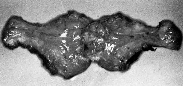
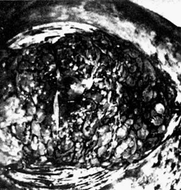 In situ hydatidiform mole in hysterectomy specimen. The dilated vesicles
are apparent. The outer membranes surrouding each of these vesicles
are made up of the trophoblastic layer.
In situ hydatidiform mole in hysterectomy specimen. The dilated vesicles
are apparent. The outer membranes surrouding each of these vesicles
are made up of the trophoblastic layer.
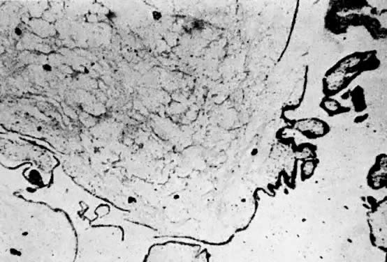 Microscopic view of hydatidiform mole with a low order of trophoblastic
proliferation. Edema of the villus stroma and loss of vascular support
are evident. Despite the low activity of the trophoblast from this tissue, the
patient from whom this was taken developed metastatic choriocarcinoma.
Microscopic view of hydatidiform mole with a low order of trophoblastic
proliferation. Edema of the villus stroma and loss of vascular support
are evident. Despite the low activity of the trophoblast from this tissue, the
patient from whom this was taken developed metastatic choriocarcinoma.
 Microscopic view of hydatidiform mole showing moderate trophoblastic proliferation
along with the other usually seen histopathologic signs. This
patient developed invasive mole.
Microscopic view of hydatidiform mole showing moderate trophoblastic proliferation
along with the other usually seen histopathologic signs. This
patient developed invasive mole.
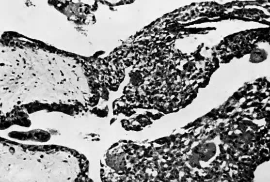
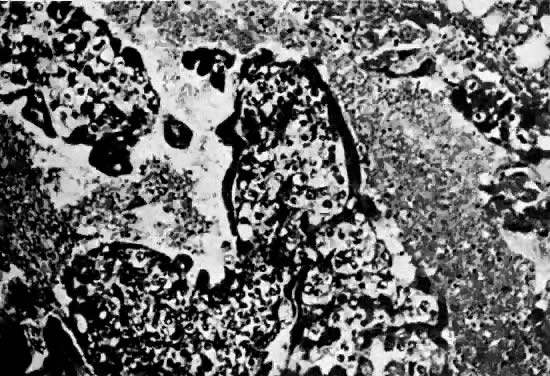
 Histopathologic view of hydatidiform mole evacuated by curettage. In this
particular view only the trophoblast is apparent, the underlying villus
being outside this field. There is moderate to severe trophoblastic
proliferation, but this patient was cured by uterine evacuation alone.
Histopathologic view of hydatidiform mole evacuated by curettage. In this
particular view only the trophoblast is apparent, the underlying villus
being outside this field. There is moderate to severe trophoblastic
proliferation, but this patient was cured by uterine evacuation alone.
Back to Top
Invasive Mole
 Uterus containing invasive hydatidiform mole (chorioadenoma destruens). The
nodule of disease is high in the uterine fundus, and on careful inspection
several dilated vesicles can be seen in the myometrium.
Uterus containing invasive hydatidiform mole (chorioadenoma destruens). The
nodule of disease is high in the uterine fundus, and on careful inspection
several dilated vesicles can be seen in the myometrium.
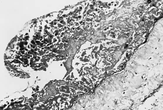
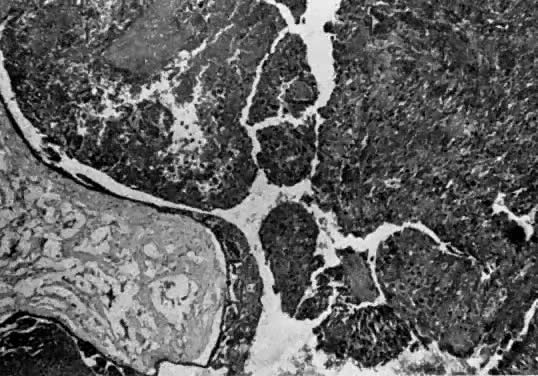 Microscopic view of invasive mole (chorioadenoma destruens), showing retention
of the villus pattern and trophoblastic proliferation deep in
the myometrium. This section was taken from the uterus shown in Figure 6.
Microscopic view of invasive mole (chorioadenoma destruens), showing retention
of the villus pattern and trophoblastic proliferation deep in
the myometrium. This section was taken from the uterus shown in Figure 6.
Back to Top
Partial Mole
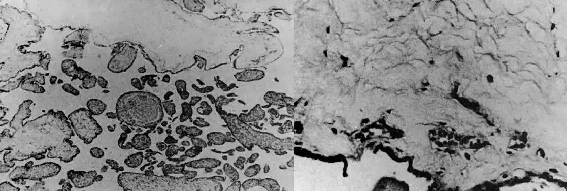 Microscopic view of partial hydatidiform mole. Left. At the upper edge is a large central cistern. Smaller villi show edema
and various degrees of scalloping. The fetus was alive. Trophoblastic
hyperplasia was inconspicuous but present. Right. The wall of a large cistern formed in the presence of a functioning fetoplacental
circulation. Note patent, well-formed villous vessels filled
with fetal erythrocytes.(Sulzman A, Buchsbaum HJ: Gestational trophoblastic disease. In Clinical
Perspectives in Obstetrics and Gynecology, p. 38. New York, Springer-Verlag, 1987.
Microscopic view of partial hydatidiform mole. Left. At the upper edge is a large central cistern. Smaller villi show edema
and various degrees of scalloping. The fetus was alive. Trophoblastic
hyperplasia was inconspicuous but present. Right. The wall of a large cistern formed in the presence of a functioning fetoplacental
circulation. Note patent, well-formed villous vessels filled
with fetal erythrocytes.(Sulzman A, Buchsbaum HJ: Gestational trophoblastic disease. In Clinical
Perspectives in Obstetrics and Gynecology, p. 38. New York, Springer-Verlag, 1987.
Back to Top
Choriocarcinoma
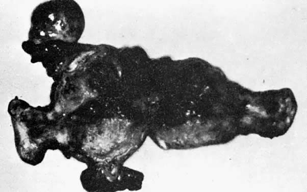 Uterus removed for choriocarcinoma. The extensive necrosis, vascular penetration, and
hemorrhage are evident from this specimen.
Uterus removed for choriocarcinoma. The extensive necrosis, vascular penetration, and
hemorrhage are evident from this specimen.
 Microscopic view of choriocarcinoma taken from the uterus illustrated in Figure 9. Sheets of anaplastic trophoblastic cells are noted without the maintenance
of the pattern of the villi. Extensive necrosis is evident.
Microscopic view of choriocarcinoma taken from the uterus illustrated in Figure 9. Sheets of anaplastic trophoblastic cells are noted without the maintenance
of the pattern of the villi. Extensive necrosis is evident.
Back to Top
| 