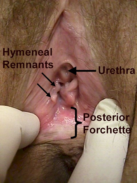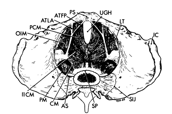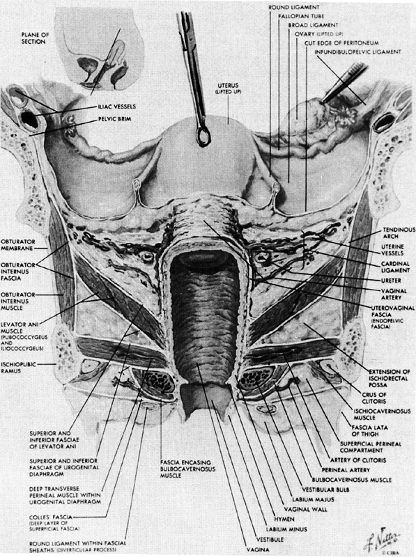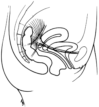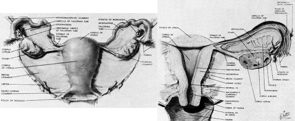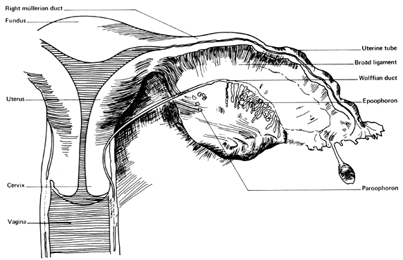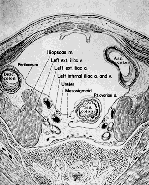
Fig. 1. Normal Anatomy of a 30-year-old multipara.
(Reproduced, with permission from Michael Hughey, MD, All rights reserved.)
|
Anatomy Michael John Hughey |
|
|
Michael John Hughey, MD |
|
Vulva Pelvis Pelvic Support Uterus, Fallopian Tubes and Ovaries Anorectal Canal |
Breasts Abdominal Wall Nerves Blood Vessels Video: Vulvar Anatomy Video: Pelvic Anatomy |
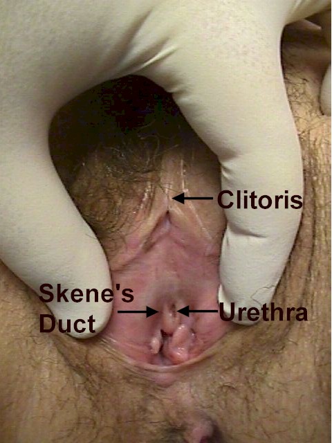
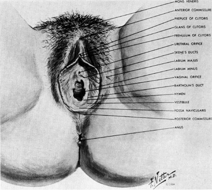
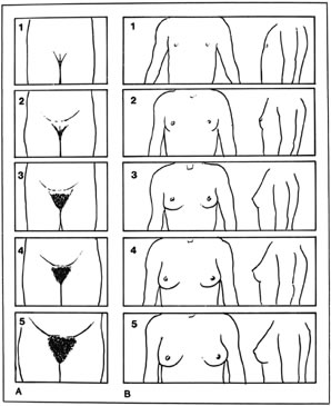
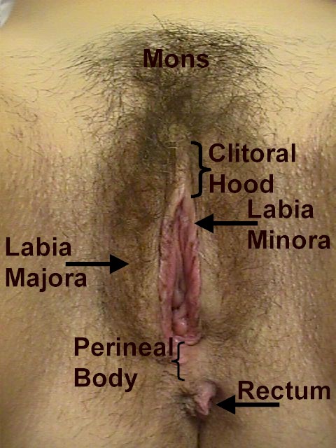
| Vulva |
|
|
|
|
|
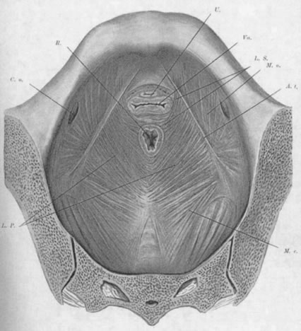
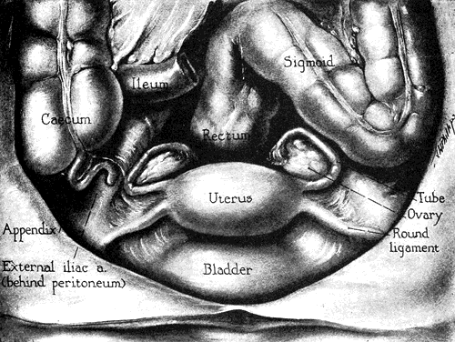
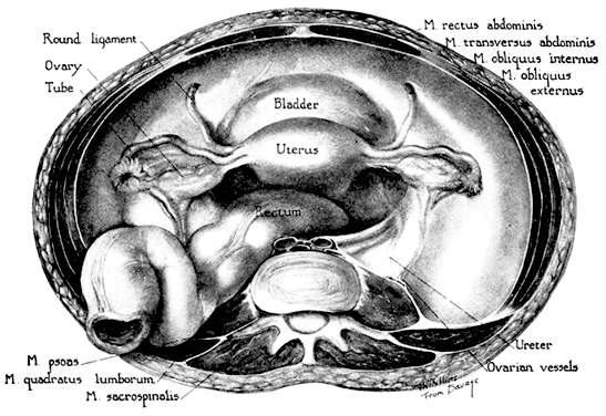
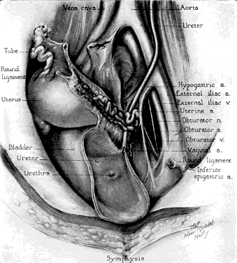
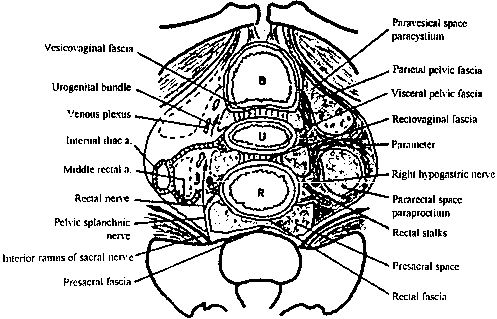
| Pelvis |
|
|
|
|
|
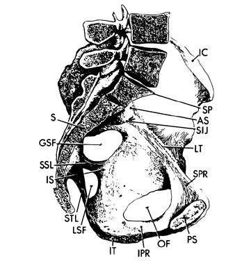
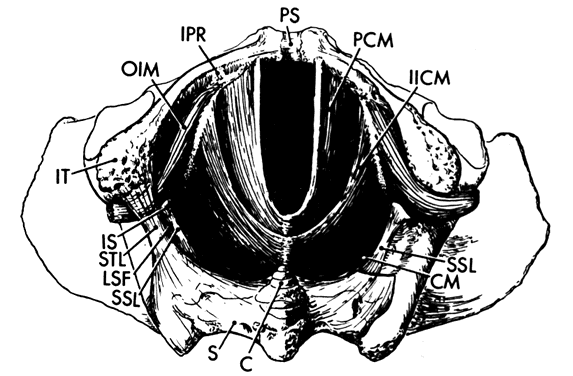
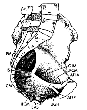
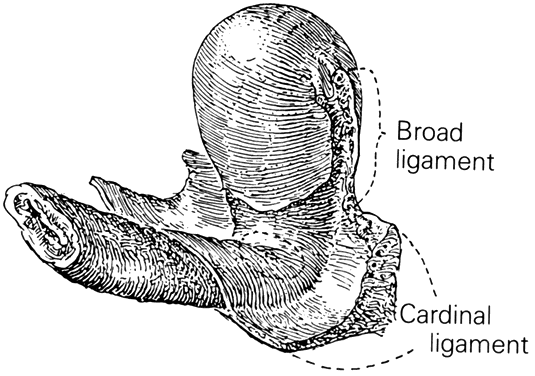
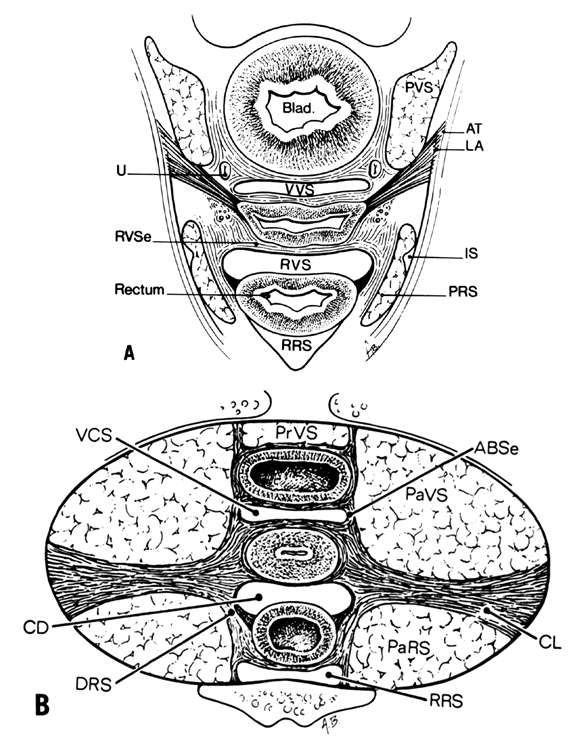
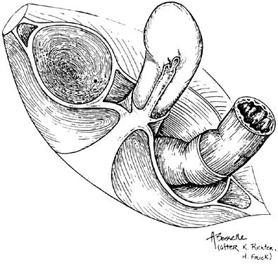
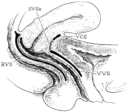
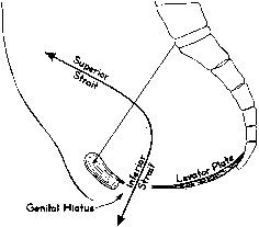
| Pelvic Support |
|
|
|
|
|
|
|
|
|
|
|
|
|
|
|
|
|
|
|
|
|
|
|
|
|
|
|
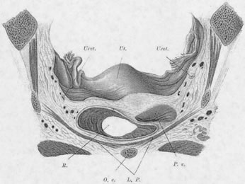
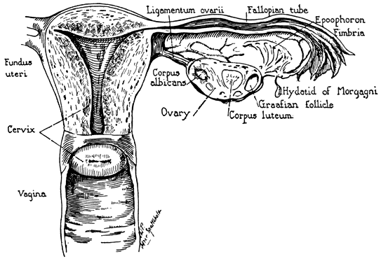
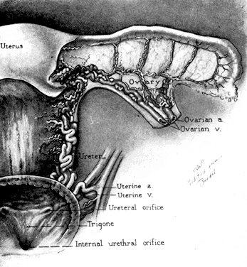
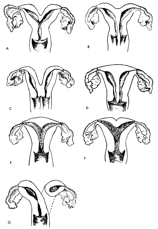
| Uterus, Fallopian Tubes and Ovaries |
|
|
|
|
|
|
|
|
|
|
|
|
|
|
|
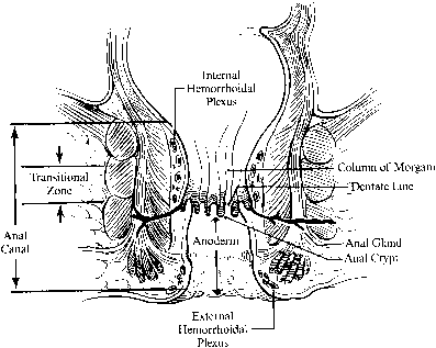
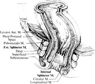
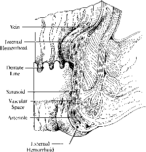
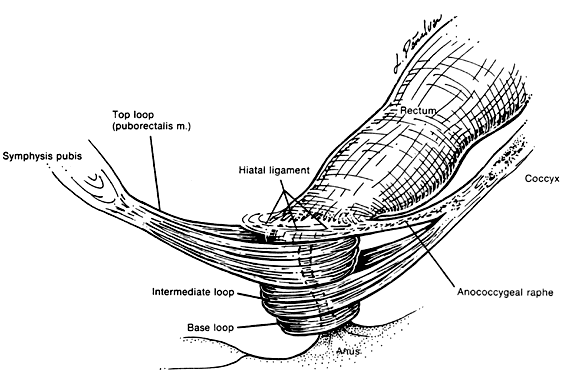
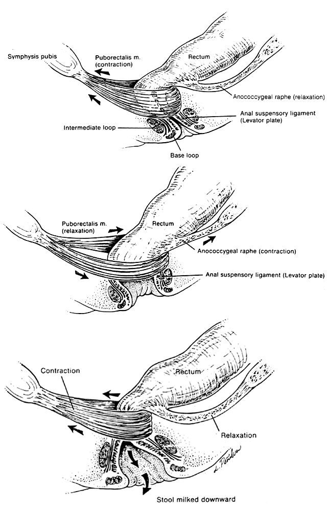
| Anorectal Canal |
|
|
|
|
|
|
|
|
| Breasts |
|
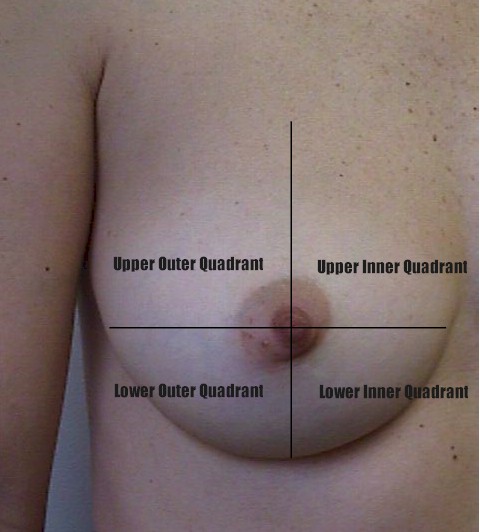
Fig. 2. Breast quadrants. (Reproduced, with permission from Michael Hughey, MD, All rights reserved.) |
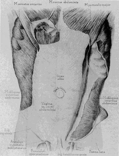
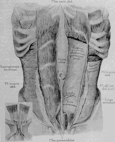
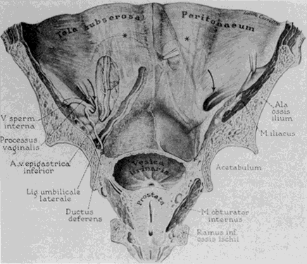
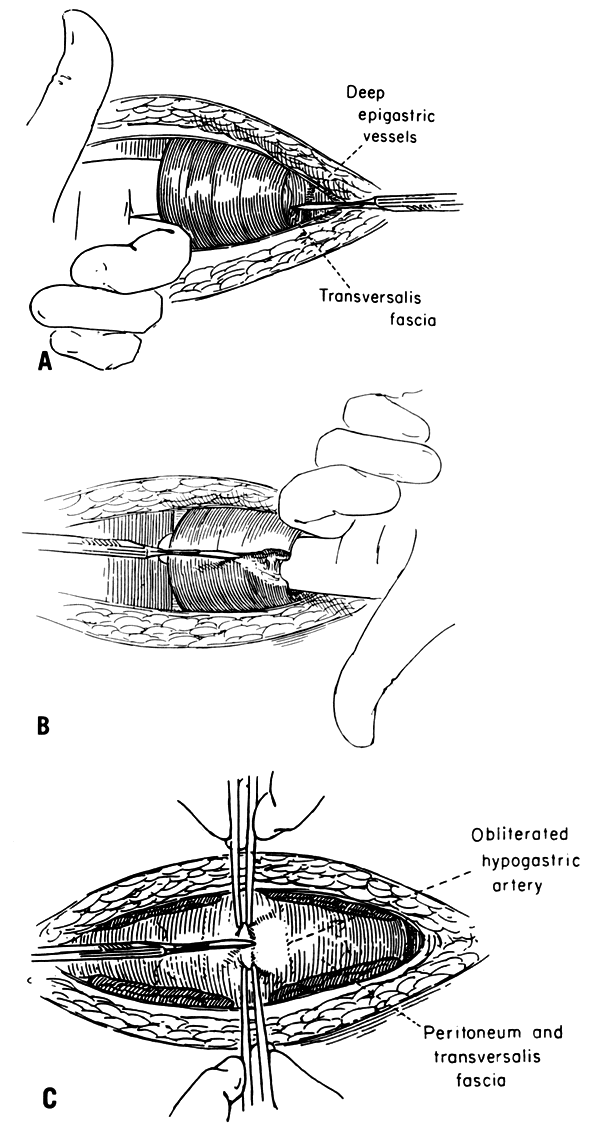
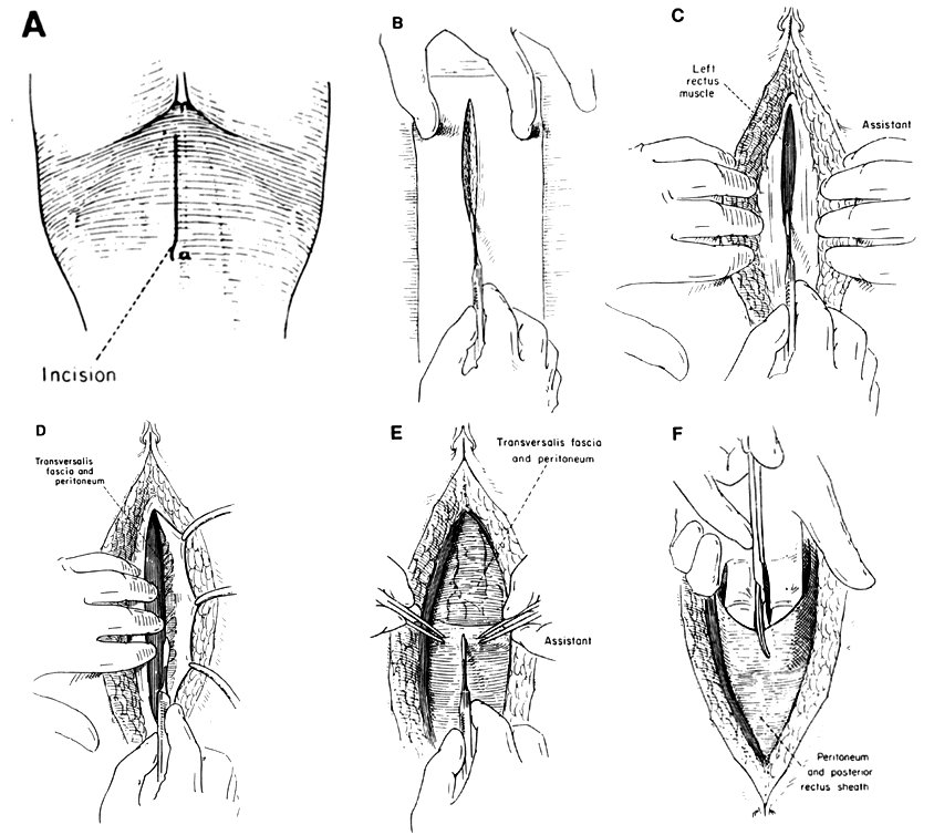
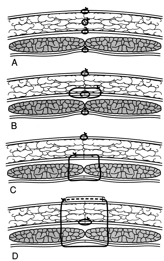
| Abdominal Wall |
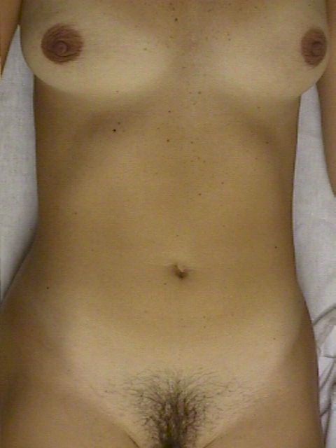
Fig. 1. Torso of a 30-year-old multipara. (Reproduced, with permission from Michael Hughey, MD, All rights reserved.) |
|
|
|
|
|
|
|
|
|
|
| Nerves |
|
|
|
|
|
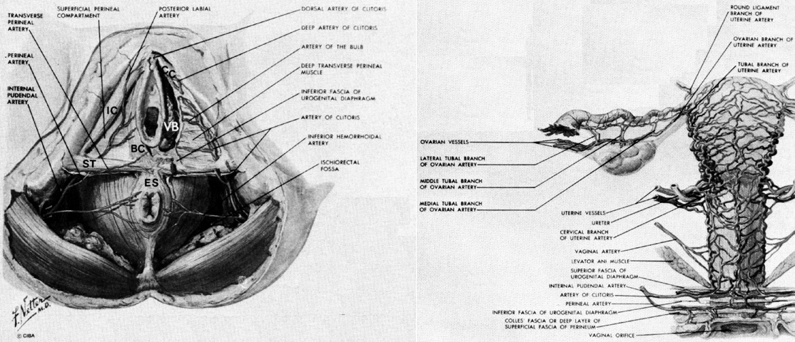
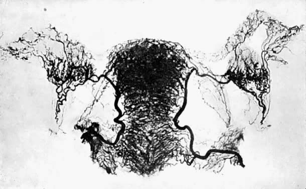
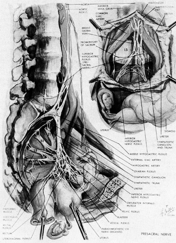
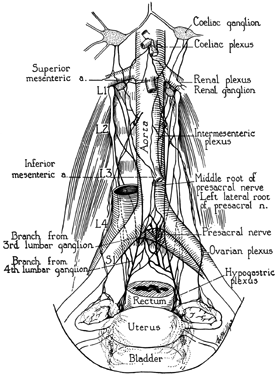
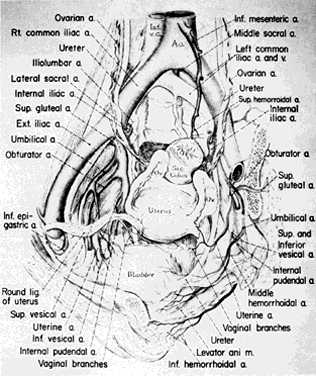
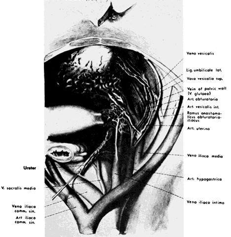
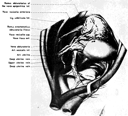
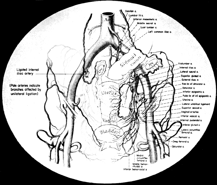
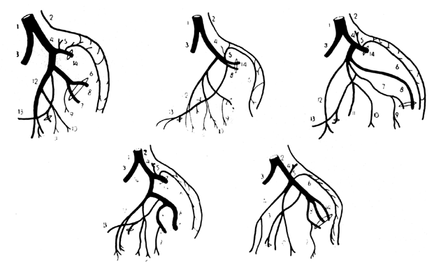
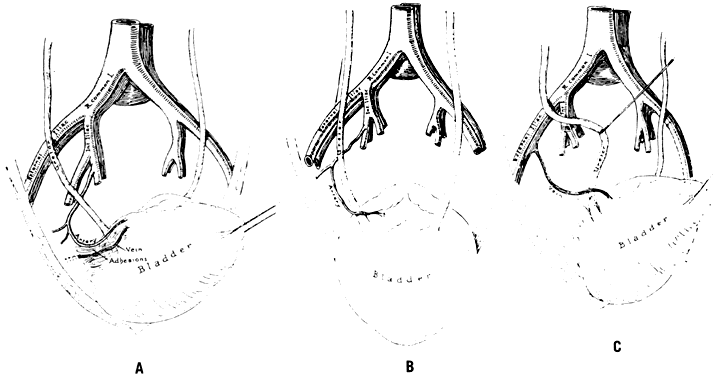
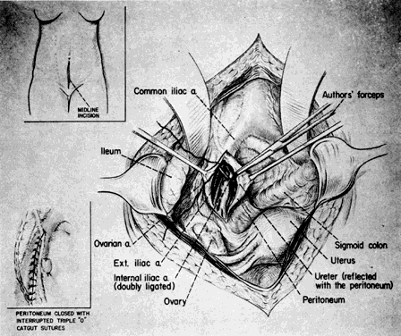
| Blood Vessels |
|
|
|
|
|
|
|
|
|