Pathology Atlas: Vagina
Michael John Hughey
Main Menu   Table Of Contents

Search

Michael John Hughey, MD
Associate Clinical Professor, Department of Obstetrics and Gynecology, Northwestern University Medical School, Chicago, Illinois; Adjunct Associate Professor, Department of Obstetrics and Gynecology, Uniformed Services University of the Health Sciences, Bethesda, MD; Senior Attending Physician, Evanston Northwestern Healthcare, Evanston, IL.

Pathology Texts from Lippincott Williams & Wilkins

Squamous Cell Carcinoma
Clear Cell Carcinoma
Vaginal Melanoma
Sarcoma Botryoides
Squamous Cell Carcinoma
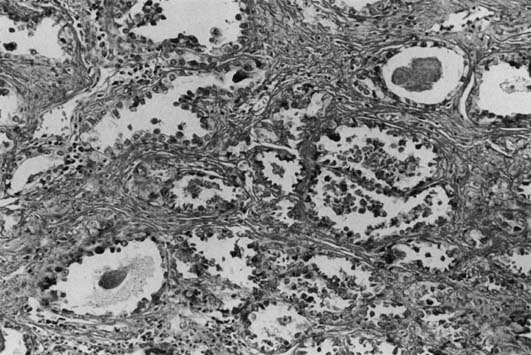
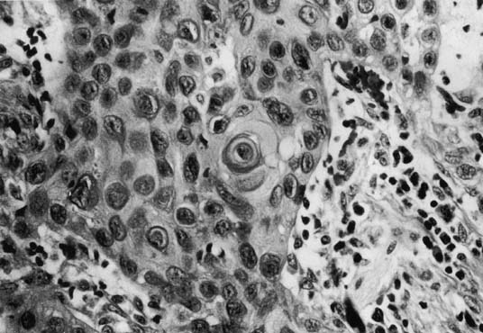
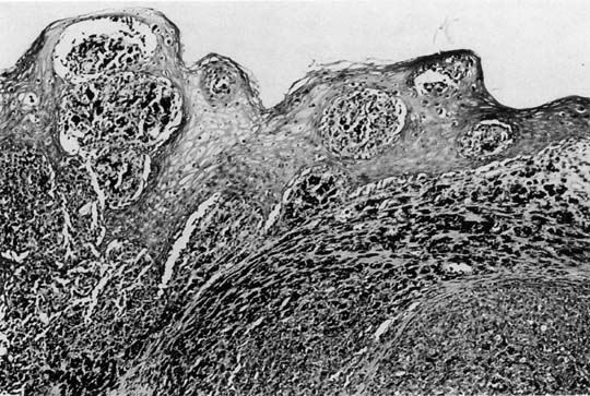
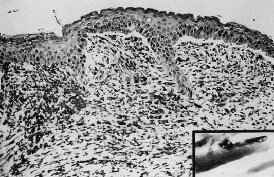
Fig. 1. Squamous cell carcinoma of the vagina (X200 magnification) demonstrating keratin pearl formation. Volume 4, Chapter 44

Back to Top

Clear Cell Carcinoma

Fig. 2. Vaginal clear-cell adenocarcinoma (X200 magnification) from a DES-exposed patient. Volume 4, Chapter 44

Back to Top

Vaginal Melanoma

Fig. 3. Vaginal melanoma (X100 magnification). Junctional melanocytic proliferation and infiltrate of pigmented (epithelioid) melanoma. Volume 4, Chapter 44

Back to Top

Sarcoma Botryoides

Fig. 4. Sarcoma botryoides (X100). Cambium layer beneath epithelium. Loose spindle cell infiltrate. Inset (X400) demonstrates cross-striation in cytoplasm of spindle cells. Volume 4, Chapter 44


Fig. 12. Sarcoma botryoides arising from the vagina of a 2-year-old girl, showing the squamous epithelium ( A) and the subepithelial cambium zone ( B ). Notice the central myxoid loose stroma ( C) (× 360) Volume 4, Chapter 22

Back to Top