Pathology Atlas: Ovary
Michael John Hughey
Main Menu   Table Of Contents

Search

Michael John Hughey, MD
Associate Clinical Professor, Department of Obstetrics Northwestern University Medical School, Chicago, Illinois; Adjunct Associate Professor, Department of Obstetrics and Gynecology, Uniformed Services University of the Health Sciences, Bethesda, MD; Senior Attending Physician, Evanston Northwestern Healthcare, Evanston, IL.

Pathology Texts from Lippincott Williams & Wilkins

Normal Growth and Development
Follicular Cyst
Teratoma
Immature Teratoma
Serous Cystoma
Mucinous Cystadenoma
Brenner Tumor
Fibroma
Granulosa Cell Tumor
Hilus Cell Tumor
Thecoma
Sertoli-Stromal Cell Tumors (Androblastomas)
Gonadoblastoma
Dysgerminoma
Endodermal Sinus Tumor
Low Malignant Potential:
  Serous Cystadenoma
  Mucinous Cystadenoma
  Endometrioid Tumor
  Clear Cell Tumor
  Brenner Tumor
  Cystadenofibroma with Atypia
  Pseudomyxoma Peritonei
  Borderline Serous Tumor
Malignancy
  Serous Adenocarcinoma
  Endometrioid Carcinoma
  Clear Cell Carcinoma
  Embryonal Carcinoma
  Choriocarcinoma
  Mixed Germ Cell Tumor
  Krukenberg Tumor
Normal Growth and Development

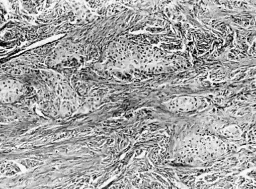
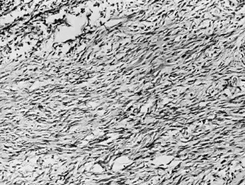
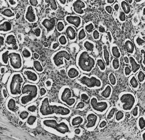
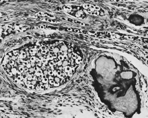
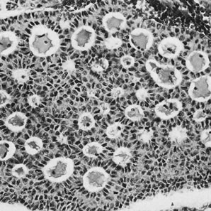
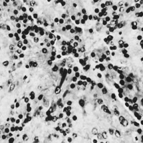
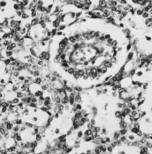
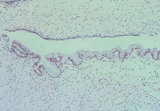
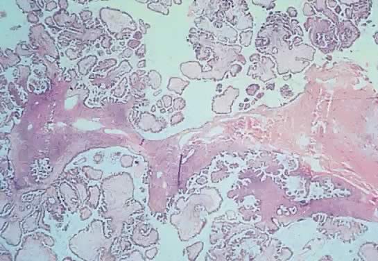
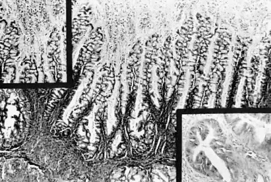
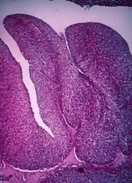
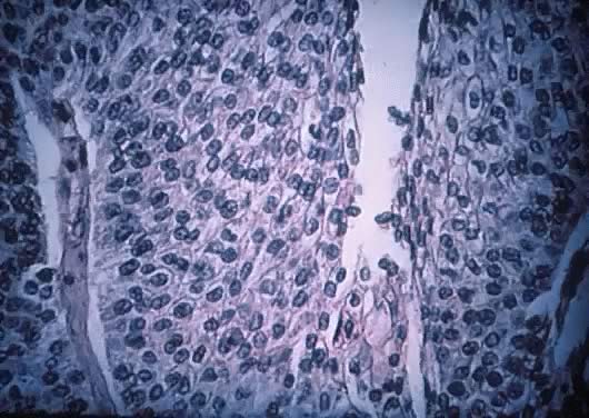
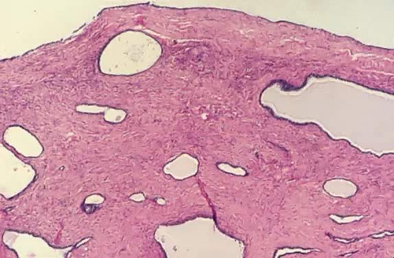
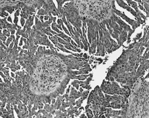
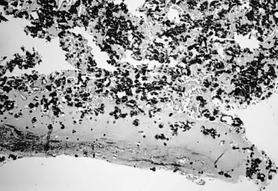
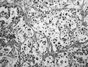
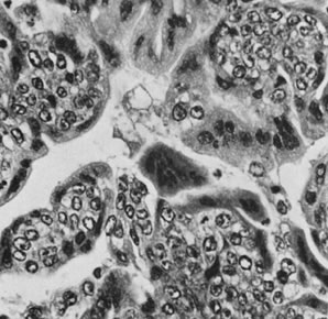
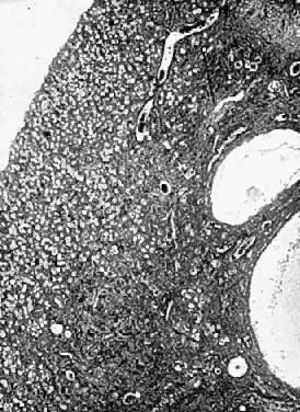
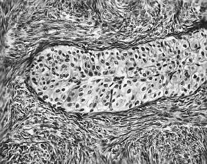
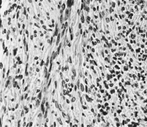
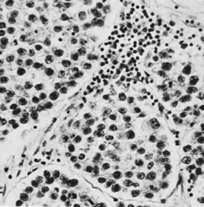
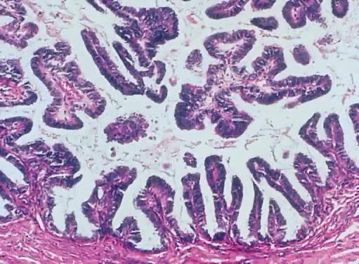
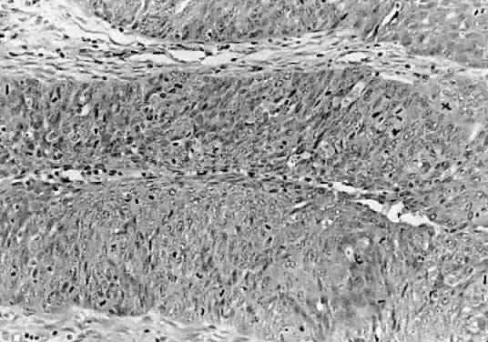
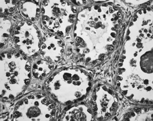
Fig. 13. Photomicrograph (low power) of the cortex of the ovary of a human infant. The cortex of the ovary has numerous primordial germ cells with relatively little stroma. The ovarian stroma is more abundant in the medulla, where the larger follicles are seen. Volume 1, Chapter 2.
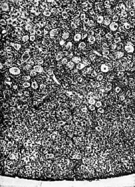
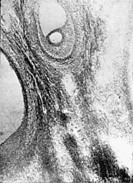
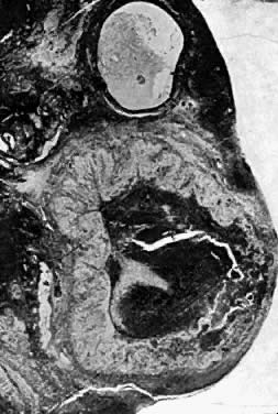
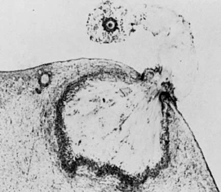
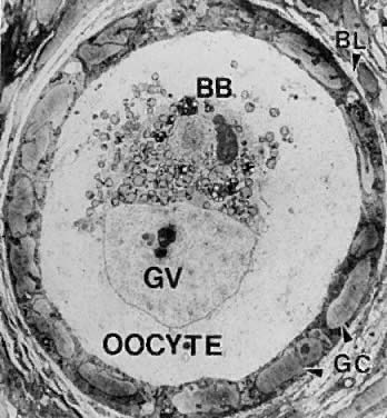
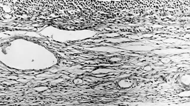
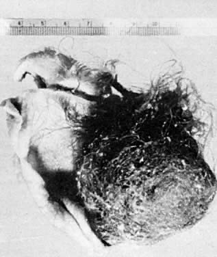
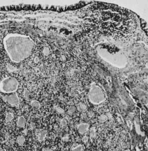
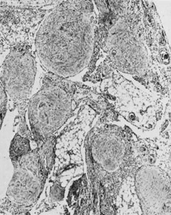
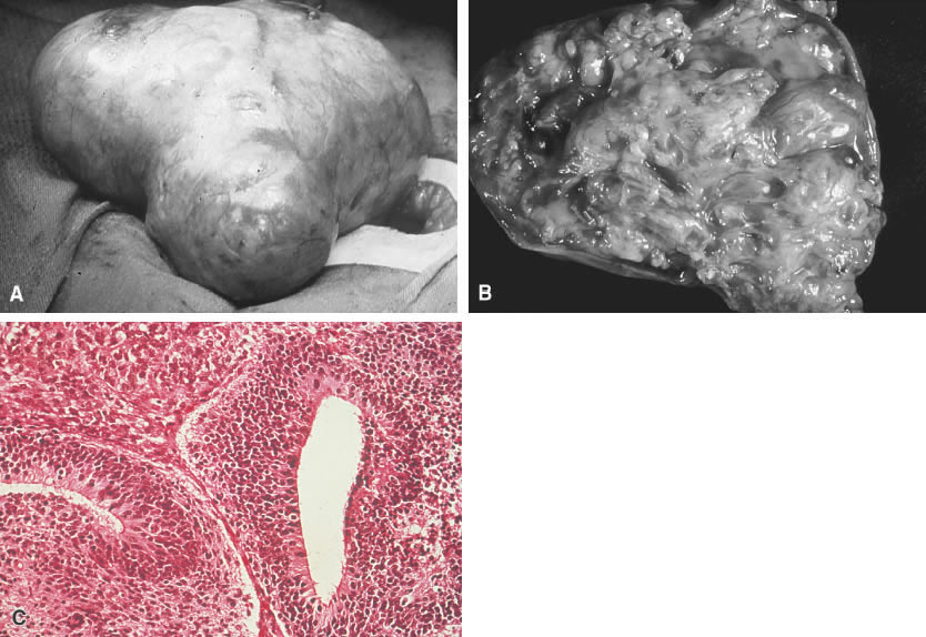
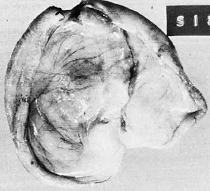
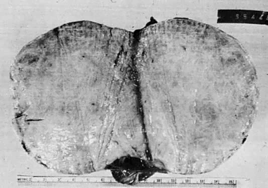
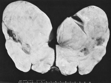
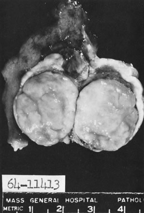
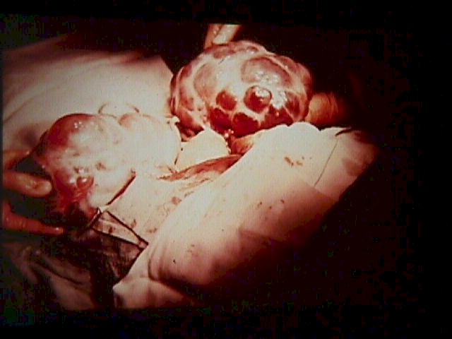
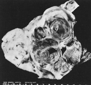
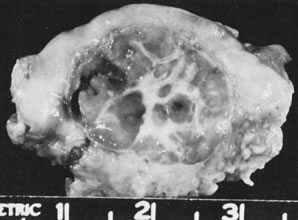
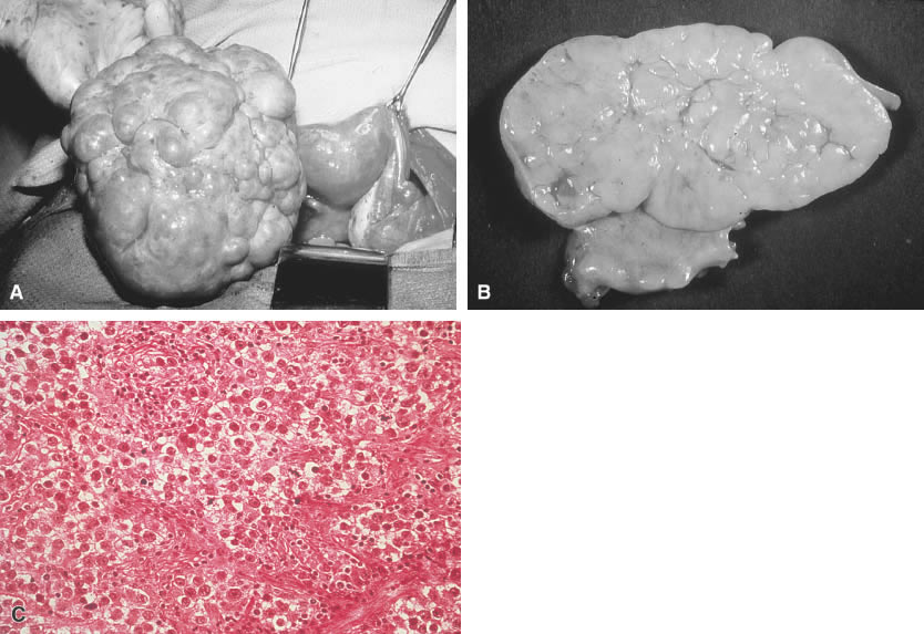
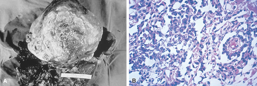
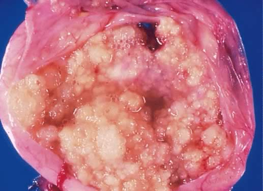
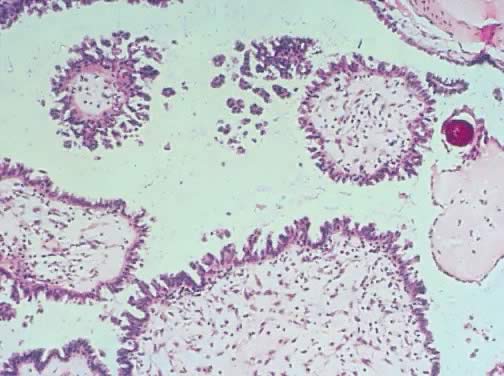
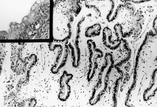
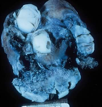
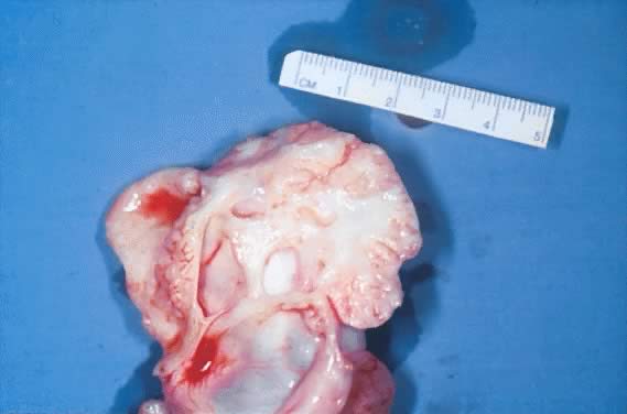
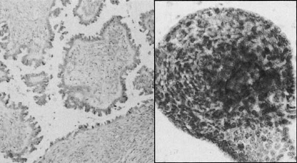
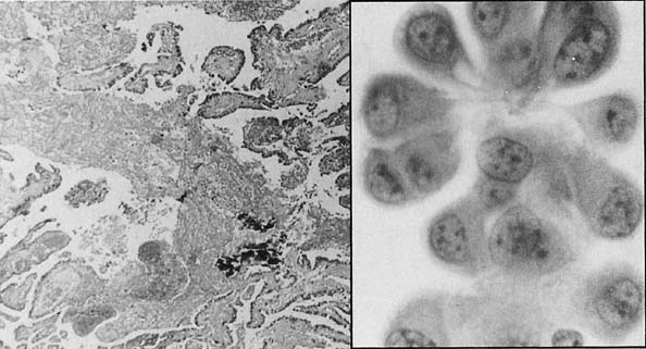
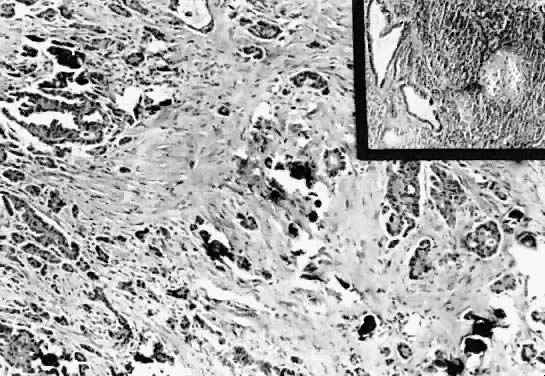
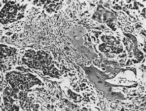
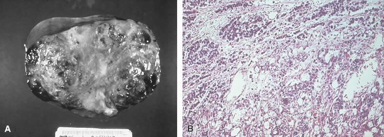
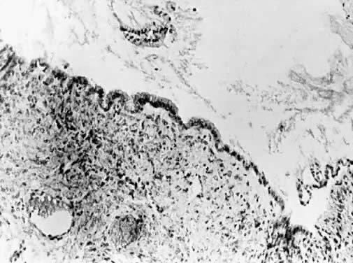

Fig. 14. Photomicrograph (medium power) of the human ovary. The germinal epithelium of the ovary rests upon the ovarian stroma. The primordial germ cells embedded in the stroma are in the cortex of the ovary. Volume 1, Chapter 2.


Fig. 15. Life history of the ovarian follicle. Starting with the primordial follicle, we have next the maturing follicle. Approximately 1 of 300 follicles fully (as shown on lower line), ruptures, and a corpus luteum. The other 299 become atretic. The final stage of both the artetic follicle and the corpus luteum is the corpus atreticum, with eventual reabsorption of this scar into the stroma of the ovary. (Frank.) Volume 1, Chapter 2.


Fig. 16. Photomicrograph (low power) of the graafian follicle of the human ovary. The eccentric location of the primordial germ cell is seen in the graafian follicle. Volume 1, Chapter 2.


Fig. 17. Photomicrograph (low power) of the corpus luteum of the human ovary. The developing corpus luteum with the central hemmorrhagic area is contiguous to a graafian follicle. Volume 1, Chapter 2.


Fig. 1. Photomicrograph of ovulation shows the expanded egg-cumulus complex leaving the follicle through the stigma. The remaining cells in the follicle wall ( i.e. granulosa, membrana and periantral, theca, the theca interna and externa) develop into the corpus luteum.(From Blandau RJ: Growth of the ovarian follicle and ovulation. Prog Gynecol 5:58, 1970.) Volume 5, Chapter 12


Fig. 3. Photomicrograph of an adult primate ovary. Follicular and luteal units are seen in the cortex and large blood vessels and nerves in the medulla. se, serous or surface epithelium; ta, tunica albuginea; pf, primary follicle; sf, secondary follicle; tf, tertiary follicle; gf, graafian follicle.(From Bloom W, Fawcett DW: A Textbook of Histology. Philadelphia: WB Saunders, 1975.) Volume 5, Chapter 12


Fig. 4. Electron micrograph of a human primordial follicle shows the flattened granulosa cells (GC), the oocyte with its germinal vesicle (GV) or nucleus, the Balbiani body (BB), with all the oocyte organelles gathered at one pole of the GV, and basal lamina (BL).(From Erickson GF: The ovary: Basic principles and concepts. In Felig P, Baxter JD, Frohman L (eds): Endocrinology and Metabolism. New York: McGraw-Hill, 1995.) Volume 5, Chapter 12


Fig. 13. A typical healthy secondary follicle contains a fully grown oocyte surrounded by the zona pellucida, five to eight layers of granulosa cells, a basal lamina, and developing theca tissue with numerous blood vessels.(From Bloom W, Fawcett DW: A Textbook of Histology. Philadelphia: WB Saunders, 1975, with permission from Arnold Ltd.) Volume 5, Chapter 12

Back to Top

Follicular Cyst

Fig. 1. Follicle cyst. Inner lining composed of granulosa cells with outer layer of luteinzed theca interna cells. (hematoxylin and eosin stain, x25) Volume 1, Chapter 17

Back to Top

Teratoma

Fig. 3. Benign cystic teratoma (dermoid cyst). Opened cyst containing hair and sebaceous material. Volume 1, Chapter 17


Infarction of a Dermoid Cyst (From Operational Obstetrics & Gynecology - 2nd Edition, The Health Care of Women in Military Settings, CAPT Michael John Hughey, MC, USNR, NAVMEDPUB 6300-2C, Bureau of Medicine and Surgery, Department of the Navy, 2300 E Street NW, Washington, D.C. 20372-5300, January 1, 2000. Original image courtesy Armed Forces Institute of Pathology)


Fig. 2. Benign cystic teratoma. Cyst lining composed of keratinizing squamous epithelium with sebaceous glands, smooth muscle, and sweat glands in the wall (hematoxylin and eosin stain, x60). Volume 1, Chapter 17


Fig. 28. Sectioned surface of dermoid cyst. Volume 4, Chapter 31


Fig. 29. Dermoid cyst with hair follicle ( left ), sebaceous glands, and sweat glands (magnification, ×80).(Serov SF, Scully RE, Sobin LJ: Histological Typing of Ovarian Tumours. Geneva, World Health Organization, 1973.) Volume 4, Chapter 31


Fig. 27. Mature teratoma (magnification, x50).(Serov SF, Scully RE, Sobin LH: Histological Typing of Ovarian Tumours. Geneva, World Health Organization, 1973.) Volume 4, Chapter 31


Fig. 30. Struma ovarii in wall of mucinous cystadenoma (magnification, ×80). The appearance is similar to that of a thyroid adenoma. Volume 4, Chapter 31


Fig. 26. Mature glial implants of immature teratoma on omentum (magnification, ×50).(Robboy SJ, Scully RE: Ovarian teratoma with glial implants on the peritoneum. Hum Pathol 1:643, 1970.) Volume 4, Chapter 31

Back to Top

Immature Teratoma

Fig. 2. Immature teratoma. A. An immature teratoma is encapsulated and grossly similar to its benign counterpart, the mature teratoma. The main difference is the immature teratoma is solid rather than cystic. B. An immature teratoma reveals a variegated appearance with areas of necrosis and cystic changes. C. An immature teratoma reveals mixtures of mature and immature tissues reminiscent of the developing stages of embryonic tissues. The most common pattern is seen here (e.g., an immature neural epithelium [neuroepithelial rosettes]). Volume 4, Chapter 34


Fig. 25. Sectioned surface of immature teratoma. Volume 4, Chapter 31


Fig. 24. Immature teratoma (magnification, ×120). Immature glial with central area of necrosis contains neuroepithelial rosettes.(Scully RE: Recent progress in ovarian cancer. Hum Pathol 1:73, 1970.) Volume 4, Chapter 31

Back to Top

Serous Cystoma

Fig. 4. Simple serous cystoma. Opened cyst demonstrating uniloculation with a smooth surface. Volume 1, Chapter 17

Back to Top

Mucinous Cystadenoma

70 pound mucinous cystadenoma (From Operational Obstetrics & Gynecology - 2nd Edition, The Health Care of Women in Military Settings, CAPT Michael John Hughey, MC, USNR, NAVMEDPUB 6300-2C, Bureau of Medicine and Surgery, Department of the Navy, 2300 E Street NW, Washington, D.C. 20372-5300, January 1, 2000. Original image courtesy Michael John Hughey, MD. All rights reserved.)


Fig. 5. Mucinous cystadenoma. Cyst lining composed of a single layer of columnar cells with basal nuclei and cystoplasmic vacuoles containing mucin (hematoxylin and eosin stain, x125). Volume 1, Chapter 17

Back to Top

Brenner Tumor

Fig. 6. Brenner tumor. Islands of uniform epithelial cells in dense fibrous stroma (hematoxylin and eosin stain, x125). Volume 1, Chapter 17


Fig. 14. Benign transitional cell (Brenner) tumor. Rounded nests of transitional-type epithelium are embedded in a fibrotic stroma resembling ovarian stroma. Volume 4, Chapter 30


Fig. 15. Benign transitional cell (Brenner) tumor. Ovoid nuclei with longitudinal grooves characterize the transitional cell type. Volume 4, Chapter 30

Back to Top

Fibroma

Fig. 7. Fibroma. Solid circumscribed tumor composed of firm, uniform white tissue. Volume 1, Chapter 17


Fig. 8. Ovarian fibroma. Spindle-shaped cells in a collagenous stroma (hematoxylin and eosin stain, x125). Volume 1, Chapter 17


Fig. 5. Sectioned surface of fibroma with areas of cystic degeneration.(Scully RE: Sex cord-stromal tumors. In Blaustein A [ed]: Pathology of the Female Genital Tract, pp 505–526. New York, Springer-Verlag, 1977.) Volume 4, Chapter 31


Fig. 6. Fibroma (magnification, ×325).(Serov SF, Scully RE, Sobin LH: Histological Typing of Ovarian Tumours. Geneva, World Health Organization, 1973.) Volume 4, Chapter 31

Back to Top

Thecoma

Fig. 7. Sectioned surface of thecoma within ovary with attached fallopian tube.(Scully RE: Sex cord-stromal tumors. In Blaustein A [ed]: Pathology of the Female Genital Tract. New York, Springer-Verlag, 1977.) Volume 4, Chapter 31


Fig. 8. Thecoma (magnification, ×130).(Morris JM, Scully RE: Endocrine Pathology of the Ovary. St. Louis, CV Mosby, 1958.) Volume 4, Chapter 31


Fig. 9. Luteinized thecoma (magnification, ×430). A nest of lutein cells is surrounded by fibroblasts. Volume 4, Chapter 31


Theca Lutein Cyst (From Operational Obstetrics & Gynecology - 2nd Edition, The Health Care of Women in Military Settings, CAPT Michael John Hughey, MC, USNR, NAVMEDPUB 6300-2C, Bureau of Medicine and Surgery, Department of the Navy, 2300 E Street NW, Washington, D.C. 20372-5300, January 1, 2000. Original image courtesy Armed Forces Institute of Pathology)

Back to Top

Sertoli-Stromal Cell Tumors (Androblastomas)

Fig. 10. Sertoli-Leydig cell tumor, well differentiated (magnification, ×100). (Morris JM, Scully RE: Endocrine Pathology of the Ovary. St. Louis, CV Mosby, 1958.) Volume 4, Chapter 31


Fig. 11. Sertoli-Leydig cell tumor of intermediate differentiation (magnification, ×240).(Morris JM, Scully RE: Endocrine Pathology of the Ovary. St. Louis, CV Mosby, 1958.) Volume 4, Chapter 31

Back to Top

Gonadoblastoma

Fig. 33. Gonadoblastoma with calcification (magnification, ×130).(Serov SF, Scully RE, Sobin LH: Histological Typing of Ovarian Tumours. Geneva, World Health Organization, 1973.) Volume 4, Chapter 31


Fig. 34. Gonadoblastoma with dysgerminoma (magnification, ×160).(Serov SF, Scully RE, Sobin LH: Histological Typing of Ovarian Tumours. Geneva, World Health Organization, 1973.) Volume 4, Chapter 31

Back to Top

Granulosa Cell Tumor

Fig. 7. Granulosa cell tumor. A. Tumor is usually solid but may be cystic. Primarily areas of a solid tumor are shown with areas of hemorrhage. B. Large, round and ovoid nuclei with a longitudinal groove arranged in patterns are referred to as Call-Exner bodies (microglandular pattern). C. Similar cells in a stromal matrix are arranged in cords and ribbons (trabecular pattern). D. A variation of cells shows some cellular atypia (sarcomatoid pattern). Volume 4, Chapter 34


Fig. 1. Sectioned surface of granulosa cell tumor with solid and cystic components.(Scully RE: Ovarian tumors with estrogenic manifestations. Contemp Ob Gyn 10:83, 1977.) Volume 4, Chapter 31


Fig. 2. Granulosa cell tumor, microfollicular pattern with Call-Exner bodies (magnification, ×290).(Scully RE, Morris JM: Functioning ovarian tumors. In Meigs JV, Sturgis SH [eds]: Progress in Gynecology, Vol 3. New York, Grune & Siralton, 1957, by permission.) Volume 4, Chapter 31


Fig. 3. Granulosa cell tumor, trabecular pattern (magnification, ×200).(Serov SF, Scully RE, Sobin LH: Histological Typing of Ovarian Tumours. Geneva, World Health Organization, 1973.) Volume 4, Chapter 31


Fig. 4. Granulosa cell tumor, diffuse pattern (magnification, ×200).(Scully RE: Ovarian tumors with estrogenic manifestations. Contemp Ob Gyn 10:83, 1977.) Volume 4, Chapter 31

Back to Top

Hilus Cell Tumor

Fig. 14. Sectioned surface of hilus cell tumor.(Scully RE: Sex cord-stromal tumors. In Blaustein A [ed]: Pathology of the Female Genital Tract. New York, Springer-Verlag, 1977.) Volume 4, Chapter 31


Fig. 15. Hilus cell tumor (magnification, ×320). Note large crystal of Reinke near center.(Scully RE: Androgenic lesions of the ovary. In Grady HG, Smith DE [eds]: The Ovary, International Academy of Pathology, Monograph no. 3. Baltimore, Williams & Wilkins, 1962.) Volume 4, Chapter 31

Back to Top

Dysgerminoma

Fig. 16. Sectioned surface of dysgerminoma. Note lobulated appearance and foci of caseation-like necrosis.(Scully RE: Recent progress in ovarian cancer. Hum Pathol 1:73, 1970.) Volume 4, Chapter 31


Fig. 17. Dysgerminoma with lymphocytes in stroma (magnification, ×300).(Scully RE: Germ cell tumors of the ovary and fallopian tube. In Meigs JV, Sturgis SH [eds]: Progress in Gynecology, Vol 4. New York, Grune & Stratton, 1963, by permission.) Volume 4, Chapter 31


Fig. 1. Dysgerminoma. A. In situ picture of a unilateral dysgerminoma shows it is bosselated and surrounded by a dense capsule. B. Grossly, a cut surface of this dysgerminoma reveals a yellow-tan, soft, homogeneous, and brainlike consistency. C. Histologic picture of a dysgerminoma shows cells are analogous to the undifferentiated germ cells of an embryonal gonad (i.e., well-defined clusters of cells separated by a fibrovascular system, infiltration [lymphocytes]). Volume 4, Chapter 34

Back to Top

Endodermal Sinus Tumor

Fig. 18. Endodermal sinus tumor, reticular pattern with Schiller-Duval body (magnification, ×300).(Scully RE: Germ cell tumors of the ovary and fallopian tube. In Meigs JV, Sturgis SH [eds]: Progress in Gynecology, Vol 4. New York, Grune & Stratton, 1963, by permission.) Volume 4, Chapter 31


Fig. 19. Endodermal sinus tumor with hyaline bodies (magnification, ×750).(Serov SF, Scully RE, Sobin LH: Histological Typing of Ovarian Tumours. Geneva, World Health Organization, 1973.) Volume 4, Chapter 31


Fig. 20. Endodermal sinus tumor, polyvesicular vitelline pattern (magnification, ×190). Secondary yolk sac at right is budding from larger primary yolk sac.(Serov SF, Scully RE, Sobin LH: Histological Typing of Ovarian Tumours. Geneva, World Health Organization, 1973.) Volume 4, Chapter 31


Fig. 21. Sectioned surface of yolk sac tumor.(Scully RE: Recent Progress in Ovarian Cancer. Hum Pathol 1:73, 1970.) Volume 4, Chapter 31


Fig. 3. Yolk sac (endodermal sinus) tumor. A. Yolk sac tumor is classically yellow-red-gray, soft, and friable with multiple cystic areas representing areas of hemorrhage and necrosis. B. A pattern is shown of pleomorphic, poorly differentiated cells with papillary clusters and a pseudopapillary projection with a vascular component within the center (Schiller-Duvall body). Volume 4, Chapter 34

Back to Top

Serous Cystadenoma of Low Malignant Potential

Fig. 2. External surface of a serous cystadenoma of low malignant potential showing a smooth, tense capsule with prominent veins and thickened fibrous areas.(Courtesy of Dr. Richard Stock, Naval Hospital, Portsmouth, VA.) Volume 4, Chapter 29


Fig. 3. Cystadenoma exhibiting lining consisting of cuboidal epithelium serous type ( top) and mucus-producing tall, columnar, endocervical-type epithelium with basally arranged nuclei ( bottom ). The mixture of epithelia frequently is found in serous or mucinous cystadenomas of the ovary (hematoxylin and eosin, 520). Volume 4, Chapter 29


Fig. 4. Serous cystadenoma of low malignant potential demonstrating an exuberant growth of papillary projections arising from the internal surface of the tumor. Bubbly serous fluid is present in the lumen.(Courtesy of Dr. Richard Stock.) Volume 4, Chapter 29


Fig. 5. External surface of an ovary showing a serous cystadenoma of low malignant potential exhibiting papillary growth. The lateral aspect of the uterus and the entire fallopian tube are seen clearly. The papillary growth does not represent invasion.(Courtesy of Dr. Richard Stock.) Volume 4, Chapter 29


Fig. 6. Photomicrograph of a serous cystadenoma of low malignant potential. The tumor is composed mainly of papillary structures of different sizes resting on stroma made up of fibrous tissue. Tufting is observed in the interpapillary space (100). Volume 4, Chapter 29


Fig. 7. Inner lining of a serous cystadenoma of low malignant potential showing papillations with epithelial pluristratification, hyperchromasia, and tufting; there is some degree of nuclear atypia (420). Volume 4, Chapter 29


Fig. 8. Serous cystadenoma of low malignant potential demonstrating papillae, stratification of epithelial lining, tufting, stromal edema with hyalinization, and focal acute inflammatory cell infiltrate. On the upper right-hand corner, a psammoma body is clearly seen (hematoxylin and eosin, 420). Volume 4, Chapter 29


Fig. 9. Photomicrograph of a serous tumor of low malignant potential showing a focus of microinvasion represented by small nests of tumor cells penetrating the tumor stroma near the epithelial-stromal interphase. The nests are surrounded by a small clear space that should be filled with serous fluid. There is no stromal necrosis or inflammation around the tumor nests (400). Volume 4, Chapter 29


Fig. 11. Omental invasive implants associated with a serous ovarian tumor of low malignant potential. The malignant histologic characteristics of the epithelium, its stromal invasion by single and papillary nests of cells, the inflammatory response, and the fibroblastic proliferation are evident (hematoxylin and eosin, 420). Volume 4, Chapter 29

Back to Top

Mucinous Cystadenoma of Low Malignant Potential

Fig. 16. Uterus with bilateral ovarian mucinous cystadenomas of low malignant potential showing multioculations and thick cyst walls.(Courtesy of Dr. Richard Stock.) Volume 4, Chapter 29


Fig. 17. Gross appearance of a mucinous cystadenoma of low malignant potential. The external surface of the tumor is irregular and multilobulated. The lobules are of different size and shape. The tumor is reddish brown. The lobules were fluctuant on palpation. Some areas of the tumor are firm. On cut section, multiple mucus-filled cysts are seen. Numerous small tan papillary structures are present in the inner surface of some of the cysts. Volume 4, Chapter 29


Fig. 18. Mucinous cystadenoma of low malignant potential, endocervical or müllerian type. Observe the marked papillation similar to that seen in serous tumors. Tufting and inflammatory exudate in the cyst lumen are seen (hematoxylin and eosin, 212). Inset, upper left. Papillation and inflammatory exudate. Inset, lower right. Epithelial atypia of gland-like spaces (420). Volume 4, Chapter 29


Fig. 19. Mucinous cystadenoma of low malignant potential, endocervical type, exhibiting a filigree pattern, pluristratification with atypia, stroma with mucinous material and inflammatory cell infiltration at the upper right-hand corner, and cellular debris with inflammatory cell exudate in the lumen (hematoxylin and eosin, 520). Volume 4, Chapter 29


Fig. 20. Papillations and tufting in a mucinous cystadenoma of low malignant potential, intestinal type. Spaces are lined with a mixture of goblet cells and tall, columnar intestinal-type epithelium. The stroma is edematous and exhibits fine capillaries. Areas with metaplastic changes are present on the left side and upper right-hand corner (hematoxylin and eosin, 212). Inset. Higher magnification illustrating goblet cells (hematoxylin and eosin, 420). Volume 4, Chapter 29


Fig. 21. Stroma of an ovarian mucinous cystadenoma of low malignant potential showing pools of mucus and atypical mucus-producing columnar epithelial cells of intestinal type in the stroma. Goblet cells are seen easily. The mucus pools are mostly acellular, although three hyperchromatic atypical nuclei are seen at the upper right-hand corner (hematoxylin and eosin, 420). Volume 4, Chapter 29

Back to Top

Endometrioid Tumor of Low Malignant Potential

Fig. 22. Photomicrograph of an endometrioid tumor of low malignant potential. The tumor is composed of endometrial-like glands simulating atypical complex hyperplasia. The glands are arranged in groups separated by dense fibrous stroma (100). Volume 4, Chapter 29


Fig. 23. Photomicrograph of an endometrioid tumor of low malignant potential. Glands similar to those observed in atypical complex, and atypical simple hyperplasia are observed. The glands are irregular. The epithelium form, in some areas, solid masses of atypical cells showing glands within glands. The stroma is formed by dense fibrous tissue. No stromal necrosis is observed (200). Volume 4, Chapter 29

Back to Top

Clear Cell Tumor of Low Malignant Potential

Fig. 24. Photomicrograph of a clear cell tumor of LMP showing tubular structures lined by low columnar epithelium. The cytoplasm in most epithelial cells is clear. Some nuclei are hyperchromatic. The intertubular stroma is dense (200). Volume 4, Chapter 29

Back to Top

Brenner Tumor of Low Malignant Potential

Fig. 25. Gross appearance an ovarian transitional cell tumor of low malignant potential (Brenner tumor of LMP). Notice irregular cystic spaces and two round-oval masses approximately 1.5 cm in diameter. These masses are solid. In the lower mass, a small cyst space is noted. The inner lining of the cystic spaces is shaggy. Volume 4, Chapter 29


Fig. 26. Papillations showing a thin, central fibrovascular core present in a proliferating Brenner tumor. The epithelial lining consists of 10 to 20 layers of transitional cell-type epithelium. The cellular atypia and pattern resemble those of transitional cell carcinoma (grades 1 to 2) of the urinary tract (hematoxylin and eosin, 400).(Courtesy of Dr. Laurence Roth.) Volume 4, Chapter 29


Fig. 27. Transitional cell-like epithelium of proliferating Brenner tumor showing atypical nuclear changes similar to those of transitional cell carcinoma of the urinary bladder (grades 1 to 2) (hematoxylin and eosin, 700).(Courtesy of Dr. Laurence Roth.) Volume 4, Chapter 29


Fig. 28. Section illustrating Brenner tumor of low malignant potential with histologic characteristics simulating a transitional carcinoma grade 3. Observe the number of layers, loss of polarity of nuclei, hyperchromasia, and abundant and abnormal mitosis (hematoxylin and eosin, 420). Volume 4, Chapter 29

Back to Top

Cystadenofibroma with Atypia

Fig. 31. Gross appearance of a cystic adenofibroma of LMP. The tumor involves part of the ovary. It is slightly lobulated and made up of cystic and solid structures. The tumor is firm and hard on cut section. It is yellowish gray. The cystic spaces show a smooth inner lining. Volume 4, Chapter 29


Fig. 29. Cystadenofibroma with atypia. The stroma is composed of dense, fibrous tissue and dilated cyst-like glands lined with cuboidal epithelial cells. Papillary areas at the top demonstrate dense, fibrous stroma lined with atypical epithelial cells (hematoxylin and eosin, 170). Volume 4, Chapter 29


Fig. 30. Close-up of cystadenofibroma with atypia showing dense, fibrous stroma and dilated cystic glands lined with cuboidal epithelial cells showing nuclear hyperchromasia (hematoxylin and eosin, 420). Volume 4, Chapter 29

Back to Top

Borderline Serous Tumor

Fig. 38. Left. Histologic section of a borderline serous tumor. (Hematoxylin and eosin, × 520). Right. A cluster of small monomorphic cells from the neoplasm.(Papanicolaou, × 900). Volume 1, Chapter 29


Fig. 1. Atypical proliferative serous tumor. Hierarchical branching pattern with detachment of cell clusters and without invasion characterizes this tumor, which is also referred to as serous borderline tumor. Volume 4, Chapter 30

Back to Top

Pseudomyxoma Peritonei

Fig. 9. Disseminated peritoneal adenomucinosis. In this form of pseudomyxoma peritonei, large pools of acellular mucin dissect through tissues. Occasional mucinous glands, seen at upper left, display minimal cytologic atypia. Volume 4, Chapter 30


Fig. 10. Peritoneal mucinous carcinomatosis. In this form of pseudomyxoma peritonei, angulated glands with moderate-to-severe nuclear atypia invade tissues. Volume 4, Chapter 30

Back to Top

Serous Adenocarcinoma

Fig. 39. Left. Histologic section of an invasive serous adenocarcinoma. (Hematoxylin and eosin, × 260). Right. A cluster of cells from the tumor, showing marked pleomorphism and nuclear atypia.(Papanicolaou, × 1200). Volume 1, Chapter 29

Fig. 6. Serous carcinoma. Markedly complex and stratified papillary proliferation with moderate-to-severe nuclear atypia. Volume 4, Chapter 30

Back to Top

Serous Psammocarcinoma

Fig. 12. Gross view of an ovarian psammocarcinoma. Multiple tumor nodules are noted. The largest one is yellowish. The cut surface has a gritty and solid consistency. Volume 1, Chapter 29


Fig. 13. Serous psammocarcinoma of the ovary. The section shows numerous psammoma bodies in the stroma, cyst lumen, and papillae (hematoxylin and eosin, 85). Volume 1, Chapter 29


Fig. 14. Photomicrograph of a psammocarcinoma to illustrate psammoma bodies in the papillary component of the tumor. Atypical cuboidal epithelial cells with prominent nucleoli line the papillae (200). Volume 1, Chapter 29


Fig. 15. Serous psammocarcinoma. The epithelial component diffusely infiltrates the desmoplastic stroma. Few psammoma bodies are present (hematoxylin and eosin, 212). Inset. Section of a periaortic lymph node involved by tumor. Observe the atypical glandular epithelium. The glands in the center exhibit atypical papillary structures (hematoxylin and eosin, 212). Volume 1, Chapter 29

Back to Top

Endometrioid Carcinoma

Fig. 11. Endometrioid carcinoma. Crowded, back-to-back glands lined by tall columnar epithelium resembling endometrial epithelium invade the stroma. Volume 4, Chapter 30

Back to Top

Clear Cell Carcinoma

Fig. 12. Clear cell carcinoma. Sheets of cells with clear cytoplasm and mild-to-moderate nuclear atypia are characteristic. Volume 4, Chapter 30


Fig. 13. Clear cell carcinoma. Glands lined by epithelium displaying severe nuclear atypia and a hobnail pattern characterized by nuclei protruding into the lumena of the glands are present. In this pattern, clear cytoplasm is less conspicuous. Volume 4, Chapter 30

Back to Top

Embryonal Carcinoma

Fig. 22. Embryonal carcinoma with syncytiotrophoblast cells (magnification, ×34). AFIP Neg. 75-14208.(Kurman RJ, Norris HJ: Embryonal carcinoma of the ovary. A clinicopathologic entity distinct from endodermal sinus tumor resembling embryonal carcinoma of the adult testis. Cancer 38:2420, 1976.) Volume 4, Chapter 31


Fig. 4. Embryonal carcinoma. Histologic pattern resembles glomeruli, vacuolated cytoplasm, and glandlike spaces. Volume 4, Chapter 34

Back to Top

Choriocarcinoma

Fig. 23. Choriocarcinoma (magnification, ×350).(Serov SF, Scully RE, Sobin LH: Histological Typing of Ovarian Tumours. Geneva, World Health Organization, 1973.) Volume 4, Chapter 31


Fig. 5. Choriocarcinoma. Microscopic view of nongestational choriocarcinoma reveals cells resembling syncytiotrophoblasts and cytotrophoblasts. There is usually extensive hemorrhage and necrosis. Volume 4, Chapter 34

Back to Top

Mixed Germ Cell Tumor

Fig. 6. Mixed germ cell tumor. A. A nondescript solid tumor has no defining characteristics. B. Dysgerminoma elements are associated with a yolk sac carcinoma. Volume 4, Chapter 34

Back to Top

Krukenberg Tumor

Krukenberg Tumor (From Operational Obstetrics & Gynecology - 2nd Edition, The Health Care of Women in Military Settings, CAPT Michael John Hughey, MC, USNR, NAVMEDPUB 6300-2C, Bureau of Medicine and Surgery, Department of the Navy, 2300 E Street NW, Washington, D.C. 20372-5300, January 1, 2000. Original image courtesy Armed Forces Institute of Pathology)