Chapter 1
Clinical Anatomy of the Vulva, Vagina, Lower Pelvis, and Perineum
David H. Nichols and Paul S. Milley
Main Menu  

Search

David H. Nichols, MD
Visiting Professor of Obstetrics, Gynecology, and Reproductive Biology, Harvard University School of Medicine; Chief of Pelvic Reconstructive Surgery, Massachusetts General Hospital; Professor and Past Chairman of Obstetrics and Gynecology, Brown University School of Medicine, Providence, Rhode Island (Vol 1, Chap 1)

Paul S. Milley, MD
Clinical Assistant Professor of Anatomy, State University of New York at Buffalo School of Medicine, Buffalo, New York (Vol 1, Chap 1)

INTRODUCTION
GENERAL CONCEPTS OF PELVIC CONNECTIVE TISSUE HISTOLOGY
VAGINA
PERINEAL BODY
UROGENITAL DIAPHRAGM AND PUBOURETHRAL LIGAMENTS
THE ARCUS TENDINEI
PELVIC DIAPHRAGM
CARDINAL AND UTEROSACRAL LIGAMENTS
THE BROAD LIGAMENTS
CONNECTIVE TISSUE PLANES AND SPACES OF THE PELVIS
REFERENCES

INTRODUCTION

Clinical anatomy is concerned with the anatomic interrelationships between various structures of the living human organism. These relationships in the pelvis and their physiologic alterations along with the degree of independent function of each organ system are the concern of the gynecologist. The anatomic interrelationships in the living differ significantly from those in the cadaver, the study of which forms the basis for descriptions in standard textbooks of gross anatomy. Since the earliest days of medicine, dissections have been performed on, texts have been written about, and anatomy has been studied from elderly, often debilitated, and malnourished female cadavers. The anatomic interrelationships in such bodies are quite different from those found in the healthy, living, well-nourished, younger female. Furthermore, the standard anatomy textbook gives extensive description of only a small number of dissections and bases broad generalizations on this small number. There is no recognition of the large amount of variation that occurs normally and frequently between individuals and at different times of life.

The anatomic relationships of tissues and organs of an anesthetized patient are not the same as those of the patient who is wide awake; this is because of muscle paralysis from anesthesia, resting or baseline underdistention of the various organs, and the change in statics caused by position. The horizontal position of the immobile surgical patient provides statics different from those of the vertical and active patient, whose pelvic organs are in varying stages of function and distention.

Vaginal reconstructive surgery is concerned with the return of abnormal organ relationships to a usual or normal state. There is no one site or degree of damage that must be repaired or restored; there are many, and they occur in various combinations at various times of life, from different etiologic factors, in varying degrees, and with varying degrees of symptomatology and disability.

For years there has often been heated discussion as to whether the more important factor concerned with vaginal position within the pelvis is that of suspension from above (cardinal ligament complex), a view championed by Fothergill, or of support from below (levator ani-pelvic diaphragm), as emphasized by Paramore and Halban. Mengert's classic contribution in this area was to report an experiment whereby a tenaculum was applied to the cadaver cervix, a cord was attached to the tenaculum and run through a fixed pulley, and a 1-kg weight was attached to the opposite end of the cord.1 One by one, starting at the top of the fundus, the lateral supports of the uterus and vagina were cut until finally prolapse of the uterus developed. It was only when the paravaginal tissues had been cut that prolapse developed. “Marked descent of the uterus amounting to actual prolapse never occurred so long as any part of the upper two thirds of the paravaginal or lower two thirds of the parametrial tissues were intact.”1

This experiment convincingly emphasized the importance of the suspensory apparatus. Bonney strongly defended the position that both points of view were correct, that is, the vagina is both suspended from above and supported from below, and that each system could be damaged either singly or together, with the type of resulting genital prolapse reflecting the site or sites of primary damage. It is important for the gynecologic surgeon to recognize the primary site of damage and its etiology; therefore, appropriate steps can be taken in surgical reconstruction to minimize the chance of postoperative recurrence of the genital prolapse. Damage to the suspensory system can give rise to inversion of the upper vagina, often with elongation of the cervix and cul-de-sac hernia; damage to the lower supporting system is more likely to be associated with eversion of the lower vagina, including cyctocele and rectocele.

It is evident that there are at least five different anatomic systems responsible for varying degrees of support of the birth canal. These can be injured or damaged separately or in various combinations, but they must be individually recognized and identified if restorative surgery is to achieve its stated goal. The anatomic support systems considered in greater detail include the perineum, including the perineal body; the urogenital diaphragm, including the pubourethral ligaments; the pelvic diaphragm, particularly the pubococcygeus component and the levator plate; the cardinal and uterosacral ligament complex; and the broad ligaments, including the smooth muscle components and the round ligaments.

Although each of the above is a separate anatomic unit, they are often interrelated, and additional components may exert synergistic, supportive, or even sphincterlike action, for example, the intact bulbocavernosi muscles contracting in concert with the pubococcygei muscles exert an almost sphincterlike effect on the vaginal outlet. It is uncommon for any of these anatomic units to be individually defective, other than by congenital anomaly. With this exception, damage to these individual units may be either primary or secondary, generally in combination. The combinations are many and must be recognized by the responsible surgeon if correct diagnosis and appropriate surgical remedy are to achieve the desired goal. Equally important is the recognition of active etiologic agents to which corrective attention can be drawn.

Back to Top
GENERAL CONCEPTS OF PELVIC CONNECTIVE TISSUE HISTOLOGY

Before evaluating the specific structures concerned with the constituent and supporting tissues of the female pelvis, one must bear in mind the highly specialized purposes for which each component has been designed. The organs of the pelvis must be capable of individual distention to certain maximal limits in the course of performance of their natural duties, and yet they must be endowed with the ability to return to their original state. The components contain admixtures of various amounts of smooth muscle, striated muscle, elastic tissue, and collagen. These act in concert, reinforcing one another but differing in certain specific functions and capability.

Smooth muscle fibers are in a constant state of activity. They help to maintain tone, respond rapidly to stresses applied to them, and are mediated through the autonomic nervous sytem (involuntarily), spinal reflex arcs, and chemical or mechanical stimuli. Smooth muscle also shows rhythmic contractility. The number of cells present within given tissues appears likely to be constant throughout the mature age of an individual, that is, it does not regress with age.

Striated muscle also responds rapidly to stress; it aids in the maintenance of tone and equilibrium, but is under voluntary control, and lacks inherent rhythm.

Elastic tissue is made up of fibers constructed in irregular networks especially well developed in tissues usually subject to stress. These fibers respond to stress with stretching, but they also resist stretching by a natural tendency to return to their original state, like a rubber band. They quantitatively decrease with age, but the extent to which this is hormone related and reversible is not known. For that matter, the actual histogenesis of these fibers is unknown, although it is assumed that they are produced by fibroblastic cells. The decrease in their number with aging probably partially accounts for the differences in composition and recurrence rate of cystoceles in women long past menopause in contrast to those still in the reproductive years.

Collagen fibers are also arranged in interlacing meshworks but, in contrast to elastic tissue fibers, are not stretchable. They are, however, flexible, permitting movement in areas in which stretching is not intended, like a piece of string or the ropes on a swing.

Back to Top
VAGINA

The vagina is a fibromuscular tube, the walls of which are normally in apposition in the relaxed state; it is H-shaped in its central portion, the side walls being suspended by their attachment to the paravaginal lateral connective tissue from which they receive their blood supply. It is lined by a stratified squamous epithelium thrown up into rugal folds, giving the epithelium accordianlike distensibility without laceration. The stratified squamous epithelium is rich in glycogen during the reproductive years. A dense, thin layer of elastic fibers is found immediately beneath the epithelium. Beneath this is a well-developed fibromuscular layer. Jacoby has described a muscular meshwork of smooth muscle fibers predominantly oriented in a longitudinal direction in the innermost component but arranged circularly toward the periphery. The fibrous capsule external to this muscular coat is rich in elastic fibers and large venous plexuses. The vagina is attached to the lateral pelvic wall by condensations of connective tissue and smooth muscle intimately adherent to the adventitia of the vaginal blood vessels. This tends to fix it in position from side to side, and the muscle elements supply a certain amount of tone, permitting it to adapt to changes in intravaginal and extravaginal pressure.

There is a large amount of elastic tissue mingling with the fibromuscular connective tissue elements of the vaginal capsule. These are structurally designed to permit distensibility with return to the previous state. In the midline, the vagina is permitted a great deal of freedom in distensibility from both bladder and rectum by the relatively avascular vesicovaginal space and rectovaginal spaces, which permit these organs to expand, contract, and slide somewhat independently of one another; thus, each interferes minimally with the function of the other. The rectovaginal septum is fused with the posterior vaginal wall as the anterior lining of the rectovaginal space. Since the vagina is a distensible organ, its depth is best measured when it is in a relaxed or resting state. The posterior vaginal wall is approximately 10 cm long. Since the cervix is incorporated in the anterior vaginal wall, the length of the anterior vaginal wall plus cervix approximates the length of the posterior wall. The connective tissue adventitia of the vagina is continuous with that of the cervix. The vagina is largest in its middle and upper thirds. The connective tissue lateral to the lower third is attached to fibers of the pubococcygeal muscle (fibers of Luschka) and to fibers fixing it to the urogenital diaphragm.2 Luschka described the connection as follows: “In the female the fibers originating from the upper pubic ramus pass alongside the vagina and are connected with it through strong connective tissue but do not end in the vagina.”

Blood Vessels

In addition to the vaginal blood supply received from branches of the internal pudendal artery, diffuse anastomoses form between these and branches of the uterine, inferior vesical, and vaginal arteries. The confluence of these anastomotic branches forms longitudinal azygos vaginal arteries in the midline of the anterior or posterior vaginal walls, or both, according to Smout and co-workers3 and Quinby.4

A right and left vaginal artery, or occasionally two, arise (independently in most instances) from each internal iliac artery slightly cephalad and posterior to the origin of each uterine and inferior vesical artery. Occasionally, the vaginal artery arises as a division of a short common trunk with the uterine artery. This branching, however, occurs at the lateral extremity of each cardinal ligament and has great clinical significance. Alarming arterial hemorrhage may thus follow laceration or surgical trauma to the vagina, especially in the vault of the vagina, even though the uterine artery has been securely ligated. Occasional postoperative arterial vaginal hemorrhage coexistent with intact ligation of the uterine artery may thus require separate isolation and ligation of the vaginal artery or, failing this, hypogastric or internal iliac ligation.

Normal Vaginal Depth and Axis and the Supports of the Vagina

The vagina in the cadaver is usually depicted as an almost straight hollow tube extending vertically upward toward the sacral promontory. In the living healthy female, however, the upper vaginal axis lies in an almost horizontal plane when the patient is in a standing position. Radiographic colpography has demonstrated a distinct, superiorly convex, perineal curve in the lower vagina (Fig. 1).5 The upper vagina lies on the rectum, which, in turn, lies on and parallel to the levator plate. It is this almost horizontal position of the supporting levator plate that accounts for a similar axis to the vagina. The levator plate is formed by the fusion of the levator ani muscles posterior to the rectum, from just behind the levator hiatus to their coccygeal insertion (Fig. 2). The rectum, vagina, and urethra pass through the levator hiatus, and if the levator ani muscle is defective, the inclination of the plate will be downward and the hiatus will sag. Although the cervix and upper vagina have considerable mobility, they are more or less anchored in position over the levator plate by the cardinal ligaments. The length and flexibility of these ligaments normally permit the cervix and upper vagina to be moved in any direction over the rectum on the levator plate but not anterior to the margin of the genital hiatus.

Fig. 1. A and B. Normal vaginal depth and axis. Colpogram of healthy 25-year-old nulligravida. The vaginal walls are painted with barium paste. The patient is standing at rest (A). The position of the symphysis is outlined to the left. The effect of straining by a Valsalva maneuver is shown (B). The perineal curve of the lower vagina is unchanged, but the horizontal axis of the upper vagina is accentuated with straining.(Nichols DH, Milley PS, Randall CL: Significance of restoration of normal vaginal depth and axis. Obstet Gynecol 36:251, 1970)

Fig. 2. The vaginal axis of the living human female. Notice the almost horizontal upper vagina and rectum lying on and parallel to the levator plate. The latter is formed by fusion of the pubococcygeal muscles (A) posterior to the rectum. The anterior limit of the point of fusion is the margin of the genital hiatus, immediately posterior to the rectum.

The vagina is maintained in depth and axis by different anatomic supports at different vaginal depths. The lower third is supported predominantly by connections between fibers received from the pelvic diaphragm and the urogenital diaphragm. The most posterior portion of the lower third is further attached to the perineal body and indirectly receives contributions from the various other muscles and connective tissue thickenings to which the perineal body itself is attached. The middle third of the vagina receives some contribution from lateral fusion with a lesser number of fibers from the pelvic diaphragm, but even more lateral support is obtained from the lateral and most inferior portion of the cardinal ligaments, which carry the main vaginal blood vessels both to and from their origin from the hypogastric sheaths. The upper third of the vagina receives almost no significant support from direct attachment to the fibers of the levator ani or pelvic diaphragm, but in fact rests on the rectum, which in turn rests on the fused pubococcygei of the levator plate. The upper vagina and the cervix together are maintained in position over the levator plate by their lateral attachments to the upper cardinal ligaments.6

Back to Top
PERINEAL BODY

The perineal body is a fibromuscular elastic structure found in the midline between the rectum and the vagina on a line between the ischial tuberosities; it contains much elastic tissue. It is subject to individual variation in tone, thickness, and composition. It is somewhat like the hub of a wheel into which various muscles—the superficial and deep transverse muscles of the perineum, the bulbocavernous muscle, the sphincter ani externus, and some fibers of the levator ani—are inserted like spokes. It is bounded anteriorly by the vagina and posteriorly by the rectum. Studdiford7 found considerable smooth muscles in the perineal body serving as a distensible attachment to the levator ani and to the vagina. He felt this distensibility was an important function which, when lost through unrepaired perineal laceration, gave rise to rectocele and posterior vaginal eversion.

The apex of the perineal body is continuous with the rectovaginal septum (the fascia of Denonvilliers), as shown in Fig. 3. When this attachment is avulsed, the weakness thus created favors the formation of a low or mid vaginal rectocele. The apex of the perineal body must be reattached to the underside of the posterior vaginal wall and rectovaginal septum by separate stitches to lessen the chance of recurrence of the rectocele.

Fig. 3. Sagittal section shows the relationship between the rectovaginal septum (RVS) as it blends with the superior border of the perineal body (PB). (Nichols DH, Randall CL: Vaginal Surgery, 3rd ed, p 42. Baltimore, Williams & Wilkins, 1989, with permission)

Ranney8 pointed out that the perineal body is not a keystone, since its wide base points down, not up, and that such a keystone, by its very nature, would fall down if pressure were applied to it.

Blood Vessels of the Female Perineum

The blood supply to the perineal structures is derived from the branches of the internal pudendal artery, which arises from the anterior trunk of the internal iliac, leaves the pelvic cavity through the greater sacrosciatic foramen, passes around the ischial spine, and enters the perineum by means of the lesser sacrosciatic foramen. As it ascends along the pubic ramus, it pierces the posterior layer of the urogenital diaphragm, travels for a short distance within the diaphragm and perforates the anterior layer, and gives off its terminal branches—the artery to the bulbocavernous muscle and the dorsal artery of the clitoris. The branches given off by this large vessel within the perineum include the external or inferior hemorrhoidal arteries given off as the vessel rises anterior to the ischial tuberosity. The external hemorrhoidal arteries run across the ischiorectal fossa and are distributed to the anal sphincter and levator ani muscles. They are the chief sources of hemorrhage from all superficial wounds of the anus or ischiorectal fossa. These vessels have accompanying veins that empty into the pudendal veins.

The superficial perineal or vulvar artery is given off anterior to the preceding branch. It is distributed to the vulva, with branches to the muscles, and is a source of arterial hemorrhage in wounds of the vulva.

The transverse perineal artery is somewhat smaller, supplies the cutaneous surface of the perineum, and is therefore a source of hemorrhage from laceration of the perineal body. The fourth branch is the artery of the bulb, a vessel of considerable diameter but of short length. It sends branches to the bulbocavernous muscle.

The terminal branches of the internal pudendal artery, the artery of the corpus cavernosum, and the dorsal artery of the clitoris are the supplying vessels of the erectile tissue of the clitoris. When the clitoris is amputated, the two dorsal arteries may require ligation. Bleeding from the vessels of the corpora cavernosa can usually be controlled by pressure, since the trabeculae favor coagulation of the blood.

The veins of the perineum are valveless and have free anastomosis with the large intrapelvic venous plexuses. This situation permits alarming hemorrhage from wounds of the vulva and vagina and the possibility of massive hematomas.8

Special properties of pelvic and perineal blood vessels include the following:

  1. Although there are many large venous networks within the pelvis that are capable of considerable venous distention, these veins are almost entirely without valves.
  2. Abundant smooth muscle fibers associated with adventitia of pelvic blood vessels probably account for at least part of the impressive quantity of smooth muscle found in the extraperitoneal connective tissue of the pelvis.
  3. The warmth and heat of tissues undergoing erection (clitoris, bulbocavernous muscle) demonstrate that most of the blood involved in the erectile process comes in fact from arteriolar dilation and that the venous congestion is probably a secondary phenomenon.

Back to Top
UROGENITAL DIAPHRAGM AND PUBOURETHRAL LIGAMENTS

Milley and Nichols9 studied the connective tissue supports of the urethra and confirmed the observations of Zacharin10 that the urethra is suspended from the pubic bone for most of its length by arched, bilaterally symmetric, anterior, posterior, and intermediate pubourethral ligaments. Our studies further showed, as was suggested by Curtis and associates,11 that the anterior and posterior ligaments are formed by reflections of the inferior and superior fascial layers of the urogenital diaphragm (Fig. 3). The intermediate ligament represents a fusion of these fascial layers. No transverse perineal ligament was found in any of our examinations, suggesting that this term, in the female, is a misnomer.

Histologic section showed the pubourethral ligaments to consist of dense collagen, both smooth and striated muscle, and elastic fibers. (The striated muscle may represent a continuation of the pubourethral muscle.) When studied by electron microscopy and neurohistochemistry, the smooth muscle bundles in this tissue are associated with numerous nerve fibers. The fine structure and enzyme content of these tissues were similar to those believed to represent cholinergic autonomic nerve tissue (acetylcholinesterase-positive). The term ligament is therefore a misnomer, because these structures contain contractile elements under neural control.12

The remainder of the urogenital diaphragm is sandwichlike, composed of superior and inferior fascial layers separated from one another by a layer of striated muscle (the deep transverse perineal muscle, the sphincter of the membranous urethra, or the sphincter vaginae). There is substantial evidence that the so-called sphincter of the membranous urethra has very minimal, if any, actual sphincter function.13 In general, sphincters of the body under voluntary control are formed by concentric layers of striated muscle, and the sphincter of the membranous urethra does not have such a morphologic arrangement of its fibers. More significantly, it has been demonstrated that sectioning the nerve supply to this group of muscle bundles has essentially no effect on the voluntary control of micturition.

The diaphragm runs between the inner surfaces of the ischiopubic rami and is pierced in the midline by the urethra and vagina; by attachment to these structures, it assists in holding them in place. The posterior fibers of the urogenital diaphragm are fixed to the perineal body. When one is in the standing position, the urogenital diaphragm is almost horizontal in sagittal section; for this reason its fixation to the perineal body contributes to the support of the urethra and vesicourethral junction, lessening the tendency of these structures to rotate around the attachment of the pubourethral ligament to the pubis. The superficial transverse muscle of the perineum, and the ischiocavernous and bulbocavernous muscles are located superficial to the urogenital diaphragm deep within the soft vulvovaginal tissues; they appear to be considerably less important in urogenital support.

Back to Top
THE ARCUS TENDINEI

There are two arcus tendinei on each side of the pelvis. The arcus tendineus of the levator ani runs from the back of the pubis to the ischial spine (Fig. 4). Medial to this is the arcus tendineus of the endopelvic connective tissue. Although there is individual variation in the distance between these two arci at their origin and lateral extent, they come together at the ischial spine. There is a connective tissue bundle running between the anterior vaginal sulcus and the arcus tendineus (Fig. 5 and Fig. 6). The urethra is held in place by two systems. It is suspended by the urogenital diaphragm and its attachment to the pubis, and supported by the connective tissue attachment between the anterior sulcus and the arcus tendineus (Fig. 7). A hypermobile urethra may be surgically remedied either by suspension or by support or by both, depending on the site of primary damage that permitted the hypermobility.

Fig. 4. Drawing showing the normal relationship between the vagina and the arcus tendinei. The arcus tendinei run from the back of the pubis to the ischial spine on each side of the pelvis.(Nichols DH, Randall CL: Vaginal Surgery, 3rd ed, p 18. Baltimore, Williams & Wilkins, 1989, with permission)

Fig. 5. Fortuitous section through the urethra (left) and vagina (below) shows some of their distinct fibromuscular support. The ventral support of the anterior sulcus by its attachments to the arcus tendineus and cardinal ligament are shown on the right.(Nichols DH, Randall CL: Vaginal Surgery, 3rd ed, p 19. Baltimore, Williams & Wilkins, 1989, with permission)

Fig. 6. Photomicrograph of cross section of pelvis of a 33-year-old cadaver is perpendicular to the intramural urethra. Coarse detrusor fibers of the bladder (D) surround the urethra, and the outer circular smooth muscle of the urethra (CMU) is seen surrounding the internal longitudinal smooth muscle layer (LMU); note the attachment of the anterior sulcus of the vaginal wall (VW) to the arcus tendineus (AT) which overlies the levator ani (LA). (DeLancey JOL: Correlative study of paraurethral anatomy. Obstet Gynecol 68:91, 1986. Courtesy of Dr. Thomas M. Oelrich, with permission of the American College of Obstetricians and Gynecologists)

Fig. 7. The urethra is both suspended ( central arrows show the attachments and pull of the pubourethral “ligament” portions of the urogenital diaphragm) and supported ( lateral arrows indicate the attachments of the vagina by intermediate connective tissue to the arcus tendineus).(Nichols DH, Randall CL: Vaginal Surgery, 3rd ed, p 21. Baltimore, Williams & Wilkins, 1989, with permission)

Back to Top
PELVIC DIAPHRAGM

The pelvic diaphragm is composed of the levator ani muscle and its superior and inferior fascial covering. This muscle functioned primarily as a tail-wagger in pronograde, four-legged animals. The assumption of the upright posture by man was accompanied by a loss of the tail as a functional appendage and the appropriation of this muscle for an entirely different purpose, that is, the support of the pelvic viscera; in the erect posture the viscera have lost their previous inferior support by the pubis. The comparative anatomy of this evolutionary process is fascinating, and for further study the reader is referred to the works of Power,14 Smout and co-workers,3 and Thompson.15

The levator ani muscle, acting in reciprocal concert with the muscles of the abdominal wall, has assumed much of the responsibility not only for support of both pelvic and abdominal contents but also for the maintenance of equilibrium of intra-abdominal pressure. Long ago Dickinson16 wrote:

I venture to affirm that there is no considerable muscle in the body the form and function of which are more difficult to understand than those of the levator ani, and about which such nebulous impressions prevail…. The muscle sling, attached to the pubis in front, encircles like a collar the rectum and vagina. Its action in women is to drag the lower end of the vagina and rectum forward, level to the symphysis.

Sturmdorf17 summarized its function as follows:

The levator ani diminishes the force of intra-abdominal pressure upon the pelvic contents by deflecting the direction of that pressure, augments the resistance to the pressure by closing the uterovaginal angle, and obstructs the pelvic outlet against the pressure by compressing the vaginal canal. It is the tensor of the pelvic fascia, the antagonist of the diaphragm and the abdominal muscles, contracting when these opposing muscles contract and relaxing when they relax. When intact, it maintains the equilibrium of the pelvic organs; when its integrity is impaired, equilibrium is disturbed.

Anatomy of the Levator Ani Muscle

The levator ani is composed of three general portions named according to the origin and insertion of each. The medial and anterior division is the pubococcygeal muscle, a somewhat V- or U-shaped sling that takes its origin from the back of the pubis on each side approximately 1.5 cm from the center of the symphysis. This portion is of the greatest importance to the gynecologist; the muscle is usually thicker along this medial margin than are the other two major divisions. The bellies of this 1- to 2-cm thick muscle sweep down and posteriorly along the sides of the urethra, the vagina, and then the rectum to insert into a fused median plate that runs from the tissue posterior to the rectum to the coccyx, the so-called levator plate. The most medial fibers form a loop behind the rectum. This U-shaped sling is called the puborectalis muscle.

Some fascial and muscular fibers from the most anterior and medial portions intermingle with those at the sides of the urethra (pubourethral muscle), lower vagina (pubovaginal muscle), and perineal body, but some stronger bundles attach to the posterior lateral sides of the rectum (puborectal muscle), and some fibers attach to the external sphincter ani. The thickness of this muscle is quite variable; it is stronger and thicker in women accustomed to heavy labor and athletic activity.

The intermediate portion of the levator ani has been called the iliococcygeal muscle; it is somewhat thinner and flatter than the pubococcygeus and measures generally 0.5 to 1 cm in thickness. It takes its origin from the surface of the fascia of the obturator internus muscle along a line running from the posterior pubis to the ischial spine, the so-called white line or the tendinous arch of the levator ani muscle. It is inserted into the lateral margin of the coccyx and lower sacrum.

The last major division, and the most posterior, is called the coccygeal (ischiococcygeal) muscle; it takes its origin from the ischial spine and inserts on the lateral margin of the coccyx and lower sacrum. It follows the course of the sacrospinous ligament; the muscle is in fact found on the superior aspect of this strong ligament.

Physiology of the Levator Ani Muscle

The levator ani is a most unique and highly specialized voluntary muscle. Its various components including the puborectalis muscle are innervated by the pudendal nerve on each side, which supplies the external anal sphincter as well, hence these muscles tend to function in concert. Different constituent parts of the levator ani muscle perform different functions according to their anatomic location.18 This may be demonstrated in the arrangement and composition of fibers of the periurethral parts in contrast to those of the perianal parts, which may be reflected by differences in the respective electromyography from each area. These muscles are in a constant state of tone, which mediates the control of rectal continence. Neuromuscular pressure receptors within the striated muscular content of the levator ani are responsible for mediating this tone, and they apparently communicate with the central nervous system by way of the pudendal nerve on each side of the body. Congenital or acquired pathology of the pudendal nerve can alter the efficiency of its work, and thus influence the ability and efficiency of these neuromuscular receptors to maintain this responsive muscular tone. Acquired damage may result from stretching of the pelvic floor during childbirth or the chronic habit of excessive straining at stool. Similarly there may be congenital malformation affecting the pudendal nerves, most frequently from spina bifida.19 Damage to these neuromuscular receptors is not only slowly progressive over the years, but is virtually irreversible.20 Abnormalities in positions of the muscles can be corrected surgically, but abnormalities of innervation, either congenital or acquired, are refractory to treatment. Prevention of neuropathy by skillful management of labor, and the elimination of the stretching coincident with chronic efforts at bearing down to relieve long-standing constipation will do much to prevent this pathology and defective physiology.

Although the levator ani, especially the iliococcygeal portion, is usually thought of as having a concave shape, pressure from fat within the ischiorectal fossa pushes the soft, yielding belly of the muscle upward and medially to a pouting convex shape when pressure is applied to the ischiorectal fat from below, as by sitting or reclining (Fig. 8).4

Fig. 8. Cross section of female pelvis through lower midportion of vagina. Note the convex configuration of the pubococygeus (PC). The rectovaginal space (RVS), as well as the position of the rectovaginal septum (RVSe), is indicated between the rectum and vagina. The blood vessels (bv) in connective tissue lateral to the vagina are shown. These tend to give the vagina its H-shape configuration. The fibers of Luschka (FL) are shown as they attach the paravaginal connective tissue to the sheath of the pubococcygeus.

In the usual laboratory anatomic dissection after death, the perineum is dissected first and the ischiorectal fat is removed. With the inferior support of the pelvic diaphragm removed, the diaphragm (minus the tonus of the living muscle) becomes concave in appearance. When loss of ischiorectal fat occurs in the living person as part of massive weight loss, the undersupport of the pelvic diaphragm is similarly removed, predisposing toward tipping of the levator plate and subsequent prolapse.

Halban and Tandler21 defined the importance of the levator plate in providing pelvic support, but although cadaver dissection clearly demonstrated the plate, the total absence of tone and the effect of rectal distention and other factors made demonstration of the function of this plate difficult. Berglas and Rubin22 were able to prove the existence of this plate in the horizontal state in the normal patient and to demonstrate how pathologic tipping from defects in this plate contributed to various degrees of genital prolapse. Their rather astounding work, functional anatomy in vivo, was accomplished by the direct injection of radiopaque contrast medium into the levator muscle and plate with simultaneous placement of contrast material in the vagina, cervix, and uterus. Radiographs were taken of the patients at rest and while straining; the results clearly showed the integrity and horizontal position of the normal plate even while straining and the tipping of the abnormal plate coincident with genital prolapse, elegantly illustrating the importance of the pelvic diaphragm in maintaining the support of the pelvic viscera in the female.

Influence of the Pubococcygeal Muscle on the Mechanism of Voiding

The function of the pubococcygeal muscle in the normal voiding mechanism is beautifully described by Muellner.23 This concept emphasizes the importance of voluntary skeletal muscle in the mechanism of continence.

Before urination begins, the diaphragm and the muscles of the abdominal wall contract, the intra-abdominal pressure rises, and the pubococcygei muscles relax. As the pubococcygei relax, the neck of the bladder moves downward. This downward movement activates or initiates contraction of the detrusor muscle. At the same time, the longitudinal fibers of the urethra, which are continuous with those of the detrusor, contract and shorten the urethra, thereby opening and widening the internal urethral orifice. Urine is then expelled from the bladder.

At the conclusion of voiding, a contraction of the pubococcygei raises the neck of the bladder, the detrusor and the urethral musculature relax, the urethra lengthens, the internal urethral orifice narrows and closes, and urination stops.

Back to Top
CARDINAL AND UTEROSACRAL LIGAMENTS

The cardinal and uterosacral ligaments are part of the suspensory apparatus that serves to hold the uterus and upper vagina over the levator plate.

The blood vessels and lymphatics from the hypogastric plexus enter and leave the uterus and vagina along their lateral margins, as the vessels connect with their origin from the main internal iliac (hypogastric) vessels. Around these vessels are strong perivascular fibroareolar sheaths closely attached to their adventitia. The histology of these so-called ligaments has been studied repeatedly by many observers, but the findings of Range and Woodburne24 are probably the most accurate. They found that these ligaments consist principally of blood vessels (largely veins), nerves, lymphatic channels, and areolar connective tissue; the connective tissue is more dense lateral to the cervix and vagina. Collagen bundles parallel the veins, and the connective tissue contains many smooth muscle fibers associated with the adventitia of the blood vessels. They found that the loosely arranged connective-tissue mesh strands become stretched or elongated longitudinally in the direction of a force applied to them (Fig. 9).

Fig. 9. The effect of traction on the connective tissue fibers of the cardinal and uterosacral ligaments. Traction applied by forceps placed in the center of a piece of plastic net demonstrates the distortion of the pelvic tissues resulting from traction on the cervix. Condensation and obliteration of interareolar spaces account for ligaments apparent at operation, reinforced by blood vessels, lymphatics, and nerves and their sheaths, which both enter and exit along the lateral margin of the upper vagina and cervix.(Adapted from Range RL, Woodburne RT: The gross and microscopic anatomy of the transverse cervical ligament. Am J Obstet Gynecol 90:460, 1964)

Amreich25 realized that this rich network of blood vessels lateral to each side of the upper vagina and cervix is strenghtened by the connective tissue and muscle sheaths surrounding the valveless blood vessels; he named this the horizontal connective tissue ground bundle. At the cervix of an anteverted uterus this lateral paravaginal condensation of tissues makes a rather abrupt turn anteriorly, following, as it turns, the axis of the lateral side of the cervix (Fig. 10); thus, the cardinal ligament is in reality the same as the horizontal connective tissue ground bundle and serves to supply and to hold both cervix and upper vagina in place over the levator plate.

Fig. 10. The cardinal ligament is shown as it attaches to the lateral portions of both cervix and upper third of the vagina. Notice that it follows the angulation of the intersecting axes of these two organs.

Campbell26 studied the anatomy and histology of the uterosacral ligaments. He found that they are attached to the posterolateral aspect of the cervix at the level of the internal os. There are fibrous attachments from the anterior third of the ligaments that course downward to attach to the lateral vaginal fornices. Near the cervix these ligaments are definite bands of peritoneum-covered tissue. As they course posteriorly, forming the superior boundary of the cul-de-sac of Douglas, they become thinned out with less definite peritoneal ridging. The posterior third of the ligament is fan-shaped and is composed of more delicate strands of tissue that attach to the presacral fascia opposite the lower portion of the sacroiliac articulation. He found there is much individual variation in the thickness and length of these ligaments and recognized that the ligaments do increase in prominence when tension or traction is applied to them.

Microscopically, the anterior or cervical third of these ligaments contains, in order of prominence, smooth muscle, fibroelastic connective tissue, blood vessels, sympathetic and parasympathetic nerves, and lymphatics. The intermediate third is made up of connective tissue meshwork with prominent sympathetic nerve ganglia and nerve elements, blood vessels with a few scattered strands of smooth muscle, and some lymphatics. The posterior or sacral third is composed almost entirely of loose strands of connective tissue and intermingling fat; a few vessels, nerves, and lymphatics are present.

The uterosacral ligaments thus are folds of peritoneum covering predominantly the pelvic parasympathetic fibers that pass anteriorly from the sacral plexus to the lateral aspects of the uterus. It is very unlikely that, under physiologic conditions, these ligaments have any significant supportive function except possibly to assist in the positioning of the uterus over the supporting levator plate. A general teleologic principle of human anatomy is that nerves usually are arranged in positions where they are protected from trauma. It therefore is unlikely that in the pelvis the nerve-containing uterosacral ligaments should normally function as guy wires to suspend the uterus.

The proliferation of connective tissue in the cardinal ligaments and uterosacral ligaments observed during surgery in patients with genital prolapse is probably a secondary pathologic hypertrophy formed in an attempt to compensate for the deficient support of the weakened levator plate.

Back to Top
THE BROAD LIGAMENTS

Whereas the broad ligaments provide routes for entrance and egress of blood vessels and lymphatics supplying the organs they ensheath, they supply minimal significant support of the genital system except when they are pathologically strengthened, as from severe fibrosis and scarring that may result from endometriosis, previous infection, cancer, previous surgery, or radiation therapy.

When such fibrosis is present, descent of the uterus may be arrested. Prolapse of the lower birth canal and cervix may develop, being then associated with pronounced and often extreme elongation of the cervix. Some relative independence of these various levels of support may explain, in the reverse situation, why independent surgical suspension or fixation of the uterus may not arrest the development and progression of cystocele, rectocele, and descent of the cervix. The round ligaments themselves provide only accessory support toward maintaining anteversion of the uterus, permitting the stability of a uterine axis at a narrow angle to that of the upper vagina under normal circumstances (Fig. 11).

Fig. 11. Fibrosis, scarring, and thickening of the broad ligaments is, as indicated by heavy arrows, of greater magnitude than that exerted by the round ligaments, shown by smaller arrows.

Back to Top
CONNECTIVE TISSUE PLANES AND SPACES OF THE PELVIS

Much of the beauty of soft tissue pelvic architecture derives from the abilities of the organs of the three primary systems in this area—urinary, reproductive, and rectal (gastrointestinal)—to function independently of one another. Each is capable of the limits of its normal range of function without permanent alteration of the anatomy or function of its neighbors, the organs are capable of independent expansion and contraction.

There are connective tissue spaces between these organs that permit this relatively independent function.25 These spaces are divided by connective tissue septa that not only afford mechanical support but also provide the physical routes of blood vessels, lymphatics, and nerve tissues to and from the pelvic organs. These structures are contained within the septa along reasonably constant routes and do not trespass on the connective tissue spaces. Although their location is quite regular within the septa, individual variations as to the site of origin and their relative size are occasionally seen. The anatomic ligaments form natural barriers to the spread of infection, cancer, and hematomas. The septa, on the other hand, through their blood vessels and lymphatics, form natural routes for the transmission of infection and malignancy arising from the pelvic organs. A detailed knowledge of the anatomy of these spaces and partitioning septa is essential to the understanding of their actual and potential functional importance in both health and disease. From accurate knowledge and experience, the surgeon can know not only where to find major vessels and so avoid unnecessary blood loss but also how to avoid unnecessary surgical penetration of adjacent organs. To the oncologic surgeon, this anatomic knowledge helps to demarcate the likely limits and routes of direct spread of malignant disease and to determine the extent of necessary extirpation. To the surgeon concerned with pelvic reconstruction, the implications are obvious in the need to reestablish original relationships between the organs.

The connective tissue capsules or adventitia of the bladder, birth canal, and rectum are attached to the pelvis, and at certain points to one another, by condensation of connective tissue that contain the principal blood vessels and lymphatics to and from these organs. Although these septa vary in strength and thickness from person to person, their relation and position are constant.

Potential spaces exist between these septa, and the spaces are filled with fat and loose alveolar tissue but are essentially free of blood vessels and lymphatics (Fig. 12, Fig. 13, Fig. 14). These areas become actual spaces only by dissection, but this is easily accomplished bloodlessly and bluntly once access to the space has been gained by surgical penetration through a septum.

Fig. 12. A. Connective tissue planes and spaces of the female pelvis. Frontal section through female pelvis near upper third of vagina. The paravesical (PVS) is shown lateral to the bladder (Blad.). The vesicovaginal space (VVS) is seen between the bladder and vagina, and the rectovaginal space (RVS) is shown between the vagina and the rectum. The paired pararectal spaces (PRS) are seen lateral to the rectum. Note that the ischial spines (IS) are found in the lateral wall of the pararectal spaces. The cardinal ligaments of the vagina (horizontal connective tissue ground bundle) are shown extending from the sides of the vagina to the pelvic wall. The tissue fuses laterally to the connective tissue capsule of the levator ani (LA), which itself takes origin from the fascia of the obturator internus muscle along a white line identified as the arcus tendineous (AT). The rectovaginal septum (RVSe) is noted between the vagina and the rectovaginal space. The ureters (U) can be seen in the tissue between the paravesical space and the vesicovaginal space. Note the retrorectal space (RRS). B. Diagrammatic cross section of the female pelvis through the cervix. The prevesical space (PrVS) is seen anterior to the bladder. It is separated from the paravesical space (PaVS) by the ascending bladder septum (ABSe). The latter also separates the paravesical space from the vesicocervical space (VCS). The descending rectal septum (DRS) separates the retrorectal space (RRS) from the pararectal space (PaRS). Note the posterior cul-de-sac (CD) and cardinal ligament (CL). (Adapted from von Peham H, Amreich J: Operative Gynecology. LK Ferguson (trans): Philadelphia, JB Lippincott, 1934)

Fig. 13. Stereograph showing the connective tissue septa and paravaginal spaces in relation to the bladder, uterus, and rectum. The spaces permit these three organs to function independently of one another.(Nichols, DH, Randall CL: Vaginal Surgery, 3rd ed, p 42. Baltimore, Williams & Wilkins, 1989, with permission)

Fig. 14. Median sagittal section through the female pelvis showing the midline connective tissue spaces between bladder, vagina, and rectum. The vesicocervical space (VCS) is separated from the vesicovaginal space (VVS) by fusion between the adventitia of the cervix and bladder, called the supravaginal septum (SVSe). The rectovaginal space (RVS) is shown between the rectum and the vagina, extending from the perineal body to the bottom of the cul-de-sac of Douglas. The rectovaginal septum is a condensation of tissue attached to the posterior vaginal wall along the full length of the rectovaginal space.

Safe extirpative or reconstructive surgery for benign pelvic disease requires identification, penetration, and invasion of the midline anterior and posterior spaces, but the oncologic surgeon requires penetration and dissection of the lateral spaces as well.

Vesicovaginal Space

The vesicovaginal space lies in the midline and is bounded anteriorly by the bladder adventitia, laterally by the bladder septa, or pillars, and posteriorly by the adventitia of the vagina. Superiorly it ends at the point of fusion between the adventitia of the bladder and vagina. This point of fusion is called the supravaginal septum or vesicocervical ligament.27 From our dissections we have found that this point of fusion occasionally contains multiple fasciculi, oriented in the same general direction but occurring at slightly different levels (Fig. 15). Inferiorly, the vesicovaginal space is limited by the fusion of the urethral and vaginal adventitia.

Fig. 15. Left. Site and direction of the anterior peritoneal incision often used in the so-called endofascial type of abdominal hysterectomy is shown by solid arrow in drawing. This dissection following the route of the broken line is often beneath the connective tissue capsule of the uterus and must cut across the lower part of the supravaginal septum to reach the vagina, or may enter the vagina behind most of the supravaginal septum, as shown by dotted line. The open arrow shows direction of removal of the uterus. Right. A desirable route of incision and dissection with vaginal hysterectomy is shown by solid arrow. The supravaginal septum may be incised immediately after opening the vagina, and the dissection may be carried superiorly between the connective tissue capsules of the uterus and bladder (the so-called vesicocervical space) until the anterior peritoneal plication is reached. Should the operator's dissection be beneath the connective tissue capsule of the uterus, he will find himself tunneling interior to (and failing to recognize) the peritoneum on the anterior surface of the uterus well above the anterior peritoneal fold. The open arrow shows direction of removal of the uterus.

Supravaginal Septum

Anterior entry between the vagina and the peritoneal cavity is often through anatomic areas somewhat different, depending on whether the approach is from the vaginal or from the abdominal side. This structural difference may help explain why the surgeon who customarily operates by the abdominal route may experience unexpected difficulty in separating bladder from cervix when he approaches a hysterectomy vaginally; similarly, the surgeon who is more comfortable with performing a hysterectomy through the vagina may wonder why unfamiliar difficulty may arise during the course of abdominal hysterectomy.

This anatomic difference may be explained in Figure 15. A customary route of dissection is identified by the arrows. The vaginal operator may incise directly through the point of fusion between the bladder and the vagina, providing ready access to the anterior vesicouterine perineal fold. When this is not promptly evident, the physician may well have carried this dissection beneath the connective tissue capsule of the uterus, well above the anterior peritoneal reflection, and succeeded in peeling the peritoneum along with this uterine connective tissue capsule from the anterior surface of the uterus. The abdominal operator, on the other hand, will incise first directly into the anterior peritoneum, continuing the dissection beneath the connective tissue capsule of the uterus beneath or through the so-called supravaginal septum to the vagina. The former is the essence of the so-called endofascial hysterectomy. Recognizing these differences and becoming comfortable with both techniques will provide valuable surgical experience and enable one to find the anterior vesicouterine peritoneal fold when operating through the vagina, as well as finding the longitudinal muscle layer of the vagina more safely when operating for benign disease 
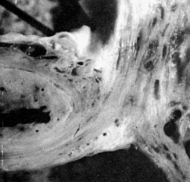
through a transabdominal approach.

That the connective tissue capsule of the vagina is continuous with that of the bladder is seen in the accompanying photomicrograph (Fig. 16) in which the sagittal section had been obtained postmortem through the vagina, cervix, and bladder of an aging patient with procidentia. Notice the continuity of this layer which must be traversed, as well as the looseness of the areolar tissues filling the potential vesicovaginal and vesicocervical spaces.

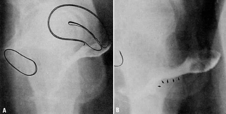
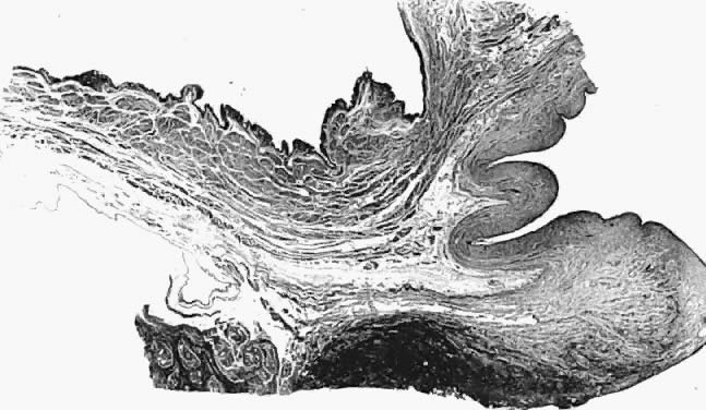
Fig. 16. Photomicrograph of sagittal section through bladder (top left), vagina (top right), and cervix (lower right) of autopsy specimen of elderly patient with untreated genital prolapse. Descent of the cervix has drawn it and the vagina away from the bladder. An attachment (supravaginal septum) of fibromuscular connective tissue capsule of the bladder to that of the vagina is shown.

Vesicocervical Space

The vesicocervical space is the continuation of the vesicovaginal space superiorly above the supravaginal septum. The posterior border becomes the connective tissue adventitia of the cervix, with which the adventitia of the vagina is continuous. The superior border is the peritoneum lining the vesicouterine peritoneal pouch. Cutting the supravaginal septum establishes communcation between the vesicovaginal space and the vesicocervical space.

Ascending Bladder Septa

Although the ascending bladder septa are weak cephalad, they become the stronger bladder pillars (which contain efferent veins from the vesical plexus and ureter) by the addition of the lateral strong connective tissue portions of the cardinal ligament. Medially, they are loose in texture and contain fat and ureter. These septa contain the lateral inferior extensions of the bladder and connect it to the upper surface of the cardinal ligament, lateral to the cervix.

Prevesical Space of Retzius

The prevesical space of Retzius is in the form of a triangle extending from the umbilicus laterally to the lateral umbilical ligament (obliterated hypogastric artery). Anteriorly, the transversalis fascia extends from the umbilicus to the pubis; it extends inferiorly to the cardinal ligament and the supravaginal septum. It is separated from the paravesical spaces by the ascending bladder septa. The prevesical space thus includes the area between the pubis and the anterior vesical wall roofed by the fascia between the medial umbilical ligaments.

The ascending bladder septum above the ureter contains many blood vessels including the inferior vesical artery and large veins of the vesical plexus. Below the ureter, however, blood vessels are scant and the tissues between bladder and vagina can be easily separated here without hemorrhage.

Paravesical Spaces

The paired paravesical spaces, right and left, are natural, fat-filled, preformed spaces that lie above the cardinal ligament and its prolongation (horizontal connective tissue ground bundle); they are bounded medially by the bladder pillars and laterally by the pelvic walls, the internal obturator muscle, and the levator ani. The roof is formed by the lateral umbilical ligament (vesicohypogastric fascia).

Descending Rectal Septa

The descending rectal septa run alongside the vagina from the undersurface of the cardinal ligament and its vaginal prolongation to the lateral surface of the rectum and thence to the sacrum.

Retrorectal Space

The retrorectal space lies in the midline between the sacrum and the adventitia of the rectum, between the posterior portion of the rectal pillars. This space communicates with the pararectal spaces above the uterosacral ligaments.

Pararectal Spaces

The paired pararectal spaces are only potential and are not preformed. They lie below the cardinal ligament and its vaginal prolongation. The medial border is formed by the rectal pillar, the lateral by the levator ani. The posterior portion extends backward above the ischial spine but under the cardinal ligament to the anterior surface of the lateral part of the sacrum. Behind the cardinal ligament the independent caudal portion of each side becomes continuous with the cranial portion of the opposite side.

The upper rectum is surrounded by a single circular pararectal space. The boundaries of this space, formed by communication of two pararectal spaces and the retrorectal space, are formed laterally and below by the cranial surface of the levator, above and medially by the rectum, descending rectal septa, and the cardinal ligament. It is made L-shaped by the horizontal part below the cardinal ligament and the cranial and ascending portion behind the cardinal ligament. The cranial portion of the space is bounded anteriorly by the cardinal ligament and posteriorly by the lateral part of the sacrum. The sheaths of the great vessels of the pelvic wall form the lateral border; the pararectal space is bordered medially by the rectal septa and ureteric sheath. The inferior or horizontal division is bounded below by the levator ani, above by the cardinal ligament, and medially by the rectal septum. The two pararectal spaces communicate with each other posterior to the rectum, where there is no limiting membrane.

Rectovaginal Septum and Space

Centered in a relatively avascular rectovaginal space, the posterior vaginal wall and anterior rectal wall have functional independence of one another. This space permits the two organs to glide over one another with considerable mobility. The anterior wall of this space is formed by a specialized connective tissue layer of fused peritoneum, the rectovaginal septum.28,29

As seen in sagittal section, this septum appears as a distinct, strong connective tissue layer between the vagina and rectum, oriented in a curved sagittal plane following the curvature of the pelvis. It is attached cranially to the caudal end of the peritoneum of the rectouterine pouch and extends inferiorly to the caudal attachment to the pelvic floor in the area of the perineal body. Anteriorly, the rectovaginal septum is always intimately attached to the posterior aspect of the vaginal connective tissue adventitia. Transversely, the septum curves posterolaterally, paralleling the course of the paracolpium (Fig. 17). Histologically, it consists of a fibromuscular elastic layer of dense collagen, abundant muscle, and coarse elastic fibers; the latter are best demonstrated with specialized orcein staining.

Fig. 17. The rectovaginal septum. Sections showing the partly dissected rectovaginal septum. It extends from the pouch of Douglas to the perineal body and forms the anterior surface of the rectovaginal space. Its adherence to the posterior vaginal wall is illustrated along with its posterolateral curve.(Nichols DH, Milley PS: Surgical significance of the rectovaginal septum. Am J Obstet Gynecol 108:215, 1970)

The rectovaginal space is in the midline between the rectovaginal septum, which is attached to the posterior vaginal wall, and the fat-covered rectal adventitia. The lateral walls are separated from the pararectal spaces by a descending rectal septum (rectouterine) on each side. The roof is the peritoneum and rectouterine peritoneal pouch (cul-de-sac of Douglas), and the inferior margin of this space is the perineal body.

Back to Top
REFERENCES

1. Mengert WF: Mechanics of uterine support and position. Am J Obstet Gynecol 31: 775, 1936

2. Goff BH: A practical consideration of the damaged pelvic floor with a technique for its secondary reconstruction. Surg Gynecol Obstet 46: 855, 1928

3. Smout CFV, Jacoby F, Lillie EW: Gynaecological and Obstetrical Anatomy pp 101–102. London, HK Lewis, 1969

4. Quinby WC: The anatomy and blood vessels of the pelvis. In Meigs JV (ed): Surgical Treatment of Cancer of the Cervix, p 32. New York, Grune & Stratton, 1954

5. Nichols DH, Milley PS, Randall CL: Significance of restoration of normal vaginal depth and axis. Obstet Gynecol 36: 251, 1970

6. Amreich J: Aetiologie und Operation des Scheidenstumpf-prolapses. Wien Klin Wochenschr 63: 74, 1951

7. Studdiford WC: The involuntary muscle fibers of the pelvic floor. Am J Obstet 60: 23, 1909

8. Ranney AL: The Topographical Relations of the Female Pelvic Organs, pp 107–110. New York, Wood, 1883

9. Milley PS, Nichols DH: The relationship between the pubourethral ligaments and the urogenital diaphragm in the human female. Anat Rec 170: 281, 1971

10. Zacharin RF: The suspensory mechanism of the female urethra. J Anat 97: 423, 1963

11. Curtis AH, Anson BJ, McVay CB: The anatomy of the pelvic and urogenital diaphragms in relation to urethrocele and cystocele. Surg Gynecol Obstet 68: 161, 1939

12. Wilson PD, Dixon JS, Brown ADG et al: Posterior pubourethral ligaments in normal and genuine stress incontinent women. J Urol 130: 802, 1982

13. Gardner E, Gray DJ, O'Rahilly R: Anatomy, 3rd ed, p 516. Philadelphia, WB Saunders, 1969

14. Power RMH: Embryological development of the levator ani muscle. Am J Obstet Gynecol 55: 367, 1948

15. Thompson P: On the arrangements of the fasciae of the pelvis and their relationship to the levator ani. J Anat Physiol 35: 127, 1901

16. Dickinson RL: Studies of the levator ani muscle. Am J Obstet 22: 897, 1889

17. Sturmdorf A: Gynoplastic Technology, pp 109–114. Philadelphia, FA Davis, 1919

18. Critchley HOD, Dixon JS, Gosling JA: Comparative study of the periurethral and perianal parts of the human levator ani muscle. Urol Int 35: 226, 1980

19. Swash MM, Henry M: Colpoproctology and the Pelvic Floor, 2nd ed. London, Butterworth-Heinemann 1992

20. Parks AG: Anorectal incontinence. Proc R Soc Med 68: 21, 1975

21. Halban J, Tandler J: Anatomie and Atiologie der Genital Prolapse beim Weibe. Vienna and Leipzig, Wilhelm Braumiller, 1907

22. Berglas B, Rubin IC: Study of the supportive structures of the uterus by levator myography. Surg Gynecol Obstet 97: 677, 1953

23. Muellner SR: The anatomies of the female urethra. Obstet Gynecol 14: 429, 1959

24. Range RL, Woodburne RT: The gross and microscopic anatomy of the transverse cervical ligament. Am J Obstet Gynecol 90: 460, 1964

25. von Peham H, Amreich J: Operative Gynecology, vol 1, pp 166–197. LK Ferguson (trans): Philadelphia, JB Lippincott, 1934

26. Campbell RM: The anatomy and histology of the sacrouterine ligaments. Am J Obstet Gynecol 59: 1, 1950

27. Shaw W: A study of the surgical anatomy of the vagina, with special reference to vaginal operations. Br Med J 1: 477, 1947

28. Milley PS, Nichols DH: A correlative investigation of the human rectovaginal septum. Anat Rec 163: 443, 1969

29. Nichols DG, Milley PS: Surgical significance of the retrovaginal septum. Am J Obstet Gynecol 108: 215, 1970

Back to Top