Chapter 30
Colposcopy
Kenneth L. Noller and Arnold Wagner
Main Menu   Table Of Contents

Search

Kenneth L. Noller, MD
Chair, Department of Obstetrics and Gynecology, University of Massachusetts Medical School, Worcester, Massachusetts (Vol 1, Chap 30)

Arnold L. Wagner, Jr., MD
Assistant Clinical Professor of Obstetrics and Gynecology, Northwestern University Medical School, Chicago; Director, Colposcopy-Laser Clinic, Evanston Hospital, Evanston, Illinois (Vol 1, Chap 30)

INTRODUCTION
SELECTION OF PATIENTS
THEORY OF THE TRANSFORMATION ZONE
ATYPICAL TRANSFORMATION ZONE
COLPOSCOPIC TECHNIQUE
SPECIAL CONSIDERATIONS
IMMUNOSUPPRESSION/HIV
POSTMENOPAUSAL CYTOLOGY AND COLPOSCOPY
COMMON ERRORS
REFERENCES

INTRODUCTION

Colposcopy is an office-based technique originally designed to diagnose preclinical cervical cancer. As such, it was in direct competition with cervical cytology screening for many years. Eventually, it was determined that the best method for screening for early cervical neoplasia was by the Papanicolaou method (Pap smear), and that the best diagnostic method was through the use of colposcopy and cervical biopsy. Thus, the techniques became complementary rather than competitive.

In 1925, it was theorized that the earliest cancers of the cervix might be visualized as ulcers or tumors if adequate magnification and illumination were available. It was soon recognized that the earliest premalignant conditions were not gross ulcerations or tumors but changes in the vascular pattern of the epithelial capillaries by the dense crowding of epithelial cell nuclei. It is the vascular pattern and epithelial opacity that colposcopists now study for evidence of intraepithelial neoplasia.

Colposcopy was introduced into mainstream gynecology in the United States in the late 1960s. Currently, it is a universally accepted gynecologic tool. Although the technique was developed to study cervix cancer by examination of the surface of the cervix (the term colposcopy literally means to look into the vagina), it has been found to be useful also in the diagnosis of various vaginal lesions, and in the evaluation of the vulva. Nonetheless, its primary purpose continues to be evaluation of the patient with an abnormal Pap smear.

To perform colposcopy, it is necessary to understand the small physiologic area of the cervix that colposcopists call the transformation zone (TZ), and the natural history of cervical intraepithelial neoplasia (CIN). Because colposcopy is a technique of pattern recognition, similar to other pattern recognition techniques (e.g., interpretation of histology, interpretation of electrocardiograms, etc.), it can be learned only by repeated exposure to the patterns and their variations. In addition, in concert with similar techniques, continued visualization of normal and abnormal patterns is necessary to maintain skills. To learn colposcopy, one must perform several hundred examinations, and must perform several examinations each month to maintain skills.

Back to Top
SELECTION OF PATIENTS

A woman that has a Pap smear suggesting the possible presence of cervical neoplasia is a candidate for colposcopic examination. At present, virtually all cytology laboratories in the United States have chosen to use “The Bethesda System for Reporting Cervical/Vaginal Cytologic Diagnoses.”1 The major categories of diagnosis in The Bethesda System (TBS) are shown in Table 1. Currently, women with Pap smears suggesting either a high grade squamous intraepithelial lesion (HGSIL) or squamous carcinoma must have colposcopic evaluation. No other first step is appropriate.

TABLE 1. The Bethesda System for Reporting Cervical/Vaginal Cytology Diagnoses: Summary of Major Diagnostic Categories

  Normal
  Benign epithelial changes
  Epithelial abnormalities

  Squamous

  Atypical squamous cells of undetermined significance (ASCUS)
  Low grade squamous intraepithelial lesion (LGSIL)
  High grade squamous intraepithelial lesion (HGSIL)
  Squamous cancer


  Glandular

  Atypical glandular cells of undetermined significance (AGCUS)
  Adenocarcinoma


Cytology reports other than HGSIL and squamous cancer are open to controversy concerning the appropriateness of colposcopic evaluation. In TBS, evidence of inflammation, infection, and repair are properly reported as “benign epithelial abnormalities.” None of the cellular findings in the benign epithelial category suggest the presence of neoplasia. Based on her symptoms, the patient may be treated for one or more infections, but colposcopy is not recommended. However, during the era when the Papanicolaou classification system was in use, those patients who had evidence of inflammation on three or more smears were shown to be at increased risk for CIN. It is unclear now whether women with several reports of benign epithelial abnormalities should have colposcopic evaluation.

TBS introduced a new term, “atypical squamous cells of undetermined significance” (ASCUS). This category was developed for smears that present the cytologist with a true diagnostic dilemma. Only those smears for which the cytologist is unable to decide whether or not a dysplastic process is present should be categorized as ASCUS. ASCUS means “I don't know.” Unfortunately, many laboratories have misused the ASCUS category. They have misinterpreted the intent of TBS and include smears consistent with inflammation and repair. They have assumed that those findings that were previously labeled class II in the Papanicolaou system belonged in the ASCUS category.

Laboratories have varied considerably in their frequency of use of ASCUS. The best laboratories in the United States currently use the ASCUS category for approximately 3% to 4% of all cytology reports. This appears to be appropriate, and a National Cancer Institute expert panel has stated that ASCUS should be used in fewer than 5% of cytology reports except in populations at high risk for cervical neoplasia where the percentage might be slightly higher.2 In general, ASCUS reports should not exceed twice the total rate of low grade squamous intraepithelial lesion (LGSIL) and HGSIL reports. Each clinician should be aware of the rate of ASCUS reports from the laboratory usually used.

There is currently no clear decision regarding the appropriateness of colposcopy for patients with ASCUS Pap smears. Many clinics repeat such a smear within a few months.3,4,5,6 Others refer all women with ASCUS smears for colposcopy. The appropriateness of colposcopy may depend most on the use of the ASCUS terminology by the reporting laboratory. For example, if a laboratory reports only 3% to 4% ASCUS, colposcopy is appropriate since a considerable fraction of these women will have significant SIL. On the other hand, if a laboratory is reporting 15% to 30% ASCUS, the chance of a patient with such a report having a HGSIL is minuscule. Large prospective studies must be completed to determine the true significance of ASCUS reports.

LGSIL reports are also controversial. Most women with such reports have only human papillomavirus (HPV) infection, and most will spontaneously resolve. Indeed, it is not possible, by light microscopy, to determine whether a woman has only an HPV infection or has an early neoplastic process that has the potential to progress through the various stages of SIL and develop into carcinoma. In the United States, most women with LGSIL reports have colposcopy. Until large-scale prospective studies are completed that determine whether or not women with LGSIL reports should have colposcopy, it is appropriate to follow such patients with either cytology or colposcopy.2,3

Back to Top
THEORY OF THE TRANSFORMATION ZONE

Embryology

In the female fetus, the paired paramesonephric ducts fuse, lose their common wall, and eventually develop into the fallopian tubes, uterus, uterine cervix, and some portion of the upper vagina (Fig. 1). The epithelial covering of these organs is initially of a simple columnar type. However, that portion of the paramesonephric ducts (more commonly called the müllerian ducts) that impinges on the urogenital sinus is eventually covered by nonglycogenated stratified squamous epithelium.4 Most of the process of conversion of the upper vaginal and cervical epithelium from columnar to stratified squamous type occurs in utero. However, virtually all females are born with a small area of columnar epithelium surrounding the anatomic external cervical os. Thus, in the female child, the vagina and most of the cervix are covered with squamous epithelium, yet a small rim of columnar epithelium surrounds the os. These areas do not change during prepubertal childhood.5

Fig. 1. Fusion of sinovaginal bulbs and formation of primitive vaginal plate at 14 weeks (140 mm).(Prins RP, Morrow CP, Townsend DE et al: Vaginal embryogenesis, estrogens, and adenosis. Obstet Gynecol 48:246, 1976)

At the time of puberty, probably coincident with increasing circulating levels of estrogens, the cervical epithelium begins to change.6 The pH of a child's vagina is neutral or basic, whereas the pH of an adult woman's vagina is very acidic, commonly in the range of pH = 3. This change in pH occurs at approximately the time of the estrogen surge during puberty.

Columnar epithelium is composed of a single layer that is easily injured (Color Plate 1A and B). Squamous epithelium, on the other hand, is much tougher, is usually 20 to 30 cell layers thick, and is relatively resistant to a low pH. Thus, the area of columnar epithelium on the cervix gradually changes to squamous epithelium through a process called squamous metaplasia (Fig. 2). This is a normal occurrence in virtually all females, and the presence of cells showing squamous metaplasia on cervical cytology should be considered normal (Fig. 3).

Color Plate 1A and B. Normal cervix, 19-year-old nulliparous patient. The opened speculum has exposed the columnar epithelium surrounding the os and is in the endocervical canal. Higher magnification shows the junction between columnar and squamous epithelia. The faint white line is the transformation zone, which is quite narrow in this patient. Color Plate 1C. Acetowhite epithelium surrounding an area f columnar epithelium. The border, with the surrounding squamous epithelium is irregulare. Biopsies confirmed infection with human papilloma virus. D. Atypical transformation zone (ATZ) showing an area of punctation in the center of the photograph and a separate island of mosaic in the upper right corner. Note the varying size of the mosaic tiles, but uniform size of the punctate vessels. Biopsy showed CIN II. Color Plate 1E. ATZ. The acetowhite lesion shows mosaic tiles. The border is distinct, but irregular. Biopsy showed CIN I. F. Large acetowhite lesion showing predominantly mosaic changes. Note the distinct border in the upper right as well as the varying intercapillary distances and punctate vessels within the mosaic tiles ( “umbilication” ). Biopsy showed CIN III. Patient refused therapy. Microinvasive carcinoma was diagnosed 4 years later.

Fig. 2. Normal process of squamous metaplasia. The villi covered by columnar epithelium are replaced by metaplastic epithelium. Capillary structures in the villi are compressed and reduced in height, ultimately forming a network under the epithelium indistinguishable from the capillary network of normal epithelium.(Kolstad P, Stafl A: Atlas of Colposcopy. Baltimore, University Park Press, 1972)

Fig. 3. Original squamocolumnar junction. An oblique line separates original squamous epithelium on the right from metaplastic squamous epithelium on the left. Note the well-glycogenated, “chicken-wire” appearance of original squamous epithelium, compared with dense packing of metaplastic cells.(Coppleson M, Pixley E, Reid B: Colposcopy: A Scientific and Practical Approach to the Cervix in Health and Disease. Springfield, IL, Charles C Thomas, 1971)

The process of change from columnar epithelium to squamous epithelium requires rapid division of cells. Perhaps because of this rapid cell turnover, the TZ—the place where metaplasia occurs—is the site of the occurrence of squamous cervical neoplasia in greater than 90% of all cases. Perhaps a carcinogen can more easily transform a metaplastic cell than a mature columnar or squamous epithelial cell (Fig. 4).

Fig. 4. Mechanisms of cervical epithelial change from columnar epithelium to squamous epithelium or neoplasia.

When a woman becomes pregnant, the cervix markedly increases in size and weight. Concomitant with the size increase, there is usually an eversion of the endocervical (columnar) epithelium. This epithelium is then subjected to the low pH of the vagina for the first time, and rapidly undergoes squamous metaplasia. Thus, after adolescence, the most common time to see large areas of squamous metaplasia is during the first pregnancy.

In menopause, when the cervix is no longer stimulated by high levels of circulating estrogen, the cervix decreases in size and weight. The exocervical squamous epithelium tends to migrate up the canal. Thus, in postmenopausal women, the presence of columnar epithelium on the cervix (commonly called cervical ectropion) is a rarely observed phenomenon.

Normal Elements of the Transformation Zone

The TZ is a narrow area, circumferentially surrounding the external cervical os. In most women, it is 2 mm to 6 mm wide, though in some cases it can be several centimeters wide (see DES-exposed women). This zone is a dynamic area with many changes in cell type and architecture. Because columnar epithelium is, by nature, a villous, folded epithelium, and squamous epithelium presents a flat smooth surface, considerable architectural change must occur. Typically, the tips of the columnar epithelial fronds or villae are the first to exhibit changes of squamous metaplasia. Whereas columnar epithelium presents as “grapelike” structures covered by completely transparent epithelium surrounding a stromal core with one capillary loop, when squamous metaplasia begins to occur, the tip of each villous becomes somewhat opaque, particularly after the application of 3% acetic acid. As squamous metaplasia progresses, the entire columnar villous structure undergoes metaplasia and the areas between the villous fronds begin to fill in with squamous cells. If normal maturation occurs, the central capillary cores become nearly totally subepithelial.

Thus, at any one time, the TZ may have columnar epithelium, squamous epithelium, and squamous metaplasia present (Fig. 5 and Fig. 6). At times, squamous metaplasia occurs so fast on the surface that contiguous columnar epithelial villae coalesce while mucus-producing columnar epithelium is still present in the covered crypts. If the process is complete, a nabothian cyst may be formed (Fig. 7). These are completely normal findings in the transformation zone. They are more commonly seen in parous women than in nonparous women. On occasion, dilated vessels may be seen stretching across these cysts but the vascular pattern always has normal branching.

Fig. 5. Normal cervix demonstrating columnar epithelium of the endocervix in characteristic grapelike appearance, tongues of squamous metaplasia, and “gland openings.” The bright patches are artifacts caused by light reflection.

Fig. 6. Anterior lip of the cervix demonstrating coalescence of tips of columnar-covered papillae on the ridges of endo-cervical rugae in early squamous metaplasia. Behind this area is an area of maturing squamous metaplasia.

Fig. 7. Parous cervix showing multiple nabothian cysts surrounding a central area of columnar epithelium and squamous metaplasia.

If metaplasia occurs normally, fully mature squamous epithelium eventuates. Even though it is many cell layers thick, squamous epithelium, like columnar epithelium, is entirely transparent. Thus, the colposcopist observing normal squamous epithelium is actually looking through the epithelium at the subepithelial vascular plexus. When this area is carefully viewed under magnification, it can be seen that each tiny vessel branches like a tree (arborizes). Each successive branch is smaller than the one from which it is generated. There are no epithelial tufts (pseudoglomeruli) and no vessel ever changes from large caliber to small back to large caliber.

Back to Top
ATYPICAL TRANSFORMATION ZONE

In a patient with an abnormal Pap smear, colposcopy can be expected to identify an abnormal area in the TZ that corresponds to the Pap smear findings. In most cases, the diagnosis of an abnormal TZ is based on the identification of vascular patterns, which suggest cervical neoplasia (Fig. 8). Most of these patterns are not visible to the naked eye and, indeed, are not visible at the time of colposcopic examination unless a solution of 3% to 5% acetic acid is first applied to the cervix. Although the exact mechanism of action of acetic acid is not known, any area with an increased nuclear/cytoplasmic ratio will appear whiter than the surrounding area after the application of dilute acetic acid. Because squamous metaplasia has an increased nuclear/cytoplasmic ratio, it will appear faintly white. However, neoplasia is much more distinct from the 
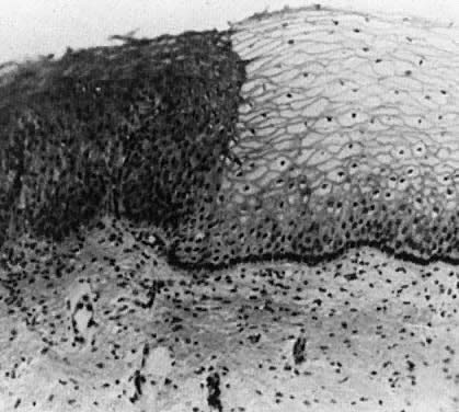
background than is squamous metaplasia.

Fig. 8. Atypical squamous metaplasia. The metaplastic epithelium for some unknown reason starts to grow in buds or blocks. The individual cells become indistinguishable from those of frank invasive carcinoma. The central vascular network of the villi of columnar epithelium remains as punctate or mosaic vessels that extend close to the surface of the epithelium. The mosaic vessels form basketlike structures around the blocks of abnormal cells.(Kolstad P, Stafl A: Atlas of Colposcopy. Baltimore, University Park Press, 1972)

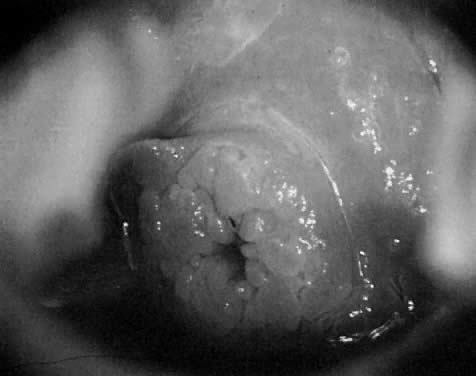
Acetowhite Epithelium (White Epithelium)

Areas that appear white after the application of acetic acid are called acetowhite epithelium or white epithelium. These areas may be observed either inside or outside of the TZ. When located outside of the TZ, they 
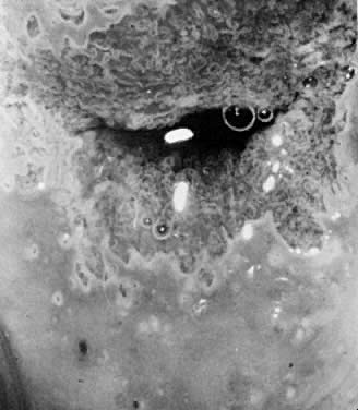
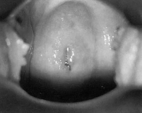
often represent areas of HPV infection, trauma, or repair (Fig. 9).

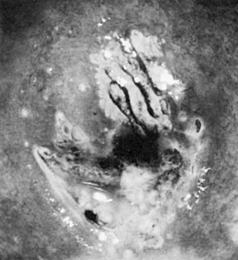
Fig. 9. The anterior lip of the cervix demonstrates gland openings, squamous metaplasia, and a localized area of white epithelium with a very fine vascular mosaic. Biopsy revealed only chronic cervicitis and squamous metaplasia.

White epithelium can be graded based on its surface contour, its whiteness, and its border with surrounding tissues. In general, neoplasia has a distinct border, is grayish-white rather than pearly-white, and is somewhat raised from the surrounding tissues. Areas of HPV infection only are generally much whiter with indistinct borders (Color Plate 1C). Areas of HPV may or may not be raised above surrounding tissues. Most areas of white epithelium have vascular changes (see below).

When a white area is observed on the cervix prior to the application of acetic acid, it is referred to as leukoplakia. These areas represent hyperkeratosis (Fig. 10) and are often caused by infection with the HPV virus. Unfortunately, without biopsy, it is impossible to know whether the epithelium underneath the hyperkeratotic area is abnormal. Therefore, it is generally recommended that at least one representative leukoplakic area be biopsied 
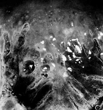
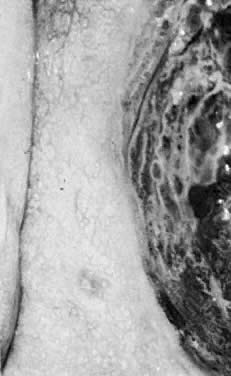
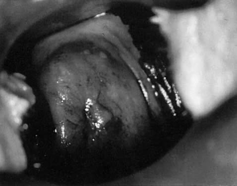
during a colposcopic examination.

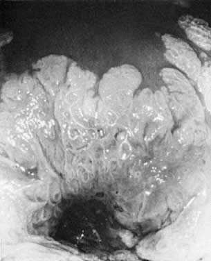
Fig. 10. The cervix prior to application of acetic acid. A very dense hyperkeratotic pattern is noted surrounding the cervical os. No comment can be made about the underlying epithelial vascular pattern.

Vascular Patterns

If a neoplastic stimulant (some combination of HPV infection, immune status, and genetics) is applied to an area of metaplasia, the resultant neoplastic epithelium causes blood vessels to grow into the epithelium. Some authors suggest that neoplasia induces neovascularization, whereas others believe that neoplasia merely influences the capillary vessels found in columnar epithelium to persist and grow.7 Regardless of the cause, the most common finding when CIN* is present is a change in the vasculature. With CIN, the vessels are no longer confined to the area under the transparent squamous epithelium but are found within the epithelium itself. The increased nuclear/cytoplasmic ratio in CIN causes the epithelium to lose its transparency after the application of acetic acid.

Sometimes the vessels of CIN remain as distinct capillary loops, but extend to the surface. In higher grade lesions (CIN III), these vessels may actually extend a millimeter or two above the surface. This can be recognized since the tips of these vascular loops often reflect light. When these loops are the predominant pattern, it is termed punctation. (Fig. 11, Fig. 12, and Fig. 13)

Fig. 11. Punctate vessels as seen in CIN.(Kolstad P, Stafl A: Atlas of Colposcopy. Baltimore, University Park Press, 1972)

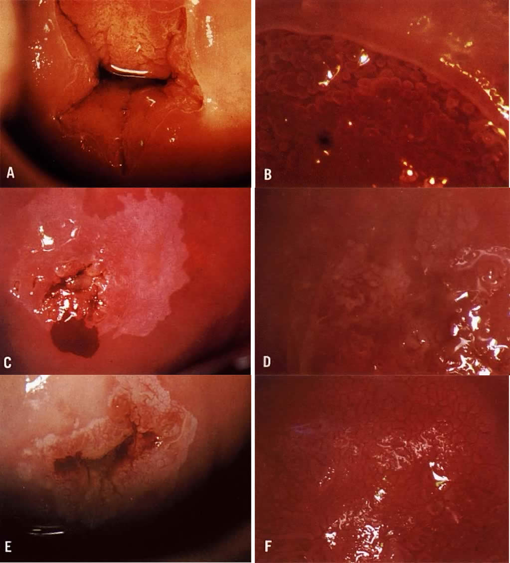
Fig. 12. A. Prior to the application of acetic acid. B. After the application of acetic acid. The posterior lip of the cervix demonstrates a coarse white epithelium with punctation and a sharply contrasting border with normal tissue. Biopsy demonstrated CIN III.

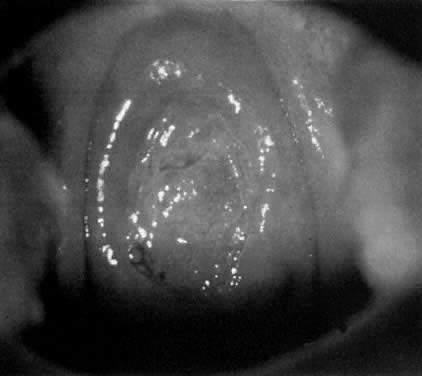
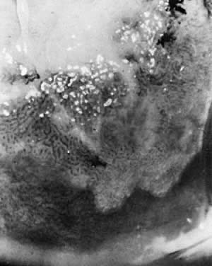
Fig. 13. A wide transformation zone demonstrates white epithelium with coarse punctation. Mucus obscures the cervical os at upper left. Biopsy showed CIN III.

In other cases of neoplasia, the intraepithelial vessels do not form simple loops but communicate with each other, forming a mosaic pattern around an epithelial core (Fig. 14). This pattern is termed mosaic. It does not appear that punctate patterns become mosaic patterns, but rather the time at which the neoplastic insult occurs determines which pattern will be present. Both mosaic and punctation can be present in lesions of CIN I, CIN II, and CIN III (Fig. 15, Fig. 16, Fig. 17, and Color Plate 1D and Color Plate 1E), that is, neither is more severe than the other.

Fig. 14. Mosaic vessels. Common finding in all grades of CIN. A greater distance between the capillaries suggests higher grade disease.(Kolstad P, Stafl A: Atlas of Colposcopy. Baltimore, University Park Press, 1972)

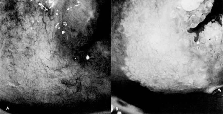
Fig. 15. A. Prior to application of acetic acid. The cervix demonstrates a fine network capillary pattern. The cervical os is at upper right. B. After application of acetic acid. White epithelium with a fine mosaic is noted. Biopsy demonstrated CIN I.

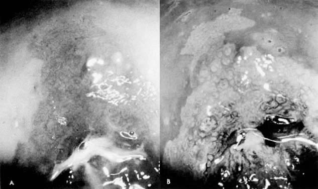
Fig. 16. A. Prior to application of acetic acid. The cervical os is at lower center. B. After application of acetic acid. A sharp-bordered white epithelium with fine punctation and a coarse mosaic is seen. Biopsy demonstrated CIN III.

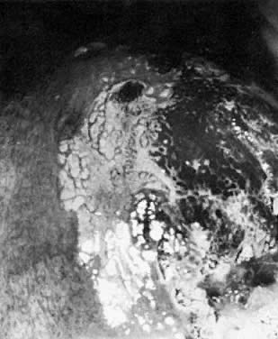
Fig. 17. The cervix reveals a localized area of dense white epithelium at the 9- to 11-o'clock position. A moderately coarse mosaic is noted, as well as punctation. The cervical os is at lower right. Biopsy demonstrated CIN III.

In general, when the capillary loops of punctation or the tiles of mosaic produce vessels that are more than 200 microns apart (0.2 mm), this suggests a more severe lesion. In addition, if the vascular loops or mosaic tiles vary in size, shape, and density, a more severe lesion is 
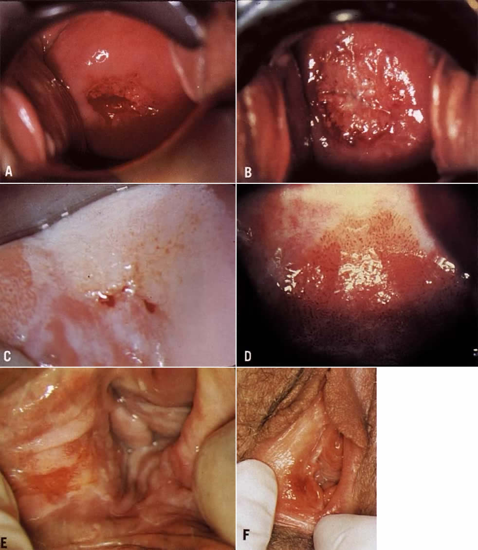
suspected (Color Plate 1F).

The term “atypical vessels” refers only to vasculature that suggests the presence of invasive cancer. This term should not be used to describe punctation or mosaic. Atypical vessels are never seen in normal epithelium, squamous metaplasia, or Cin. These vessels are typically large, branch in unusual, irregular ways, often run horizontally through the epithelium, and form bizarre shapes (Fig. 18, Fig. 19, and Color Plate 2A). Because invasive cervix cancer is a rare finding, colposcopists should have reference materials available to review whenever a bizarre vessel is seen. If there is any question about the pattern, biopsy or referral to a colposcopist with more experience is wise.

Color Plate 2A. Atypical vessels. The lesion on the anterior lip of the cervix exhibits large vessels running horizontal to the surface. The border is very distinct and the edge is raised. The lesion color is more yellow than white. Biopsy revealed invasive squamous carcinoma to a depth of 4 mm. Conization was consistent and radical hysterectomy found no evidence of disease spread. Patient is alive and well 17 years later. B. Normal network of capillaries beneath normal epithelium, which is transparent. Color Plate 2C. Bacterial vaginosis. Note the blurring of the margins of the subepithelial vessels. The squamous epithelium remains transparent. D. Trichomonas cervicovaginitis. The “strawberry spots” are actually dilated vessels. They do not exhibit abnormal branching. Color Plate 2E. White epithelium on the posterior lip of the cervix. The examinations unsatisfactory because the entire transformation zone cannot be see and the extent of the lesion is not known. F. An endocervical speculum is used to expose the upper extent of the lesion Although seen entirely, an excisional procedure rather than ablative is suggested due tot he endocervical extension.

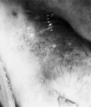
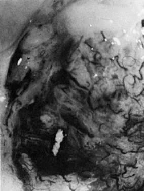
Fig. 18. The external os of the cervix is filled with an irregular-surfaced, sessile polypoid structure demonstrating grossly distorted, dilated, and irregular capillaries. Biopsy confirmed invasive squamous cell carcinoma.

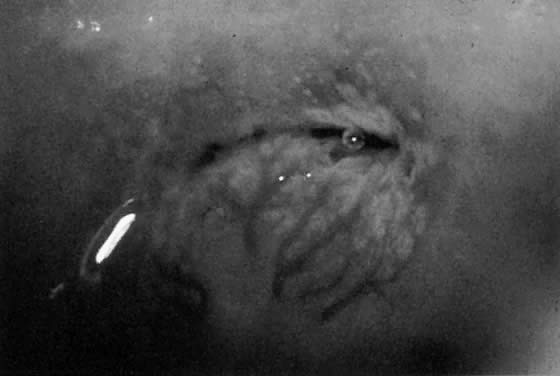
Fig. 19. Normal transformation zone with large vessels overlying nabothian cysts. Note that these vessels branch normally (arborize). They do not resemble the vessels in Figure 18 or Color Plate 2A.

Surface Contour

A smooth, regular surface contour is normal in mature, squamous epithelium and squamous metaplasia. When CIN occurs, as it progresses in severity, the surface may become rougher and more irregular as mosaic tiles push up between vascular margins. In areas of invasive carcinoma, the surface contour may be grossly uneven, having a cauliflower-like appearance. Areas of hyperkeratosis may be grossly irregular in contour, but these areas lack any of the accompanying vascular aberrations seen in CIN.

Border with Normal Tissue

Areas with significant CIN demonstrate a sharp border with the surrounding pale pink normal tissue. A sharply contrasting border around a geographic area of white epithelium signifies an area of epithelial abnormality (the sharper the border, the more marked is the histologic abnormality). Areas of squamous metaplasia or HPV demonstrate a diffuse, poorly defined border with normal tissue. The border contrast often can be enhanced by use of the green filter.

Inflammation

The presence of infection or inflammation may complicate the colposcopic examination. When bacterial vaginosis, trichomoniasis, or candidiasis is present, there is often considerable discharge, as well as some hyperemia that may make evaluation of the cervix difficult (Fig. 20 and Color Plate 2B and Color Plate 2C). Particularly when dealing with Pap smears suggesting less significant lesions, it is suggested that cervicovaginitis be cleared before colposcopy is performed. Trichomoniasis may cause dilated capillary loops (strawberry spots) that make evaluation of the TZ difficult (Color Plate 2D).

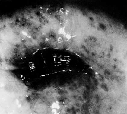
Fig. 20. Typical blotchy appearance of the inflamed cervix.

Natural History of CIN

Colposcopists must thoroughly understand the natural history of CIN lesions, as well as the multiple terminologies that have been used to describe them. Figure 21 shows the relationship of the terminologies used to describe intraepithelial neoplasia of the cervix. In the United States, the dysplasia/carcinoma in situ terminology was used for many years. However, it was convincingly demonstrated that light microscopy could not distinguish between severe dysplasia and carcinoma in situ (CIS). Therefore, the CIN terminology, which combines severe dysplasia and CIS, was developed by Dr. Ralph Richart. The CIN terminology is still most widely used to describe histopathologic changes.

Fig. 21. Dysplasia, CIN, and SIL terminologies for cervical neoplasia.

The Bethesda System for Cytology Reporting developed the high grade and low grade squamous intraepithelial lesion terminology.1 This terminology is also rapidly being adopted for histopathology reports. It has been demonstrated that cytology using light microscopy cannot distinguish between an HPV infection and a lesion that has the ability to progress (mild dysplasia). It now appears that the distinction also cannot be made on histologic specimens.

Indeed, it is not important to distinguish between HPV and mild dysplasia. What the clinician needs to know is whether an early lesion has the ability to progress. Unfortunately, light microscopy cannot make this distinction. At present, there is no technique that can.

Figure 22 graphically demonstrates the chance of spontaneous resolution of various intraepithelial problems. In general, HPV infections are believed to be totally reversible, though they may also persist indefinitely.8 The body does seem to be able to make antibodies that can successfully clear the virus in some individuals. However, in many cases the virus persists, and in a few women some cells may undergo a change that begins the cascade that can ultimately lead to cervical cancer. This change cannot be detected by light microscopy, DNA probes, or HPV subtyping at present.

Fig. 22. HPV infections are usually reversible. However, the chance of regression decreases as the severity increases.

The body does seem to be able to clear many intraepithelial neoplastic lesions. However, once the lesion has progressed to a full thickness change (CIN III/severe dysplasia/CIS), unless the lesion is treated, it will likely become invasive cancer.

The role of cytology and colposcopy, then, is to detect focal intraepithelial lesions, thus allowing the clinician to treat them before invasive cancer occurs. In most cases, treatment of CIN is an office-based procedure. Hysterectomy is not indicated for CIN except in the most unusual circumstances.

Etiology of CIN

Since 1842 investigators have searched for the etiology of cervical cancer.9 Initial crude epidemiologic studies discovered that women who never had sexual intercourse never developed invasive squamous cancer of the cervix. More sophisticated studies continue to suggest that neoplasia of the cervix is a sexually transmissible disease. That is, some factor or factors transmitted during sexual activity eventuates in the development of cervical cancer.

In the 1960s and early 1970s, it appeared that herpes simplex virus-2 (HSV-2) might be the transmissible factor causing cervical neoplasia.10 Later studies showed that an HSV-2 infection was largely an indicator of increased risk based on sexual activity rather than the true etiologic agent of cervical neoplasia.

In 1977 Meisels and associates reported that the vast majority of cases previously diagnosed as mild dysplasia were human papilloma virus infection.11 This remarkable discovery—as with most newly discovered biologic events—prompted an immediate, far reaching, and overzealous response. It was observed that koilocytes, squamous cells with a clear halo surrounding each nucleus, were indicative of HPV infection. It was later shown that these clear areas often were filled with HPV. Many substances can cause koilocytosis (though HPV infection is most common). However, when koilocytes also have wrinkling and clumping of the nuclear DNA (raisinoid nuclei), the changes are almost always evidence of HPV infection.12

Considerable time was wasted for nearly a decade trying to decide whether a patient had “just HPV infection” or “CIN I.” Originally both entities were treated most often by the newly available ablative technique, CO2 laser. Not surprisingly, it was eventually reported that ablative techniques often did not cure a patient of an HPV infection. Clinicians, therefore, began to treat the two entities differently, that is, patients with “just” HPV were not treated whereas those with CIN I were treated.

Unfortunately, it has now become clear that light microscopy cannot distinguish between HPV infection and CIN I. For this reason, TBS places evidence of both HPV infection and mild dysplasia into the same category, LGSIL. It is also agreed by most histopathologists that it is impossible to tell the difference between these two entities on biopsy specimens. Thus, the clinician cannot distinguish the patient with only evidence of HPV infection from the patient who has the earliest changes of neoplasia.

HPV is a DNA virus that appears to be specific to humans. There are now more than 70 subtypes identified; most tend to be localized to certain areas of the body. For example, one subtype tends to produce plantar warts, another common warts on the fingers and hands, and several types tend to be found in the genital area. HPV infections have been found in the oral cavity, in the esophagus, in the anal region, and many other sites.13

HPV types 6 and 11 tend to cause common genital warts. These subtypes have only rarely been identified in invasive cancers of the lower genital tract except for verrucous carcinoma of the vulva. HPV 6 and 11 are rarely found in CIN II and III.14

Genital warts usually become apparent to the patient and her physician only when present on the vulva (Fig. 23). However, whenever one genital site is involved (vulva, vagina, or cervix), there is a greater than 80% chance that the entire lower genital tract harbors the HPV virus. That is, HPV tends to be a field infection rather than single site. Infections of the cervix usually result in acetowhite changes (Fig. 24). Occasionally, a discrete wart is observed on the cervix (Fig. 25).

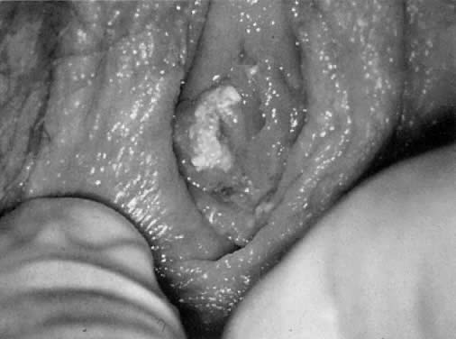
Fig. 23. Typical genital wart. This lesion is adjacent to the urethra.

Fig. 24. HPV infection of the anterior lip of the cervix.

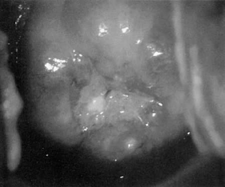
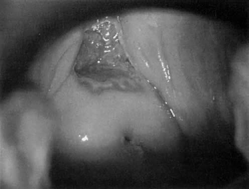
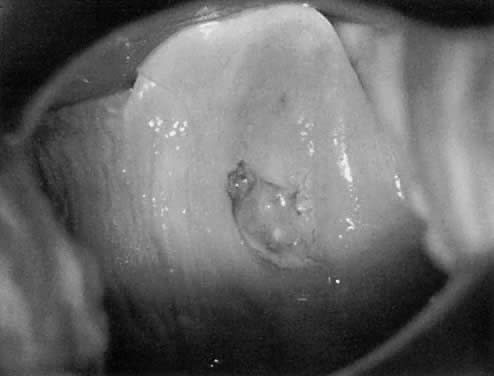
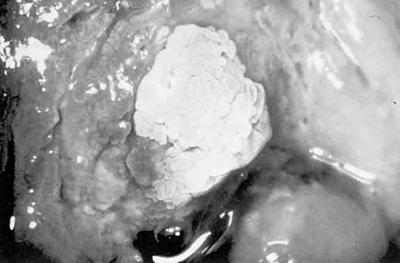
Fig. 25. Cervical wart. A discrete exophytic wart of the cervix is a relatively uncommon finding.

Many HPV types have been isolated from lower genital tract invasive carcinomas. By far, the most commonly isolated type in squamous lesions is HPV 16. HPV 18 is also found with some frequency in squamous lesions, and commonly in adenocarcinomas of the endocervix.15 HPV DNA also can be found incorporated into the cellular DNA of most cases of CIN III and in many cases of CIN II. In invasive cancers, the HPV DNA is incorporated directly into the cell genome, and through various interruptions of the normal cell cycle cause a cell line (clone) to develop which is immortal. The exact mechanism by which such immortality is caused by the virus is currently under study.

HPV cannot be grown under usual conditions in a microbiology or virology laboratory. Several sophisticated laboratory systems have been developed for determining the presence of HPV and assigning the appropriate subtype. For several years, the “Southern blot” technique has been the one most often used by research laboratories. More recently, polymerase chain reaction (PCR) techniques have been able to identify low levels of viral DNA. Although early PCR reports showed an extremely high prevalence of the virus in the general population, these reports are now known to have been caused by contamination of the identification system. Nonetheless, PCR remains a powerful and important tool in the investigation of these viruses.

The prevalence of HPV among women in the United States is not well understood. Studies of samples obtained from young, sexually active women suggest that HPV prevalence might be as high as 30%.16 These populations may or may not be representative of United States women, in general. Additionally, it has been shown that the prevalence of HPV identified in any group is a function of the number of times the group has been studied.17 Although PCR techniques should have the ability to identify latent infections that are not histologically or clinically apparent, it appears that any identification technique misses some infections. For example, a population might demonstrate a 20% prevalence of HPV infection if sampled once, but the same population may have a total 25% prevalence if resampled.

With the discovery that HPV (particularly subtypes 16, 18, 30, 45, 56, and several others) can be found in virtually all cases of squamous cancer of the cervix, research attention was focused on this agent. It now appears that, in almost all cases of cervical neoplasia, prior infection with HPV is necessary. When multivariate analyses have been performed, all other risk factors, except smoking, disappear. That is, early sexual intercourse, multiple sexual partners, and exposure to HSV-2 are not important when compared with HPV infection.

However, HPV is not a complete carcinogen, nor is HPV infection a sufficient cause for the development of neoplasia. That is, of the millions and millions of women who have HPV infection, few ever develop cervical neoplasia, and almost none ever develop cervix cancer. A combination of events is probably responsible for this observation. First, the body's immune system may defeat the HPV virus and/or may attack and destroy early neoplastic cell clones. In addition, many early intraepithelial neoplastic lesions are destroyed by office treatment and thus never lead to invasive cancer of the cervix.

Smoking appears to be an independent risk factor for the development of cervical neoplasia among women with HPV infections.18 Cervical neoplasia is increased approximately twofold, and vulvar neoplasia is increased approximately fourfold. Some of the carcinogens in inhaled smoke appear in high concentration in the cervical mucus. In addition, there is a loss of immune-competent cells in the cervical epithelium and stroma when biopsies of smokers are compared to nonsmokers.

Back to Top
COLPOSCOPIC TECHNIQUE

Equipment

A colposcope is a low-power binocular microscope with a focal length of approximately 30 cm. This extended focal length is necessary to be able to see into the vagina. A coaxial light source is also focused the same distance in front of the instrument so that optimal vision is obtained. Most colposcopes have a magnification of approximately 2 to 40x. The range between 4 and 15x is most useful. In the past, some colposcopes had a fixed magnification, usually 13.5x. These have limited usefulness, particularly when it is necessary to scan the cervix, vagina, and vulva.

All colposcopes should be fitted with a red-free (green) filter. Use of this filter allows better visualization of the blood vessels. Indeed, many colposcopists only view the cervix with the green filter in place.

It is very useful to have the colposcope attached to a small video camera and monitor. With such a setup, the patient may view the examination. All women who have an abnormal Pap smear are quite anxious about their medical condition.19 They have been taught throughout life that Pap smears detect cancers, and if their smear is abnormal they fear that they have a cancer already growing in the genital area. By demonstrating the epithelial nature and small size of most lesions, patients can be reassured. In addition, it is possible to record the examination and to use the monitor for teaching.

Photographic equipment can also be attached to the colposcope. Various cameras are available which can use slide, print, or instant film. While these are useful for developing sets of slides for teaching, they are less useful for documentation purposes. Even the best colpophotographer finds that many photographs are of poor quality.

Method

After introduction of a bivalve speculum, the colposcope is brought into position and focused on the cervix. If there is excess mucus present, it can be removed with a swab dipped in saline. The cervix should then be carefully examined prior to application of acetic acid. In some cases, atypical vessels can only be seen before application of acetic acid because of the dense white color that develops after acetic acid is applied.

After thorough inspection, 3% to 5% acetic acid is applied to the cervix with a swab. It takes 30 to 60 seconds for acetowhite changes to occur, thus the examiner should not be overly quick. Repeated application of acetic acid during the course of the examination is necessary as the acetowhite changes fade within 1 to 2 minutes after application.

It is necessary, during the course of each colposcopic examination, to determine whether or not the examination is satisfactory. Figure 26 shows the algorithm for colposcopic examination. During the examination with saline and acetic acid the examiner should determine whether the examination is satisfactory and the location of the most atypical areas. In many cases if the examination is satisfactory, it is possible to perform office therapy. However, if the examination is unsatisfactory, further evaluation by means of conization or loop electroexcision must be performed.

Fig. 26. Simplified algorithm for colposcopic examination. The entire transformation zone and lesion are identified, and the endocervical evaluation is negative.*

Three elements are necessary for the examination to be satisfactory. First, the entire TZ must be seen. This means that 360 degrees of columnar epithelium and 360 degrees of normal squamous epithelium must be seen, as well as all of the area between them. Second, if a lesion is present, it must be seen in its entirety. Lesions that extend up the canal out of sight have not been seen completely (Color Plate 2E and F). Third, evaluation of the endocervix must be negative. This evaluation includes both visualizing normal columnar epithelium throughout the visible canal, and having a negative sample from the canal.

In the past, endocervical sampling meant endocervical curettage (ECC). This technique uses a small curette to obtain a circumferential sample of the epithelium. Although it is not necessary to deeply sample the epithelium, the procedure still causes pain. The purpose of sampling is to determine whether the canal is positive or negative, not the degree of intraepithelial change.

More recently, the endocervical brush has been used for evaluating the endocervical canal.20,21 Prior to obtaining biopsies, after thoroughly examining the cervix, a brush sample of the endocervix only is obtained and rolled onto a slide and fixed in the routine manner for cytology. The sample is then labeled “endocervix sample only” and sent to the laboratory. Such sampling is more sensitive though less specific than ECC. ECC misses approximately one half of all endocervical lesions, whereas brush sampling misses fewer than 10% of lesions.

Biopsy of cervical lesions follows endocervical evaluation. In general, the most abnormal lesion should always be sampled, and samples nearer the os are more likely to be high grade than those farther from the os. Depending on the extent of the lesion, one to six biopsies are usually necessary.

Many different instruments are available for obtaining cervical biopsies. In general, biopsies should be obtained only with an instrument that has two cutting edges (e.g., Tischler, Burke) rather than a knife and anvil type biopsy (e.g., Eppindorfer). Biopsy instruments that are toothed (e.g., Kevorkian) seem to cause more discomfort. Biopsy samples are typically 3 mm to 4 mm wide and 2 mm to 6 mm in length and depth. It is always necessary to take a sample of the epithelium and underlying stroma to be able to grade the dysplasia and to rule out invasive lesions.

Fortunately, the uterine cervix has few pain endings for cut. Thus, the samples may be obtained without the use of local anesthesia. (The vagina and vulva do require the use of a local anesthesia, see below). In fact, the use of local anesthesia has been shown to have no beneficial effect.22 However, the cervix has excellent stretch receptors (the reason why labor pains hurt). Therefore, it is most important to maintain sharp edges on all biopsy instruments. If instruments are heat sterilized they will rapidly become dull and must be returned to the manufacturer for sharpening.

In some cases, a large, obviously high grade lesion will be observed. If the lesion is so large that many biopsies (>6) would be necessary, or if there is genuine concern about the possibility of microinvasive cancer, consideration should be given to the use of the loop electroexcision technique. This office procedure allows the examiner to obtain tissue that is virtually identical in extent and quality to that of cold knife conization.23 The technique is essentially pain free, and is tolerated well by patients. Unfortunately, large loops are available and it is possible to perform a near trachelectomy (removal of the entire cervix) in untrained hands. Loop electroexcision should be used solely in those cases where an extensive lesion is present. Younger patients and women in whom a LGSIL is suspected should not have electroexcision. Cervical stenosis can occur following loop electroexcision, as with other methods that destroy large parts of the cervix.

Cold knife conization is a procedure which has few indications now that loop electroexcision is available.24 It is much more costly, and it provides tissue which is the same quality as that obtained from the use of loops.

After cervical biopsies have been obtained, they should be placed immediately in formalin. (An occasional pathologist will prefer Bowen's solution.) Particularly when learning, each biopsy should be labeled with its location on the cervix and placed in a separate container. By doing so, the colposcopist can compare the estimate of CIN grade with the pathology report. Such comparison allows the colposcopist to improve skills.

In most cases, bleeding after cervical biopsy is minimal. Three methods of control are commonly used: pressure, AgN03, and Monsel's solution. Monsel's is a solution of ferric subsulfate. If allowed to dehydrate in room air, the solution will eventually develop the consistency of molasses. In this state, it can be placed on a swab and used to establish hemostasis.

If a loop electroexcision procedure is performed, the specimen should be opened and prepared in the manner preferred by the local pathology department. In some cases, this requires pinning the specimen in an open position; other pathology laboratories prefer to leave the specimen intact. The upper edge of the specimen can be marked with India ink if it is the laboratory's preference.

Back to Top
SPECIAL CONSIDERATIONS

Pregnancy

There is probably no use of colposcopy which has had a bigger impact on clinical practice than its use in pregnancy. Prior to the advent of colposcopy, pregnant women with abnormal Pap smears were forced to undergo cold knife conization. This is a bloody and complicated procedure. Transfusion may be necessary and fetal loss may occur. Prolonged hospitalization is common. Fortunately, colposcopy can be performed safely during pregnancy.25,26 Indeed, in the early stages of pregnancy examination is easier because the TZ tends to be outside the endocervical canal and easily observed (Color Plate 3A and B).

Color Plate 3A. Colpophotograph of a normal nonpregnant cervix in a nulliparous patient. B. The same patient 14 weeks pregnant. Note that there has been considerable eversion of the endocervical columnar epithelium. The cervix is a “dusky” color. Color Plate 3C. Large area of acetowhite epithelium and punctation of the anterior vagina in a 49-year-old woman. Biopsy showed VAIN III. D. Large area of punctation on the anterior vaginal wall of a 67-year-old woman. In the lower left hand corner several vessels appear to be running horizontally, and microinvasion was suspected. Biopsy and lesion excision identified only VAIN III. Color Plate 3E. VIN III of right vestibular area. Note the presence of mosaic changes which are very rarely seen on the vulva. F. 34-year-old patient with chronic vestibulitis. Note the erythematous area in the vestibule surrounding the opening of the Bartholin gland. The left side was also involved.

Biopsies of lesions may be obtained in pregnancy, but may bleed more than in the nonpregnant state. The examiner is advised to hold a cotton-tipped swab next to the end of the biopsy instrument and place the swab immediately into the biopsy crater. Another swab containing Monsel's solution can then be exchanged in the biopsy location. Using this technique, it is rare to lose more than a few drops of blood. This technique is safe for office use. However, the colposcopist must have the ability to suture the biopsy site if necessary.

Examination should occur as soon in pregnancy as the abnormal Pap smear is identified. If an intraepithelial lesion is diagnosed, it can be followed without treatment throughout pregnancy, even if the lesion is CIN III. However, with high grade lesions, it is recommended that repeat examination occur approximately every 8 weeks to assure that microinvasive carcinoma does not develop. Patients that are followed in this manner may be allowed to deliver vaginally. (CIN is never an indication for a cesarean section.) If a biopsy in pregnancy shows microinvasive carcinoma, it is necessary to perform a larger excision to rule out frankly invasive cancer. Although conization has been the method used in the past, a wedge resection is now felt to be preferable. In general, less blood is lost with a wedge than with a full conization. The role of loop electroexcision in pregnancy is still being developed at this time.

Vagina: Vaginal Intraepithelial Neoplasia (VAIN)

The squamous epithelium of the vagina apparently goes through changes similar to those of the cervix before invasive disease develops. VAIN is also the result of HPV infection.27 VAIN grades I to III can be diagnosed by means of colposcopically directed biopsy, but the appearance is slightly different. Lesions in the vagina rarely show mosaic changes. Rather, acetowhite epithelium or punctation are usually seen (Color Plate 3C).

High grade VAIN lesions are extremely rare. Indeed, invasive cancer of the vagina is the single rarest genital cancer, even rarer than primary fallopian tube carcinoma. Thus, screening for VAIN is of uncertain cost-effectiveness except in women who have previously had cervical or vulvar neoplasia.

Abnormal Pap smears in women who have undergone hysterectomy rarely indicate the presence of significant intraepithelial neoplasia. More often, in premenopausal women they indicate only HPV infection and in postmenopausal women epithelial atrophy (Fig. 27). However, if cytology suggests the presence of a high grade lesion, colposcopy is indicated. Acetic acid can be used in the vagina in the same manner as on the cervix. Half-strength Lugol's solution can be useful when attempting to identify a vaginal lesion. Only glycogenated epithelium will stain with iodine.

Fig. 27. Atrophic epithelium with a fine network capillary pattern. The squamocolumnar junction is deep within the endocervix at upper right. This is an unsatisfactory examination of a cervix with atrophic epithelium.

Other lesions may occasionally be seen. For example, ulcers due to HSV-2 infection are seen on occasion, as well as tampon-induced vaginal ulcers (Fig. 28). These ulcers are not associated with toxic shock syndrome. Rather, they are macro-ulcers most likely due to pressure changes. Vaginitis is also quite apparent with colposcopy, but, obviously, there are less costly methods to make this diagnosis.

Fig. 28. Large vaginal ulcer anterior to the cervix. Such ulcers appear to be secondary to tampon usage and respond to discontinuance. They do not appear to be associated with toxic shock syndrome.

Women who were exposed before birth to DES have been found to have an increased risk of developing VAIN, as well as vaginal and clear cell adenocarcinoma.28,29 The initial evaluation of each DES-exposed woman includes both cytology and colposcopy. If colposcopic examination is normal, the patient may be safely followed with cytology alone. However, if the TZ extends off the cervix and into the vagina, the combination of colposcopy and cytology is more accurate in the diagnosis of VAIN than is cytology alone (Fig. 29 and Fig. 30).

Fig. 29. A 14-year-old girl exposed before birth to DES. The cervix is covered entirely by columnar epithelium that extends into the vagina. The cervix also has a small coxcomb.

Fig. 30. A 19-year-old woman exposed before birth to DES. Large area of columnar epithelium and squamous metaplasia is present. The cervix and vagina form a “hood.”

Various structural abnormalities of the vagina and cervix have also been described in DES-exposed women.30 These include changes in the size and structure of the cervix (hypoplastic cervix, cervical sulcus, coxcomb cervix). Vaginal changes include vaginal hoods and transverse vaginal strictures (Fig. 31, Fig. 32, Fig. 33, and Fig. 34).

Fig. 31. Woman exposed to DES, exhibiting columnar epithelium covering the entire cervix, with the transformation zone extending onto the lateral vaginal wall. A fine mosaic, white epithelium, and fine punctation are seen. The cervical os is out of the photo at right; the vaginal wall is at left.

Fig. 32. Flat “pancake” cervix in a woman exposed before birth to DES. Note the large area of squamous metaplasia.

Fig. 33. Woman exposed before birth to DES. Cervix has unusual elongated appearance.

Fig. 34. Lugol's solution has been used to stain the cervix and vagina (same patient as Fig. 33). Note the large nonstaining area in the vagina. This area is covered by squamous metaplasia and likely was covered by columnar epithelium at birth.

Vulva: Vulvar Intraepithelial Neoplasia (VIN)

For unknown reasons, VIN is becoming more common. In addition, the disease is being seen in younger and younger women. Two decades ago, VIN was rarely diagnosed in premenopausal women and invasive vulvar cancer virtually never occurred in this group. Recently, females in their teens have been diagnosed with VIN III (vulvar carcinoma in situ) and invasive vulvar cancer has been found in women in their 20s.

The colposcope can be of use in the diagnosis and treatment of VIN.31 The binocular magnified view appears to be superior to a hand-held light source and magnifying glass for the identification of atypical lesions.

Because the vulvar skin is keratizined, acetic acid only slowly produces acetowhite changes. Five percent acetic acid is superior to three percent. However, even with 5%, it is necessary to allow several minutes between application and observation. Soaking a gauze sponge in acetic acid, applying it to the vulva, and waiting approximately 10 minutes before viewing is recommended.

Acetowhite changes may be associated with VIN, HPV, and hyperkeratotic areas due to trauma, scratching, or pressure. Mosaic and punctation are rarely seen on the vulva. However, if abnormal vascular patterns are present, biopsy is always indicated.

Vulvar biopsy may be safely accomplished in the office. A local anesthetic, such as lidocaine, should be injected into the epithelium with a 30-gauge needle and a specimen including both epithelium and stroma obtained. Disposable 3 mm Keys biopsy instruments are very useful on the exterior hair-bearing surfaces. On the labia minora and vestibular area, a cervical biopsy punch can be used in many cases. Usually the application of silver nitrate to the base of vulvar biopsy sites is all that is necessary to stop bleeding. Sutures are rarely necessary.

VIN lesions may have other appearances besides acetowhitening (Color Plate 3E). A biopsy should be performed on any lesion that shows pigment incontinence (change in pigmentation of the vulva, particularly with irregular edges and contours). Many VIN III lesions are darkly pigmented.

Vulvar colposcopy may be useful in the identification of the erythematous lesions commonly associated with chronic vestibulitis/vulvodynia (Color Plate 3F). This disease of unknown etiology is treated with many different modalities.32,33 In most cases, it is possible to avoid the use of vestibulectomy.

Back to Top
IMMUNOSUPPRESSION/HIV

It has been known for some time that patients who are chronically immunosuppressed have an increased risk of developing neoplasia. Among renal transplant patients, cervical neoplasia has been found to be increased above expected.34 It was not surprising, therefore, when it became evident that women who are infected with the human immunodeficiency virus (HIV), particularly those with acquired immunodeficiency syndrome (AIDS), have an increased risk of developing cervical cancer.35 This association is concrete enough that the Centers for Disease Control have recently added cervical cancer to its list of AIDS-defining conditions.

It also appears that HIV-infected women are at increased risk for developing CIN. Although all studies reported to date have study design deficiencies, overall it appears that these women are at increased risk for CIN. They also exhibit an increased prevalence of HPV infections, which may be a partial explanation for the increased neoplasia. Thus, all HIV-infected women should be urged to have cervical cytology obtained, preferably every 6 months.

It has also been suggested in the literature that cervical cytology may not be accurate in HIV-infected women.36 Some papers have suggested that all HIV-infected women have colposcopy as a part of their routine medical care. Other investigators have suggested cytology is sufficient.37 CDC convened an expert panel to discuss this difference of opinion, and its conclusion was that cervical cytology is an adequate method of surveillance for the development of cervical neoplasia for HIV-infected women. However, cytology should be obtained at least semiannually.

Several groups are now investigating the efficacy of office methods of treatment of CIN in HIV-infected women. Although no results were published at the time this chapter was written, oral presentations at meetings have suggested that there is a much higher risk of persistence/recurrence of CIN among HIV-infected women when compared with uninfected women. If true, this suggests that surveillance must continue following CIN treatment and must be increased.

Back to Top
POSTMENOPAUSAL CYTOLOGY AND COLPOSCOPY

Cytology specimens obtained from postmenopausal women not on estrogen replacement therapy present a difficult interpretation problem. The smear generally shows a great preponderance of basal and parabasal cells, some of which may appear abnormal. Thus, it is not unusual for a postmenopausal woman to have a cytology smear that is read as ASCUS or LGSIL.

These patients also present a colposcopy dilemma. In general, the estrogen nonstimulated postmenopausal epithelium of both the vagina and cervix is extremely thin and friable. The subepithelial capillary network appears prominent (see Fig. 27). Injury and repair are common and, during the repair phase, these areas can appear white when acetic acid is applied.

In general, if a postmenopausal woman has a slightly atypical Pap smear, is not on hormone replacement therapy, and has no contraindications to estrogen use, it is advised that she receive approximately 2 weeks of nightly intravaginal estrogen cream followed, approximately 48 hours later, by colposcopic examination. Even this short course of estrogen cream helps the maturation process and makes it easier to evaluate the cervicovaginal epithelium.

If the original cytology specimen suggested only minimal changes and colposcopy following estrogen therapy is negative, the patient should be followed with cytology without treatment.38 The cytology specimens are more likely to be normal if the patient remains on either systemic or vaginal estrogen replacement therapy.

Back to Top
COMMON ERRORS

The simplicity of colposcopic examination unfortunately lends itself to errors of technique, errors of omission, and errors of interpretation, any one of which can result in failure to define the nature of the patient's disease, possibly leading to inadequate or inappropriate outpatient treatment.

Errors of Technique

To apply colposcopic criteria for epithelial or vascular atypia to the cervicovaginal tissues, these tissues must be visible and free of exudate or inflammatory vascular changes. Hence, it is appropriate to treat vaginitis prior to colposcopy.

Mechanical abrasive trauma can cause friable dysplastic epithelium to become detached from its underlying stroma or may produce bleeding from superficial capillaries. Both of these events can obscure the colposcopic view of cervicovaginal lesions. Careful and gentle placement of the vaginal speculum helps to prevent this difficulty. Iatrogenic trauma in the form of an ECC or cervical biopsy can obscure lesions so there should be a logical progression of events in a colposcopic examination: visual inspection, cytologic sampling, acetic acid application, endocervical curettage, posterior lip cervical biopsy, anterior lip cervical biopsy, vaginal wall biopsy. This sequence of events will preserve visualization for each subsequent stage.

Another error of technique is fixation on an immediately apparent lesion to the exclusion of a complete and thorough colposcopic examination. Adherence to an established routine assures evaluation of the whole lower genital tract: vulva, vagina, cervix, and endocervix.

Adequate sampling of the lesion is mandatory. Biopsies should be taken from all areas that demonstrate epithelial or vascular atypia as well as from each part of the lesion that exhibits a different appearance. By sampling all potential areas of neoplasia, the likelihood of missing a serious lesion is reduced.

The nearly universal availability of loop electroexcision is another potential source of error. Most women with abnormal Pap smears have small, low-grade lesions. Often one or two biopsies are sufficient for diagnosis, and treatment can be accomplished with cryotherapy. In these cases loop electroexcision removes far too much tissue, particularly in young, nulliparous patients. Inappropriate selection of loop candidates and use of large loops has resulted in cases of removal of most of the exocervix for non-worrisome lesions.

Potentially, the most serious error of technique is to proceed with therapy of a lesion based on the colposcopic findings alone without waiting for histologic confirmation. Most of the reported cases of invasive cancer of the cervix following outpatient diagnosis and treatment have been associated with failure to completely evaluate the area at risk, failure to obtain histologic confirmation, or failure to adequately follow up with the patient posttherapy. Both the patient and the physician must be aware that outpatient therapy for CIN is not complete until three normal Pap smears have been obtained.

Errors of Omission

Omission of any element of the colposcopic examination (naked eye inspection, Pap smear, colposcopic inspection, endocervical evaluation and directed cervical or vaginal biopsy) constitutes a breach of protocol and increases the possibility of missing a significant lesion.

Errors of Interpretation

Fortunately, errors of interpretation should not pose a problem in patient management if the principle of relying on histologic confirmation is observed.

Patients who have experienced intrauterine exposure to DES may exhibit white epithelium that has the appearance of CIN including mosaic and white epithelium. Even when histologic consultation is obtained, the initial impression might be that of CIN. The DES-exposed woman deserves careful attention in interpretation of colposcopic findings and histology.

The final common error of interpretation is failure to insist on agreement among all of the available diagnostic modalities (cytology, histology, colposcopic impression, and patient's clinical risk profile) prior to undertaking treatment. It is natural to seize an opportunity to avoid additional diagnostic procedures, particularly when there seems to be histologic support for a benign diagnosis.

In the past, when disagreement among cytology, colposcopy, and histology occurred, the patient was required to have cold knife or CO2 laser conization of the cervix, most often under general anesthesia. With the advent of the technique of loop electroexcision, both scalpel and laser cones are rarely indicated. The loop specimen, if performed correctly, provides a satisfactory sample for evaluation of the complete transformation zone.

Back to Top
REFERENCES

1. Broder S: Rapid communication—The Bethesda System for reporting cervical/vaginal cytologic diagnoses—Report of the 1991 Bethesda Workshop. JAMA 267: 1892, 1992

2. Kurman RJ, Henson DE, Herbst AL et al: The 1992 National Cancer Institute Workshop: Interim guidelines for management of abnormal cervical cytology. JAMA 271; 1866, 1994

3. Fletcher A, Metaxas N, Grubb C, Chamberlain J: Four and a half year follow up of women with dyskaryotic cervical smears. Br Med J 301: 641, 1990

4. Forsberg JG: The cervicovaginal epithelium: Its origin and development. Am J Obstet Gynecol 115: 1025, 1973

5. Pixley E: Morphology of the fetal and prepubertal cervicovaginal epithelium. In Jordan JA, Singer A (eds): The Cervix. Philadelphia, W B Saunders, 1976

6. Singer A: The uterine cervix from adolescence to the menopause. Br J Obstet Gynecol 82: 81, 1975

7. Stafl A, Mattingly RF: Angiogenesis of cervical neoplasia. Am J Obstet Gynecol 121: 845, 1975

8. Kataja V, Syrjanen K, Syrjanen S et al: Prospective follow up of genital HPV infections: Survival analysis of the HPV typing data. Eur J Epidemiol 6: 9, 1990

9. Rotkin JD: A comparison review of key epidemiological studies in cervical cancer related to current searches for transmissible agents. Cancer Res 33: 1353, 1973

10. Adam E, Kaufman RH, Melnick JL et al: Sero-epidemiologic studies of herpes virus type 2 and carcinoma of the cervix IV. Am J Epidemiol 98: 77, 1974

11. Meisels A, Fortin R, Roy M: Condylomatous lesions of the cervix. II. Cytologic, colposcopic and histopathologic study. Acta Cytol 21: 379, 1977

12. Spitzer M, Chernys AE, Hirschfield L et al: Assessment of criteria used in the histologic diagnosis of human papillomavirus-related disease of the female lower genital tract. Gynecol Oncol 38: 105, 1990

13. Greer RO Jr, Eversole LR, Crosby LK: Detection of human papillomavirus-genomic DNA in oral epithelial dysplasias, oral smokeless tobacco-associated leukoplakias, and epithelial malignancies. J Oral Maxillofac Surg 48: 1201, 1990

14. Wright TC Jr, Richart RM: Role of human papillomavirus in the pathogenesis of genital tract warts and cancer [Review]. Gynecol Oncol 37: 151, 1990

15. Lungu O, Sun XW, Felix J: Relationship of human papillomavirus type to grade of cervical intraepithelial neoplasia. JAMA 267: 2493, 1992

16. Cox JT, Schiffman MH, Winzelberg AJ, Patterson JM: An evaluation of human papillomavirus testing as part of referral to colposcopy clinics. Obstet Gynecol 80: 389, 1992

17. deVillieres EM, Wagner D, Schneider A et al: Human papilloma virus DNA in women without and with cytological abnormalities: Results of a 5 year follow up study. Gynecol Oncol 44: 33, 1992

18. Simons AM, Phillips DH, Coleman DV: Damage to DNA in cervical epithelium related to smoking tobacco. Br Med J 306: 1444, 1993

19. Nugent LS, Tamlyn-Leaman K, Isa N: Anxiety and the colposcopy experience. Clini Nurs Res 2: 267, 1993

20. Andersen W, Frierson H, Barber S et al: Sensitivity and specificity of endocervical curettage and the endocervical brush for the evaluation of the endocervical canal. Am J Obstet Gynecol 159: 702, 1988

21. Hoffman MS, Sterghos S Jr, Gordy LW et al: Evaluation of the cervical canal with the endocervical brush. Obstet Gynecol 82: 573, 1993

22. Prefontaine M, Fung-Kee M, Moher D: Comparison of topical Xylocaine with placebo as a local anesthetic in colposcopic biopsies. Can J Surg 34: 163, 1991

23. Prendiville W, Cullimore S: Large loop excision of the transformation zone (LLETZ). A new method of management for women with cervical intraepithelial neoplasia. Br J Obstet Gynecol 96: 1054, 1989

24. Matseoane S, Williams SB, Navarro C et al: Diagnostic value of conization of the uterine cervix in the management of cervical neoplasia: A review of 756 consecutive patients. Gynecol Oncol 47: 287, 1992

25. Campion MJ, Sedlacek TV: Colposcopy in pregnancy [Review]. Obstet Gynecol Clin North Am 20: 153, 1993

26. Economos K, Perez VN, Delke I et al: Abnormal cervical cytology in pregnancy: A 17 year experience. Obstet Gynecol 81: 915, 1993

27. Brinton LA, Nasca PC, Millin K et al: Case-control study of in situ and invasive carcinoma of the vagina. Gynecol Oncol 38: 49, 1990

28. Herbst AL, Ulfelder H, Poskanzer DC: Adenocarcinoma of the vagina: Association of maternal stilbestrol therapy with tumor appearance in young women. N Engl J Med 284: 878, 1971

29. Robboy SJ, Noller KL, O'Brien P et al: Increased incidence of cervical and vaginal dysplasia in 3,980 diethylstilbestrol-exposed young women. JAMA 252: 2979, 1984

30. Jefferies JA, Robboy SJ, O'Brien PC et al: Structural anomalies of the cervix and vagina in women enrolled in the Diethylstilbestrol Adenosis (DESAD) Project. Am J Obstet Gynecol 148: 59, 1984

31. Hatch K: Colposcopy of vaginal and vulvar human papillomavirus and adjacent sites [Review]. Obstet Gynecol Clin North Am 20: 203, 1993

32. Burning vulva syndrome: Report of the ISSVD Task Force. J Reprod Med 29:457, 1984

33. McKay M: Vulvodynia: A multifactorial problem. Arch Dermatol 125: 256, 1989

34. Penn I: Cancers of the anogenital region in renal transplant recipients. Cancer 58: 611, 1986

35. Centers for Disease Control: Risk for cervical disease in HIV-infected women - New York City. MMWR 39:846, 1990

36. Maiman M, Tarricone N, Vieira J et al: Colposcopic evaluation of human immunodeficiency virus-seropositive women. Obstet Gynecol 78: 84, 1991

37. Adachi A, Fleming I, Burk RD et al: Women with human immunodeficiency virus infection and abnormal Papanicolaou smears: A prospective study of colposcopy and clinical outcome. Obstet Gynecol 81: 372, 1993

38. Symmans F, Mechanic L, MacDonnell P et al: Correlation of cervical cytology and human papillomavirus DNA detection in postmenopausal women. Int J Gynecol Pathol 11: 204, 1992

Back to Top