Chapter 61
Laparoscopic Subtotal Hysterectomy
Harrith M. Hasson and G. Fernando Perego
Main Menu   Table Of Contents

Search

Harrith M. Hasson, MD
Department of Obstetrics and Gynecology, Rush University; Chairman, Weiss Memorial Hospital, Chicago, Illinois (Vol 1, Chaps 61, 122)

G. Fernando Perego, MD
Fairview University Medical Center, Minneapolis, Minnesota (Vol 1, Chap 61)

 
HISTORICAL BACKGROUND
RISKS OF RETAINING THE CERVIX
BENEFITS OF RETAINING THE CERVIX
NORMAL ANATOMY AND PHYSIOLOGY
POTENTIAL PROBLEMS OF TOTAL HYSTERECTOMY
INDICATIONS AND CONTRAINDICATIONS FOR LAPAROSCOPIC SUBTOTAL HYSTERECTOMY
PREOPERATIVE PREPARATION
SURGICAL TECHNIQUE
CLASSIC INTRAFASCIAL SUPRACERVICAL HYSTERECTOMY TECHNIQUE
CLINICAL EXPERIENCE
CONCLUSIONS
REFERENCES

Hysterectomy is the second (after cesarean section) most commonly performed major surgery in the United States. Approximately 600,000 hysterectomies are performed in the United States annually.1 Most hysterectomies are abdominal, and the most common indications are uterine leiomyomas and dysfunctional uterine bleeding. In the 1990s, laparoscopic hysterectomy emerged as an alternative to the traditional abdominal or vaginal hysterectomy. Because of its various benefits (less morbidity, less bleeding, less tissue damage, short hospital stay, quick recovery, and better cosmetic results), laparoscopic hysterectomy has attracted considerable attention. Three main types have been described: laparoscopic hysterectomy, laparoscopic subtotal (supracervical) hysterectomy (LSH), and laparoscopic-assisted vaginal hysterectomy. The first two are performed exclusively through the laparoscopic approach. Laparoscopic-assisted vaginal hysterectomy is a vaginal hysterectomy preceded by laparoscopic isolation of the adnexa and variable degrees of dissection classified into stages.2

Reports describing techniques of performing LSH and various terms coined to label it have been published. The same basic technique was described by Lyons3 as laparoscopic supracervical hysterectomy, by Hasson and colleagues4 as supracervical laparoscopic hysterectomy, and by Donnez and colleagues5 as laparoscopic assisted supracervical hysterectomy. An intrafascial cervix coring technique was described by Metler and coworkers6 initially as classic abdominal Semm hysterectomy and subsequently as classic intrafascial supracervical hysterectomy (CISH).6 It also was labeled pelviscopic intrafascial hysterectomy by Vietz and Ahn.7 Sadoghi8 described a transvaginal approach, and Pelosi and Pelosi9 reported a single-puncture technique. The American Association of Gynecologic Laparoscopists, in an effort to standardize the terminology, used the term laparoscopic subtotal hysterectomy (LSH) to describe any laparoscopic procedure in which the uterine corpus is removed and the cervix or any portion thereof is retained.2

Back to Top
HISTORICAL BACKGROUND

Before 1940, 95% of all hysterectomies performed in the United States were subtotal.10 The cervix was left behind to minimize morbidity, especially as it related to ascending infection from the vagina. In the mid-1940s, with the advent of penicillin and the increased availability of blood transfusions, total abdominal hysterectomy (TAH) began to gain popularity. Improvements in anesthesia and electrolyte management also helped improve outcomes. With scientific data estimating the incidence of carcinoma of the cervical stump at 1% to 2%,11,12 practitioners sought to prevent this cancer by removing the cervix with the uterus. Subtotal (supracervical) hysterectomy started to decline, and TAH became the standard of care. In Los Angeles in the 1940s, 64% of the hysterectomies were subtotal; in the 1950s, 29% were subtotal; and by 1975, only 5% were subtotal.10 The rate of subtotal hysterectomy reached its lowest point in the early 1990s (0.7%) but tripled over the subsequent 8 years to 2% by 1997.1

In 1942, Papanicolaou13 introduced a technique for the examination of cervical cells that provided a highly effective screening test for cervical cancer (Papanicolaou [Pap] smear). By the time this cytologic screening method was accepted, however, the switch from subtotal to total hysterectomy had taken place.

Since the introduction of laparoscopic hysterectomy by Reich and colleagues in 1988,14 the total number of TAHs performed annually has slowly declined. Between 1990 and 1997, Farquhar and Sterner1 reported that the percentage of TAH declined from 73.6% to 63%. During the same period, laparoscopic hysterectomies increased from 0.3% to 9.9%, with the most common reason for performing hysterectomy of any type being uterine fibroids. Similar trends have been seen in the Netherlands,15 Finland,16 the Czech Republic,17 and Australia.18 In addition to laparoscopic innovations, less invasive alternative approaches have been developed for the treatment of leiomyomas and dysfunctional uterine bleeding, including myolysis, uterine artery embolization, endometrial ablation, and medical therapies.19

Back to Top
RISKS OF RETAINING THE CERVIX

Other than the force of habit and tradition, the major reason for removing the cervix at the time of hysterectomy for benign disease is fear of cancer development in the cervical stump. To evaluate this risk, one needs to consider the incidence of such occurrence and whether removal of the cervix would eliminate it.

The incidence of cervical stump carcinoma is low, and such occurrences are often preventable if the established guidelines are followed.20,21 Among 1104 women who underwent supracervical hysterectomy for benign conditions, only 0.2% developed cervical stump carcinoma during 10 years of observation.22 Carcinoma of the cervical stump may be divided into two distinct groups: coincidental cases and true cases. Coincidental cases are detected within 2 years after hysterectomy and are considered to be due to pre-existing disease that escaped detection at the time of hysterectomy. True cases are detected later and considered to have arisen de novo in the stump.23 Most cervical stump carcinomas are of the squamous cell type (87.6% to 91%); adenocarcinomas account for the remainder.24,25 The long-term prognosis for radiologically treated squamous cell carcinoma of the uterine stump is similar to that of cervical carcinoma with an intact uterus. The prognosis for stump adenocarcinoma is worse, however. The mean time interval from procedure to diagnosis of cervical stump carcinoma was 17.6 years.24 This delay in diagnosis may be due to reduced frequency of Pap smear screening.

Microbiologic tests show high correlation (99.7%) between human papillomavirus (HPV) and cervical dysplasia.26 HPV types 16 and 18 have been recognized as highly carcinogenic subtypes. HPV type 16 is associated mainly with squamous differentiation, whereas HPV type 18 is associated mainly with adenosquamous differentiation. Two longitudinal Swedish studies measured the HPV type 16 viral load in multiple Pap smears from women taken over periods up to 26 years. Elevated levels of HPV type 16 predicted a high probability of the development of cervical cancer, even when cervical cytology was completely normal.27,28

Such data seem to indicate that prophylactic removal of the cervix does not eliminate the risk of cancer, but rather shifts that risk to the vaginal epithelium in high-risk patients. Kalogirou and colleagues29 followed 793 patients with previous history of total hysterectomy. During 10 years of observation, they found that 41 patients (5%) developed vaginal intraepithelial neoplasia (VaIN). Other clinical observations suggest that HPV has a role in the development of VaIN.30,31 Women who have a hysterectomy for benign reasons but who are known to be infected with carcinogenic subtypes of HPV may be at higher risk for VaIN and should be followed appropriately. Numerous studies have reported cases of VaIN III and vaginal carcinoma after hysterectomy for benign disease.23,32

Another problem with total hysterectomy is sequestration of vaginal epithelium above the suture line from improper cuff closure or healing. Should atypical epithelium develop in these inaccessible locations, it may not be available for cytologic screening or colposcopic evaluation and may progress to invasive cancer before being detected.23,32

Back to Top
BENEFITS OF RETAINING THE CERVIX

Sparing the cervix at the time of performing hysterectomy may have advantages, including less surgical trauma and blood loss, fewer incidences of vaginal vault prolapse, enterocele, and vaginal shortening. Beneficial effects of retaining the cervix on the neurophysiologic status of the pelvic organs and the psychosexual behavior of the patient have been suggested23 but not proven.

Back to Top
NORMAL ANATOMY AND PHYSIOLOGY

The endopelvic fascia is a continuous layer of connective tissue that surrounds each pelvic organ and spreads to the pelvic sidewalls, where it attaches to the parietal fascia. Medially the endopelvic fascia forms two main supportive pelvic ligaments, the cardinal and uterosacral ligaments, which hold the cervix and the vagina in place. Approximately two thirds of these supporting ligaments attach to the cervix (cardinal ligaments) and one third to the vagina (upper paracolpium); the rest of the uterus is freely mobile. The round ligaments, which attach the upper part of the uterus to the pelvic wall, are thin and play a minor role in uterine support and position.33,34 The proximal third of the vagina is supported by a well-defined paracolpium, which becomes less defined in the middle third and indistinct in the distal third. In its most caudal part, the vagina is fused with the surrounding structures without an intervening paracolpium.35

The cervix and upper vagina are intimately associated with the paracervical plexus and ganglia (Frankenhäuser plexus), which is the major relay station of all autonomic and sensory neurotransmissions from the pelvic organs, upper vagina, bladder, and proximal urethra. The bulk of this plexus consists of ganglia that lie below the broad ligament within the cardinal ligaments on each side of the cervix and upper vagina in front of the rectum.36,37 Sensory nerve fibers from the cervix, upper vagina, and proximal urethra pass through the Frankenhäuser plexus and pelvic nerves to the second, third, and fourth sacral nerves. Sensory nerves of the lower vagina, perineum, and distal urethra pass primarily through the pudendal nerve.33,35,36

Back to Top
POTENTIAL PROBLEMS OF TOTAL HYSTERECTOMY

Anatomic

SURGICAL INJURY.

In total hysterectomy, the bladder is mobilized and dissected from the cervix and the main trunk of the uterine artery, which is closely related to the ureter. These steps are associated with an increased risk of urologic injury.38,39,40 The increased dissection also may be associated with increased blood loss.

VAGINA VAULT PROLAPSE AND ENTEROCELE.

Vagina vault prolapse and enterocele are common after TAH or total vaginal hysterectomy.23

VAGINAL SHORTENING.

Jewett41 measured the vaginal depth immediately before and 6 weeks after surgery. He found that 12% of patients showed vaginal shortening after TAH.

IMPROPER CLOSURE OF THE VAGINAL CUFF.

Improper closure can lead to abnormal granulation tissue or tissue sequestration as previously discussed.23,32

RARE COMPLICATIONS.

Prolapse of the preserved fallopian tubes is a rare complication, with 80 cases reported in the literature.42 Vaginal vault rupture and intestinal herniation are other complications, with several cases reported.43

Neurophysiologic Concerns

Cervical amputation may damage the paracervical plexus, causing anorectal and urethrovesical dysfunction. Several clinical studies support this concept.44,45,46,47 The evidence to date is conflicting, however.48

Psychosexual Concerns

The cervix has few pain receptors; however, it has many pressure and temperature receptors, which may be stimulated with light pressure via the lateral vaginal fornices, eliciting pleasurable sensation.36 Loss of a major portion of the paracervical plexus, through excision of the cervix and extensive dissection, may alter a woman’s sexual response.23 Some studies support this concept.49,50,51 Other studies have failed, however, to show a difference in sexual response between total, vaginal, and subtotal CISH or subtotal abdominal hysterectomy.52,53 Grimes54 reviewed the pertinent literature and concluded that the evidence for and against a role for the cervix is weak.

Another important function of the cervix is to contribute to the vaginal lubrication at the time of intercourse.55 Decreased lubrication is a well-recognized cause of dyspareunia. Jewett41 implicated vaginal shortening, as a consequence of total hysterectomy, as a cause of dyspareunia.

Back to Top
INDICATIONS AND CONTRAINDICATIONS FOR LAPAROSCOPIC SUBTOTAL HYSTERECTOMY

The indications for LSH are identical to those of abdominal hysterectomy for benign disease. The contraindications include the following:

  1. Patient’s medical condition or degree of obesity makes it inadvisable to keep the patient in Trendelenburg position with a pneumoperitoneum for a prolonged period.
  2. Uterine size greater than 18 weeks’ gestation.
  3. Malignant or premalignant lesions of the cervix, including persistent or recurrent high-grade squamous intraepithelial lesions.
  4. Malignant or premalignant lesions of the endometrium, including atypical adenomatous hyperplasia.
  5. Adequate follow-up is not anticipated.
  6. Patient desires gender reassignment.

Low-grade squamous intraepithelial lesions are not a contraindication to LSH, provided that adequate follow-up and treatment are provided. Most patients with this associated condition may prefer total laparoscopic hysterectomy, however.

Back to Top
PREOPERATIVE PREPARATION

The procedure is explained to the patient, and informed consent is obtained. Preoperative workup includes a normal Pap smear obtained within 1year and a recent ultrasound of the pelvis. Endometrial biopsy is recommended for patients with abnormal endometrial thickening found on ultrasound. Preoperative medical therapy is used to reduce the size and vascularity of the uterine mass and to conserve menstrual blood loss before surgery, as indicated. Prophylactic antibiotics and bowel preparation also are recommended.

Back to Top
SURGICAL TECHNIQUE

Preparation

After adequate anesthesia, the patient is placed into Allen stirrups with the thighs roughly parallel to the floor and the heels comfortably resting on the foot rests. The patient’s toe, knee, and opposite shoulder are aligned in a straight line. The bladder is emptied, and a Foley catheter is left indwelling. An appropriate uterine manipulator is used to mobilize the uterus.

Access and Exploration

Our team uses the open laparoscopy approach for primary access and three secondary access points drawn along the line of an imaginary Pfannenstiel incision for manipulation. Exploration of the pelvis and abdomen is the first step in the operation. This includes systematic exploration of the upper abdomen, the diaphragm, liver and gallbladder, appendix, omentum, pelvic organs, and cul-de-sac. After exploration, the uterus and adnexa are freed from adhesions if present.

Anterior Dissection and Development of the Bladder Flap

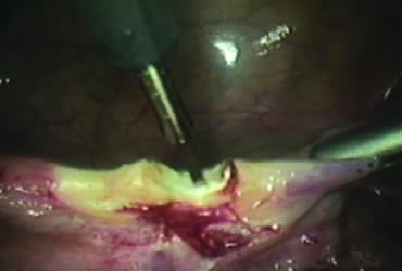
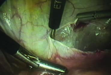
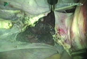
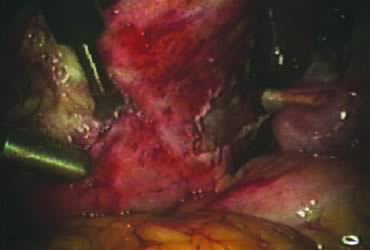
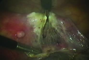
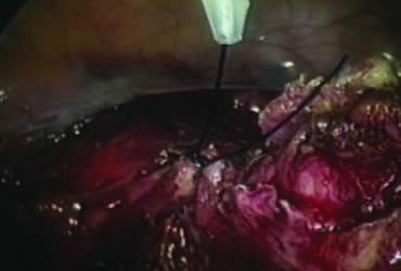
The right round ligament is held in its midportion, coagulated with bipolar forceps, and cut (Fig. 1). The anterior leaf of the right broad ligament is incised parallel to the uterus, and the incision is curved medially over the cervix (Fig. 2). The same two steps are repeated on the left side, and the incisions are connected (Fig. 3). The bladder flap is mobilized out of the surgical field by cutting the cervicovaginal septum, as needed. Coagulation and cutting the superficial upper portions of the lateral vesicouterine ligaments or bladder pillars completes the dissection.

Fig. 1. Coagulation and cutting of round ligament.

Fig. 2. Incision of the anterior leaf of the broad ligament.

Fig. 3. Creation of the bladder flap.

Posterior Dissection

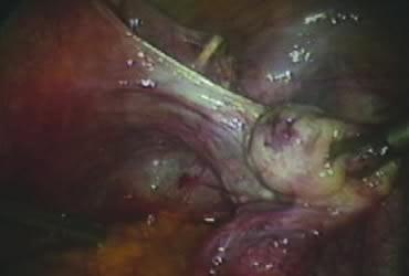
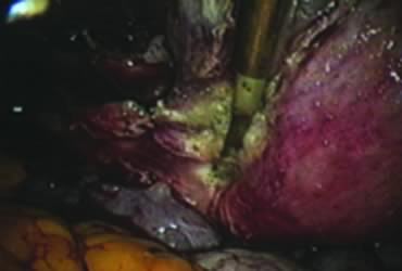
The right ovary is held with the three-prong grasper stretching and exposing the utero-ovarian ligament, which is coagulated and cut in its midportion (Fig. 4). The posterior leaf of the right broad ligament is incised downward to the level of the insertion of the uterosacral ligament in the cervix. The same two steps are repeated on the opposite side and the two sides are connected (Fig. 5).

Fig. 4. Coagulation of the utero-ovarian ligament.

Fig. 5. Incision of the posterior leaf of the broad ligament connecting both sides.

Isolation of the Adnexa

A window is created in an areolar avascular area of the broad ligament with gentle blunt dissection. The adnexal structures above the window are coagulated (or sutured) and cut, isolating the adnexa and detaching it from the uterus (Fig. 6). The procedure is repeated in the contralateral side.

Fig. 6. Creation of a window in the broad ligament.

Securing Uterine Vessels

In supracervical hysterectomy, only the ascending branch of the uterine artery is skeletonized and secured preferably with a stitch (Fig. 7). The tissues medial to the stitch are coagulated (to prevent back flow) and cut (Fig. 8). The same steps are repeated on the opposite side.

Fig. 7. Securing the uterine vessels with a stitch.

Fig. 8. Prevention of back bleeding by coagulating tissues medial to the stitch.

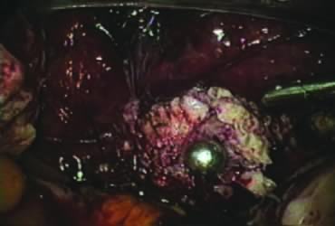
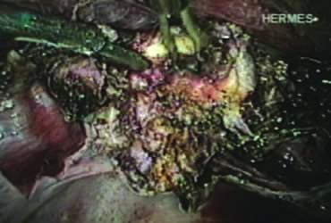
Amputation of the Uterus and Ablation of the Endocervix

After ligating both uterine arteries, the uterus becomes pale. At this time, the uterine body may be divided longitudinally using a shielded knife down to the cervix to facilitate morcellation. Each half is detached separately from the cervix 0.5 to 1 cm below the uterocervical junction and stored in the left upper abdomen. Alternatively, amputation of the uterine body off the cervix may be carried out through coagulation and cutting (above the ligated artery) proceeding from lateral to medial. The process may be initiated from the right or the left side. Bleeding points in the cervical stump are secured (Fig. 9). The upper portion of the endocervical canal is ablated circumferentially with bipolar coagulation (Fig. 10). Initially the walls of the cervix and the edges of the peritoneum were drawn together with sutures. This step was omitted, however, after noting at second look that the pelvic peritoneum heals without adhesions in the absence of sutures.

Fig. 9. Appearance of the cervical stump after uterine amputation.

Fig. 10. Ablation of the endocervix.

Concomitant Adnexectomy

If indicated, salpingo-oopherectomy can be performed at this time.

Removal of the Specimen

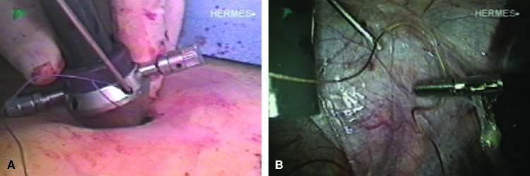
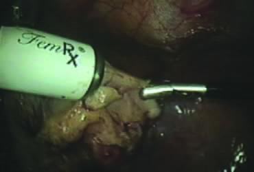
The stored uterus is removed using a mechanical morcellator, which is essentially a circular rotatory knife that slices the tissues longitudinally in the shape of a cylinder. The incision in the left lower quadrant is increased to 17 mm to accommodate a 15-mm trocar. The secure cone is mounted over the trocar, which is inserted into the abdomen.56 Sutures are passed through the tunnels of the secure cone to hold and stabilize the cannula in place (Fig. 11A). The mechanical morcellator is introduced through the 15-mm cannula, and morcellation is carried out in horizontal fashion by pulling the tissues into the morcellator from right to left to minimize the possibility of bowel injury (Fig. 12). When morcellation is complete, the trocar and the cone are removed, leaving the stay sutures in place. Tying these sutures together closes the surgical defect in the abdominal wall (Fig. 11B).56

Fig. 11. A. Secure cone introduction and cannula stabilization. B. Closure of the surgical defect.

Fig. 12. Uterine morcellation.

Lavage, Inspection, and Closure

The pelvic cavity is irrigated, the operative sites are inspected, and small bleeding points are controlled, if needed. The enlarged incision in the left lower quadrant is closed under direct vision, as previously described. Secondary access trocars are removed under direct vision. The abdomen is deflated, and the primary access cannula is withdrawn. The open laparoscopy incision is closed in layers. The Foley catheter and the uterine manipulator are removed, and the operation is completed.

Back to Top
CLASSIC INTRAFASCIAL SUPRACERVICAL HYSTERECTOMY TECHNIQUE

In the CISH technique, after the uterine body is amputated, the endocervical canal is cored out, and the transformation zone of the cervix is resected.

Back to Top
CLINICAL EXPERIENCE

Tables 1 and 2 show selected studies57,58,59,60 and outcome data for the LSH and CISH procedures. In reviewing the first 100 cases of LSH performed by our team, the average estimated blood loss was less than 100 mL with no blood transfusions given. The operative time ranged from 60 to 180 minutes; the mean length of stay was 1 day. Five patients complained of postoperative cyclic bleeding: Two of these experienced spontaneous cessation within a few months, two continued with scanty menses that did not cause them concern, and one had her cervix removed vaginally. Histology revealed endometriosis. Another patient had cervicectomy because of an abnormal Pap smear; histology revealed chronic cervicitis without dysplasia.21


TABLE 1. Comparison of Outcomes of Laparoscopic Subtotal Hysterectomy and Classic Intrafascial Supracervical Hysterectomy Procedures from Selected Studies


Procedure

Author

No. Patients

ORT (min) (range)

EBL (mL) (range)

LOS (hr) (range)

LSH

Donnez57

500

62 (30–135)

<100

42–72


Lyons58

236

85 (59–235)

55 (25–125)

17 (3–38)

CISH

Morrison59

437

70 (46–370)

68 (10–765)

22 (10–120)


Kim60

231

176.1 ± 55.1

152 ± 86

168


LSH, laparoscopic subtotal hysterectomy; CISH, classic intrafascial supracervical hysterectomy; ORT, operative time; EBL, estimated blood loss; LOS, length of stay.


TABLE 2. Comparison of Early and Late Complications of Laparoscopic Subtotal Hysterectomy and Classic Intrafascial Supracervical Hysterectomy Procedures from Selected Studies


Early Complications

Late Complications

Procedure

Author

No. Patients

Bladder Injury

Febrile Morbidity

Bleeding

Conversion

Return to OR

Cyclic Bleeding

Cervix Removal

LSH

Donnez57

500

3 (0.6%)


6* (2.4%)


Lyons58

236


1 (<1%)


1 (0.4%)

3 (1.3%)

CISH

Morrison59

437


3 (0.7%)

3 (0.7%)

1 (0.2%)

4 (0.9%)

1 (0.2)


Kim et al60

231


1 (0.4%)


3 (1.3%)


LSH, laparoscopic subtotal hysterectomy; CISH, classical intrafascial hysterectomy; OR, operating room.
* Of 243 patients who were followed.


The senior author (H.M.H.) reviewed 50 consecutive LSH cases he performed to assess quality-of-life outcomes after the procedure.61 Estimated blood loss was based on decrease in hemoglobin and hematocrit rather than the less reliable subjective estimates. The mean decrease in hemoglobin was 1.9 g (range, 0 to 6 g), and the mean decrease in hematocrit was 5.9% (range, 2% to 17%). Mean hospital stay was 1.2 days (nine patients stayed 2 days). Immediate complications included one bladder injury repaired laparoscopically, one pneumothorax in a patient with history of catamenial endometriosis, one patient with urinary retention, and one patient with occult inferior epigastric hematoma who received 2 U of packed red blood cells postoperatively. One patient experienced recurrent cyclic bleeding that did not require further treatment. The overall satisfaction rate was 94%, with one patient dead and two lost to follow-up: 89% of the patients were completely satisfied, 4% were satisfied, and one was satisfied with exception (cyclic vaginal spotting). The mean satisfaction score was 9.9 out of 10. Of patients, 80% returned to work part-time within 2 weeks and 90% within 3 weeks; 58% returned to work full-time within 2 weeks, 78% within 3 weeks, and 94% within 1 month. According to 38 responses, sexual activity was resumed in a mean time of 27 days (range, 10 to 60 days), with 72% of respondents having sexual relations within 1 month and 91% within 6 weeks.61

Other investigators reported higher rates of late complications that necessitated removal of the cervical stump. Okaro and colleagues62 reported 16 cervicectomies after 70 LSH procedures for a rate of 22.8%. Pelvic pain (75%) and cyclic bleeding (44%) were the most common indications. The pathology report showed normal cervix in six patients (35%), chronic cervicitis in one (6%), residual endometrium in four (23.5%), and endometriosis in four (23.5%). One patient had cervical dysplasia, and another had mucocele. Incomplete removal of the lower uterine segment and the presence of endometriosis accounted for almost 50% of the cases.62

Back to Top
CONCLUSIONS

In selected cases, laparoscopic subtotal (supracervical) hysterectomy is a suitable alternative to total vaginal, abdominal, or laparoscopic hysterectomy for the treatment of benign gynecologic disease. Development of carcinoma in the cervical stump may be prevented with periodic cytologic screening. Incomplete removal of the lower uterine segment and the presence of endometriosis may be associated with cyclic bleeding and pelvic pain, which may require further treatment.

Back to Top
REFERENCES

1. Farquhar CM, Sterner CA: Hysterectomy rates in the United States 1990–1997. Obstet Gynecol 99:229, 2002

2. Olive DL, Parker WH, Cooper JM, Levine RL: The AAGL classification system for laparoscopic hysterectomy. J Am Assoc Gynecol Laparosc 7:9, 2000

3. Lyons TL: Laparoscopic supracervical hysterectomy: A comparison of morbidity and mortality results with laparoscopic assisted vaginal hysterectomy. J Reprod Med 38:763, 1993

4. Hasson HM, Rotman C, Rana N, Asakura H: Experience with laparoscopic hysterectomy. J Am Assoc Gynecol Laparosc 1:1, 1993

5. Donnez J, Smets M, Polet R, et al: LASH: Laparoscopic supracervical (subtotal) hysterectomy. Zentralbl Gynakol 117:629, 1995

6. Metler L, Semm K, Lehmann-Wiellenbrrkl M, et al: Comparative evaluation of classical intrafascial supracervical hysterectomy (CISH) with transuterine mucosal resection as performed by pelviscopy and laparotomy, our first 200 cases. Surg Endosc 9: 418, 1995

7. Vietz PF, Ahn TS: A new approach to hysterectomy without colpotomy: Pelviscopic intrafascial hysterectomy. Am J Obstet Gynecol 170:609, 1994

8. Sadoghi H: Supracervical uterus amputation via the vaginal route. Geburtshilfe Frauenheilkd 54:602, 1994

9. Pelosi MA, Pelosi MA 3rd: Laparoscopic supracervical hysterectomy using a single umbilical puncture (minilaparoscopy). J Reprod Med 37:777, 1992

10. Stern E, Misczynski M, Greenland S, et al: Pap testing and hysterectomy prevalence: A survey of communities with high and low cervical cancer rates. Am J Epidemiol 106:296, 1977

11. Carter B, Thomas WL, Parker RT: Adenocarcinoma of the cervix and of the cervical stump. Am J Obstet Gynecol 57:37, 1949

12. Nielsen K: Carcinoma of the cervix following supracervical hysterectomy. Acta Radiol 37:335, 1952

13. Papanicolaou GN: A new procedure for staining vaginal smears. Science 95:438, 1942

14. Reich H, DeCaprio J, McGlynn F: Laparoscopic hysterectomy. J Gynecol Surg 5:213, 1989

15. Robertson EA, de Block S: Decrease in the number of abdominal hysterectomies after introduction of laparoscopic hysterectomy. J Am Assoc Gynecol Laparosc 7:523, 2000

16. Härkki P, Kurki T, Sjöberg J, Tiitinen A: Safety aspects of laparoscopic hysterectomy. Acta Obstet Gynecol Scand 80:383, 2001

17. Novotny Z, Rojikoval V: Complications of laparoscopic-assisted vaginal hysterectomy, a 1996 survey of the Czech Republic. J Am Assoc Gynecol Laparosc 6:459, 1999

18. Wood C, Maher P, Hill D: The declining place of abdominal hysterectomy in Australia. Gynaecol Endosc 6:257, 1997

19. ACOG: Surgical alternatives to hysterectomy in the management of leiomyomas. ACOG Practice Bulletin, no. 16, May 2000

20. ACS: ACS guidelines for early detection of cancer. CA Cancer J Clin 52:8, 2002

21. Hasson HM: Laparoscopic supracervical hysterectomy. In Liu C (ed): Laparoscopic Hysterectomy and Pelvic Floor Reconstruction. pp 238, 255 Cambridge, Blackwell, 1996

22. Storm HH, Clemmensen IH, Manders T, Brinton LA: Supracervical uterine amputation in Denmark 1978–1988 and risk of cancer. Gynecol Oncol 45:198, 1992

23. Hasson HM: Cervical removal at hysterectomy for benign disease: Risks and benefits. J Reprod Med 38:781, 1993

24. Hellström AC, Sigurjonson T, Pettersson F: Carcinoma of the cervical stump: The Radiumhemmet series 1959–1987: Treatment and prognosis. Acta Obstet Gynecol Scand 80:152, 2001

25. Hannoun-Levi JM, Peiffert D, Hoffstetter S, et al: Carcinoma of the cervical stump: Retrospective analysis of 77 cases. Radiother Oncol 43:147, 1997

26. Forbes C, Jepson R, Martin-Hirsch P: Interventions targeted at women to encourage the uptake of cervical screening. Cochrane Database of Systematic Reviews Issue 1:2002

27. Ylitalo N, Sorensen P, Josefsson AM, et al: Consistent high viral load of human papilloma virus 16 and risk of cervical carcinoma in situ: A nested case-control study. Lancet 355:2194, 2000

28. Josefson AM, Magnusson PK, Ylitalo N, et al: Viral load of human papilloma virus 16 as a determinant for the development of cervical carcinoma in situ: A nested case-control study. Lancet 355:2189, 2000

29. Kalogirou D, Antoniou G, Karakitsos P, et al: Vaginal intraepithelial neoplasia (VAIN) following hysterectomy in patients treated for carcinoma in situ of the cervix. Eur J Gynaecol Oncol 18:188, 1997

30. vanBeurden M, ten Kate FW, Tjong-A-hung SP, et al: Human papillomavirus DNA in multicentric vulvar intraepithelial neoplasia. Int J Gynecol Pathol 17:12, 1998

31. Sugase M, Matsukura T: Distinct manifestations of human papillomaviruses in vagina. Int J Cancer 72:412, 1997

32. Hoffman MS, Roberts WS, LaPolla JP, et al: Neoplasia in vaginal cuff epithelial inclusion cysts after hysterectomy. J Reprod Med 34:412, 1989

33. Romanes GJ (ed): Cunningham’s Text Book of Anatomy. pp 571, 572 Oxford, Oxford University Press, 1981

34. DeLancey JO: Anatomic aspects of vaginal eversion after hysterectomy. Am J Obstet Gynecol 166:1717, 1992

35. Baggish MS, Karram MM (eds): Anatomy of the vagina. Atlas of Pelvic Anatomy and Gynecologic Surgery. pp 336, 347 Philadelphia, WB Saunders, 2001

36. Baggish MS, Karram MM (eds): Anatomy of the cervix. Atlas of Pelvic Anatomy and Gynecologic Surgery. pp 284, 289 Philadelphia, WB Saunders, 2001

37. Mundy AR: An anatomical explanation for bladder dysfunction following rectal and uterine surgery. Br J Urol 54:501, 1982

38. Hasson HM, Parker WH: Prevention and management of urinary tract injury in laparoscopic surgery. J Am Assoc Gynecol Laparosc 5:99, 1998

39. Baggish MS, Karram MM (eds): Identifying and avoiding ureteral injury. Atlas of Pelvic Anatomy and Gynecologic Surgery. pp 204, 213 Philadelphia, WB Saunders, 2001

40. Hurd WW, Chee SS, Gallagher KL, et al: Location of the ureters in relation to the uterine cervix by computed tomography. Am J Obstet Gynecol 184:336, 2001

41. Jewett JF: Vaginal length and incidence of dyspareunia following total abdominal hysterectomy. Am J Obstet Gynecol 63:400, 1952

42. Piacenza JM, Salsano F: Post-hysterectomy fallopian tube prolapse. Eur J Obstet Gynaecol Reprod Biol 98:253, 2001

43. Nezhat CH, Nezhat F, Seidman D, Nezhat C: Vaginal vault evisceration after total laparoscopic hysterectomy. Obstet Gynecol 87:868, 1996

44. Taylor T, Smith AN, Fulton PM: Effect of hysterectomy on bowel function. BMJ 299:300, 1989

45. Prior A, Stanley K, Smith AR, Read NW: Effect of the hysterectomy on anorectal and urethrovesical physiology. Gut 33:264, 1992

46. Kilkku P, Hivornen T, Gronroos M: Supra-vaginal uterine amputation vs. abdominal hysterectomy: The effects on urinary symptoms with special reference to pollakisuria, nocturia and dysuria Maturitas 3:197, 1981

47. Parys BT, Haylen BT, Hutton JL, Parsons KF: Urodynamic evaluation of lower urinary tract function in relation to total hysterectomy. Aust N Z J Obstet Gynaecol 30:161, 1990

48. Johns A: Supracervical versus total hysterectomy. Clin Obstet Gynecol 40:903, 1997

49. Kilkku P: Supravaginal uterine amputation vs. hysterectomy: Effects on coital frequency and dyspareunia Acta Obstet Gynecol Scand 62:141, 1983

50. Kilkku P, Gronroos M, Hivornen T, Rauramo L: Supravaginal uterine amputation vs. hysterectomy: Effects on libido and orgasm Acta Obstet Gynecol Scand 62:147, 1983

51. Popov I, Stoikov S, Boiadzhieva M, Khristova P: Disorders in sexual function following hysterectomy. Akush Ginekol 37:38, 1998

52. Strauss B, Jakel I, Koch-Dorfler M, et al: Psychiatric and sexual sequelae of hysterectomy, a comparison of different surgical methods. Geburtshilfe Frauenheilkunde 56:473, 1996

53. Scott JR, Sharp HT, Dodson MK, et al: Subtotal hysterectomy in modern gynecology: A decision analysis. Am J Obstet Gynecol 176:1186, 1997

54. Grimes DA: Role of the cervix in sexual response: Evidence for and against. Clin Obstet Gynecol 42:972, 1999

55. Dennerstein L, Wood C, Burrows GD: Sexual response following hysterectomy and oopherectomy. Obstet Gynecol 49:92, 1977

56. Hasson HM: Laparoscopic cannula cone with means for cannula stabilization and wound closure. J Am Assoc Gynocol Laparosc 5:183, 2001

57. Donnez J, Nisolle M, Smets M, et al: Laparoscopic supracervical (subtotal) hysterectomy: A first series of 500 cases. Gynaecol Endosc 6:73, 1997

58. Lyons TL: Laparoscopic supracervical hysterectomy. Baillieres Clin Obstet Gynaecol 11:167, 1997

59. Morrison JE Jr, Jacobs VR: 437 Classic intrafascial supracervical hysterectomies in 8 years. J Am Assoc Gynecol Laparosc 8:558, 2001

60. Kim DH, Bae DH, Hur M, Kim SH: Comparison of classic intrafascial supracervical hysterectomy with totallaparoscopic and laparoscopic-assisted vaginal hysterectomy. J Am Assoc Gynecol Laparosc 5:253, 1998

61. Hasson HM: Quality of life after supracervical laparoscopic hysterectomy. J Am Assoc Gynocol Laparosc 8(3 suppl):S24, 2001

62. Okaro EO, Jones KD, Sutton C: Long term outcome following laparoscopic supracervical hysterectomy. Bjog 108:1017, 2001

Back to Top