Chapter 31
Thromboembolic Disease
David C. Shaver
Main Menu   Table Of Contents

Search

David C. Shaver, MD
Director, Maternal-Fetal Medicine, Presbyterian Hospital, Charlotte, North Carolina (Vol 2, Chap 31)

INTRODUCTION
PATHOGENESIS
DIAGNOSIS
DIAGNOSIS OF PULMONARY EMBOLUS
MANAGEMENT
INITIAL TREATMENT
LONG-TERM THERAPY
ALTERNATIVE THERAPIES
PREVENTION OF THROMBOPHLEBITIS
SEPTIC PELVIC THROMBOPHLEBITIS
REFERENCES

INTRODUCTION

Venous thromboembolic disorders are a leading cause of morbidity and mortality during pregnancy and the puerperium.1 The exact incidence of venous thrombosis during pregnancy varies depending on the criteria used for diagnosis. Studies using strict radiologic criteria in symptomatic patients have suggested a risk of venous thrombosis of between 1 in 1000 and 1 in 2000 pregnant women.2 The subclinical incidence of the disease, however, may be higher as suggested by prospective studies in asymptomatic women.3

Historically the risk during pregnancy was believed to increase as the pregnancy progressed, the greatest risk for the patient being in the postpartum period. Obstetric management of these patients usually included immobilization and prolonged hospitalization after delivery, particularly after an operative delivery, which may have accounted for the marked increase in thromboembolic events in the postpartum period. More recent data suggest that most clinically significant thromboembolic events occur in the antepartum period.4,5 Regardless of which pregnancy stage carries the highest risk, venous thrombosis continues to be a source of major morbidity and is the leading cause of death after a live birth in the United States.6

Back to Top
PATHOGENESIS

Numerous factors appear to contribute to the development of venous thrombosis. Virchow was the first to suggest a triad of predisposing factors: (1) alterations in the composition or chemistry of the blood, (2) venous stasis, and (3) damage to the vessel wall.

Changes in the Composition of Blood

Normal coagulation depends on a complex interplay among numerous factors within the coagulation system. The initial step in thrombus formation is usually the aggregation of the platelets in response to damage to a vessel wall. This platelet aggregation results in a release of (1) adenosine diphosphate, which attracts more platelets and (2) platelet factor 3, which initiates the coagulation system.

The coagulation system (Fig. 1) consists of numerous proteins, most of which are proenzymes that circulate in trace amounts (micrograms). Both an intrinsic and extrinsic pathway exist for initiating the coagulation system. Initiation of coagulation through either pathway ultimately involves a common pathway of activation of factor X to Xa. Factor Xa results in generation of thrombin, which converts fibrinogen into fibrin clot.

Fig. 1. Coagulation pathways.

Normal inhibitors to the coagulation system are present, characterized most importantly by antithrombin III (AT III). AT III can effectively neutralize several factors in the coagulation system, including thrombin, as well as factors XII, X, and IX.

The fibrinolytic system consists primarily of plasmin, which is formed from plasminogen. The activation of plasminogen can result from the liberation of activators at the site of thrombus formation or from activated factor XII. The final result of plasmin is the breakdown of fibrin with the release of fibrin degradation products.

The protein C pathway is also important in inhibition and fibrinolysis. Protein C activation occurs as the result of thrombin binding to the endothelial cell receptor thrombomodulin. Activation of protein C increases tissue plasminogen activator activity, resulting in its fibrinolytic activity. Protein C, in association with protein S, also has anticoagulant activity in that it inhibits factors V and VIII.

Pregnancy has a significant effect on the proteins in the coagulation system. All clotting factors except factors XII and XIII are increased during pregnancy, the most marked effect being on fibrinogen levels, which increase twofold to threefold during pregnancy.7 Lesser effects are seen on the anticoagulant or fibrinolytic system, with most studies demonstrating little change in the levels of AT III and protein C during pregnancy. Protein S levels, however, are noted to be decreased during pregnancy.8 All of these measurable changes in the coagulation system appear to increase the tendency toward thrombus formation. All parameters return to normal by 3 weeks postpartum.

Stasis

Pregnancy is known to have a marked effect on blood return from the lower extremities. The enlarging gravid uterus obstructs blood return through the inferior vena cava, with a subsequent increase in pressure and stasis below the level of obstruction. Additionally, the increase in circulating blood volume adds to the venous distention.

Vessel Damage

The distention of the veins of the pelvis and lower extremity has the potential to damage the endothelium. Additionally, damage to the venous walls during parturition or as a result of postpartum infections can occur.

Back to Top
DIAGNOSIS

The diagnosis of deep vein thrombosis (DVT) is typically made on the basis of an acutely swollen and painful leg in the absence of another explanation, such as trauma. The calf and thigh are frequently turgid and tender, and dorsiflexion of the foot results in pain in the calf (Homan's sign). Numerous studies have demonstrated, however, that clinical diagnosis of DVT is fraught with errors, and that an incorrect diagnosis can be made in 50% or more of cases.9,10 Therefore, an objective evaluation is required in all cases in which DVT is suspected. Although treatment may be initiated before the diagnosis is confirmed, long-term therapy should never be carried out in the absence of objective criteria. Numerous tests have been developed to screen for DVT.

Venography

Contrast venography is the definitive test for evaluating DVT and is considered the standard by which all other tests are judged because of its ability to completely and objectively visualize the venous system of the lower extremity.11 Contrast agents are injected to the venous system of the lower extremity, and the venous system of the leg and pelvis are evaluated both fluoroscopically and radiographically.

There are, however, a number of disadvantages to contrast venography:

  1. Obstetricians are frequently reluctant to use this test because of concern about fetal exposure; however, with shielding of the maternal abdomen except during evaluation of the pelvis, the radiation exposure to the fetus can be kept to a minimum.
  2. This is an expensive test, both time-consuming for the radiologist to interpret and economically costly.
  3. There is an inherent risk of phlebitis associated with injection of radiocontrast agents, which may be as high as 2% to 3%.12

The veins of the pelvis are difficult to evaluate with venography as well as with most of the tests used to evaluate DVT. The use of computed tomography (CT) for evaluating pelvic veins may be superior, especially in the postpartum patient,13 although the sensitivity has not been well defined. Because of its invasiveness and potential risk, contrast venography is generally not recommended as a first-line diagnostic tool. Noninvasive methods should be used first, with venography reserved for cases in which noninvasive methods are nondiagnostic.

Impedance Plethysmography

Impedance plethysmography (IPG) is a simple, noninvasive technique that has been shown to be both sensitive and specific. In IPG, the electrical impedance between two electrodes wrapped around the calf is measured. A pneumatic cuff is applied to the upper leg and, as venous outflow is reduced and the leg becomes engorged with blood, the impedance decreases. Release of the cuff results in a characteristic change in impedance if venous outflow is not compromised. IPG is useful in assessing proximal vein thrombosis but generally unreliable in evaluating the calf veins.11 False-positive results may also occur with other causes for obstruction to venous flow, including a gravid uterus.14 The test can be reliable even during the third trimester of pregnancy if the patient is placed in a lateral decubitus position.15

Studies have shown that the use of IPG serially in patients can be very reliable for excluding DVT. It is an ideal noninvasive test for following patients serially who are believed to be at high risk.

Ultrasonography

Introduction of real-time B-mode ultrasonography to assess the venous system of the lower extremities is currently the most widely used means of investigation. Although thrombosis in the vein may be frequently visualized with ultrasonography, the most sensitive finding is a failure to demonstrate compression of a vein with gentle pressure on the transducer.16 This lack of compression indicates the presence of a thrombus. Obviously, this method has limited usefulness in the evaluation of pelvic veins.

The introduction of ultrasonography supplemented by color flow Doppler imaging does allow visualization of all the vessels of the lower extremity. Meta-analysis of four studies of symptomatic patients indicated a sensitivity of 93% and a specificity of 98% in the detection of DVT.17

In a symptomatic patient with a positive IPG or an ultrasound result indicating DVT, treatment can be initiated because of the specificity of both of these tests. In this situation, confirmation by venography is not required.

Back to Top
DIAGNOSIS OF PULMONARY EMBOLUS

Pulmonary embolus complicates 1 in every 750 to 2500 pregnancies. The onset of dyspnea in a patient with DVT obviously suggests pulmonary embolus. In most cases, however, the diagnosis is more obscure, and appropriate diagnostic studies must be done before committing a patient to long-term anticoagulation.

The immediate result of a pulmonary embolus is the obstruction to pulmonary arterial flow. In addition to alteration of blood flow based purely on mechanical obstruction by the thrombus, there is concomitant vasoconstriction of additional small arterial vessels, thought to be mediated by the release of serotonin from embolized platelets. The net result is decreased perfusion of the normally ventilated portion of the lung. This effect is immediate and is complicated by an additional effect of poor pulmonary perfusion (i.e., loss of surfactant). Progressive loss of surfactant begins within a few hours after embolization, and atelectasis can be detected within 24 to 48 hours.

Infarction of lung tissue as a result of the embolus rarely occurs because of the additional supply of oxygen to the lung tissue from the bronchial arterial circulation and from direct diffusion through the alveoli. Unless there is underlying cardiopulmonary disease affecting the latter, pulmonary infarction after pulmonary embolus is rare.

Clinical Manifestations

The hallmark of pulmonary embolus is the acute onset of dyspnea. In pregnancy, however, dyspnea can be difficult to assess, and associated findings, such as tachypnea and discomfort in the lower extremities, are common. Substernal chest pain may be noted, but hemoptysis and pleuritic chest pain are unusual, occurring only with infarction. Physical examination is usually normal, except for the presence of tachycardia. An electrocardiogram may demonstrate right heart strain, and arterial blood gases may show hypoxia. It should be remembered, however, that during the third trimester arterial oxygen tension (PaO2) can be as much as 15 mmHg lower in the supine position than in the sitting position.18 Because of the implications of the diagnosis of pulmonary embolus and the need for long-term therapy, objective documentation using various tests is recommended.

Pulmonary Ventilation/Perfusion Scans

The ventilation/perfusion (VQ) scan is the primary method of screening for pulmonary embolus.1,2 Confirmation of a pulmonary embolus requires documentation of absent or decreased perfusion to a segment of the lung. Using a perfusion scan, radiolabeled microspheres are injected intravenously and are lodged in the pulmonary capillaries. Their distribution is then determined with a gamma detecting device. A normal perfusion scan virtually eliminates the possibility of an embolus. The presence of one or more defects requires further evaluation, usually a ventilation scan. The ventilation scan is performed by inhalation of radioactive xenon, which can then be compared with the perfusion scan. Certain conditions, such as pneumonia, that are associated with decreased perfusion are also underventilated, whereas lung tissue involved in embolus is poorly perfused but normally ventilated early in the course of the disease. The amount of radiation associated with the performance of a VQ scan is minimal and should not preclude its use in pregnancy.19

Pulmonary Angiography

Lung scans provide only an indirect measurement of pulmonary perfusion and in cases in which the diagnosis is uncertain, pulmonary angiography is required. Because this technique allows one to actually visualize the pulmonary vessels, it is the standard against which the other methods are evaluated. With the appropriate abdominal shielding, the amount of radiation to the fetus should be minimal and should not preclude its use in pregnancy.19

How aggressive one should be in evaluating the patient for possible pulmonary embolus depends on the individual case. However, if the patient's symptoms are serious enough to warrant contemplating long-term therapy, every effort should be made to obtain a definitive diagnosis before committing a patient to this course of therapy.

Back to Top
MANAGEMENT

The goals of treatment of thromboembolic disease in pregnancy are several. Arrest of the growth of the thrombus and prevention of pulmonary embolization are uppermost. In addition, eventual restoration of patency with resolution of the thrombus would hopefully follow with as little damage to the veins and valves as possible, thus preventing the postphlebitic syndrome. The primary management of DVT and pulmonary embolus is medical, the mainstay of treatment being anticoagulation. The anticoagulants available include heparin and warfarin (Coumadin). Other modalities are available and have a limited role, including thrombolytic agents and surgery.

Warfarin

Warfarin is the most widely used oral anticoagulant. It is a synthetic drug that is absorbed well orally, has a long half-life, and easily crosses the placenta. The anticoagulant effect is due to its ability to 
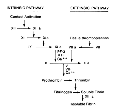
compete with vitamin K-dependent clotting factors (factors II, VII, IX, and X).

Bleeding complications are more frequent with warfarin than with heparin. The long half-life of warfarin and the mechanism of action make good control more difficult. The action of warfarin can be reversed by the administration of vitamin K.

Warfarin is contraindicated during pregnancy. Administration in the first trimester, particularly from 6 to 9 weeks' gestation, is associated with warfarin-specific embryopathy,20,21 characterized by hypoplastic nose and stippled epiphyses in 25% to 50% of cases. These abnormalities are the result of the direct teratogenic effects of warfarin, not the indirect effect of fetal anticoagulation, since clotting factors are not present in the first-trimester fetus.

Central nervous system abnormalities are associated with the use of warfarin at any time during pregnancy.22,23 Additionally, fetal hemorrhage, especially during labor and delivery, is known to occur in these patients. The exact fetal risk is unknown, but complications including intraventricular hemorrhage and stillbirths, appear to be increased in patients taking warfarin during pregnancy. Women who inadvertently take the medication during the first trimester should be counseled regarding the risk to the fetus, and the option of therapeutic termination of pregnancy should be offered.

Warfarin is detectable in only very small amounts in breast milk, and its use does not appear to be contraindicated in breast-feeding mothers.24,25

Heparin

Heparin is a high-molecular-weight mucopolysaccharide obtained from the mucosa of the lung and gut of swine and cattle. It is highly negatively charged and has a molecular weight of 3000 to 30,000 with an average of about 12,000, and therefore does not cross the placenta. In addition, it has a short half-life of about 30 to 60 minutes.26

The anticoagulant activity of heparin is derived from its interaction with the antithrombins, primarily AT III. The effect of AT III, which normally inhibits the activity of the activated proteases (clotting factors), including both thrombin and factor Xa, is markedly accelerated in the presence of heparin. The anticoagulant effect of heparin is almost instantaneous. In addition, heparin has an inhibitory effect on platelet aggregation and serotonin release.27

Heparin is considered the anticoagulant of choice in pregnancy. Because it does not cross the placenta, there should be no direct fetal effect such as that seen with warfarin. An initial report suggested an adverse effect of anticoagulants on pregnancy, regardless of which agent was used, implying that heparin was not safer than oral anticoagulants.28 However, this study did not control for the maternal conditions that were being treated. A more recent study suggested that if this prior study had controlled for the medical conditions being treated, the outcomes of the pregnancies treated with heparin would not have differed from those of the normal population.29

There are several significant side effects associated with heparin administration. As is the case with all anticoagulants, hemorrhage can occur. Significant hemorrhage is infrequent in the absence of risk factors such as surgery or trauma (probably less than 5%). Long-term administration of heparin (greater than 6 months) is associated with osteoporosis.30 The incidence of osteoporosis with long-term administration is unknown but may occur in all patients if treated long enough.31 Despite the confirmation of heparin-induced osteoporosis, few cases of symptomatic fractures have been reported.

Of potentially greater risk to the patient who is receiving heparin is the occurrence of heparin-associated thrombocytopenia. The thrombocytopenia is caused by heparin-dependent IgG antibodies.32 The onset of thrombocytopenia can be insidious but typically occurs more than 5 days after initiation of heparin therapy and is more common with higher doses of heparin compared to low-dose prophylactic therapy. Platelet counts are typically checked within 5 to 7 days after initiation of therapy, and if they remain normal for 2 weeks, development of thrombocytopenia is unlikely.1 If thrombocytopenia does occur, it can increase bleeding tendencies, but can be paradoxically associated with arterial thrombosis, presumably as a result of platelet activation. The incidence of thrombocytopenia is apparently related to the source of heparin; in one study the instance of thrombocytopenia was 26% in patients receiving bovine lung heparin versus 7% to 9% in patients receiving porcine intestinal mucosa-derived heparin.33 These differences were highly significant (p < 0.005). Most studies, however, have demonstrated a significantly lower incidence of immune-mediated thrombocytopenia, with the incidence generally reported to be less than 1% to 2%.26

Anticoagulation achieved by heparin administration can be rapidly reversed with protamine sulfate. The dosage necessary for reversal is 1 mg of protamine sulfate per 100 units of heparin. In treating patients on continuous infusion of heparin, the dose should be appropriate for the amount of heparin delivered over the previous hour, because of the very short half-life of heparin.

Low-Molecular-Weight Heparin

Low-molecular-weight (LMW) heparin is prepared by the depolimerization and fractionation of standard heparin, a process that yields chains with a mean molecular weight of 4000 to 6000.34 The unique sequence of heparin, which accounts for its binding to AT III and therefore its anticoagulant effect, is preserved. Despite its smaller size, it is still too large to cross the placenta and appears to be safe for use during pregnancy. Several advantages of LMW heparin have been demonstrated, although its superiority for use in pregnancy has not been established because of the limited number of clinical studies.

LMW heparin has been shown to have fewer hemorrhagic complications than standard heparin.35 This may be due to its less pronounced effect on platelet function and the fact that it preferentially inactivates factor Xa but not thrombin, whereas standard heparin inactivates both.

Additionally, LMW heparin has more favorable pharmacokinetics because it binds less readily to endothelial cells, macrophages, and plasma proteins than does standard heparin. This results in a longer plasma half-life and a more predictable anticoagulant response. Therefore, dosing is less frequent (once or twice per day), and the usual laboratory monitoring of anticoagulant effect is not required.

The incidence of thrombocytopenia associated with LMW heparin has been found to be decreased compared to standard heparin,36 and LMW heparin has been used efficaciously in patients who have had thrombocytopenia while on standard heparin. Although the incidence of osteoporosis has been suggested to be lower with LMW heparin, this has not been proved.

Back to Top
INITIAL TREATMENT

The initial therapy of choice for thromboembolism is intravenous heparin. Immediate anticoagulation with heparin is necessary for effective therapy, and treatment with warfarin alone is never indicated, even in the nonpregnant patient.37 Therapy should be begun in any patient who presents with signs and symptoms suggestive of DVT, and further evaluation to confirm the diagnosis can be carried out while the patient is receiving the drug. For DVT, heparin is initially given in a bolus dose of 5000 units. If pulmonary embolus is suspected, the initial heparin dose is 15,000 to 20,000 units.

After the initial bolus, heparin is generally given as a continuous intravenous infusion. Intermittent bolus therapy can also be used, although bleeding complications may be more common when heparin is given as a bolus rather than as continuous infusion. In the past, therapy was continued for 10 to 14 days; however, in a study of the nonobstetric population, it has been shown that 5 days of intravenous heparin is adequate for initial therapy.38 Similar results have not been confirmed in an obstetric population.

Before initiating heparin therapy, a baseline prothrombin time (PT), partial thromboplastin time (PTT), urinalysis, and platelet count should be obtained. In most laboratories PTT is satisfactory for monitoring heparin therapy, but because factor VIII levels are increased during pregnancy, the patient's own pretreatment values should be used as the baseline. An alternative is to monitor heparin levels; levels of 0.2 to 0.4 units/mL are considered therapeutic. If heparin is given as a continuous infusion, it should be administered by an infusion pump.

Every effort should be made to have the PTT in the therapeutic range as quickly as possible, since patients who are subtherapeutic in the first 24 hours may have rates of recurrence of DVT as much as 15 times higher than those patients in whom the PTT is not in the therapeutic range.39 A PTT value of 1.5 to 2.5 times the control value is commonly recommended as the target therapeutic value. Guidelines for adjustments in the rate of heparin infusion based on PTT levels are given in Table 1A and Table 1B.

TABLE 1A. Published Protocols for Heparin Infusion: Weight Based*


Measured Value

Adjustment

APTT <35 s (<1.2 × control)

80 U/kg bolus; increase infusion by 4 U/kg/h

APTT <35–45 s (1.2–1.5 × control)

40 U/kg bolus; increase infusion by 2 U/kg/h

APTT 46–70 s (>1.5–2.3 × control)

No change

APPT 71–90 s (>2.3–3 × control)

Decrease infusion by 2 U/kg/h

APPT >90 s (>3 × control)

Stop infusion for 1 h, then decrease by 3 U/kg/h


APTT = activated partial thromboplastin time
*Initial dose is a bolus of 80 U/kg, followed by infusion starting at 18 U/kg/h. APTT measured every 6 hours
(Raschke RA, Reilly BM, Guidry JR et al: The weight based heparin dosing nomogram compared with a “standard care” nomogram: A randomized controlled trial. Ann Intern Med 119:874, 1993)

TABLE 1B. Published Protocols for Heparin Infusion*


Rate of Change †


APTT(s)

Bolus (U)

Stop Infusion (min)

(mL/h)

Repeat APTT

<50

5000

0

+3

6 hr

50–59

0

0

+3

6 hr

60–85

0

0

0

Next AM

86–95

0

0

-2

Next AM

96–120

0

30

-2

6 h

>120

0

60

-4

6 h


APTT = activated partial thromboplastin time
*5000-U bolus, followed by 1280 U/hr
Author: Please supply credit line
†1 mL = 40 U.
(Cruickshank MK, Levine MN, Hirsh J et al: A standard heparin nomogram for the management of heparin therapy. Arch Intern Med 151:333, 1991)

If heparin is administered as an intermittent bolus, the aim is to achieve a prolongation of the PTT 1.5 to 2 times the pretreatment level, with a measurement obtained just before the next intermittent dose. In general, this will require 5000 to 7500 units of heparin every 4 hours.

Although anticoagulants are the mainstay of therapy, several additional modalities may be of benefit in therapy. In patients in whom pulmonary embolus is suspected, additional treatment with oxygen therapy is indicated, maintaining the PaO2 above 70 mmHg or oxygen saturation above 95%. Initial bed rest is indicated in all patients with thrombosis, and elevation of the lower extremities may be helpful in relieving edema. Local heat may be beneficial in alleviating pain, and narcotic agents are used as indicated. Ambulation is encouraged as soon as most of the local signs and symptoms begin resolving.

Recent studies have indicated that in the nonpregnant patient, subcutaneous doses of LMW heparin are equivalent to intravenous administration of unfractionated heparin.35 Although there are limited data on this modality during pregnancy, the use of LMW heparin in this situation has several advantages. Dosing is given only once or twice daily, monitoring anticoagulant effect is not required, and bleeding complications may be less than with standard intravenous heparin. An outpatient regimen of LMW heparin as compared to hospitalization for standard intravenous therapy in patients with proximal vein thrombosis has been reputed to be safe and effective.40 Although unfractionated heparin therapy remains the standard, the advantages of LMW heparin may prove to make it the better choice. The cost of LMW heparin is considerably greater than that of unfractionated heparin, but it may be more cost-effective because monitoring of laboratory values is not required and because therapy may potentially be administered on an outpatient basis.

Back to Top
LONG-TERM THERAPY

Intravenous heparin should be continued for at least 5 days or until symptoms improve. After the initial heparinization, long-term therapy is indicated for at least 3 months or until 6 weeks postpartum, whichever is longer. In the postpartum patient, warfarin therapy may be initiated along with the heparin, and the PT is monitored to maintain an international normalized ratio of between 2.0 and 3.0. The heparin is discontinued after 5 days if adequate anticoagulation is achieved on warfarin. Therapy is continued for at least 3 months and may be continued for 6 months or longer in patients with pulmonary embolus or recurrent DVT. If warfarin is given concomitantly with the heparin over a 3- to 5-day period, a loading dose of warfarin is unnecessary. A typical starting dose is 10 mg daily; adjustments in the dose can be made, with an average dose of 5 to 15 mg necessary to achieve an adequate prolongation of the PT. Concomitant heparin has a minimal effect on the PT, so no adjustments are necessary in interpreting the PT when both warfarin and heparin are being given.

In the pregnant patient, long-term therapy with warfarin is contraindicated. For this reason, long-term therapy is typically carried out with subcutaneous injections of unfractionated heparin. Dosage and method of administration of heparin are dependent on the site of thrombosis and whether pulmonary embolization occurred. With distal or calf vein thrombosis, low-dose therapy (5000 units every 12 hours) appears to be adequate in preventing recurrence.41 Low-dose heparin is not satisfactory in preventing recurrence of proximal vein (iliofemoral thrombosis) or pulmonary embolism.42 A satisfactory alternative to warfarin in these circumstances is adjusted-dose heparin. This technique involves subcutaneous administration of heparin given every 12 hours in a dose sufficient to prolong the midinterval (6-hour) PTT to 1.5 times the control value. In nonpregnant patients, the mean dosage with this regimen is 10,000 units every 12 hours. In the pregnant patient, the heparin should be a concentrated dose (20,000 units/ml) and a dose of up to 15,000 to 20,000 units every 12 hours may be necessary.

LMW heparin may also be effective in preventing DVT recurrence. The advantage of LMW heparin in this situation would be that laboratory monitoring of anticoagulant effect would not be required. The high cost of LMW heparin, however, makes it a less attractive alternative for long-term therapy.

Back to Top
ALTERNATIVE THERAPIES

In patients in whom anticoagulation is contraindicated, such as those who have had recent surgery or have a bleeding abnormality, filters inserted into the inferior vena cava can be used safely and effectively. Several reports on their use during pregnancy have been described, and no complications unique to pregnancy have been described. Other surgical approaches are generally restricted to those situations in which medical therapy has failed or is contraindicated. A massive pulmonary embolus in an unstable patient may require surgical removal of the thrombus. Surgical therapy for DVT has been studied much less extensively, although cases of removal of iliofemoral thrombus have been reported with good results. Generally surgical therapy for DVT is restricted to those cases in which the viability of the limb is threatened by an acute, massive iliofemoral thrombosis with development of phlegmasia cerulea dolens, which is a state of extensive venous occlusion with compromised arterial circulation.43

Thrombolytic treatment with either urokinase or streptokinase in the past, or more recently with tissue plasminogen activator, has been demonstrated to be more effective than anticoagulation alone for restoration of patency of the affected vein.44 Presumably, successful therapy would have a favorable impact on long-term sequelae of DVT, such as the postphlebitic syndrome. This might be expected to be of special benefit to obstetric patients because there is evidence that venous insufficiency is more common after DVT in pregnancy than in other groups.45 However, because of a potential risk of bleeding into the placental bed with abruptio placenta, thrombolytic therapy during pregnancy has been restricted to life-threatening conditions such as massive pulmonary emboli. Additionally, the risk in the postpartum patient is not well known, and concerns about increased risk of bleeding from the placental site or after a cesarean section typically would preclude its use in the postpartum patient.

Reports of graduated compression stockings used in nonpregnant patients after the first episode of venogram-proven proximal DVT revealed a decreased incidence of postphlebitic syndrome in the treated patients.46 Interestingly, within the first 2 years there was evidence of postphlebitic syndrome in up to one half of the patients in the control-group study, whereas those who were continued on graduated compression stockings had approximately one half this risk.

Back to Top
PREVENTION OF THROMBOPHLEBITIS

The most effective therapy for thrombophlebitis is prevention. Minidose heparin has been shown to be effective in preventing DVT in patients at risk. The rationale for minidose heparin is that smaller doses of heparin are required to prevent factor X activation than are required for therapy once the clotting cascade has been activated. When given as 5000 units subcutaneously every 12 hours, heparin is not associated with major bleeding complications and does not require monitoring of coagulation studies.

The major factor in administering low-dose heparin is determining which patients are at significant risk and would benefit from its use. Typically, in nonobstetric patients heparin is given for surgical procedures or for the immobilized patient. The data in the obstetric literature regarding the efficacy and benefit of low-dose heparin use are scarce. Empirically, patients who are considered at increased risk, particularly those with a history of thrombophlebitis, are believed to benefit from low-dose heparin therapy. Pregnancy per se is not considered to be a sufficient risk to require heparin therapy, and most pregnant patients who undergo surgery can obtain adequate prophylaxis from graduated compression stockings and early ambulation.

In some patients, the decision whether to initiate heparin therapy is difficult. An example is the patient with a previous history of thrombophlebitis not associated with pregnancy, such as occurs after trauma or with an orthopedic procedure. No clear consensus exists as to the risk for such a patient during a subsequent pregnancy and the need for prophylaxis.

Prophylactic heparin should also be considered for patients who have familial hypercoagulable states, such as a deficiency of protein C, protein S, or AT III. Recent reports have implicated factor V Leiden, which results from a point mutation in the gene coding for factor V, for the increased tendency toward venous thrombosis. This results from a resistance of factor V to activated protein C.2 Patients with a positive anticardiolipin antibody are also believed to benefit from heparin therapy throughout pregnancy, both for prevention of DVT in the mother and for thrombosis within the placental bed.

Back to Top
SEPTIC PELVIC THROMBOPHLEBITIS

Septic (or suppurative) pelvic thrombophlebitis is a relatively uncommon condition that occurs primarily in the postpartum period, although it can occur after gynecologic procedures. It is an entity distinct from DVT in that clotting in the pelvic veins is usually secondary to pelvic infection. Whereas DVT is usually associated with a single large thrombus that can result in embolization of one or more fragments, septic pelvic thrombophlebitis results from infection in the pelvic organs, with formation of an infected clot in the pelvic veins that undergo liquefaction and give rise to multiple small infected emboli. It is seen most commonly after cesarean section or septic abortion and less commonly after vaginal delivery. The risk is also increased with prolonged labor or amnionitis. The incidence appears to be decreasing in recent years, presumably as a result of more successful therapy of infectious complications of delivery.47

Diagnosis

Classically, the diagnosis of septic pelvic thrombophlebitis is one of exclusion. The patient is typically seen from days to weeks after a gestational event with a fever that is unresponsive to conventional antibiotic therapy. Most patients report having chills, and a characteristic “picket fence” (i.e. sharply spiking) appearance of the fever has been described. The pulse rate tends to be rapid and sustained. The chest X-ray is usually normal but may reveal abnormalities consistent with abscess or infarction.

Generally, the physical examination is not helpful in diagnosis. Examination of the uterus and parametrial areas may reveal no tenderness or abscess formation. Patients are typically in no distress, although patients with ovarian vein thrombosis may appear acutely ill, and the thrombosed ovarian vein may be palpable abdominally.

Debate exists as to whether thrombosis of the ovarian vein is a distinct entity from that of suppurative pelvic thrombophlebitis. Septic pelvic thrombophlebitis typically affects the iliofemoral vessels and has a more indolent and nonacute course. Ovarian vein thrombosis, on the other hand may present more acutely and earlier in the postpartum period. That they are part of the same process is suggested by clinical overlap,48 with many patients having involvement of both the iliofemoral and the ovarian veins, and the clinical course can by very similar in the two processes. Ovarian vein thrombosis can, however, occur acutely in the absence of infection and has even been described in antepartum patients.49

As noted, the diagnosis is usually one of exclusion, and it is based on response to therapy. Several radiographic techniques have been employed to aid in diagnosis. Excretory urography, sonography, and pelvic venography are occasionally of benefit when associated with a positive finding. Numerous reports have demonstrated that CT scanning of the pelvis is frequently helpful for diagnosis and has the advantage of being both specific and noninvasive, although nonocclusive thrombus in the iliofemoral veins might not always be detected.50 Ovarian vein thrombosis, however, is usually seen on CT scanning. Thrombosis of the ovarian vein is typically found on the right (90% of cases) and the thrombus can, on occasion, extend into the inferior vena cava.

Therapy

Collins,51 in 1951, was the first to describe the pathogenesis of septic pelvic thrombophlebitis. He advocated antimicrobial therapy, with surgery reserved for those patients who did not respond to, or who had evidence of, sepsis or pulmonary infarction. Subsequent reports suggested that heparin therapy was of benefit in patients who had unexplained fever and failure to respond to antibiotic therapy alone. In the largest such series reported, Josey and Staggers52 described their experience with 46 patients. In their series, heparin was added after antibiotic failure, and 42 responded and did not require surgical therapy.

A systematic approach is required for the patient with postpartum infection. Antibiotic therapy is initiated after cultures are done. If the patient fails to respond to antibiotic therapy and the physical examination is consistent with the diagnosis of septic pelvic thrombophlebitis, a trial of heparin therapy is warranted. Heparin is typically both therapeutic and diagnostic. Patients with septic pelvic thrombophlebitis typically have a favorable response to heparin within 48 to 72 hours and both antibiotics and heparin are then continued, although debate exists as to the length of therapy. Some authors have suggested that therapy be continued until the patient has a favorable response, but most authors recommend therapy for a total of 7 to 10 days. Surgical intervention is reserved only for those patients who do not respond to therapy or if there is a contraindication to anticoagulation. If surgery is performed, ligation of the vena cava or excision of the thrombosed veins would be the procedures of choice. Most cases with a typical clinical history do not require additional tests to confirm the diagnosis, such as CT scanning. However, if the course is atypical or there is no response to therapy, a CT scan of the pelvis can be helpful in confirming the diagnosis.

The need for long-term anticoagulation is debated. In most patients with rapid response to heparin therapy, long-term anticoagulation is not continued. However, patients with documented extensive thrombosis by CT scan, especially if the thrombus extends to the inferior vena cava, are often treated with long-term warfarin therapy.

Back to Top
REFERENCES

1. Barbour LA, Pickard J: Controversies in thromboembolic disease during pregnancy: A critical appraisal. Obstet Gynecol 86: 621, 1995

2. Toglia MR, Weg JG: Venous thromboembolism during pregnancy. N Engl J Med 335 (2): 108, 1996

3. Jackson P: Puerperal thromboembolic disease in high risk cases. Br Med J 1: 263, 1973

4. Bergqvist A, Bergqvist D, Hallböök T: Deep vein thrombosis during pregnancy: A prospective study. Acta Obstet Gynecol Scand 62: 443, 1983

5. Terghorn L, Bergqvist D, Mätzcht et al: Recurrent thromboembolism in pregnancy and puerperium. Am J Obstet Gynecol 28: 107, 1985

6. Rochatrw, Koonin LM, Atrash HK, Jewett JF: Maternal mortality in the United States: Report from the Maternal Mortality Collaborative. Obstet Gynecol 72: 91, 1988

7. Hellgren M: Hemostasis during pregnancy and puerperium. Hemostasis 26: 244, 1996

8. Faught W, Garner P, Jones G, Ivey B: Changes in protein C and protein S levels in normal pregnancy. Am J Obstet Gynecol 172: 147, 1994

9. Hull R, Hirsh J, Sackett DL et al: Replacement of venography in suspected venous thrombosis by impedance plethysmography and 125I-fibrinogen leg scanning: A less invasive approach. Ann Intern Med 94: 12, 1981

10. Barnes RW, Wu K, Hoak JC: Fallibility of the clinical diagnosis of venous thrombosis. JAMA 235: 605, 1975

11. Weinman EE, Salzman EW: Deep vein thrombosis. N Engl J Med 24: 1630, 1994

12. Albrechtson V, Olsson GG: Thrombotic side-effects of lower limb phlebography. Lancet 1: 723, 1976

13. Bauer AR, Flynn RR: Computed tomography of venous thrombosis of the lower extremity and pelvis with contrast material. Surg Gynecol Obstet 167: 12, 1988

14. Clarke-Pearson DL, Jelovsek FR: Alteration of occlusive cuff impedance phlebography results in the obstetric patient. Surgery 89: 594, 1981

15. Clarke-Pearson DL, Creasman WT: Diagnosis of deep venous thrombosis in obstetrics and gynecology by impedance phlebography. Obstet Gynecol 58: 52, 1981

16. Cogo A, Lensing AW, Prandoni P, Hirsh J: Distribution of thrombosis in patients with symptomatic deep vein thrombosis: Implications for simplifying the diagnostic process with compression ultrasound. Arch Intern Med 153: 2777, 1993

17. White RH, McGahan JP, Daschbach MM, Hartling RP: Diagnosis of deep-vein thrombosis using duplex ultrasound. Ann Intern Med 111: 297, 1989

18. Ang CK, Tan TH, Walters WA, Wood C: Postural influence on maternal capillary oxygen and carbon dioxide tension. BMJ 4: 201, 1969

19. Ginsberg JS, Hirsh J, Rainbow AJ, Coates G: Risks to the fetus of radiologic procedures used in the diagnosis of maternal venous thromboembolic disease. Thromb Haemost 61: 189, 1989

20. Shaul WL, Hall JG: Multiple congenital anomalies associated with oral anticoagulants. Am J Obstet Gynecol 127: 191, 1977

21. Chong MKB, Harvey D, deSwiet M: Follow-up of children whose mothers were treated with warfarin during pregnancy. Br J Obstet Gynecol 91: 1070, 1984

22. Stevenson RE, Burton OM, Ferlauto GJ, Taylor HA: Hazards of oral anticoagulants during pregnancy. JAMA 243: 1549, 1980

23. Sherman S, Hall BD: Warfarin and fetal abnormality. Lancet 1: 692, 1976

24. L'eOrme M, Lew PH, deSwiet M et al: May mothers given warfarin breast-feed their infants? Br Med J 1:1564

25. McKenna R, Cole E, Vasan U: Is warfarin sodium contraindicated in the lactating mother? J Pediatr 103: 325, 1983

26. Hirsh J: Heparin. N Engl J Med 324: 1565, 1991

27. Fernandez F, Nguyen P, vanRyan J et al: Hemorrhagic doses of heparin and other glycosaminoglycans induce a platelet defect. Thromb Res 43: 491, 1986

28. Hall JG, Pauli RM, Wilson KM: Maternal and fetal sequelae of anticoagulation during pregnancy. Am J Med 68: 122, 1980

29. Ginsberg JS, Hirsh J, Turner DC et al: Risks to the fetus of anticoagulant therapy during pregnancy. Thromb Haemost 61: 197, 1989

30. Ginsberg JS, Kowalchuk G, Hirsh J et al: Heparin effect on bone density. Thromb Haemost 64: 286, 1990

31. deSwiet U, Ward PD, Fidler J et al: Prolonged heparin therapy in pregnancy causes bone demineralization. Br J Obstet Gynaecol 90: 129, 1983

32. Kelton JG, Sheridan D, Santos A et al: Heparin-induced thrombocytopenia: Laboratory studies. Blood 72: 925, 1988

33. Bell WR, Royall RU: Heparin associated thrombocytopenia: A comparison of three heparin preparations. N Engl J Med 303: 902, 1980

34. Hirsh J, Levine MN: Low molecular weight heparin. Blood 79: 1, 1992

35. Lensing AW, Prins MH, Davidson BL, Hirsh J: Treatment of deep venous thrombosis with low-molecular weight heparins: A meta-analysis. Arch Intern Med 155: 601, 1995

36. Warkentin TE, Levine MN, Hirsh J et al: Heparin-induced thrombocytopenia in patients treated with low-molecular weight heparin or unfractionated heparin. N Engl J Med 332: 1330, 1995

37. Brankjes DPM, Heijboer H, Büller HR et al: Acenocoumarol and heparin compared with acenocoumarol alone in the initial treatment of proximal-vein thrombosis. N Engl J Med 327: 1485, 1992

38. Hull RD, Raskob GE, Rosenblood D et al: Heparin for 5 days as compared with 10 days in the initial treatment of proximal vein thrombosis. N Engl J Med 322: 1260, 1990

39. Hull RD, Raskob GE, Hirsh J et al: Continuous intravenous heparin compared with intermittent subcutaneous heparin in the initial treatment of proximal-vein thrombosis. N Engl J Med 315: 1109, 1986

40. Levine M, Gent M, Hirsh J et al: A comparison of low-molecular weight heparin administered primarily at home with unfractionated heparin administered in the hospital for proximal deep-vein thrombosis. N Engl J Med 334: 677, 1996

41. Hall R, Delmore T, Benton E et al: Warfarin sodium versus low-dose heparin in the long term treatment of venous thrombosis. N Engl J Med 301: 855, 1979

42. Hall R, Delmore T, Carter C et al: Adjusted subcutaneous heparin versus warfarin sodium in the long-term treatment of venous thrombosis. N Engl J Med 306: 189, 1982

43. Mahorner H, Castleberry JW, Coleman WO: Attempts to restore function in major veins which are the site of massive thrombosis. Am Surg 146: 510, 1957

44. Watz R, Savidge GF: Rapid thrombolysis and preservation of valvular venous function in high deep-vein thrombosis. Acta Med Scand 205: 293, 1979

45. Lindhagen A, Bergqvist A, Bergqvist D et al: Late venous function in the leg after deep venous thrombosis occurring in relation to pregnancy. Br J Obstet Gynaecol 93: 348, 1986

46. Brandjes DPM, Büller HR, Heijboer H et al: Randomised trial of effect of compression stockings in patients with symptomatic proximal-vein thrombosis. Lancet 349: 759, 1997

47. Cohen MB, Pernoll ML, Gevirtz CM et al: Septic pelvic thrombophlebitis: An update. Obstet Gynecol 62: 83, 1983

48. Duff P, Gibb RS: Pelvic vein thrombophlebitis: Diagnostic dilemma and therapeutic challenge. Obstet Gynecol Surv 38: 365, 1983

49. Simons GR, Pinnica-Worms DR, Goldhaber SZ: Ovarian vein thrombosis. Am Heart J 126: 641, 1993

50. Brown CEL, Lowe TW, Cunningham FG et al: Puerperal pelvic thrombophlebitis: Impact on diagnosis and treatment using X-ray computed tomography and magnetic resonance imaging. Obstet Gynecol 68: 789, 1986

51. Collins CG, McCallum EA, Nelson EW et al: Suppurative pelvic thrombophlebitis: I. Incidence, pathology, etiology. II. Symptomology and diagnosis. III. Surgical technique: A study of 70 patients treated by ligation of the inferior vena cava and ovarian veins. Surgery 50: 198, 1951

52. Josey WE, Staggers SR: Heparin therapy in septic pelvic thrombophlebitis: A study of forty-six cases. Am J Obstet Gynecol 120: 228, 1974

Back to Top