Chapter 32
Dermatologic Diseases in Pregnancy
Dina Yaghmai, Brian Cook and Joaquin Brieva
Main Menu   Table Of Contents

Search

Dina Yaghmai, MD
Resident, Department of Dermatology, Northwestern University Medical School, Chicago, Illinois (Vol 2, Chap 32)

Brian Cook, MD, MBA
Chair, Department of Dermatology, Northwestern University Medical School, Chicago, Illinois (Vol 2, Chap 32)

Joaquin Brieva, MD
Instructor, Department of Dermatology, Northwestern University Medical School, Chicago, Illinois (Vol 2, Chap 32)

INTRODUCTION
PHYSIOLOGIC CUTANEOUS CHANGES IN PREGNANCY
PIGMENTARY CHANGES
VASCULAR CHANGES
STRUCTURAL CHANGES
APPENDAGEAL CHANGES
SPECIFIC DERMATOSES OF PREGNANCY
OTHER DERMATOSES AND INFECTIONS AFFECTED IN PREGNANCY
REFERENCES

INTRODUCTION

Pregnancy is accompanied by changes in the skin. These can be divided into physiologic changes, which include pigmentary, vascular, structural, and appendageal changes and the specific dermatoses that are seen only during pregnancy or the postpartum period. These specific dermatoses include polymorphic eruption of pregnancy, pemphigoid gestationis, impetigo herpetiformis, and cholestasis of pregnancy. These specific dermatoses are better defined compared with the poorly defined prurigo of pregnancy, papular dermatitis of pregnancy, and pruritic folliculitis of pregnancy.

Back to Top
PHYSIOLOGIC CUTANEOUS CHANGES IN PREGNANCY

Pregnancy is a state of profound hormonal, immunologic, and metabolic changes. The specifics of the hormonal changes caused by pregnancy and their exact effects are beyond the scope of this chapter; however, these changes contribute to the spectrum of physiologic changes seen in pregnancy.1

Back to Top
PIGMENTARY CHANGES

Hyperpigmentation occurs in 90% of women during the course of pregnancy and is more pronounced in darker-haired individuals. Most pregnant women notice a mild generalized hyperpigmentation with accentuation on the nipples, areolae, and the vulvar region. The increase in melanocyte-stimulating hormone, estrogen, and progesterone are believed to play a role in these changes.2

Linea Nigra

Linea alba, a line that extends from the xiphoid to the pubic region becomes hyperpigmented during pregnancy and is renamed “linea nigra.” Regression of hyperpigmentation occurs postpregnancy in the majority of cases.

Melasma

"Mask of pregnancy," or chloasma, occurs in 50% to 70% of pregnant women during the second trimester. Macular pigmentation develops in a symmetrical pattern on the central face, forehead, temples, cheeks, upper lip, and chin (Fig. 1). Similar changes are seen in about 20% of women taking oral contraceptives. This condition usually resolves within 1 year after pregnancy or discontinuation of oral contraceptives; however, about 30% of cases persist. Ultraviolet (UV) exposure significantly contributes to the exacerbation of melasma. In patients with early signs of melasma, avoidance of ultraviolet exposure is very important. In cases in which oral contraceptives are the cause, discontinuation of the medication and other means of contraception should be considered. Treatment overall is very unsatisfactory and consists of sunscreen (UVA and UVB protection) and topical bleaching agents such as hydroquinone creams and tretinoin.2,3 There have been reports of temporally associated cases of congenital malformations with topical tretinoins; thus, use of this topical agent during pregnancy should be discouraged. Currently, retinoids are contraindicated during pregnancy because of teratogenicity and embryotoxicity.4 Conversely, hydroquinone is acceptable for use during pregnancy. Lasers such as the ruby laser (694 nm) and the Q-switched alexandrite laser (755 nm) have shown unpredictable and limited benefit in the treatment of epidermal-dermal pigmented lesions such as melasma.5

Fig. 1. Melasma, or “mask of pregnancy.”

Additionally, nevi and ephelides may arise de novo, increase in size, or darken during pregnancy.2

Back to Top
VASCULAR CHANGES

The vascular changes seen in pregnancy are greatly influenced by changes in maternal hormones such as human chorionic gonadotropin (hCG), adrenocorticotrophic hormone (ACTH)-like substance, thyrotropin-releasing hormone, and estrogen. These hormones result in an increase in cardiac output, vascular proliferation, congestion, and vasomotor instability (Table 1).1,6,7

TABLE 1. Vascular Changes in Pregnancy

  Spider nevi
  Palmar erythema
  Hemangiomas
  Granuloma gravidarum (pyogenic granuloma)
  Varicosities
  Purpura

Spider Nevi

Spider nevi are bright red, blanchable, small arterioles with fine vessels radiating from the center. They are typically seen at the end of the first trimester and gradually increase in size throughout pregnancy. These are mainly seen on the skin drained by the superior vena cava, particularly around the eyes (Fig. 2). Fair-skinned individuals have a higher incidence than African Americans (67% vs 11%, respectively). Most spider nevi regress postpartum, but a small percentage persist and require treatment with electrocautery or laser.3

Fig. 2. Spider nevi on the face.

Palmar Erythema

Palmar erythema is a mottled erythema of the thenar and hypothenar eminences with sparing of the digits. It is seen in two thirds of pregnancies during the first trimester and resolves postpartum. Palmar erythema is also seen in other conditions such as lupus erythematosus, cirrhosis, and hyperthyroidism, indicating a possible role of increased estrogen levels as a common cause.1

Varicosities

Increased distention in the superficial venous vasculature of legs (i.e. varicose veins), vagina, vestibule (i.e. Jacquemier-Chadwick sign), and rectum (i.e. hemorrhoids) are common in pregnancy. About 40% of pregnant women are affected. Hormonal factors and increased intraabdominal pressure play a role. Elevation of the legs and lying in Trendelenburg position help to decrease varicose veins. Varicose veins usually regress in the postpartum period; however, in severe cases, surgical intervention may be necessary (Fig. 3). The small, spider-like telangiectasias are often unresponsive to electrocautery and require sclerotherapy (i.e. intravascular injection of sclerosing agents such as sodium tetradecyl sulfate) or laser therapy. Hemorrhoids are often treated with stool softeners, topical antiinflammatory agents, and in severe cases, injection of sclerosing agents or rubber band ligation.1,3

Fig. 3. Varicosities of the lower extremity.

Granuloma Gravidarum or Pyogenic Granuloma

Granuloma gravidarum is a benign, rapidly proliferating vascular lesion that commonly occurs at sites of previous trauma on the face, neck, and digits (Fig. 4). Pregnancy, irritation, and increased estrogen levels are predisposing factors. Although postpartum regression is common, surgical removal followed by electrodessication may be required.8

Fig. 4. Granuloma gravidarum: rapidly growing friable vascular lesion at a site of trauma.

Hemangioma

Hemangiomas occur spontaneously in 5% of pregnancies during the second or third trimesters and particularly affect the hands and neck (Fig. 5).6 Purpura usually seen on the lower extremities is also a common finding during the second trimester of pregnancy.9 Both hemangiomas and purpura regress postpartum.

Fig. 5. Hemangioma: new growth adjacent to the areola.

Back to Top
STRUCTURAL CHANGES

Striae Gravidarum

Striae gravidarum, or “stretch marks,” occur commonly on the abdomen, thighs, and buttocks of pregnant women. These appear as atrophic, wrinkled, erythematous, purplish bands that fade over time (Fig. 6). Striae occur in 90% of fair-skinned individuals, usually in the third trimester of pregnancy, and they are less common in African American and Asian persons. Pathogenesis of striae formation is unclear. Proposed hypotheses include increased lateral stress on connective tissue and increased glucocorticoid levels due to elevated adrenocortical activity.1 Currently, there is no effective treatment for striae. Treatment options such as dietary supplements, topical low-dose tretinoin, or laser therapy with the flashlamp-pumped pulsed dye laser postpregnancy are of limited benefit.

Fig. 6. Striae distensae: atrophic purplish bands on the medial aspect of the thigh.

Molluscum Fibrosum Gravidarum

Molluscum fibrosum gravidarum are benign, small, pedunculated, tan-to-brown, fleshy papules similar to acrochordons (skin tags) that are commonly seen on the neck, axilla, inner thighs, and inframammary folds. They frequently occur during the second half of pregnancy and regress postpartum. Their etiology is unclear; however, a potential role of elevated hormone levels during pregnancy has been proposed. Treatment options include shave excision, electrocautery, and scissors removal.

Back to Top
APPENDAGEAL CHANGES

Eccrine Sweat Glands

Eccrine glands, which are involved in the regulation of body temperature through sweating, show a gradual increase in activity during pregnancy. This, along with an increase in thyroid activity, results in hyperhidrosis and increased miliaria.1

Apocrine Glands

Apocrine glands are confined to the axillae and perineum. These glands become functional close to puberty. Pregnancy results in a decrease in activity of the apocrine glands and improvement of conditions such as hidradenitis suppurativa.1

Sebaceous Glands

Sebaceous glands are sebum-producing glands associated with hair follicles. These glands demonstrate increased activity in pregnancy, resulting in new-onset or exacerbation of preexisting acne. However, a minority of cases show improvement of acne during pregnancy.3,7 Treatment of acne during pregnancy is difficult. Most oral and topical agents used to treat this condition are contraindicated during pregnancy. Based on the severity of the acne, agents commonly prescribed include oral or topical erythromycin preparations, topical benzoyl peroxide, and topical sulfur. Preventive measures such as use of noncomedogenic makeup and moisturizers are an important part of the treatment regimen.

Hair

Pregnancy is associated with a decrease in the percentage of hair follicles in the telogen (shedding) phase.10 Many women notice increased thickness and body of scalp hair. Hirsutism, with increased hair growth on the face, limbs, and back, may also be seen. Telogen effluvium, which is a delayed telogen response, results in hair loss within 1 to 2 months postpartum. Hair regrowth is commonly seen at approximately 6 months postpartum; however, complete recovery of hair is not always obtained. No medical intervention is necessary, and treatment should consist of patient education and reassurance.11

Nails

Nail changes such as onycholysis, transverse grooving, brittleness, and subungual keratosis have been reported in pregnancy. The cause of these uncommon findings is unclear.1

Back to Top
SPECIFIC DERMATOSES OF PREGNANCY

There has been great controversy and confusion in the literature when discussing the dermatoses that are unique to pregnancy. Many different names have been used to define clinically similar disorders. Different classifications have been proposed ever since the first article was written by Besnier in 1904 (Table 2).12

TABLE 2. Dermatoses of Pregnancy


Classification

Synonyms

Herpes gestationis

Pemphigoid gestationis

Pruritic uricarial papules and plaques of pregnancy

Polymorphic eruption of pregnancy


Toxemic rash of pregnancy


Toxic erythema of pregnancy


Late onset prurigo of pregnancy

Cholestasis of pregnancy

Intrahepatic cholestasis of pregnancy


Obstetric cholestasis


Prurigo gravidarum


Jaundice of pregnancy

Impetigo herpetiformis


Prurigo of pregnancy

Early onset prurigo of pregnancy

Papular dermatitis of pregnancy


Pruritic folliculitis of pregnancy


Polymorphic Eruption of Pregnancy

Polymorphic eruption of pregnancy (PEP) was first described by Lawley in 197912A as pruritic urticarial papules and plaques of pregnancy (PUPPP). Both PEP and PUPPP are terms that are interchangeably used, with PEP being preferred in the current literature. PEP is the most common of the dermatoses unique to pregnancy, with an incidence of 1 in 160. Seventy-five to eighty-five percent of cases occur in primigravidas, who experience an abrupt pruritic onset in the third trimester of pregnancy, most commonly in the 35th to 39th week of gestation or immediately postpartum.13 The eruption begins on the abdomen along the striae distensae, sparing the umbilical and immediate periumbilical area (Figs. 7 and 8).14 This is in contrast to pemphigoid gestationis, in which the majority of cases arise in the umbilical area. PEP may spread to the thighs, buttocks, and extremities, but facial involvement is rare. As the name implies, the skin manifestations are quite variable. These include vesicular, target-like, annular or polycyclic papules or plaques that become confluent over time (Figs. 9 and 10). In a recent study by Aronson and colleagues, three categories were defined15: type I, urticarial papules and plaques; type II, nonurticarial erythema, papules, or vesicles; and type III, a combination of types I and II. The cause of PEP is unknown. One proposed theory is the rapid stretching of the skin late in pregnancy; this hypothesis is supported by the initial presentation of the eruption along the striae distensae. Increased maternal and newborn weight gain lends support to this theory; there is a higher incidence of PEP in twin pregnancies.14,16,17

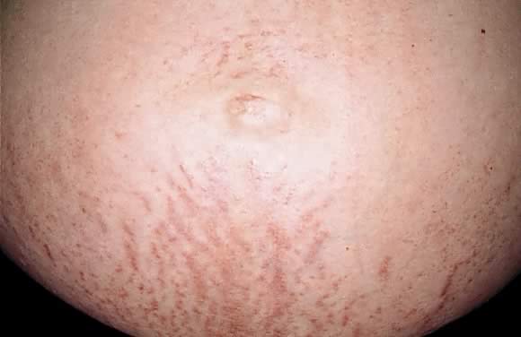
Fig. 7. Polymorphic eruption of pregnancy: erythematous papules along the striae distensae.

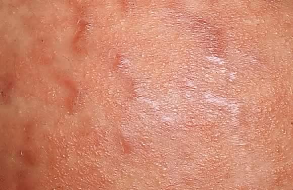
Fig. 8. Polymorphic eruption of pregnancy. Close-up view of Figure 7 showing the erythematous papules along the striae distensae.

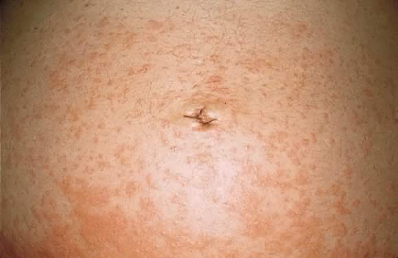
Fig. 9. Polymorphic eruption of pregnancy morphology: coalescing urticarial papules on the abdomen.

Fig. 10. Polymorphic eruption of pregnancy morphology: urticarial plaques on the lateral surface of the upper thigh.

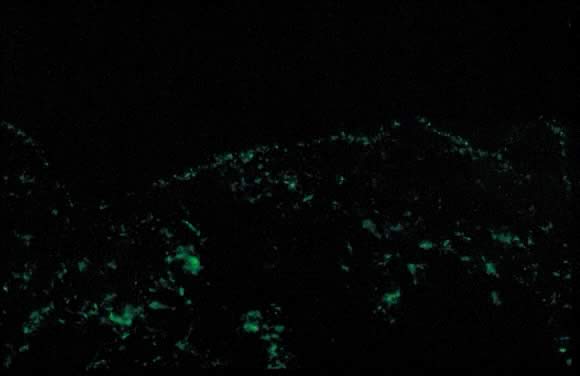
Fig. 11. Polymorphic eruption of pregnancy direct immunofluorescence: granular band of C3 along the skin basement membrane zone.

Dermatopathology shows variable epidermal spongiosis with a perivascular inflammatory infiltrate in the dermis composed of lymphocytes, histiocytes, and a variable number of eosinophils. Direct immunofluorescence (DIF) is negative for a linear band of C3 or IgG along the skin dermoepidermal junction (DEJ); however, there have been reports of deposition of IgM, C3, and IgA along the DEJ and blood vessels on DIF (Fig. 11).15,18 Differential diagnosis of PEP includes pemphigoid gestations (PG), contact dermatitis, drug eruption, and viral exanthems. DIF of skin is necessary to differentiate PEP from PG. The clinical course of PEP is usually self-limiting, with a mean duration of 6 weeks. Pruritus is most severe in the first week of onset, with spontaneous remission occurring within days of parturition. Maternal and fetal mortalities are unaffected. PEP rarely occurs in subsequent pregnancies; however, a few cases of reoccurrence are reported in the literature.13 Treatment is symptomatic relief of pruritus with topical corticosteroids of low- to mid-potency (use of ultra-high-potency corticosteroids for an extensive period of time should be avoided) and pregnancy category B antihistamines such as loratadine and cetirizine. Hydroxyzine and diphenhydramine are pregnancy category C antihistamines that have been used to relieve pruritus. In cases of severe pruritus unresponsive to conservative measures, systemic corticosteroid administration or induced delivery is considered.

Pemphigoid Gestationis

Initially described by Milton in 187218A as “herpes gestationis,” this condition was renamed pemphigoid gestationis (PG) in 1982 due to its clinical and immunofluorescence similarities with bullous pemphigoid.19 Both names continue to be used; pemphigoid gestationis is more common in the United Kingdom.20,21

Estimated incidence of PG is 1 in 50,000 cases. Pemphigoid gestationis most commonly occurs in the second or third trimester of pregnancy; about 25% of cases may have an initial presentation immediately postpartum. Clinical presentation is an abrupt onset of an intensely pruritic, urticarial 
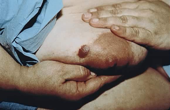
eruption on the trunk that forms tense vesicobullous lesions (Figs. 12 and 13). About 50% of cases have an initial presentation on the abdomen. Umbilical involvement accounts for a significant number of cases of PG. As in PEP, facial and mucosal membrane involvement is rare.21

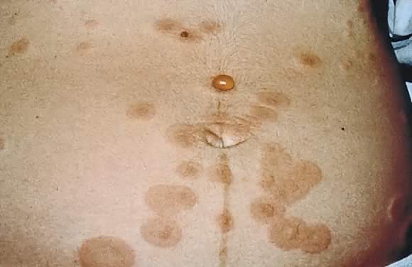
Fig. 12. Pemphigoid gestationis: urticarial plaques in the periumbilical area with tense blisters.

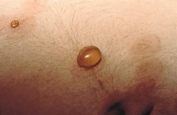
Fig. 13. Pemphigoid gestationis: close-up view of the tense blister in Figure 12.

Accurate diagnosis is crucial in light of the variable clinical course of this disorder. Spontaneous resolution over weeks to months postpartum is a common finding. About 75% of cases of PG present with flares immediately postpartum. Recurrence in subsequent pregnancies with an earlier onset and more severe clinical course is a common feature. Disease-free pregnancies (i.e. “skip pregnancies”) with no cutaneous involvement in patients with a history of PG have also been reported in the literature. There have also been reports of PG flares occurring with menstruation and use of oral contraceptives (25% of cases).20,21

PG is an autoimmune disorder caused by aberrant expression of the MHC II class antigen on the chorionic villi of the placenta which triggers an allogenic response to the placental basement membrane zone and subsequent cross-reaction with maternal skin through the maternal decidua.22,23 There are reports of occurrence of PG in association with hydatidiform mole and choriocarcinoma.24,25 Association of PG with other autoimmune diseases such as Graves' disease has also been reported.26 Studies also show an increased incidence in HLA DR3 and DR4. HLA DR3 occurs in the same percentage of white persons as in African American persons; however, the percentage of DR4 is lower in African Americans, and this may explain the rarity of PG in this population.27

Dermatopathology of PG shows subepidermal vesicle formation with focal necrosis of keratinocytes. The dermis shows papillary edema and a perivascular infiltrate consisting mainly of eosinophils and few lymphocytes. An occasional finding on histopathology is the alignment of eosinophils along the dermoepidermal junction. DIF shows a characteristic linear band of C3 along the skin basement membrane zone of patients with PG (Fig. 14). Linear C3 deposition on DIF is diagnostic of PG in the correct clinical setting and is used to differentiate PEP from PG.28 Using this method, about 25% of cases also present with IgG deposits along the basement membrane zone. Indirect immunofluorescence (IIF) demonstrates the “PG factor,” which consists of circulating IgG complement-fixing anti-basement membrane zone antibodies in serum of patients with PG. Complement-activated IIF using monoclonal antibodies directed against IgG1 demonstrates this factor in all PG patients.29 Clinically, titers of PG factor do not correlate with disease severity. The PG factor is an IgG directed against a 180-kd hemidesmosomal (transmembrane) component of the basement membrane zone.30 Electron microscopy also demonstrates the C3 and IgG deposits in the lamina lucida.31

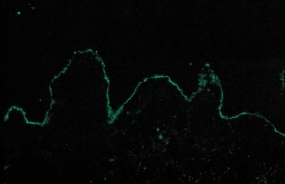
Fig. 14. Pemphigoid gestationis by direct immunofluorescence: linear band of C3 along basement membrane zone of the skin.

In PG 10% of the infants born to affected mothers have skin lesions that resemble PG; this is not the case with PEP. DIF and IIF studies done on some of these infants are consistent with a diagnosis of PG.21 There has been considerable controversy in assessing fetal morbidity and mortality associated with PG.32 Recent consensus on infant morbidity indicates a slight increase in prematurity and small size for gestational age.33,34 The differential diagnosis of PG includes PEP, allergic contact dermatitis, and drug eruption. A precise clinical history accompanied by diagnostic tests such as histopathology and DIF help to distinguish among these disorders. Treatment options include oral steroids (0.5 mg/kg daily) with a possible increase in dose around the time of delivery to avoid postpartum exacerbations. Other options include plasmapheresis, topical corticosteroids, and antihistamines, all of which offer limited benefit. After delivery, depending on breastfeeding status, alternative treatments include dapsone, methotrexate, and cyclosporine.21

Impetigo Herpetiformis

Impetigo herpetiformis is an acute eruption of pustular psoriasis during pregnancy (most often presenting in the third trimester) in individuals with no prior history of psoriasis. The first case was described by Von Hebra in 1872; since then, about 100 cases have been reported.35 Clinical presentation involves sterile pustules on an erythematous base that progressively become more confluent. This eruption commonly begins on the flexural and inguinal skin and gradually spreads to the trunk and involves the periumbilical skin (Figs. 15 and 16). Mucous membrane involvement of the oropharynx and the esophagus is also seen. Impetigo herpetiformis is associated with constitutional symptoms such as elevated temperature; gastrointestinal symptoms including nausea, vomiting, and diarrhea; central nervous system symptoms such as delirium; and musculoskeletal manifestation of tetany due to hypocalcemia.2,36 Recurrent eruptions in subsequent pregnancies usually present with an earlier onset and more severe course.36 There have also been reports of exacerbation of this condition in affected patients associated with later use of oral contraceptives.37

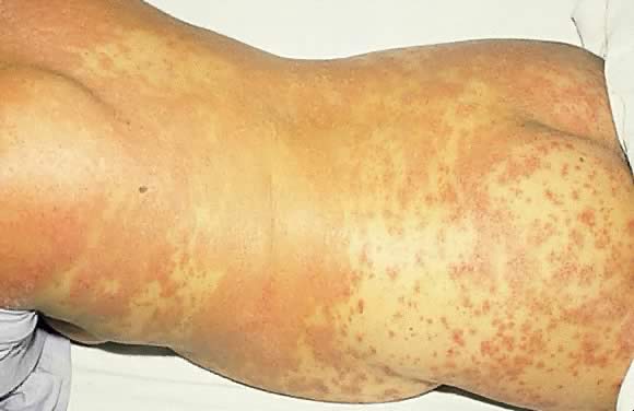
Fig. 15. Impetigo herpetiformis: diffuse erythematous papulosquamous eruption on the trunk and extremities.

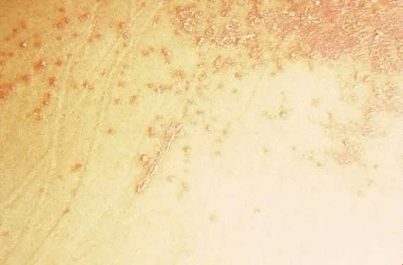
Fig. 16. Impetigo herpetiformis: coalescing erythematous papules with silvery scale.

Early diagnosis and treatment is crucial. The few cases that have been reported in the literature are associated with increased risk of fetal mortality due to placental insufficiency, increased stillbirths, and fetal abnormalities.36 Laboratory findings include evidence of leukocytosis, elevated erythrocyte sedimentation rate, hypoalbuminemia, and hypocalcemia.2 Histopathology of skin biopsy specimens are consistent with pustular psoriasis. The epidermis shows parakeratosis and elongation of rete ridges with spongiform pustules of Kogoj. DIF, as in psoriasis, is negative.36

Treatment involves oral corticosteroids (with limited benefit), correction of hypocalcemia, supportive measures, and antibiotics to prevent secondary infections. Termination of pregnancy is usually curative.36,37 Retinoids (isotretinoin) and light therapy are more effective means of treatment that can be used postpartum.

Cholestasis of Pregnancy

Cholestasis of pregnancy was initially described by Svanborg38 and Thorling39 in 1954. Cholestasis of pregnancy has been referred to by many other names, including prurigo gravidarum, intrahepatic cholestasis of pregnancy, jaundice of pregnancy, and obstetric cholestasis. The etiology is believed to be multifactorial, and the condition occurs in 0.02% to 2.4% of pregnancies. Studies show an increased incidence among certain ethnic groups, such as some South American Indians. There is also a seasonal variation in the prevalence of this condition, with a higher incidence in the winter months. Fifty percent of cases are believed to be familial, and a higher association has been seen in twin pregnancies.40,41 Another possible factor that contributes to the pathogenesis of this condition is the effect of estrogen and other female hormones on the metabolism and secretion of hepatic bile.2,40

Cholestasis of pregnancy, as the name implies, occurs only in pregnancy (most commonly during the third trimester) and resolves after delivery, with a 40% to 60% rate of recurrence in subsequent pregnancies. Clinical presentation includes severe generalized pruritus with no primary skin lesions. Secondary excoriations due to patient's scratching may be the only skin findings. The extent and severity of pruritus fluctuates until the time of delivery.40,42 Most severe pruritus occurs at night.9 About 20% of patients present with mild jaundice. This condition is the second most common cause of gestational jaundice; viral hepatitis is the most common cause.21 Laboratory values demonstrate elevated levels of bile salts, serum aminotransferases, alkaline phosphatase, and γ-glutamyl transpeptidase.40,42 Because there are no primary skin lesions, skin biopsy results for histology and DIF are normal.

Pruritus greatly improves after delivery, and complete resolution is achieved within a few days. In cases in which symptoms continue to persist, other causes of cholestasis must be addressed.40 There have been reports of recurrence of symptoms with use of oral contraceptives.42 Differential diagnosis of pruritus in pregnancy should include parasitic infections, allergic skin reactions, and other metabolic disorders.

Fetal and maternal prognosis shows an increase in premature labor and low birth weight. The fetus and the mother are at an increased risk of intracranial and postpartum hemorrhage, respectively, due to deficiency in vitamin K, which results in cases of prolonged fat malabsorption.21,42 Treatment options range from bed rest, a low-fat diet, and topical emollients in mild cases to the use of agents such as cholestyramine, ursodeoxycholic acid (UDCA), and S-adenosyl-L-methionine in more severe cases. Studies show better outcomes for both mother and infant with administration of UDCA compared with placebo. In severe cases, fetal monitoring and cesarean section may be required.42

Prurigo of Pregnancy

Prurigo of pregnancy was initially described by Besnier in 190412 as prurigo gestationis. It commonly occurs in the second to third trimester of pregnancy as discrete erythematous excoriated papules on the trunk and the extensor aspect of the lower extremities (Fig. 17). Incidence is roughly 1 in 300 pregnancies. Pathogenesis of this eruption is believed to be the presence of atopy in the pregnant woman. Dermatopathology of skin biopsy specimens shows parakeratosis and mild acanthosis with a mixed inflammatory infiltrate of neutrophils and eosinophils in the perivascular area. DIF results and laboratory values are normal. There is no increased fetal or maternal risk, and treatment is symptomatic relief with topical corticosteroids and antihistamines.2,21

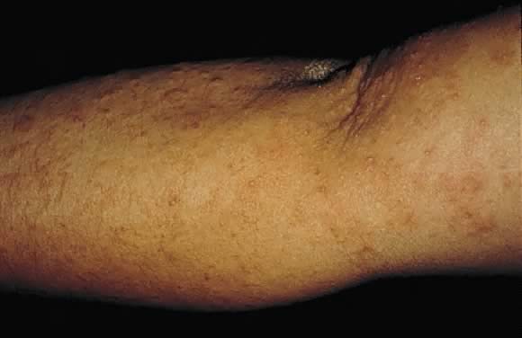
Fig. 17. Prurigo of pregnancy: discrete erythematous excoriated papules on the extensor surface of the arm.

Papular Dermatitis of Pregnancy

Papular dermatitis of pregnancy was initially described by Spangler in 196243 as a generalized papular erythematous and pruritic eruption with central crust. The distribution is on the abdomen with spread to the extremities. An increased level of urine hCG and a decrease in the urinary estriol level, in combination with a significant increase in fetal morbidity and mortality, was initially described. Many believe that papular dermatitis of pregnancy and prurigo of pregnancy are similar entities. Histopathology and DIF findings are similar. The high fetal risk initially reported by Spangler43 has not been reproducible in other studies.44

Pruritic Folliculitis of Pregnancy

Pruritic folliculitis of pregnancy was first described by Zoberman and Farmer in 1981.45 Onset of eruption most commonly occurs in the second or third trimester of pregnancy as small erythematous papules around follicles. The eruption is typically monomorphic with distribution on the trunk and extremities (Fig. 18). Histopathology resembles a folliculitis, and the DIF is negative. Differential diagnosis involves papular dermatitis or steroidinduced acne. The fetus is unaffected, and the treatment is topical benzoyl peroxide.2,45

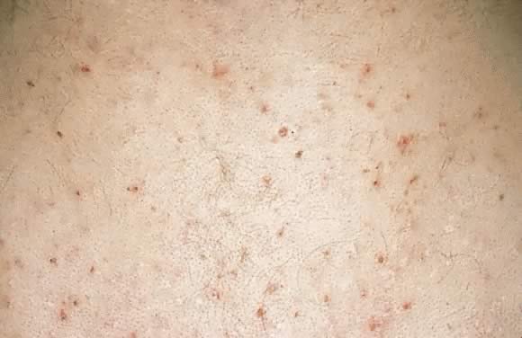
Fig. 18. Pruritic folliculitis of pregnancy: erythematous papules centered around hair follicles on the back.

Back to Top
OTHER DERMATOSES AND INFECTIONS AFFECTED IN PREGNANCY

In addition to the dermatoses that are specific to pregnancy, many common dermatoses and infections are affected by the hormonal and immunologic changes seen in pregnancy. These include conditions such as atopic dermatitis, seborrheic dermatitis, psoriasis, condyloma acuminata, and genital herpesvirus infection.

Atopic Dermatitis

Atopic dermatitis (atopic eczema) is a chronic inflammatory skin condition that presents as pruritic papules or papulovesicular lesions with a predilection for flexural surfaces of the body. These lesions are often excoriated, lichenified, or secondarily infected. Affected individuals often have associated conditions such as asthma, allergic rhinitis (hay fever), xerosis, or drug sensitivities. Atopic dermatitis usually improves during pregnancy. Treatment regimens depend on the severity of the eruption and the extent of body involvement. Topical corticosteroids and emollients are the mainstay of treatment. Avoidance of aggravating factors such as soaps, detergents, and hot showers and baths are also very important.

Seborrheic Dermatitis

Seborrheic dermatitis is a chronic erythematous eruption with greasy-appearing scale in areas rich in sebaceous glands such as the scalp, face, and upper trunk. The yeast Malassezia furfur is a causative agent. Treatment includes low-potency topical corticosteroids and topical ketoconazole cream.

Psoriasis

Psoriasis is a chronic inflammatory and proliferating skin condition that presents as sharply demarcated erythematous plaques with silvery scale. The effects of pregnancy on psoriasis are variable. Retrospective studies usually show improvement or no change in this skin condition with pregnancy.46,47 Treatment options include topical corticosteroids, calcipotriol, and tar. Oral cyclosporine should be used with caution. Oral agents such as methotrexate, hydroxyurea, and retinoids are contraindicated during pregnancy.

Human Papillomavirus

Human papillomavirus (HPV) constitutes a group of numerous viral strains that cause clinical and subclinical infections. Warts are the most common clinical manifestation of these infections. In the anogenital area, these are commonly referred to as condylomata acuminata, which often present as flesh-colored, exophytic, cauliflower-like masses. Immunologic changes during pregnancy result in exacerbation of previous lesions or new onset of subclinical infections. The amount of viral DNA is also greatly increased during pregnancy. Recognition of HPV infection is important because certain strains can be transmitted to the fetus through an infected birth canal, with subsequent association with juvenile respiratory papillomatosis in infants born to infected mothers. Respiratory papillomatosis in infants is rare compared with the extent of genital condylomas in child-bearing women. Thus, performance of cesarean section in this situation is controversial because of the inherent risks of the procedure itself. Treatment options during pregnancy include trichloroacetic acid topical preparations, cryotherapy, cautery, or laser ablation. Other topical agents such as podophyllin are contraindicated during pregnancy. None of the treatments for HPV are curative, and latent infection persists.48

Herpes Simplex Virus

Herpes simplex virus (HSV) is a common cause of viral infections worldwide. HSV-1 and HSV-2 cause both primary and recurrent infections; primary infections are more severe. Infection clinically presents as grouped vesicles on an erythematous base that may erode and form ulcerations. Asymptomatic shedding of the herpesvirus has also been shown in the absence of any skin findings. Genital herpes infection at the time of delivery is associated with a high risk of neonatal infection. Even in the absence of skin lesions in infected newborns, neurologic and visceral organ damage can be severe. Thus, recognition and treatment of herpes infection during pregnancy is very important. Patients considered high risk for HSV infection should be tested weekly with viral cultures, and if there is evidence of active infection or viral shedding, cesarean delivery should be performed.49 Acyclovir is a pregnancy category C antiviral agent that is used for primary or symptomatic infections. Valacyclovir (pregnancy category B) has also been used in the treatment of HSV during pregnancy.

Back to Top
REFERENCES

1. Elling SV, Powell FC: Physiological changes in the skin during pregnancy. Clin Dermatol 15: 35– 43, 1997

2. Lawley TJ, Yancey KB: Skin changes and diseases in pregnancy. In Freedberg IM, Eisen AZ, Wolff K (eds): Fitzpatrick's Dermatology in General Medicine, 5th ed, pp 1963-1969. New York, McGraw-Hill, 1999

3. Winton GB, Lewis CW: Dermatoses of pregnancy. J Am Acad Dermatol 6: 977– 998, 1982

4. Colley SM, Walpole I, Fabian VA, Kakulas BA: Topical tretinoin and fetal malformations. Med J Aust 168 (9): 467, 1998

5. Dover JS, Kane KS: Lasers for the treatment of cutaneous pigmented disorders. In Arndt KA, Dover JS, Olbricht SM (eds): Lasers in Cutaneous and Aesthetic Surgery, pp 165–187. Philadelphia, Lippincott-Raven, 1997

6. Hellreich PD: The skin changes of pregnancy. Cutis 13: 82– 86, 1974

7. Brenner S, Politi Y: Dermatologic diseases and problems of women throughout the life cycle. Int J Dermatol 34: 369– 379, 1995

8. Graham-Brown RAC: The ages of man and their dermatoses. In Rook A, Wilkinson DS, Ebling FJB (eds): Textbook of Dermatology, pp 3259–3277. London, Blackwell Scientific Publications, 1998

9. Murray JC: Pregnancy and the skin. Dermatol Clin 8: 327– 334, 1990

10. Lynfield YL: Effect of pregnancy on the human hair cycle. J Invest Dermatol 35: 323– 327, 1960

11. Headington JT: Telogen effluvium. Arch Dermatol 129: 356– 363, 1993

12. Besnier E, Brocq L, Jacquet L: La Pratique Dermatologique, p 75. Paris, Masson et Cie, 1904

12A. Lawley TJ, Hertz KC, Wade TR, et al: Pruritic urticarial papules and plaques of pregnancy. JAMA 241: 1696– 1699, 1979

13. Holmes RC: Polymorphic eruption of pregnancy. Semin Dermatol 8: 18– 22, 1989

14. Yancey KB, Hall RP, Lawley TJ: Pruritic urticarial papules and plaques of pregnancy. J Am Acad Dermatol 10: 473– 480, 1984

15. Aronson IK, Bond S, Fiedler VC, et al: Pruritic urticarial papules and plaques of pregnancy: Clinical and immunopathologic observations in 57 patients. J Am Acad Dermatol 39: 933– 939, 1998

16. Cohen LM, Capeless EL, Krusinski PA, Maloney ME: Pruritic urticarial papules and plaques of pregnancy and its relationship to maternal-fetal weight gain and twin pregnancy. Arch Dermatol 125: 1534– 6, 1989

17. Beckett MA, Goldberg NS: Pruritic urticarial plaques and papules of pregnancy and skin distention. Arch Dermatol 127: 125– 126, 1991

18. Toussaint S, Kamino H: Noninfectious erythematous, papular, and squamous diseases. In Elder D, Elenitsas R, Jaworsky C, Johnson B (eds): Lever's Histopathology of the Skin, 8th ed, p 153. Philadelphia, Lippincott-Raven, 1997

18A. Milton JL: The Pathology and Treatment of Diseases of the Skin. London, Robert Hardwicke, 1872

19. Holmes RC, Black MM: The specific dermatoses of pregnancy: A reappraisal with emphasis on a proposed simplified clinical classification. Clin Exp Dermatol 7: 65– 73, 1982

20. Shornick JK, Bangert JL, Freeman RG, Gilliam JN: Herpes gestationis: Clinical and histologic features of twenty-eight cases. J Am Acad Dermatol 8: 214– 224, 1983

21. Shornick JK: Dermatoses of pregnancy. Semin Cutan Med Surg 17: 172– 181, 1998

22. Borthwick GM, Holmes RC, Stirrat GM: Abnormal expression of class II MHC antigens in placentae from patients with pemphigoid gestationis: Analysis of class II MHC subregion product expression. Placenta 9: 81– 94, 1988

23. Ortonne JP, Hsi BL, Verrando P, et al: Herpes gestationis factor reacts with the amniotic epithelial basement membrane. Br J Dermatol 117: 147– 154, 1987

24. Tindall JG, Rea TH, Shulman I, Quismorio FP: Herpes gestationis in association with a hydatidiform mole. Arch Dermatol 117: 510– 512, 1981

25. Slazinski L, Degefu S: Herpes gestationis associated with choriocarcinoma. Arch Dermatol 118: 425– 428, 1982

26. Shornick JF, Black MM: Secondary autoimmune diseases in herpes gestationis (pemphigoid gestationis). J Am Acad Dermatol 26: 563– 566, 1992

27. Shornick JK, Meek TJ, Nesbitt LT, Gilliam JN: Herpes gestationis in blacks. Arch Dermatol 120: 511– 513, 1984

28. Holmes RC, Black MM, Dann J, et al: A comparative study of toxic erythema of pregnancy and herpes gestationis. Br J Dermatol 106: 499– 510, 1982

29. Kelly SE, Cerio R, Bhogal BS, Black MM: The distribution of IgG subclasses in pemphigoid gestationis: PG factor is an IgG1 autoantibody. J Invest Dermatol 92: 695– 698, 1989

30. Morrison LH, Labib RS, Zone JJ, et al: Herpes gestationis autoantibodies recognize a 180-kd human epidermal antigen. J Clin Invest 81: 2023– 2036, 1988

31. Honigsmann H, Stingl G, Holubar K, Wolff K: Herpes gestationis: Fine structural pattern of immunoglobulin deposits in the skin in vivo. J Invest Dermatol 66: 389– 392, 1977

32. Lawley TJ, Stingl G, Katz SI: Fetal and maternal risk factors in herpes gestationis. Arch Dermatol 114: 552– 555, 1978

33. Holmes RC, Black MM: The fetal prognosis in pemphigoid gestationis (herpes gestationis). Br J Dermatol 110: 67– 72, 1984

34. Shornick JK, Black MM: Fetal risks in herpes gestationis. J Am Acad Dermatol 26: 63– 68, 1992

35. Breier-Maly J, Ortel B, Breier F, et al: Generalized pustular psoriasis of pregnancy (impetigo herpetiformis). Dermatology 198: 61– 64, 1999

36. Lotem M, Katzenelson V, Rotem A, et al: Impetigo herpetiformis: A variant of pustular psoriasis or a separate entity? J Am Acad Dermatol 20: 338– 341, 1989

37. Oumeish OY, Farraj SE, Bataineh AS: Some aspects of impetigo herpetiformis. Arch Dermatol 118: 103– 105, 1982

38. Svanborg A: A study of recurrent jaundice in pregnancy. Acta Obstet Gynecol Scand 33: 434– 444, 1954

39. Thorling L: Jaundice in pregnancy: A clinical study. Acta Med Scand 151 (Suppl): 1– 123, 1955

40. Reyes H: The spectrum of liver and gastrointestinal disease seen in cholestasis of pregnancy. Gastroenterol Clin North Am 21: 905– 921, 1992

41. Gonzalez MC, Reyes H, Arrese M, et al: Intrahepatic cholestasis of pregnancy in twin pregnancies. J Hepatol 9: 84– 90, 1989

42. Reyes H: Review: Intrahepatic cholestasis. A puzzling disorder of pregnancy. J Gastroenterol Hepatol 12: 211– 216, 1997

43. Spangler AS, Reddy W, Bardawil WA, et al: Papular dermatitis of pregnancy. JAMA 181: 577– 581, 1962

44. Michaud RM, Jacobson D, Dahl MV: Papular dermatitis of pregnancy. Arch Dermatol 118: 1003– 1005, 1982

45. Zoberman E, Farmer ER: Pruritic folliculitis of pregnancy. Arch Dermatol 117: 20– 22, 1981

46. Boyd AS, Morris LF, Phillips CM, Menter MA: Psoriasis and pregnancy: Hormone and immune system interaction. Int J Dermatol 35: 169– 172, 1996

47. Mowad CM, Margolis DJ, Halpern AC, et al: Hormonal influences on women with psoriasis. Cutis 61: 257– 260, 1998

48. Greenberg MD, Rutledge LH: Understanding human papillomaviral infections in women. Semin Dermatol 11: 241– 146, 1992

49. Freij BJ, Sever JL: Infectious complications of pregnancy. Clin Perinatol 15: 202– 213, 1988

Back to Top