Chapter 75
Cesarean Hysterectomy
Grainger S. Lanneau, Patrick Muffley and Everett F. Magann
Main Menu   Table Of Contents

Search

Grainger S. Lanneau, Jr, MD
Clinical Instructor, Departments of Family Practice/Ob/Gyn, Puget Sound Family Practice Residency, Bremerton, Washington, Ob/Gyn Residency, Madigan Army Medical Center, Tacoma, Washington, Staff, Department of Ob/Gyn, Naval Hospital Bremerton, Bremerton Washington (Vol 2, Chaps 74, 75)

Patrick Muffley, MD
(Vol 2, Chaps 74, 75)

Everett F. Magann, MD
Professor, Department of Obstetrics & Gynecology, University of Western Australia, Perth Australia, Head, Department of Labor & Delivery, King Edward Memorial Hospital, Perth, Australia (Vol 2, Chaps 74, 75, 97)

 
SURGICAL PITFALLS
SURGICAL TECHNIQUE
INTRAOPERATIVE COMPLICATIONS
POSTOPERATIVE MORBIDITY AND MORTALITY
DISCUSSION
REFERENCES

The concept behind cesarean hysterectomy dates back to descriptions of the procedure on laboratory animals from the mid 1700s. In 1869, Storer performed the first cesarean hysterectomy in the United States. Soon thereafter, Porro of Milan described the first cesarean hysterectomy in which the infant and mother survived; the procedure is frequently referred to as the Porro operation in his honor.1,2 This chapter uses data from the University of Mississippi and Louisiana State University (LSU), as well as that obtained from an extensive review of the current literature.

Cesarean hysterectomies may be classified as emergent, indicated nonemergent, and for elective sterilization. Each of these categories presents different management problems for the obstetric surgeon.3

The classic historical indications for emergency cesarean hysterectomy are life-threatening hemorrhage and infection. With the advent of broad-spectrum antibiotics and improved pharmaceutical therapy for hemorrhage, the incidence of emergent cesarean hysterectomy has decreased. Currently, most cesarean hysterectomies are performed for recalcitrant hemorrhage.

Table 1 represents a series of 145 cases from LSU from 1975 through 1985. Most cesarean hysterectomies performed emergently were the result of abnormal placentation or abruption. Uterine rupture and placenta accreta, particularly previa with accreta, have been encountered with increasing frequency in recent years as the number of cesarean sections and attempted vaginal deliveries after cesarean section increases.4,5,6 In cases of uterine rupture, the necessity of hysterectomy has decreased as authors have shown that successful suture repair of the defect can be safely performed. In patients who have experienced uterine rupture or dehiscence of a previous scar, management in subsequent pregnancies should be similar to that of patients who have had a previous classical cesarean section. Ritchie noted a repeat rupture rate of 10%.8 Unless the tear is in the lower uterine segment and easily repaired, a repeat cesarean section appears prudent in these cases.9 Additionally, the use of radiologic embolization procedures has become an important tool for the obstetric surgeon and may serve a critical role in the patient with persistent bleeding after surgery.10,11,12


Table 1. Indications for Cesarean Hysterectomy at Louisiana State University Service—Charity Hospital of New Orleans, 1975–1985 (n = 145)


Emergency Cases (32% of total) Indicated Nonemergencies (31% of total) Elective Sterilization (37% of total)
Uterine hemorrhage (26%) Leiomyomata uteri (10%) Multiple repeat cesarean section plus sterilization (20%)
Placental problems (previa, abruptio, accreta) (11%) Cervical intraepithelial neoplasia (10%) Primary elective sterilization (17%)
Uterine rupture (7%) Adnexal disease (3%)  
Postpartal atony (5%)    
Extension of cesarean section incision into uterine vessels (3%)    
Infection    
Chorioamnionitis with sepsis (6%)    


Nonemergent indicated cesarean hysterectomies are considered by some to be controversial. It is the current policy to consider cesarean hysterectomy for patients who have a valid obstetric indication for cesarean section and a concurrent valid indication for hysterectomy. The primary indications at our institution include high-grade squamous intraepithelial lesions, leiomyomas.

Small myomas commonly regress in size after pregnancy and seldom require cesarean hysterectomy for their management. However, some patients have multiple large myomas that can obstruct the pelvic outlet. Others can experience painful degeneration of myomas or menorrhagia. Such patients are frequently best served by an operation that accomplishes both delivery and definitive treatment of their symptomatic myomas.

Squamous intraepithelial lesions of the cervix are a particularly serious indication for cesarean hysterectomy. During pregnancy, it can be difficult to adequately evaluate the endocervical canal, and the physiologic changes in the cervical epithelium make it difficult for the cytologist to evaluate the condition. Occasionally, dysplastic or malignant changes may progress in the interval between initial diagnosis and delivery; staging of the disease process may then be inaccurate and lead to inappropriate therapy. Of added importance is the sometimes difficult removal of the entire cervix at the time of cesarean hysterectomy. Despite these concerns, there are cases in which cesarean hysterectomy or radical cesarean hysterectomy may be the best management plan, as they can provide definitive, curative therapy for these persistent and potentially evolving lesions.

The most controversial indication for cesarean hysterectomy is elective sterilization. In a multiparous patient who desires permanent sterility and is undergoing a repeat cesarean section, it could be argued that cesarean hysterectomy may be an appropriate option. When surgical sterilization alone is the indication, cesarean section followed by a tubal ligation is a far safer procedure than cesarean hysterectomy. Although a comprehensive review indicates that 17% of women undergoing postpartum sterilization ultimately undergo hysterectomy,13 cesarean section and desire for sterilization alone do not warrant cesarean hysterectomy.14,15

Back to Top
SURGICAL PITFALLS

The physiologic changes in the maternal pelvis that accompany pregnancy are responsible for many of the surgical challenges of cesarean hysterectomy. The vessels that supply the uterus, ovaries, and bladder are remarkably larger and more tortuous in pregnancy than they are in the nonpregnant state. Careless manipulation of clamps, cutting of pedicles, or placement of sutures may precipitate severe bleeding. Edema of the structures surrounding the uterus allows easy dissection of surgical planes but produces large pedicles from which blood vessels may escape. Special attention must be given to the proper size of pedicles and careful hemostatic suturing techniques.

Scarring from previous surgery, particularly previous cesarean sections, is a common complicating feature of cesarean hysterectomy. Great care is necessary to avoid injury to organs that are adherent to the uterus, bladder, adnexa, and abdominal wall. The surgeon must be prepared to recognize and repair injuries of the urinary tract. Cesarean hysterectomy is often performed in the presence of uterine trauma or rupture in which hematomas of the broad ligament and neighboring structures make visualization difficult and distort anatomic relations. Careful exposure, skilled assistants, and attention to hemostasis are of primary importance.14

Back to Top
SURGICAL TECHNIQUE

There are several ways to perform cesarean hysterectomy successfully. Any competent gynecologic surgeon should be able to accomplish the task with dispatch and should respect, but not fear, the special problems posed by the operation. When cesarean hysterectomy is planned in advance, the surgeon has the luxury of a complete preoperative evaluation of the patient and her hematologic and coagulation status. The surgeon can prepare to fill the bladder to test its integrity, should that become necessary. The surgeon can choose experienced assistants and discuss procedures, necessary instruments, and sutures before the operation. In emergent cases, such luxuries do not exist, and the scene often becomes chaotic.16,17

Cesarean hysterectomy is really two operations: cesarean section and hysterectomy, one following the other in an orderly fashion. Haste is required only briefly at the beginning of each operative segment. Efforts at speed that replace careful surgical technique court disaster. The surgeon should strive for steady, deliberate progress through the successive steps of the operation.

Cesarean hysterectomy can be accomplished through any commonly used abdominal wall incision. A vertical incision provides excellent exposure; however, many patients and surgeons are not satisfied with the cosmetic effects. A transverse incision may be desirable, especially if a previous transverse scar is present. This incision can provide adequate exposure, and, if needed, it can be converted into a Cherney incision by transecting the rectus muscle at its insertion. An alternative approach used specifically in patients with previous transverse incisions is division of the rectus muscle converting a standard Pfannenstiel to a modified Maylard incision. The following description of cesarean hysterectomy technique is that used by the authors for emergent procedures.

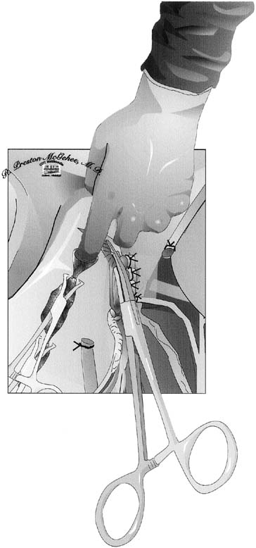
The type of uterine incision used is guided by obstetric indications; however, a low vertical incision is less likely to extend and lacerate the uterine vasculature. After the infant is delivered and the decision is made to proceed with cesarean hysterectomy, the placenta is expressed unless there is a placenta accreta. The fundus is grasped, and the uterus is removed from the pelvis and placed on the abdominal wall. Using the uterine incision for traction is not advisable, because it may lead to extension of the incision. From this point forward, the uterus should be kept under upward traction to constrict the uterine vasculature and diminish blood loss. The hysterotomy incision is then closed with a running suture or towel clips (Fig. 1). Reapproximating the myometrium optimizes uterine contractility and decreases the occurrence of uterine atony. If there is considerable bleeding from the placental site, the uterus can be packed with one or two laparotomy pads above the site before uterine closure; this may serve to tamponade the bleeding.

Fig. 1. Closing of the hysterotomy incision with a running suture or towel clip. The round ligaments are divided between Kocher clamps and ligated.

The round ligaments are divided between Kocher clamps and ligated (see Fig. 1). Care should be taken to place the tips of the clamps in the avascular portion of the broad ligament to avoid lacerating Sampson's artery. The broad ligament is then incised anteriorly to the point where the vesicouterine peritoneum was dissected off the lower uterine segment to create the bladder flap. Posteriorly, the broad ligament is incised laterally and parallel to the infundibulopelvic ligament to expose the retroperitoneum. The loose areolar tissue encountered in this space, which is more pronounced than usual because of the enlarged uterus, can be carefully dissected parallel to the course of the ureter. This allows visualization of the retroperitoneal space and the ureter throughout its course.

Before approaching the uterine arteries, the bladder must be dissected free and displaced below the operative field. Because most patients undergoing cesarean hysterectomy have had previous surgery, significant adhesive disease is frequently encountered; as a result, sharp dissection is the technique of choice. It is extremely important to avoid lateral dissection into the highly vascular bladder pillars. It is also wise not to extend the dissection farther than is necessary to safely ligate the uterine arteries, because excessive dissection can cause additional bleeding and waste time.

Fig. 2. A Heaney clamp is placed with the distal portion perpendicular to the vessels at the level of the internal cervical os.

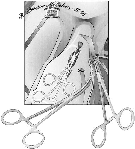
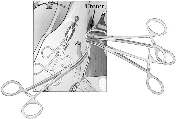
Attention is then directed toward the uterine arteries. The uterine vessels are significantly enlarged in pregnancy, and care must be taken in isolating or skeletonizing them. A Heaney clamp is placed with the distal portion perpendicular to the vessels at the level of the internal cervical os (Fig. 2). Once the same procedure is repeated on the contralateral side, the uterine arteries are secure, and blood loss should rapidly diminish. The use of a second clamp on the specimen side reduces back-bleeding from the uterus, which can obstruct the operative field. Additionally, until the utero-ovarian ligament is ligated, a substantial blood supply to the uterus persists. We suture ligate the uterine arteries using an 0 or 00 polyglycolic suture using a Heaney ligature; others advocate passing a suture beyond the tip of the clamp and tying behind the clamp. Using a Heaney ligature divides the pedicle in half and may cause less chance of the vessels retracting. Another technique to prevent retraction of the vessels is to incorporate the leaves of the broad ligament into each pedicle. Care must be taken not to place lateral or downward traction on these clamps, which might tear friable tissues and cause bleeding that cannot be easily controlled. These clamps should be supported and not manipulated.

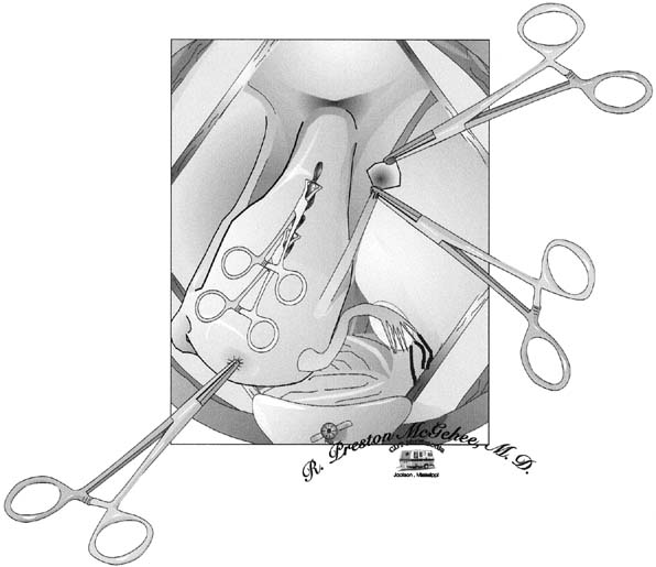
A second Heaney clamp is placed medial to the first clamp. Careful placement of clamps medially, moving down the parametrial tissue and hugging the cervix, will allow ligation of any branches of the uterine artery not ligated with the first clamp and ensure the safety of the ureter. After the uterine arteries are ligated, the utero-ovarian ligament can be approached. An avascular window in the peritoneum is created, thus allowing the pedicle to be isolated. Heaney clamps are placed close to the uterus to ensure that the ovary is not damaged (Fig. 3). Once divided, a free tie is placed on the specimen side, the utero-ovarian ligament is suture ligated. Because most patients in our series required oophorectomies because of infundibulopelvic ligament hematomas, we place a free tie proximal to the suture ligated pedicle.

Fig. 3. To ensure that there is no damage to the ovary, Heaney clamps are placed close to the uterus.

With the entire blood supply to the uterus now secured, a critical decision must be made in performing an emergent cesarean hysterectomy. If the patient is unstable or if the amount of bleeding has been excessive, the surgeon should consider to a subtotal hysterectomy, which shortens operating time while still accomplishing the primary goal of hemostasis. Although most surgeons prefer to remove the cervix, subtotal hysterectomy should not be dismissed, because it can be life-saving. If subtotal hysterectomy is performed, the uterus is amputated with a scalpel; if possible, this is achieved by cutting superiorly to the ligated uterine arteries while angling the scalpel blade medially and downward. This technique allows removal of an inverted cone of the cervix, which will facilitate approximation of the edges of the cervical stump regardless of the degree of dilation. Once amputated, the cervical stump can be approximated in an anterior-to-posterior fashion using interrupted figure-of-eight ligatures. Special care should be taken to avoid the bladder. If the patient is stable and the cervix can be removed, the task can prove to be difficult. As the cardinal ligament dissection proceeds downward, dissection of the bladder must also advance. Sharp dissection with Metzenbaum scissors (with tips pointing downward) ensures that no harm comes to the friable posterior bladder wall. Bladder dissection need never progress more than 1 cm distal to the current cardinal ligament dissection.

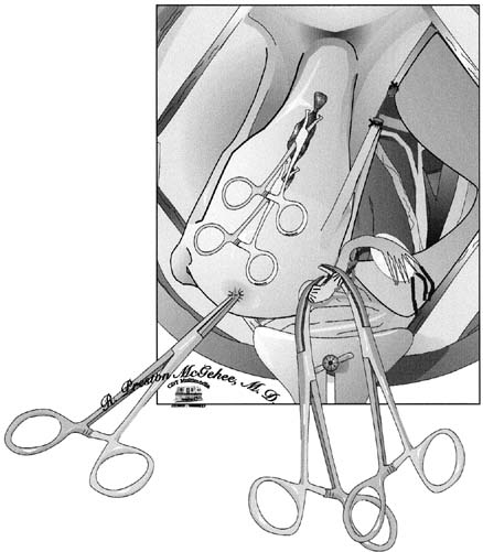
The next step is the identification of the lowest extent of the dissection: the junction of the cervix and vagina. This junction can usually be felt between the thumb and forefinger by palpating the upper vaginal walls and encountering the thickened cervix. When the cervix is dilated, it can be difficult to discern its lower end. The best approach to this problem is to place each clamp of the cardinal ligament complex medial to the preceding pedicle and roll it off the cervix to lie exactly against the lateral cervical wall. Each pedicle should be no longer than the distal third of the clamp used to grasp it. This allows the surgeon to progress down the cervix while each pedicle falls laterally, and the safety of the ureter is further ensured. In some cases, the uterosacral ligaments are quite prominent and may need to be individually clamped to completely ligate them. As the surgeon approaches the external cervical os, the towel clamps or suture line approximating the uterine incision can be released, and the cervix can be palpated from within the uterus (Fig. 4). Once the cardinal ligament dissection reaches the lower limit of the cervix, the vagina is entered, usually at the last pedicle. Curved scissors are used to amputate the cervix. The inside blade of the scissors is placed just beneath the cervix in the vaginal fornix and circumscribes the upper vagina to complete the dissection under direct vision. Allis or Kocher clamps maintain traction on the anterior, posterior, and lateral angles of the vaginal wall. The vaginal epithelium is very friable and must be handled gently.

Fig. 4. The towel clamps or suture lines approximating the uterine incision can be released as the external cervical os is approached, and the cervix can be palpated from within the uterus.

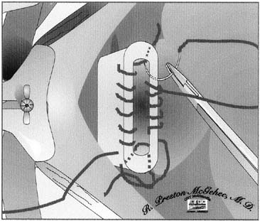
Surgical management of the vaginal cuff begins with supporting angle sutures that incorporate the lateral vaginal angles, the lower cardinal ligament pedicles, and the uterosacral pedicles. We usually hold this suture as a stay suture for subsequent identification of the vaginal angles. The surgeon may now choose to close the vaginal cuff with continuous or interrupted sutures or to leave the cuff open and to secure hemostasis of the anterior and posterior cuff edges with a continuous nonlocking suture (Fig. 5). This will allow for drainage until spontaneous reperitonealization occurs.

Fig. 5. To allow drainage, the surgeon chooses either to close the vaginal cuff with continuous or interrupted sutures or to leave the cuff open and to secure hemostasis of both the anterior and posterior cuff edges with a continuous nonlocking suture.

If the surgeon is suspicious that the integrity of the bladder wall has been breached at any time during the operation, the bladder may be filled with an opaque solution and the operating field inspected for extravasation of the solution. A second choice would be to administer intravenous indigo carmine and examine the area of the bladder for extravasation. If there has been an inadvertent cystotomy, the area is dissected so that two rows of sutures can be placed without tension. The bladder is closed with two continuous layers of 4.0 polyglycolic sutures, with the second layer imbricating the first. Permanent suture material should never be chosen for bladder closure. The bladder is then refilled to ensure its integrity. When bladder repair is necessary, postoperative antibiotic coverage and Foley catheter drainage of the bladder should be continued for 7 to 10 days. A Jackson-Pratt drain is placed in the retroperitoneal space near the bladder repair and led out through a separate stab wound in the lower abdomen.

All pedicles are individually reinspected for hemostatic security. The pelvis is copiously washed with warm saline and water. Sponges and retractors are removed, and instrument, needle, and sponge counts are checked. The abdominal incision is closed in the routine fashion.

Back to Top
INTRAOPERATIVE COMPLICATIONS

The principle problems encountered by surgeons performing cesarean hysterectomy are those of bleeding and urinary tract injury. Intraoperative bleeding difficulties from the adnexal pedicles, the uterine vascular pedicles, the cardinal ligaments, or angles of the vagina arise at the time of bladder dissection. When dissecting the bladder inferiorly, the surgeon is cautioned to avoid lateral dissection, which may disrupt dilated veins of the plexus of Santorini.

In our review of cesarean hysterectomies at University of Mississippi Medical Center, bleeding from adnexal pedicles was a common cause of intraoperative bleeding and early postoperative bleeding and an indication for removal of one or both adnexa. These edematous pedicles are under considerable tension, which causes vessels to retract and escape their ligatures.14

The principles of skeletonizing and transfixing the uterine vessels has reduced the incidence of postoperative retroperitoneal bleeding from retracted uterine vessels. Pedicles that are too large and clamp manipulation that tears pedicles away from the uterine wall are the most common continuing problems that result in uterine vascular and cardinal ligament bleeding. Bleeding at the vaginal angles is often caused by injudicious traction and manipulation of stay sutures that open ascending branches of the vaginal arteries. Vigorous use of the suction tip and retractors in the region of the bladder pillars, angles of the vaginal cuff, and beneath the base of the bladder can cause troublesome venous bleeding for which it is often difficult to achieve hemostasis.18

Emergent cesarean hysterectomy presents special bleeding problems. Coagulopathies may be present when the patient is first encountered or may develop as the case progresses. There are often large hematomas in the broad ligament and other retroperitoneal spaces that distort anatomic relations and defy attempts at direct clamping and suturing. It is often possible to find a free dissecting space near the lateral pelvic wall where the uterine vessels can be ligated near their origin from the internal iliac artery or, failing this, the internal iliac (hypogastric) artery itself can be isolated and ligated. In drastic emergencies, the aorta may be compressed for a time to stop copious bleeding while the field is cleared for a more direct attack on the bleeding problem.

The use of surgical packs is a frequently overlooked adjunct in obtaining hemostasis. Temporary packing of a bleeding area can offer time, similar to that of compressing the aorta, and allow blood component replacement to catch up with a possible coagulopathy. Packs can also be used in a more definitive manner. In areas of diffuse venous bleeding, packs can be left in the abdomen after closure. Each pack should be brought out through the skin through a separate incision. They should be tagged and numbered so that they can be removed in the opposite order that they were placed. A Logalopolas pack can be used in a similar setting. A Logthatopulos pack or umbrella pack is a bowel bag containing a number of packs with the opening protruding through the vagina. The entire pack can then be placed on traction, compressing the vasculature of the pelvis. Whenever we leave a pack in the abdomen, we administer broad-spectrum antibiotics until the packs are removed, usually within 72 hours.

The use of interventional radiology can be an invaluable tool. Prophylactic hypogastric artery embolization can be performed before cesarean hysterectomy without putting the fetus at risk and can potentially prevent surgical hemorrhage. Radiologic embolization can have an even larger role in cases of postoperative bleeding. Selective embolization can stop bleeding without the need for a second surgical procedure.10,11,12

Training in cesarean hysterectomy is often a weakness in most obstetrics and gynecology residency programs. However, this does not imply that the risks of the procedure are analogous with those of a vaginal delivery and subsequent hysterectomy.14

The bladder and ureters are at risk during the performance of cesarean hysterectomy, particularly in emergent cases in which trauma and hematomas distort expected anatomic relations. The bladder is at greatest risk during its initial dissection from the anterior cervical wall. We describe techniques that make this dissection safer and allow easy identification of bladder injury (see previous).

Some methods of cesarean hysterectomy include the technique of cross-clamping the upper vagina just before excision of the specimen. This technique must be used with caution in the obstetric patient with edematous tissues, because vesicovaginal fistulas have resulted from incorporation of the tented corners of the bladder in these clamps or sutures.18 We recommend the direct visualization technique described in this chapter for ligation of the vaginal cuff. Other causes of bladder fistulas include postoperative abscess formation, which can also contribute to ischemic necrosis at the base of the bladder and may produce a fistula.

The processes of uterine rupture and cesarean section scar dehiscence may directly injure the bladder and/or ureters. The surgeon must look for these, as well as inadvertent surgical injuries. It is usually the unrecognized bladder injury that results in vesicovaginal fistula; the properly repaired bladder usually heals without difficulty. Haynes and Martin19 reported 149 cesarean hysterectomies of which 10 were recognized and repaired cystotomies. No fistulas occurred in this series. Mickal and associates20 reported a series of 383 cesarean hysterectomies from the LSU service of which 20 cystotomies were recognized and repaired. Only one vesicovaginal fistula developed.

Fig. 6. Without direct visualization, the ureter, which can inadvertently be lifted into the operative field, can be ligated or transected.

The ureters are most closely approached at three points during the dissection in cesarean hysterectomy: the infundibulopelvic ligament ligation site (when adnexal structures are removed), the uterosacral ligament dissection, and the uterine vascular and cardinal ligament pedicles. The most important principle in approaching the ureter in complicated cesarean hysterectomy cases is direct visualization. Without direct visualization, the ureter, which can inadvertently be lifted into the operative field, can be ligated or transected (Fig. 6). The ureter is avoided at the level of the uterine artery and cardinal ligament pedicles by placing the clamps exactly against the lateral wall of the uterus and cervix. When the uterosacral ligament is divided as a separate pedicle, it must be carefully identified and accurately clamped and ligated without endangering the ureter, which passes just lateral to this dissection. Table 2 summarizes the incidence of major intraoperative and postoperative problems from a review of more than 5000 cases from the cesarean hysterectomy literature of the past 30 years.


Table 2. Major Operative and Postoperative Complications: Review of 5185 Cesarean Hysterectomy Cases, 1951–1984


Complication Percentage of Cases
Postoperative hemorrhage 3.3
Bladder laceration 2.8
Ureteral injury 0.44
Fistula (total) 0.57
 Vesicovaginal 0.46
 Ureterovaginal 0.1
Rectovaginal 0.02
Thromboembolism 0.52
Overall morbidity 35.3
Maternal mortality 0.70


Back to Top
POSTOPERATIVE MORBIDITY AND MORTALITY

Postoperative complications after cesarean hysterectomy are similar to those after any abdominal hysterectomy performed for gynecologic indications. The differential diagnosis of postoperative fever must include pulmonary atelectasis and pneumonia; wound seroma, hematoma, and infection; vaginal cuff hematoma and infection; urinary tract infection; and breast engorgement mastitis. The most frequently occurring complications patients who are febrile after cesarean hysterectomy are cuff cellulitis and pelvic hematoma or abscess formation.

We encourage a complete workup and examination of the postoperative patient, including appropriate cultures, whenever significant fever exists. We use antibiotic prophylaxis with one to two doses of a second-generation cephalosporin for all elective procedures. In patients with evidence of chorioamnionitis at the time of cesarean hysterectomy, broad-spectrum antibiotic coverage including ampicillin, gentamicin, and clindamycin is continued postoperatively.

The maternal mortality rate in our literature review series was 7 per 1000 cases (see Table 2). The LSU series of 943 cesarean hysterectomies revealed a maternal mortality rate of 6.3 per 1000 cases (Table 3). Review of these six maternal deaths indicates that half of the cases were very complex management problems in which the cesarean hysterectomy played little role in the death of the patient. The three deaths directly associated with the operation were from unrecognized retroperitoneal postoperative hemorrhage, sepsis with its attendant complications, and advanced pelvic malignancy.


Table 3. Postoperative Complications of Cesarean Hysterectomy at the Louisiana State University Service—Charity Hospital of New Orleans (943 cases)


Complications Percentage of Cases
Urinary tract infection 17.7
Vaginal cuff hematoma/infection 14.1
Atelectasis/pneumonia 4.9
Wound infection/dehiscence 5.0
Laparotomy for bleeding 1.8
Fistula 1.1
 Vesicovaginal 0.88
 Ureterovaginal 0.22
Intestinal obstruction 0.33
Pulmonary embolism 0.22
Overall morbidity 30.1
Mortality* 0.63

*6 cases; 1 death in past 15 years.


Back to Top
DISCUSSION

The incidence of rupture of the previously scarred uterus with placenta accreta or placenta previa with accreta is rising, as the number of cesarean sections that have occurred in the past decade have also increased. These patients require cesarean hysterectomy. All physicians who care for pregnant patients must prepare themselves to manage these problems and to perform cesarean hysterectomy when it becomes necessary.

Cesarean hysterectomy remains an operation with indications that arise infrequently in obstetric practice. When the occasion arises, it often does so under adverse circumstances. Cesarean hysterectomy is one of the most difficult of obstetric operations when it is performed as an emergent procedure. Some experience with uncomplicated, unhurried, preplanned cesarean hysterectomy is valuable in the educational development of obstetrics and gynecology specialists so that they can safely accomplish this operation under formidable emergency conditions.14,16

Back to Top
REFERENCES

1. Park RC, Duff WP: Role of cesarean hysterectomy in modern obstetric practice. Clin Obstet Gynecol 23:(2):601, 1980

2. Porro E: Dell'amputazionne utero-ovarica come complemento di taglio cesareo. Ann Univers Med Chir 237:289, 1876

3. Plauche Plauché WC, Wycheck JS, Iannessa M et al: Cesarean hysterectomy on LSU Service of Charity Hospital. South Med J 76:1261, 1983

4. Clark SL, Yeh ZY, Phelan JP, et al: Emergency hysterectomy for obstetrics hemorrhage. Obstet Gynecol 64:376, 1985

5. Chestnut DH, Eden RD, Gall SA et al: Peripartum hysterectomy: A review of cesarean and postpartum hysterectomy. Obstet Gynecol 65:365, 1985

6. Raimer KA, O'Sullivan MJ: Cesarean section: History, incidence, and indications. In Plauche Plauché WC, Morrison JC, O'Sullivan MJ (eds): Surgical Obstetrics. p 407, Philadelphia, WB Saunders, 1991

7. Baker ER, D'Alton ME: Cesarean section birth and cesarean hysterectomy. Clin Obstet Gynecol 37:806, 1994

8. Ritchie EH: Pregnancy after rupture of the pregnant uterus. J Obstet Gynaecol Br Commonwealth 78:642, 1971

9. Plauche Plauché WC: Surgical problems involving the pregnant uterus: Uterine inversion, uterine rupture and leiomyomas. In: Plauche Plauché WC, Morrison JC, O'Sullivan MJ (eds): Surgical Obstetrics. p231, Philadelphia, WB Saunders, 1991

10. Mitty HA, Sterling KM, Alvarez M et al: Obstetric hemorrhage: Prophylactic and emergency arterial catheterization and embolotherapy. Radiology 188:183, 1993

11. Alvarez M, Lockwood CJ, Ghidini A et al: Prophylactic and emergent arterial catheterization for selective embolization in obstetric hemorrhage. Am J Perinatol 9:(5/6): 441, 1992

12. Martin JN, Ridgway LE, Connors JJ et al: Angiographic arterial embolization and computer tomography-directed drainage for the management of hemorrhage and infection with abdominal pregnancy. Obstet Gynecol 76:941, 1990

13. Hills SD, Marchbanks PA, Tylor LR et al: Tubal sterilization and long-term risk of hysterectomy: Findings from the United States Collaborative Review of Sterilization. Obstet Gynecol 89:609, 1997

14. Seago DP, Roberts WE, Johnson VK et al: Can scheduled cesarean hysterectomies be justified. ? Am J Obstet Gynecol 180:1385, 1999

15. Bey MA, Pastorek JG II, Lu Py et al: Comparison of morbidity of cesarean section hysterectomy versus cesarean section tubal ligation. Surg Gynecol Obstet 177:357, 1993

16. Gonsoulin W, Kennedy R, Guidry K: Elective versus emergency cesarean hysterectomy cases in a residency program setting: A review of 129 cases from 1984 to 1988. Am J Obstet Gynecol 165:91, 1991

17. Yancey MK, Harlass FE, Benson et al: The perioperative morbidity of scheduled cesarean hysterectomy. Obstet Gynecol 81:206, 1993

18. Plauche Plauché WC, Gruich FC, Bourgeois MD: Hysterectomy at the time of cesarean section: Analysis of 108 cases. Obstet Gynecol 58:459, 1981

19. Haynes DM, Martin BJ: Cesarean hysterectomy: A 25 year review. Am J Obstet Gynecol 137:393, 1979

20. Mickal A, Begneaud WP, Hawes TP: Pitfalls and complications of cesarean section hysterectomy. Clin Obstet Gynecol 12:660, 1969

Back to Top