Chapter 81
Multiple Gestation: Antenatal Care
Louis G. Keith, Timothy R. B. Johnson and Geoffrey Machin
Main Menu   Table Of Contents

Search

Louis G. Keith, MD
Professor, Department of Obstetrics and Gynecology, Northwestern University Medical School, Chicago, Illinois (Vol 1, Chap 73; Vol 2, Chaps, 74, 81, 82)

Timothy R. B. Johnson, MD
University of Michigan, Department of Obstetrics and Gynecology, Ann Arbor, Michigan (Vol 2, Chaps 81, 82)

Geoffrey Machin, MD, PhD
Permanente Medical Group, Northern California Region, Department of Pathology, Oakland, California (Vol 2, Chap 81)

INTRODUCTION
DEMOGRAPHIC CONSIDERATIONS
THE CLINICAL CHALLENGE
CARE BEFORE 20 WEEKS
CARE BETWEEN WEEKS 20 AND 26
CARE BETWEEN WEEKS 26 AND 32
DETECTION OF TWIN-TO-TWIN TRANSFUSION SYNDROME
CARE AFTER 32 WEEKS
REFERENCES
APPENDIX: MULTIPLE BIRTH RESOURCE GUIDE

INTRODUCTION

The high-risk nature of multiple pregnancy had long been a feature of scholarly reviews of this topic. Past interpretations of this risk, however, often were affected by a variety of biases, including small numbers, faulty ascertainment methodologies, insufficient analytic power, use of inappropriate outcome measures, or a combination of these factors. More recent publications on twin gestations have utilized national data obtained from birth certificates or representative national population samples. These data have provided a far more accurate picture of specific aspects of the risks associated with multiple pregnancy than could have been attained in the past. Moreover, these samples have been sufficiently large so that appropriate comparison groups could be constituted and clinical differences characterized by their statistical validity or lack thereof.

One prime example of the “new research” in multiple gestations is an analysis of the 1985 to 1988 US linked birth/infant death data sets by Powers and Wampler.1 A total of 138,779 twin pregnancies, each ending in the delivery of two liveborn infants, were described. The large numbers of both white and black women included in the study serve to refine prior estimations of risk by using twin pair gender and maternal age/race as study variables. According to this methodology, male/female pairs born to white women more than 28 years old have the lowest risk, whereas male/male pairs born to black women less than 28 years old are at higher risk.1 Analyses such as these will be increasingly useful in the future, not only to clarify our concept of “risk,” but also to introduce programs to compensate for it. Unfortunately, these large analyses have studied factors such as maternal age, race, and sex of twins and have failed to analyze twin outcomes by chorionicity (see later discussion), which is a major factor leading to adverse outcome.

Back to Top
DEMOGRAPHIC CONSIDERATIONS

According to Boklage,2 “most human conceptions fail before clinical recognition and thus never reach term.” This is true for singletons as well as twins, although the risk of total pregnancy loss is disproportionately weighted against twins. Available data indicate survival for no more than 1 in 4 natural singleton conceptions; in contrast, survivability declines drastically to 1 in 50 natural twin conceptions.2 Boklage's projections further suggest that 1 of every 8 human conceptions begins as a twin conception, and that for each twin pair born alive 10 to 12 twin pregnancies that began as twins result in single births. An extensive discussion of this natural reduction process, popularly known as the vanishing twin syndrome (or phenomenon), is beyond the scope of this chapter, as is discussion of the question of whether twins are “accidents of nature” or “congenital anomalies."3,4 For completeness, however, the commonly quoted disappearance rate assessed by Landy and associates is 21%.4

In the United States, comprehensive data on birth plurality have been compiled, albeit in an irregular manner, from live birth certificates since 1917. Summaries of these data have been published annually since that time, incorporating a minimum lag time of 2 or more years. Live birth and fetal death records were matched before 1959. Since then, however, this process has been abandoned. The last revision of the standard US birth certificate was promulgated in 1989. Although extensively modified, it failed to inquire whether conceptions had been achieved by natural or artificial means.5 The next revision of this document is not expected until 1999.

In the United States, the number of live twin births and the ratio of twin births per 1000 live births began to increase in the early 1970s. This trend continues and holds true for all races combined, whites and/or blacks separately and the category of “all others.” Detailed analyses of these changes have been prepared by Taffel from unpublished governmental sources.6 Table 1 summarizes this information and demonstrates that all racial groups contribute a portion of the general increase in twin births, albeit not to the same degree. Such findings support the contention that the increases in the number of multiple births are not restricted to racial and social groups with better access to medical care, especially fertility-enhancing agents, but rather represent a broad global phenomenon.

TABLE 1. Changes in the Absolute Numbers and Ratio of Twin Births, USA, 1973 and 1992
Click here to view Table 1.

Figure 1 provides an additional perspective of the changes occurring in recent years. Between 1960 and 1973, singleton and twin birth rates declined in a parallel fashion (twins declined from 1 in 49 to 1 in 55 births), whereas triplet births were fairly constant (1 in 3624 to 1 in 3323). The conclusion of the “baby booms” that followed World War II and the Korean War, respectively, was followed by the present “epidemic” in multiple births starting in 1973. The rate of twin births increased at twice that of singletons (65% vs 32%); the rate of births of triplets and higher order multiples increased at seven times that of singletons (221% vs 32%).7

Fig. 1. Number of live births in the United States by plurality.(Luke B: The changing pattern of multiple births in the United States: Maternal and infant characteristics, 1973 and 1990. Obstet Gynecol 84:101, 1994)

As depicted in Table 1, the changes in the total numbers of twins did not affect all groups of the population equally.7 Whereas the multiple birth ratio increased in every age category in whites and blacks, there were substantial differences between these two racial groups (Fig. 2). Of equal or greater importance, as shown in Figure 3 and Figure 4, respectively, a 25% increase in the proportion of preterm births was recorded among multiple births (from 39.3% to 49.1%) compared to singleton births (a 13% increase, from 8.6% to 9.7%).7 Much of this overall increase, however, may have related to the growing number of triplet and higher order births, because the characterization of “preterm” was assigned to 87.8% of triplet and higher order births, compared to 47.9% of twin births and 9.7% of singleton births, respectively, in 1990.7

Fig. 2. Multiple birth ratio by maternal age among births to all women ( A ), by maternal age among births to white women ( B ), and by maternal age among births to black women ( C ), USA, 1973 and 1990.(Luke B: The changing pattern of multiple births in the United States: Maternal and infant characteristics, 1973 and 1990. Obstet Gynecol 84:101, 1994)

Fig. 3. Cumulative distribution of live births by gestational categories and plurality, USA, 1990.(Luke B: The changing pattern of multiple births in the United States: Maternal and infant characteristics, 1973 and 1990. Obstet Gynecol 84:101, 1994)

Fig. 4. Cumulative birth weight distribution of live births by plurality, USA, 1973 and 1990.(Luke B: The changing pattern of multiple births in the United States: Maternal and infant characteristics, 1973 and 1990. Obstet Gynecol 84:101, 1994)

Figure 4 demonstrates differences in birth weight distributions by plurality.7 The obvious differences in gestational age and weight at birth based on plurality represent a principal feature of the risks attributable to multiples compared to singletons—risks that continue later in life.8 Simply stated, the relative risks of adverse health outcomes are heavily weighted against multiples compared to singletons (Table 2). Two independent investigations, both using US linked birth/infant death data sets, augment the findings shown in Table 2. The first relied on data from the 1985 and 1986 birth cohorts and used singletons as the referent group.9 Despite contributing only 2.79% of live births, twins had population-attributable risks for very low birth weight (VLBW), low birth weight (LBW), neonatal, postnatal, and infant deaths of 15.8%, 13.7%, 11.2%, 3.4%, and 8.4%, respectively.

TABLE 2. Relative Risks for Adverse Health Outcomes by Race and Plurality, USA, 1988


Birth Weight

Infant

Handicap

Race and Plurality

VLBW

LBW

Mortality

Severe

Moderate

Overall

Singletons

1.0

1.0

1.0

1.0

1.0

1.0

Twins

9.6

10.3

6.6

1.7

1.3

1.4

Triplets

32.7

18.8

19.4

2.9

1.7

2.0


LBW = low birth weight; VLBW = very low birth weight.
(Adapted from Luke B, Keith LG: The contribution of singletons, twins and triplets to low birth weight, infant mortality and handicap in the United States. J Reprod Med 37:661, 1992)

The second investigation described mortality rates by gestational age, again using singletons as the referent group and including birth cohorts from 1983 through 1986.10 The 1983 to 1986 fetal death tapes were also used in this analysis. Results are shown in Table 3. Before the publication of these two investigations, comprehensive national data were unavailable. Chorionicity was not considered in either of these two investigations.

TABLE 3. Mortality Rates and RRs (±95% CIs) by Gestational Age and Plurality, USA, 1983-1986
Click here to view Table 3.

Although Figure 2 clearly demonstrates the effect of increasing maternal age on the US multiple birth ratio, no data presently exist for accurate assessment of the direct contribution of reproductive technologies on these changes. Indirect data, biased by admitted self-selection, come from Bleyel at the Triplet Connection.11 As of February 1996, 7064 responses were available to a question on fertility contained in a survey instrument distributed to all expectant parents who requested information from that organization over a 12-year period. The frequency of use of artificially reproductive technology increased from 73.6% among 1863 triplet mother respondents to 92.8% among 600 quadruplet mother respondents to 95.7% among 93 quintuplet mother respondents. Additional data from the population-based East Flanders Prospective Twin Study, reported by Derom and colleagues,12 corroborated these observations. In this registry, not only did the percentage of iatrogenic twins increase from less than 10% in 1976 to more than 50% in 1993, but the frequency of monozygotic splitting after ovulation almost tripled (from 4 to 10 per 1000).13 This latter and as of yet unexplained biologic phenomenon has been confirmed by others who have used in vitro fertilization and embryo transfer technology.14 Although most authors stress differential mortality between multiples and singletons,15 it is equally important to take into account the differential mortality between monochorionic and dichorionic twin pairs. In a recent review of published rates of mortality, Machin (G.M., personal communication) found that the perinatal death rates varied by chorionicity as follows: dichorionic, 11%; monochorionic-diamniotic, 23%; and monochorionic-monoamniotic, 45%. These figures may be high because the data were collated from referral centers. Nonetheless, the pattern of differential mortality is valid.

The preceding paragraphs stress fetal morbidity and mortality, but it is equally important to remember that maternal complications are increased in twin pregnancy. Pregnancy loss and threatened and actual abortions are common. The “vanishing phenomenon” and an arguably higher rate of congenital anomalies are also present. Hydramnios, pregnancy-induced hypertension, antepartum bleeding from abruptio placentae or placenta previa, anemia, and postpartum hemorrhage all are mentioned in classic discussions of the twin condition. As of yet, however, some of these widely recognized concerns have not been refined by any consideration of how the process of “vanishing” might cause bleeding or how the occurrence of twin-to-twin transfusion syndrome (TTTS) might act as an initiating event for the onset of hydramnios. Because operative delivery of twins and other multiples is so prevalent in the United States, the risks inherent to the administration of any anesthetic or operative procedure are also present.

Back to Top
THE CLINICAL CHALLENGE

Previously described clinical challenges for the care of multiples remain valid and are as follows: (1) to enhance early diagnosis of multiple pregnancy, specifically the diagnosis of chorionicity; (2)to institute intensive prenatal, antepartum, and intrapartum care; and (3) to reduce the risk of adverse maternal, fetal, and neonatal outcomes.

These general goals have recently been enhanced by the description of a plan of prenatal care using specific target goals based on risks known to occur at clearly defined gestational stages.15 Such a concept of “tight control” backed up by an extensive educational program is not dissimilar to the concept of tight control for diabetic pregnancies, which has led to a reduction in the rate of stillbirths and macrosomic infants.

In the case of multiples, this new construct of prenatal care is quite simple. Sufficient data currently exist so that a rational plan of prenatal and intrapartum care can be developed and implemented, either in the setting of a special twin clinic or in the office of an individual practitioner. This plan can and should be based on an awareness of the frequency and the timing of potential adverse events and the need for special educational preparation of mothers. Special written materials about twin pregnancy can be prepared and given to parents, along with recommendations for books to read. A similar suggestion has been successful for singleton pregnancies.16

Back to Top
CARE BEFORE 20 WEEKS

Diagnosis

The concept that early diagnosis of twin pregnancy leads to a decreased likelihood of perinatal mortality was described as early as 1979.17 Today, consensus exists that early diagnosis not only decreases mortality and morbidity rates, but is the key to providing optimal antepartum care and the cornerstone of effective management of labor and delivery. Among the most important advantages of early diagnosis is elimination of the possibility that the second twin will remain unrecognized until after the birth of the first. When the senior author (LK) initially reviewed data from the Department of Obstetrics and Gynecology at Northwestern University and the 13 hospitals that then referred their newborns for care from 1970 to 1975, he found that approximately 40% of twin gestations were unknown or undetected either until the mother arrived at the delivery room or until after the delivery of the first twin.18 This figure had declined to 10% by 1984, when Chervenak and co-workers19 published data on a series of 385 consecutive twin deliveries. Recent data from Sweden showed that the majority (97%) of all pregnant women received an ultrasound screen in the year 1990.20 Of these, 90% were performed from 16 to 18 weeks and 10% at 10 to 12 weeks. Thus the likelihood of undiagnosed twins was remote in these circumstances. Population-based figures such as these can be extrapolated to the United States or, for that matter, to any other country.

Whether or not population-based ultrasound should be the standard of care remains a matter of debate. European countries, most notably France and England, consider it as such. In the United States, however, opinions are divided based on whether even a single ultrasound is a cost-effective means to reduce morbidity and mortality. One prospective randomized study suggests that it is not,21 but this opinion is not accepted by all,22 especially when multiples are concerned. Opponents of routine ultrasound suggest that numerous clinical criteria suggestive of multiple pregnancy exist and that one or more should be present to trigger the decision to order an ultrasound. These clinical indicia include: (1) fundal height greater than expected for dates; (2) simultaneous auscultation of two or more fetal heart tones (FHTs) (an attempt to find a second FHT is rarely, if ever, made); (3) presence of hydramnios; (4) unexplained excessive maternal weight gain; (5) unexpected severe anemia; (6) increased fetal activity; (7) early onset of pregnancy-induced hypertension; (8) personal or family history of twins; and (9) an abnormally elevated alpha-fetoprotein or triple screen in the second trimester. It is important to remember, however, that all of these clinical indicia were well recognized in the era before the advent of ultrasound. Without doubt, their lack of use has contributed to the underdiagnosis of twins.

The use of first-trimester ultrasound for diagnosis of twins is intuitively attractive, but unfortunately not without certain limitations (see later discussion). In either the first or second trimester, the diagnosis of a twin pregnancy is based on the simultaneous visualization, on the same ultrasound section, of the corresponding body parts of two fetuses.23 Some years ago this observation was mainly restricted to the simultaneous visualization of two fetal heads as they often were the only easily identifiable parts using the technology of the time. In contrast, current technology permits easy identification of complete embryos (Fig. 5). Ideally both embryos should be visualized at the same initial exploratory examination. However, once a twin gestation has been identified, the examination should continue in order to avoid missing a higher order pregnancy.24 Although multiple sacs are clearly identifiable at 5 weeks of amenorrhea (3 weeks postfertilization), embryonic visualization is usually possible only at week 7 of amenorrhea (depending on operator experience and technical capacity).

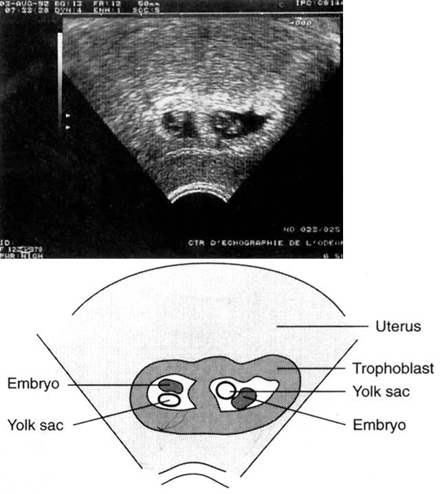
Fig. 5. Dichorionic twin pregnancy (6 weeks). (Bessis R: Ultrasound scanning techniques. In Keith LG, Papiernik E, Keith DM, Luke B [eds]: Multiple Pregnancy: Epidemiology, Gestation and Perinatal Outcome. London, Parthenon Publishing Group, 1995)

In the opinion of most experts, the major advantage of first or very early second trimester diagnosis is the confirmation or adjustment of the estimated date of conception as well as the clear delineation of chorionicity. The confirmation of estimated date of conception is particularly valuable because of the high rate of fetal growth abnormalities in twins as well as higher order multiple pregnancies. It is important to distinguish monochorionic from dichorionic placentas because of the far greater rates of morbidity and mortality for monochorionic compared to dichorionic twins (see earlier discussion). The question of zygosity is related, but of much less importance. It should be recalled that monozygotic twinning in pregnancies produced via artificially reproductive technology is almost triple the rate seen in spontaneous gestations.13,14 Many of these may be monochorionic. The presence of a webbed, lambda-shaped structure at the end of the interovular membrane is pathognomonic of dichorionic (or multichorionic) pregnancies (Fig. 6).25 This structure is particularly visible between 8 and 13 weeks of amenorrhea, and its presence or absence should be mentioned in any clinical report. The thickness of the intertwin membrane is also indicative of chorionicity. To some degree, the reliability of this measurement is as dependent on the technical conditions of the examination as it is on the actual thickness of the structure being examined (Fig. 7). The following is a useful clinical tool:

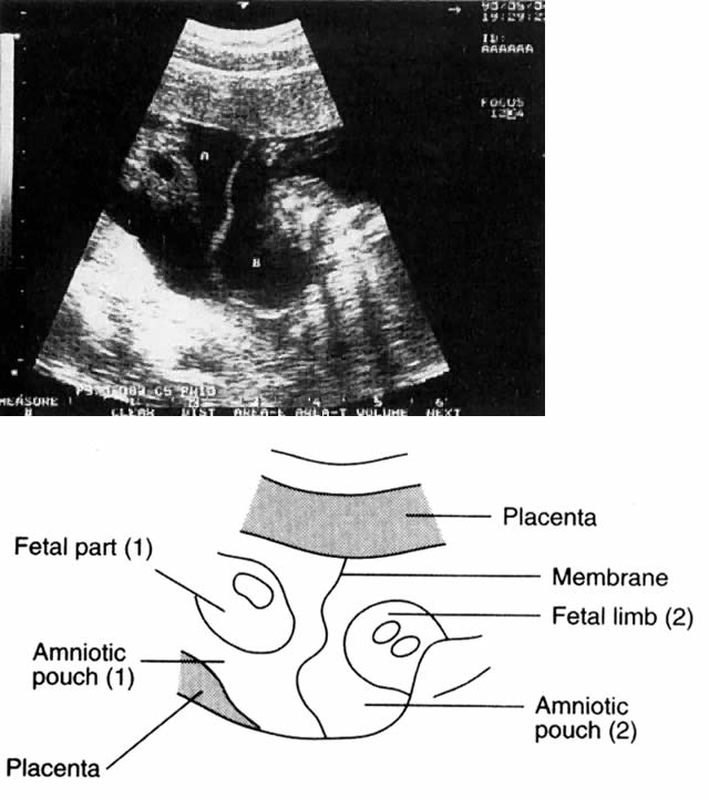
Fig. 6. Lambda-shaped membrane.(Bessis R: Ultrasound scanning techniques. In Keith LG, Papiernik E, Keith DM, Luke B [eds]: Multiple Pregnancy: Epidemiology, Gestation and Perinatal Outcome. London, Parthenon Publishing Group, 1995)

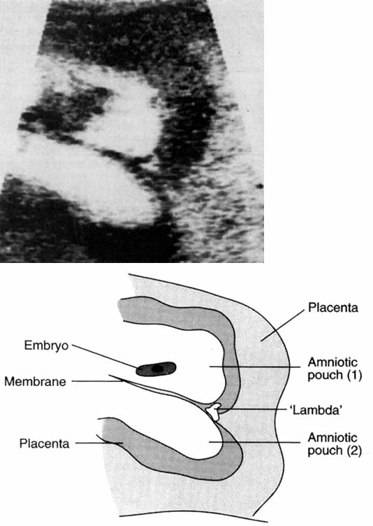
Fig. 7. Interovular membrane in a monochorionic pregnancy.(Bessis R: Ultrasound scanning techniques. In Keith LG, Papiernik E, Keith DM, Luke B [eds]: Multiple Pregnancy: Epidemiology, Gestation and Perinatal Outcome. London, Parthenon Publishing Group, 1995)

  In the presence of a single placenta, a membrane thickness of 2 mm or less favors the likelihood of monochorionic-diamniotic placentation.
  If, however, the membrane thickness is 4 mm or greater, fused dichorionic-diamniotic placentation is indicated in the presence of a single placental disc.26

In the second trimester, in addition to the detection of the twin condition, advantages of ultrasound examination include reliable estimation of gestational age; exclusion of placenta previa; detection of major structural anomalies, such as spina bifida, acrania, or conjoined twinning; and an opportunity for early bonding between a mother and her children. It is important to remember that later ultrasound is more productive for the detection of structural and/or body stalk abnormalities and progress of growth than for the optimal detection of chorionicity. For this reason, some experts believe that the improved outcomes that accrue from early detection of any deviation from normal more than compensate for the extra cost of multiple ultrasound examinations.22 An extensive review on the use of ultrasound in multiple gestation is provided by Lopez-Zeno and colleagues27 as well as by Bessis.23

As suggested above, clinical methods of diagnosing twin pregnancy are underutilized. More important, each method is fraught with inherent inaccuracies. For example, examination of fundal height is not only dependent on operator experience and skill, but may be reflective of the thickness of the maternal abdominal wall or whether the uterus is in a state of relaxation or contraction at the time of the examination. Because auscultation of the FHT is considered successful once heart tones are found, it is speculative how often this test is used to the point of calling another person to listen for a second sound. Further, unless the onset of hydramnios is acute and severe enough to cause symptoms, the quantity of fluid may be insufficient to allow its detection without the use of an ultrasound scan. Although excessive weight gain may be the first clinical sign of a twin gestation, lack of a precise definition of “excessive” may hinder its clinical utility. Recently the National Academy of Sciences (NAS) recommended an ideal total weight gain of 35 to 45 lb for twin pregnancy,28 but provided no definition of excessive weight gain. The NAS recommendation is useful, however, because it can be the basis of early intervention after initial diagnosis. Indeed, Luke29 and others15 have refined this recommendation to include the necessity of gaining 24 lb by the 24th week. Before recommending a comprehensive plan of weight gain, care givers should take into account the maternal prepregnancy body mass index and stature as well as the fact that about one half of twin pregnancies deliver at the 37th week of gestation or earlier. This latter point mandates an early gain, as opposed to waiting until after 32 weeks to try to augment weight.

Unfortunately, anemia is clearly too ubiquitous a sign to relate solely to multiple gestation. At the minimum, a complete blood count should be obtained and a stool sample studied for the presence of blood and/or parasites if and when severe anemia is present. Because maternal perception of increased fetal activity is uniquely subjective, an ultrasound examination may help determine whether a perception of “too much movement” is indeed a result of twins. The use of a personal or familial history of twins as a screen for obtaining an ultrasound is paradoxical: many women with a strong family history of twinning do not have twins, and many without it do. Of course, such clinical considerations are superfluous in patients who have undergone any form of artificial reproductive technology. In these instances, both the patient and the clinician want to know about the existence of pregnancy and its type as soon as possible. Without ultrasound, their wishes would be frustrated.

Although specific biochemical parameters, such as human chorionic gonadotropin and human placenta lactogen levels, exhibit slow and predictable increases as pregnancy progresses, these cannot be accurately applied to twins.30 This statement may be modified by the recent observation of the use of the three components of the pan - alpha-fetoprotein test.31 In the past, the use of any biochemical marker for the detection of twin pregnancy was hampered by day-to-day fluctuations in individual patients; the possibility of inaccurate conclusions based on miscalculations of gestational age; and the need to confirm any abnormal test with another of greater sensitivity. Because the finding of any abnormal biochemical marker is almost invariably followed by an ultrasound examination, many practitioners (at least at Northwestern) prefer to obtain an ultrasound in the first trimester and biochemical assessments in the second. In contrast, the policy at the University of Michigan is to obtain an ultrasound routinely at 16 to 18 weeks (see earlier discussion). This may preclude determination of chorionicity to some degree. Regardless, in institutions where first-trimester ultrasound is underutilized, the initial evidence suggesting the presence of twins may be the alpha-fetoprotein test.

Nutritional Considerations

The importance of prenatal nutrition cannot be overstressed. Having said this, until recently it was difficult for practitioners to develop practical guidelines to share with their patients. Although a variety of recommendations on the composition of the prenatal diet have been put forward since antiquity, specific recommendations are of fairly recent origin. Moreover, until 1990, recommendations were based purely on the needs of a singleton pregnancy.

The origins of dietary recommendations in pregnancy until at least the late 1950s were based on the writings of the German obstetrician Prochownik, and his predecessor Glassner, both of whom practiced in the latter half of the 19th century.32,33 Their suggestions were based on the fact that weight restriction was beneficial in producing smaller infants who were more likely to pass through deformed, contracted, rachitic pelves. The Prochownik diet, as well as other forms of weight restriction, which allegedly had no adverse effect on small infants born at term, influenced medical thinking until the middle of the 20th century.32,33 Writers inferred that the fetus and its mother were locked into a parasitic/symbiotic relationship in which the maternal dietary intake or nutritional status was immaterial. These opinions, though vehemently opposed by more physiologically oriented authors, were the basis of the widely accepted recommendation of a total weight gain of 20 lb. This dictum remained unchanged until around the late 1970s, despite the fact that several large hospital series published after 1940 suggested that many patients did not adhere to the 20 lb recommendation and actually gained more (20 to 30 lb) during their pregnancies.32,33 Among the deficiencies of these early hospital studies, however, was a general lack of consideration of preconceptional weight or anthropometric factors, such as short stature and low weight for height (body mass index), along with gestational weight gain as they may have influenced birth weight and length of gestation. After the association between pregravid weight and birth weight was recognized, gestational weight gain began to be viewed as a therapeutic intervention that not only augmented deficiencies of pregravid weight in the mother, but led to increased fetal growth and higher birth weights.32,33

Unfortunately, the “starvation mentality” initiated in the late 1800s was apparent as late as 1980. Taffel34 analyzed the gestational weight gain component of the 1980 National Mortality Survey and the National Fetal Mortality Survey. A total of 23% of women giving birth in 1980 gained less than 21 lb, and 12% less than 16 lb. Black mothers were more likely to gain less than 21 lb and were twice as likely to gain less than 16 lb compared to white mothers. Fortunately, the 1990 NAS publication, Nutrition During Pregnancy, served as a crucial turning point in developing rational guidelines for weight gain in pregnancy.35 This report reviewed all available data on gestational weight gain. It recommended, for the first time, ranges of gain by pregravid weight categories, including 28 to 40 lb for underweight women, 25 to 35 lb for normal-weight women, and 15 to 25 lb for overweight women.28 It also recommended, for the first time, a 35- to 45-lb weight gain in normal women with twin pregnancies.

Two independent studies paralleled the NAS publication and provided an indication of the future direction of subsequent clinical investigations. In one, based on birth/death certificates from the Office of Vital Statistics, Kansas Department of Health and Environment, from the years 1980 to 1996,36 a total of 922 twin gestations delivered at term were described. It was determined that the proportion of infants born with LBW declined as maternal prepregnancy weight status increased. It was further determined that infant birth weights increased linearly with prenatal weight gain for women who entered their pregnancy either underweight or at normal weight, but not for those who were overweight or obese at the start of their pregnancy. In another study, optimal pregnancy outcome (greater than 37 weeks' gestation; both infants greater than 2500 g each, with Apgar scores greater than 7) was associated with gestational weight gains of 44 lb (20 kg) compared to 37 lb (16.8 kg) for women with less-than-optimal outcomes.37

The concept of ideal or optimal twin pregnancy vis-à-vis maternal weight gain was studied extensively by Luke and co-workers38 in a series of papers published between 1991 and 1995. These investigations were based on all twin pregnancies of 28 or more weeks' gestation delivered between January 1, 1979, and December 31, 1989, at the Johns Hopkins Medical Institutions in Baltimore. Pregnancies complicated by extreme maternal obesity or pathologic weight gain were excluded in order to facilitate the evaluation of the maternal anthropometric characteristic of pregravid weight and subsequent gestational weight gain. Of the 274 available pregnancies, 111 were excluded because of one or more exclusion factors, and 163 twin pairs remained for analysis. When total weight gain was investigated in terms of early (before 24 weeks) and late (after 24 weeks) gain, twins with higher birth weights and shorter lengths of stay were delivered of mothers with higher late rates of gain. Luke and colleagues39 suggested that an early rate of gain of 1 lb/week before the 24th week and a late rate of gain of 1.5 lb per week might have therapeutic advantage. These analyses support the recommendations of the National Academy of Sciences. More important, however, they clarify the relationship between early and late rate of weight gain, total weight gain, and perinatal outcomes, including birth weight and length of stay (LOS), as shown in Table 4.

TABLE 4. Comparison of Perinatal Outcomes by Term vs Preterm and by Average of Twin Birth Rates <2500g vs 2500g


Pregravid Weight


Gain

Length of

Birth

Length

Birth

for Height and Birth


Rate of Gain (lb/wk)

Gestation

Weight

of Stay

Weight

Weight Categories

n

Early

Late

Total (lb)

(weeks)

(g)

(days)

Ratio

Preterm*

90

0.93

1.13

33.2

33.6

2024

16.3

0.973

Term†

73

0.9

1.31

40.5

38.4

2641

7.8

0.836

p value


NS

0.03

0.001

<0.001

<0.001

<0.001

<0.001

Preterm


 <2500 g

78

0.92

1.1

32.2

33.3

1921

18.1

0.948

 2500 g

12

1.03

1.35

39.6

35.1

2693

4.5

1.135

 p value


NS

NS

NS

0.01

<0.001

<0.001

0.01

Term


 <2500 g

27

0.87

1.05

35.7

38.2

2184

10.6

0.704

 2500 g

46

0.92

1.47

43.3

38.5

2910

6.2

0.913

 p value


NS

<0.001

0.02

NS

<0.001

<0.001

<0.001

2500 g


 Preterm

12

1.03

1.35

39.6

35.1

2693

4.5

1.135

 Term

46

0.92

1.47

43.3

38.5

2910

6.2

0.913

 p value


NS

NS

NS

<0.001

0.01

0.03

<0.001


* Preterm is defined as <37 complete weeks of gestation.
† Term is defined as {ewc MVIMG, MVIMAGE,!greateq.bmp}37 completed weeks of gestation.
NS not significant at the 0.05 level (two-tailed).
(Luke B: Maternal characteristics and prenatal nutrition. In Keith LG, Papiernik E, Keith DM, Luke B [eds]: Multiple Pregnancy: Epidemiology, Gestation and Perinatal Outcome, p 303. London, Parthenon Publishing Group, 1995)

A companion investigation was conducted by Luke and associates at the Twins Day Festival in Twinburg, Ohio, in the years 1989, 1990, 1991, and 1993. A total of 924 mothers of twins were interviewed, and data were obtained on their 1848 twin children. Study variables were compared by “ideal outcomes” (2500 to 2800 g birth weight and 35 to 38 weeks' gestational age) versus “nonideal outcomes” (birth weight above or below 2500 to 2800 g and/or gestational duration less than 35 weeks). Ideal twin outcomes had significantly fewer birth weights below the 10th percentile (11% vs 28%; p < 0.0001).40 Further, a significantly greater number of mothers with ideal outcomes did not smoke during their pregnancies (95.5% vs 17.3%; p = 0.01); they gained significantly more weight than mothers with nonideal outcomes (44.8 vs 41.1 lb [p = 0.005] and 1.23 lb/week vs 1.14 lb/week [p = 0.02]). Finally, it was determined that both body mass index and weight gain were positive factors affecting outcome. There was a progressive increase in the odds ratio of an ideal twin outcome with increasing weight gain. Weight gains below 35 lb were significantly associated with a nonideal outcome, whereas gains of 35 lb or more, or even more than 45 lb, were associated with ideal outcomes.40 The study also compared actual weight gains with those gains that the mothers had been advised by their respective care givers. Actual gains averaged closer to 45 to 50 lb, an amount substantially higher than the advised average of 30 lb. In this study, the NAS recommendation of a 25- to 45-lb weight gain was not significantly associated with ideal twin outcome (adjusted odds ratio, 1.22; 95% confidence interval, 0.85 to 1.77), whereas weight gain greater than 45 lb was (adjusted odds ratio, 1.67; 95% confidence interval, 1.17 to 2.39; p = 0.005).40 The weight gains of twins studied at the Johns Hopkins Institution are graphed alongside the NAS recommendations for singletons. Triplet weight gains are also shown (Fig. 8). This figure emphasizes the gain required by the 24th week of gestation in order to achieve “optimal gain” in the shortened average gestational time that is common in twins and triplets.

Fig. 8. Recommendations for gestational weight gain by plurality.(*, National Academy of Science) (Luke B, Johnson TRB, Petrie R: Clinical Maternal-Fetal Nutrition, p 287. Boston, Little, Brown & Co, 1993)

The enormous potential impact of aggressive efforts at maximizing maternal weight gain was described by Luke and colleagues41 in a theoretic model in which the birth weights of twins were shifted upward by 500 g at the lower side of the gaussian distribution of all twin birth weights, while the upper end of the distribution curve was maintained in its regular position. The net effect was to narrow the curve and the area under it. Luke and associates calculated that if such a shift could be implemented on a national basis by improved nutrition or prolongation of pregnancy it would reduce VLBW and perinatal mortality by 59.5% and 84.6%, respectively. It would also result in a lifetime cost saving of $219 million annually for each year's birth cohort by reducing physical handicap among survivors. Increasing birth weights and gestation length also have dramatic effects on LOS. In the Johns Hopkins database, for infants born at less than 37 weeks of gestation, LOS was 1.5 to 2.0 times greater for twins with intrauterine growth retardation (IUGR) compared to non-IUGR twins, whereas no significant differences were found between non-IUGR twins and singletons.42 At weights below 2500 g, IUGR twins had significantly higher LOS compared to singletons.42 Specifically, for non-IUGR twins compared to singletons, LOS was nearly 50% and 30% shorter, respectively, for the birth weight categories of 1000 to 1499 g and 1500 to 1999 g.

In contrast to the growing consensus regarding total weight gain and rate of gain considered optimal in twin pregnancies, little consensus exists regarding dietary supplementation with iron, vitamins, minerals, and folic acid, although the value of folic acid in terms of its ability to prevent neural tube defects is now widely appreciated. The recommendations contained in Nutrition During Pregnancy35 are as follows:

  1. Beginning at the 12th week of gestation, routine use of 12 mg of ferrous iron per day is recommended. Ferrous sulfate or fumarate are preferable because the former contains 20% iron by weight (60 mg of elemental iron in a 300 mg tablet). Ferrous fumarate contains 32% iron by weight (96 mg elemental iron in a 300 mg tablet).43
  2. Supplemental medication should be free of additional ingredients, such as calcium, magnesium, or zinc, all of which reduce the absorption of iron.
  3. Supplemental iron should be taken between meals with liquids other than milk, tea, or coffee.
  4. Hemoglobin level should be determined early to detect preexisting anemia. Levels below 11 g/dL during the first and third trimesters or below 10.5 g/dL during the second trimester are characteristic of anemia.

Table 5 shows the cutoff values for anemia in women—pregnant and nonpregnant, smokers and nonsmokers—adapted from the Centers for Disease Control criteria for anemia in childbearing-aged women.44 When accompanied by a serum ferritin concentration of less than 12 μg/dL, iron deficiency anemia can be diagnosed with certainty. Required treatment consists of 60 to 120 mg/day of ferrous iron until the hemoglobin concentration becomes normal. At this time, the dose of iron can be decreased to 30 mg/day. No specific prenatal dietary guidelines have been published for expectant mothers of twins. In the absence of such recommendations, Luke and co-workers suggested that a 50% to 100% increase in the recommended dietary allowances for singleton pregnancies be used for women with twins. Their adaptation of the 10th edition of The Pregnancy RDAs in National Academy of Sciences is shown in Table 6.44

TABLE 5. Cutoff Values for Anemia in Women: Nonpregnant and Pregnant, Smokers and Nonsmokers


Hemoglobin (g/100 mL)

Hematocrit (%)


Smokers

Smokers


Nonsmokers

10–20*

21–40*

Nonsmokers

10–20*

21–40

Nonpregnant

12.0

12.3

12.5

36.0

37.0

37.5

Pregnant


 1st Trimester

11.0

11.3

11.5

33.0

34.0

34.5

 2nd Trimester

10.5

10.8

11.0

32.0

33.0

33.5

 3rd Trimester

11.0

11.3

11.5

33.0

34.0

34.5


* Number of cigarettes smoked per day.
(Luke B, Johnson TRB, Petrie R: Clinical Maternal-Fetal Nutrition, p 196. Boston, Little, Brown & Co, 1993)

TABLE 6. Summary of RDAs* for Women Aged 25 and Older, Pregnant with Singletons vs Twins


Singleton

Twin/Triplet


Nutrient

Pregnant

Pregnant

Dietary Sources

Folic Acid

400 mg

800 mg

Leafy vegetables, liver

Vitamin D

10 μg

15 μg

Fortified dairy products

Iron

30 mg

50 mg

Meats, eggs, grains

Calcium

1200 mg

1800 mg

Dairy products

Phosphorus

1200 mg

1800 mg

Meats

Pyroxidine

2.2 mg

4.0 mg

Meats, liver, grains

Thiamine

1.5 mg

3.0 mg

Enriched grains, pork

Zinc

15 mg

30 mg

Meats, seafood, eggs

Riboflavin

1.6 mg

3.0 mg

Meats, liver, grains

Protein

60 mg

120 mg

Meats, fish, poultry, dairy

Iodine

175 mg

300 mg

Iodized salt, seafood

Vitamin C

70 mg

150 mg

Citrus fruits, tomatoes

Energy

2500 kcal

3000 kcal

Protein, fat, carbohydrate

Magnesium

320 mg

450 mg

Seafood, legumes, grains

Niacin

17 mg

25 mg

Meats, nuts, legumes

Vitamin B12

2.2 mg

3.0 mg

Animal proteins

Vitamin A

800 μg

1000 μg

Dark green, yellow, or orange fruits and vegetables, liver


* Recommended dietary allowances.
(Luke B, Johnson TRB, petrie R: Clinical Maternal Fetal Nutrition, p 285. Boston, Little, Brown & Co, 1993)

Schedule of Visits

A key element of intensive prenatal care for multiples should be the scheduling of more frequent visits: every 2 weeks until 20 weeks, and weekly thereafter. An alternative scheduling schema has been presented by others.15 The rationale behind this precept is the fact that the mother must receive not only the routine care provided to any pregnant woman (e.g., testing for neural tube defects and abnormalities of carbohydrate metabolism, amniocentesis ), but also the additional tests mandated by the presence of multiple gestation (e.g., repeat ultrasound to clarify the type of placentation or membrane status, or for surveillance for signs of preterm labor) as well as crucial educational counseling.

Regardless of the socioeconomic status of the parents and whether the pregnancy was planned or unplanned, natural or assisted, the reactions of prospective parents of twins are not the same as in a singleton pregnancy. Surprise and delight are often admixed with apprehension and fear.

Physicians and staff should guard themselves from being swept along in the parents' euphoria; instead, they should present a rational and balanced account of the increased risks associated with twin pregnancy and what can be done to prevent them. Parents often leap from diagnosis to delivery planning in their enthusiasm. They must be informed, and physicians need to continually remind themselves and their staffs that good prenatal care for twins consists of a multitude of special considerations designed to bring the pregnancy to an optimal outcome and that despite such efforts adverse events may occur before delivery. In the vast majority of instances, the prospective parents worry about their ability to cope with the physical, emotional, and financial demands of a multiple pregnancy.45 In addition, parents often bombard physicians and their staffs with questions, many of which are based on reading or conversations with other parents of multiples. More frequent visits not only facilitate obtaining requisite examinations in a timely fashion, but also help establish the rapport that is so helpful in guiding these pregnancies to successful outcomes. More frequent visits also benefit monochorionic twins who are clearly at higher risk as TTTS may be appreciated from 18 weeks onward. Therefore, weekly visits and liberal use of ultrasound is indicated for these patients.

More frequent visits also allow the mother to avail herself of information about self-help groups, which can provide invaluable assistance for prospective parents. For twin gestations, the National Organization of Mothers of Twins Clubs is unique because it has numerous chapters located throughout the country. As the mother is adjusting to the many changes that will occur in the near future, she can also obtain valuable information on breast feeding from La Leche League. A list of useful addresses and phone/FAX numbers of some of the most important organizations is provided at the end of the reference section (see Appendix).

Selective Fetal Reduction in Multiple Pregnancy

Most twin pregnancies will not benefit from selective reduction. Even in higher order gestations, its use is controversial. The timing of selective reduction in multiple pregnancy varies. In some instances, it is performed in the first trimester, usually but not invariably for the purpose of reducing the number of fetuses. Second-trimester reductions are usually, but also not invariably, performed because one or more fetuses are found to to have abnormalities (structural, chromosomal, or mendelian). Access to this technique is not universal. In the United States, nine centers have reported outcome data to date, but it cannot be stated with certainty how frequently this operation is performed outside of these reporting centers or the results of these endeavors. It is clear, however, that this technique is not without risks (e.g., loss of the entire pregnancy, occasional infection). A learning period is required before the technique is mastered, and the decision to resort to reduction is never easy for the patient, especially if she has been infertile for a period of years and only became pregnant as a result of prolonged and costly efforts. Two summary chapters on the operative technique of selective reduction are available for further reference.46,47

Two specific outcome variables associated with fetal reduction merit special attention: (1) maternal serum alpha-fetoprotein level after second-trimester reduction; and (2) rate of preterm delivery subsequent to reduction. With regard to the first point, according to Lynch and Berkowitz,48 maternal alpha-fetoprotein in the second trimester is always elevated after a reduction procedure and is not necessarily indicative of fetal defects. This elevation is probably caused by release of tissue or serum from the dead fetus or fetuses. Amniocentesis is not indicated in these cases; rather, ultrasonography should be used to search for the presence of fetal defects. Lynch and Berkowitz further noted that although some patients may have laboratory evidence of disseminated intravascular coagulation after reduction procedures, such changes generally resolve spontaneously.48 With regard to the second point, in another report from the same institution, Berkowitz and associates49 described the outcomes of 200 cases. In this series, higher order pregnancies (three to nine fetuses) were reduced to 189 sets of twins, 5 sets of triplets and 6 singletons. A total of 19 (9.5%) patients lost their remaining fetuses before the 24th week of gestation. The mean gestational age at delivery was 35.7 weeks for the pregnancies that continued. However, gestational age at delivery varied inversely with the initial number of fetuses from 36.1 weeks for women who presented with triplets to 33.8 weeks for women who had six or more fetuses. This trend was statistically significant. Of the 19 complete pregnancy losses, 16 occurred more than 4 weeks after the reduction procedure.

Back to Top
CARE BETWEEN WEEKS 20 AND 26

Education

Educational processes begun during the first 20 weeks must continue thereafter; however, the focus should change to incorporate those events the patient is likely to encounter during the second half of the pregnancy. Foremost among these is a consideration of the nature and risk of preterm labor and delivery and the means that might be employed to reduce this risk. Patients should be instructed about the signs and symptoms of preterm labor, including vague signs such as backache and lower abdominal pressure. More important, they should be empowered to request medical assistance immediately to confirm the presence or absence of preterm contractions.50 In addition, they should be informed about the available means to reduce the risk of preterm labor or treat it should it occur. The distinction between true preterm labor and the not infrequent need to deliver infants prematurely for complications, such as bleeding, pregnancy-induced hypertension, and premature rupture of the membranes, should be spelled out in clear terms understandable to a person who is not trained in medical terminology. The patient should also be informed of the survival rates at given weeks of gestation at the institution where delivery is contemplated. If this institution does not have a level III nursery, the implications of this deficiency should be explained in detail, as should the standing arrangements of the health-care provider and the hospital with consulting institutions and individual consultants, be they physicians or allied medical personnel. The patient and her partner should also know exactly whom to call in the event that an emergency arises and/or the regular health care provider is unavailable.

Bed Rest and Lifestyle Modifications

The value of bed rest in the prevention of preterm labor has been and remains controversial. Indeed, some would say that it is the most controversial aspect of antepartum care. A major reason underlying this controversy is the fact that “bed rest” has never been adequately defined, even in studies that advocate its use. Clearly the concept of bed rest means different things to different people, physicians and patients alike. The issue is further clouded by the inability to monitor compliance with prescriptions for bed rest unless the patient is hospitalized and the nursing staff has little else to do but observe and chart their inactivity. Moreover, chorionicity was not considered in any of these studies. The relatively few prospective randomized trials of routine hospitalized bed rest do not support this practice as being efficacious. However, some unrandomized studies with historical controls that emanated from communist countries in the late 1960s and early 1970s do suggest its efficacy, albeit based on their methods of analysis. A meta-analysis of four prospective randomized trials published in 199151 confused rather than clarified the issue: it determined increased rates of preterm delivery and perinatal death in mothers who underwent bed rest. The reasons for these unexpected observations are not clear. If there is any point of agreement on the purported beneficial effect of bed rest, it is that birth weight is increased.17,52 Unfortunately it is currently impossible to determine the number of variables that are related to outcome. Other factors, such as improved uterine blood flow (see later discussion), changes in diet, and probable restrictions on the use of drugs, alcohol, and tobacco, obviously come into play when a patient undergoes hospitalized bed rest.

The major deterrent to the routine implementation of in-hospital bed rest is its cost. As a result, clinicians have begun to look for alternatives that might provide the same effect. Among the most popular is the combination of increased “rest” at home (defined as getting off one's feet) along with a reduction in the physical activities of daily life. The value of this latter intervention is well proven in singleton gestations.53 A recent evaluation of data on twins from France suggests that it is equally efficacious in twins.54 The lifestyle modifications proven valuable in reducing the preterm delivery rate in France included the following: (1) reduced hours of paid employment outside of the home and/or modification in work load; (2) reduction or elimination of specific activities, such as walking stairs, carrying groceries, or doing the laundry; and (3) avoiding chores such as house painting or nursery decoration in anticipation of the forthcoming delivery.

Newman and Ellings15 recommend a 2-hour rest period three times a day (morning, afternoon, and after the evening meal); this advice is reasonable. These authors also recommended,15 and we agree, that the health-care providers institute a thorough evaluation of the stresses involved in the patient's employment outside of the home. This can be accomplished most easily by speaking frankly with the patient. To our knowledge, no validated question set is available. The list of circumstances suggested by Newman and Ellings15 for which the discontinuance of work is recommended is heavily influenced by the French model53,54 and supported by American data obtained in a study of working nurses.55 The following are among the reasons for discontinuing outside employment: (1) work that involves standing more than 3 hours/day or carrying loads of more than 20 lb; (2) work on industrial machines, especially those with incessant vibrations or that involve conveyor belts; (3) work involving continuous, low-level physical exertion or intermittent, high-level exertion (low-level exertion is seemingly worse if it involves repetition without stimulation); (4) military duty; and (5) environmental stresses involving cold, loud noise, or exposure to toxic substances. Newman and Ellings15 also advocated, and we agree, that work should cease regardless of the occupation if the woman has a prior history of preterm labor or if she is carrying three or more fetuses.

Although the precise mechanisms by which bed rest and reduction of physical activity exert their effect are not completely clear, some information is available. Several investigators have reported that standing for long periods is associated with preterm birth.56,57 Others have shown the same association for increased uterine contractions.58,59 For preterm birth associated with prolonged standing, Launer and associates60 reported an adjusted odds ratio (AOR) of 1.56. An even higher AOR (2.7) was reported by Teitelman and colleagues,61 as was a higher proportion of preterm births among women whose jobs require prolonged standing (7.7%) compared to those with sedentary or active jobs (4.2% and 2.8%, respectively). Using a definition comparable to that of Teitelman and co-workers,61 Klenbanoff and associates62 reported an AOR of 1.3 for preterm birth among women whose work involved standing for more than 8 hours/day.

The work of Schneider and colleagues58,59 in Switzerland is particularly instructive for understanding the physiology behind these differences. Simply stated, their experimental data support the concept that standing for long hours is a vertical form of the supine hypotensive syndrome. Gravity pulls the pregnant uterus deeper into the pelvis with prolonged standing. The bony pelvic girdle forces the uterus to impinge on the inferior vena cava and impede venous return from the lower extremities. The only way the pressure can be relieved is for the uterus to contract. From a clinical point of view, this scenario seems plausible.

Baseline Cervical Evaluation

Among the common preterm-labor risk factors that appear during pregnancy are bleeding, shortening or dilation of the cervix, expansion of the lower uterine segment, and descent of the presenting part. All may appear with or without the presence of uterine contractile activity. The most complete discussion of the effect of these signs on the risk of preterm labor and the rate of preterm delivery is contained in the monograph by Papiernik and co-workers.53 Although the Papiernik data pertain to singleton pregnancies, they are instructive for multiple gestations as well. In particular, two signs—shortening of the cervix and prelabor dilation—are especially common in multiple gestations. Both are indicative of changes in the lower uterine segment that may precede the onset of preterm labor, and both can be recorded with ease at each office visit, if vaginal examinations are conducted.

Newman and colleagues63 conducted an extensive review of the effects of cervical change in multiple gestation. Among their observations is the fact that numerous processes inherent to multiple gestation result in advanced stages of cervical maturation. However, because multiple gestation acts as an independent factor in the classification of risk for preterm labor and delivery, these authors strongly advocated the use of repeated cervical examinations at each visit as part of ongoing risk assessment and the incorporation of these data into a “cervical score.”

A number of investigators, including the French group headed by Papiernik,53 have used changes in antepartum cervical status to predict the risk of preterm delivery in multiple pregnancy. Central to most of these reports is the calculation of a score. Newman and associates63 calculated their scores in the manner described by Houlton and colleagues. Simply stated, the score is based on the length of the cervix minus the dilation of the internal os. Weekly cervical assessment in more than 100 multiple gestations documented a progressive fall in the cervical score throughout the second half of gestation. As the score declined, so did the time to delivery. Figure 9 is a graphic representation of the cervical score (mean ± SD) and baseline uterine contraction frequency (mean ± SD) for multifetal gestations at 20 to 36 weeks' gestation.

Fig. 9. Changes in cervical score and contraction frequency for multifetal gestations. = cervical scores; = contraction frequency.(Newman RB, Godsey RK, Ellings JM: Assessment of cervical change. In Keith LG, Papiernik E, Keith DM, Luke B [eds]: Multiple Pregnancy: Epidemiology, Gestation and Perinatal Outcome. London, Parthenon Publishing Group, 1995)

Use of a score helps to define the actual risk of preterm labor and delivery for a given patient, as noted in Table 7. The data of Newman and co-workers63 suggest that the earlier a low (-1, -2, or -3) score is detected, the more likely the patient will deliver prematurely. Regrettably, the greatest impediment to the use of a scoring technique is the strong reluctance of many American clinicians to perform vaginal examinations. As Papiernik and colleagues53 noted, it is paradoxical that an examination considered essential by European (and some American) clinicians is totally disfavored by others, despite the fact that the majority of reports on this subject have failed to associate cervical examination with any adverse outcome.63

TABLE 7. Sensitivity, Specificity, and Relative Risk of Premature Delivery (<37 Weeks' Gestation), Based on Cervical Score 0 on or Before 34 Weeks' Gestation


Patient Group

Sensitivity (%)

Specificity (%)

Relative Risk (RR)*

p Value

All patients

88

62

3.9 (1.8 < RR < 8.7)

0.0001

At 20–28 wk

33

96

2.0 (1.4 < RR < 2.9)

0.01

At 29–34 wk

87

81

3.8 (1.7 < RR < 8.5)

0.0001

Nulliparas

83

75

3.3 (1.0 < RR < 11.4)

0.02

Paras

91

58

4.3 (1.5 < RR < 12.4)

0.001


* Relative risk with 95% confidence limits.
(Newman RB, Godsey RK, Ellings JM: Assessment of cervical change. In Keith LG, Papiernik E, Keith DM, Luke B(eds): Multiple Pregnancy: Epidemiology, Gestation and Perinatal Outcome, p 464. London, Parthenon Publishing Group, 1995)

The major value of cervical examination lies in its ability to alert the clinician to the probability of impending preterm labor. As is the case with home uterine monitoring, assessment of cervical change does not prevent either preterm labor or delivery, but rather it is a valuable predictor of these events. Thus the presence of cervical change, especially early in gestation, allows the physician to reassess all aspects of the pregnancy and, in some instances, to prescribe therapeutic interventions.

Back to Top
CARE BETWEEN WEEKS 26 AND 32

Without minimizing the importance of anything that might be required before week 26, the care provided between weeks 26 and 32 is a unique opportunity to institute measures designed to protect the fetuses, enhance maternal and fetal well-being, and to prolong pregnancy. In this regard, it is important to recognize that obstetricians and neonatologists often differ in their characterization of the earliest safe moment for delivery. The fact that infants may survive at 28 to 32 weeks is not, in our opinion, a reason for not attempting to prolong pregnancy unless mitigating circumstances are well documented (e.g., physical deterioration of the maternal or fetal condition, especially if this latter is due to TTTS or other complications of monochorionic placentation). It behooves each obstetric unit to be fully aware of singleton/twin survival outcomes in their own neonatal intensive-care unit on a week-to-week basis after week 24. This is because national data on neonates reporting morbidity and mortality generally lag 5 to 10 years behind the impact of continual improvements in the care of premature infants. Therefore, it follows that obstetricians should obtain antenatal consultation with neonatologists on a case-by-case basis on the benefits and risks of preterm delivery versus continuation of pregnancy in given instances.

The protective measures that should be stressed between weeks 26 and 32 fall into four general categories: (1) reassessment of the adequacy or lack thereof of maternal weight gain; (2) prophylactic administration of steroids; (3) changes in lifestyle and activity reduction; and (4) serial ultrasound assessment. A fifth protective measure, over which much controversy exists, is home uterine activity monitoring.

Weight Gain

In the event that the patient has not gained 24 lb by the 24th gestational week or 32 lb by the 32nd gestational week, the health-care provider should seriously question, why not? Nutritional consultation is generally available at most hospitals, although such services are notoriously underutilized. Measurement of the fat stores in the upper area of the mother's arm is an easy way to assess the quantity of fat deposited during the pregnancy, but most obstetricians are unfamiliar with this type of office evaluation. Because glucose intolerance is thought to be more common in multiple pregnancies, it is suggested that a 1-hour, 50-g glucose load be administered between the 24th and 28th week of gestation to all patients. If the plasma glucose result is greater than 145 mg/dL, a standard 3-hour glucose tolerance test can be scheduled.

Steroid Administration

Data presented by the National Institutes of Health consensus conference on the antepartum use of corticosteroids for the induction of fetal lung maturity are consistent with the recommendation that steroids be administered to women with a multiple gestation if they experience premature labor at less than 32 weeks' gestation or premature rupture of the membranes, or if they require preterm delivery for maternal or fetal indications.64 In addition, Newman and Ellings15 advised that a single 12-mg dose of betamethasone be administered intramuscularly in women whose cervical score is less than or equal to 0 at 32 weeks' gestation, although they admitted that they had no scientific basis for their therapeutic advice. One of the authors (TRBJ) prefers to restrict steroids to patients with documented preterm labor accompanied by cervical changes.

Modification of Activity

Ideally, continued modification of the patient's activity level and the frequency of periods of recumbent rest can be rationally planned on the basis of repeated cervical assessment in combination with maternal symptomatology (see earlier discussion). It is crucial to remember, however, that uterine activity in twin gestations is most often insensible (hence the argument in favor of home uterine activity monitoring). It is most helpful if cervical examinations are conducted by the same examiner on a repetitive basis. During these examinations, symptomatology can be reassessed by careful questioning (hence the argument in favor of repetitive nursing contact). Newman and Ellings15 noted that increasing symptomatology, accompanied by palpable or recorded uterine contractions in the face of a cervical score of less than or equal to 0 before 34 weeks' gestation place the patient at risk for preterm delivery.

Serial Ultrasound Examinations

The care provided to multiples has been revolutionized by the availability of ultrasound examinations; nowhere is this more apparent than during the period in question, 26 to 32 weeks' gestation. Serial ultrasound examinations not only permit the documentation of abnormal growth patterns, but allow assessment of discordance, recognition of TTTS, and detection of previously overlooked or late-appearing congenital anomalies. In the case of TTTS, diagnosis can occur in mid second trimester, well before 26 weeks (i.e., 18 to 20 weeks). The finding of discordance for a major congenital anomaly presents both a diagnostic and a therapeutic pitfall. In the first instance, discordance cannot be equated with dizygosity, as numerous reports of monozygous discordance exist.65 In the second, procedures for selective reduction are particularly hazardous, as the death of the affected twin will affect the cotwin if the twins are monozygous (see later discussion). Ultrasound examinations also are extremely useful in detecting disparities in the quantity of amniotic fluid in each sac. Without doubt, ultrasound examinations during these weeks are more complex, require longer examination times, and are technically more demanding. Much of the material that follows in this subsection is adapted from a recent work by one of the authors (JA L-Z).66

GROWTH IN TWINS.

At one time, the necessity of constructing special growth curves for twins was a matter of debate; this is no longer the case. Several large studies have evaluated this question from different perspectives. Not unexpectedly, their conclusions differed. A major point of difference has centered around the exact point in time when weight differences become evident in twins compared to singletons. Conclusions have ranged from the 27th through the 35th week of gestation.33 Three studies published during the 1980s all concluded that by the 38th week of gestation, the 90th percentile for twins is equal to the 50th percentile for singletons and that the 50th percentile for twins is equal to the 10th percentile for singletons.

Measurement of the abdominal circumference is the most reliable parameter for assessing normality or discordance in multiple gestations. Even a difference as small as 20 mm may be indicative of significant discordance, despite the fact that the abdominal circumference begins to decrease in twin gestation and may reach as much as 1 cm by the 37th week of gestation.66 Fetal weight estimations should be derived from mathematic formulations incorporating the head circumference, abdominal circumference, and femur length. Several general formulas have been proposed, but that of Sabbagha and colleagues is targeted to gestational age and fetal size.66 The causes of growth discordance vary according to chorionicity. For example, growth discordance in monochorionic twins may be due to onset of TTTS or simply unequal sharing of the placental parenchyma between the twins.

OTHER CONSIDERATIONS.

The biophysical profile score extends the regular ultrasound examination by combining five separate parameters evaluating biophysical function into a single test conducted over a 30-minute interval. These parameters include, in addition to the nonstress test, determination of the amniotic fluid volume, as well as assessments of fetal breathing movements, fetal body motion, and fetal tone. When the function under study is normal, it is assigned a value of 2; when it is absent a score of 0 is recorded. The biophysical profile score is used in twins just as it is in singletons.66 It is often used if questions exist about nonstress test reactivity or if there is concern about fetal well-being (e.g., IUGR).

Data are slowly accumulating on the use of Doppler velocimetry indices in twin gestations. There exists some question whether the standard deviations differ significantly between singletons and twins. Based on a study of 248 twin pregnancies at the Antoine Beclere Hospital in Clamart, France, Bessis23 concluded that the curves established for singleton pregnancy are entirely appropriate for monitoring twin pregnancies. His observations are supported by others in the literature.66 It is not yet clear, however, how this new information might be put to practical use.66 In any event, the use of Doppler velocimetry studies is not the standard of care, although some investigators suggest that they are useful in conjunction with an abnormal nonstress test. Results of Doppler studies are particularly complex in monochorionic twins, and interpretations must be made with caution because of the vascular status of the placenta.

AMNIOCENTESIS.

It is axiomatic that any amniocentesis should be performed under ultrasound guidance. After placental localization, the ultrasonographer must first search for the dividing membrane. The transducer is then directed toward one sac, a local anesthetic is injected into the skin and subcutaneous tissue after appropriate antiseptic preparation, and finally a spinal-type needle is inserted into the designated sac as it is being visualized on the ultrasound screen. After removing the requisite amount of fluid, 1 mL of indigo carmine is instilled. The use of methylene blue is no longer advocated because of its association with fetal hemolysis, hypoxia, and duodenal atresia.67 The needle can then either be withdrawn and reinserted under ultrasound guidance or redirected into the second sac via a puncture of the membranes. This latter technique is not without risk, however, and is discouraged by many experts. The membranous division between the two sacs may be torn, thereby converting a diamniotic pregnancy into a pseudomonoamniotic pregnancy. Should this occur, the potential for cord accidents is greatly increased. Regardless of which insertional technique is used, the fluid obtained from the second sac must be free of indigo carmine dye in order to ensure that both sacs indeed have been sampled. Motew and Ginsberg68 suggested that the syringe be shaken vigorously so that microbubbles appear immediately prior to the injection of the indigo carmine. Once injected, these bubbles diffuse evenly throughout the sac and demarcate it sharply on the ultrasound picture. This technique is also useful to delineate diamniotic from monoamniotic gestations.

Home Uterine Activity Monitoring

To say that opinions are vociferously divided about the usefulness of this technology is a gross understatement. Generalist obstetricians and maternofetal specialists seeking evidence based data before establishing practice guidelines are in what appear to be opposing camps. Arguments center around two questions: (1) Is there any utility to the procedure?; and (2) If there is, is it related to the machine and the technology or to the human contact provided by the nurses? Unfortunately, these arguments often lose sight of the primary reasons why this technology may have particular utility in multiple gestations, which relate to the following well-recognized facts:

  1. Antepartum uterine activity is related to increased risk of preterm labor in twins as well as higher order gestations.
  2. Compared to singletons, baseline uterine activity is increased, regardless of whether preterm labor is present.
  3. The increase in contraction activity occurs at two distinct points in time—weeks before the onset of preterm labor and then again within 24 hours of the onset of preterm labor.15

In the twins studied by Newman and Ellings,15 increased uterine activity was correlated with decreasing cervical score (see Fig. 9). Despite the clear association between antepartum activity and cervical change, women with twins frequently are not able to perceive their contractions as easily as women with singletons. Indeed, less than 15% of objectively recorded contractions are perceived by women with twins, and many women are totally unaware of their occurrence.15

Numerous trials of home uterine activity monitoring have been published, but they differ enormously in design, outcome variables, statistical interpretation, and agreement or disagreement with other published reports. To our knowledge, the issue has not been addressed by a global analysis, in which all data from all studies are treated as if it were one study. A useful review was provided by Newman and Ellings.15 We agree with the opinions expressed in this review. Possible situations where home uterine activity monitoring may be useful include (1) if the patient has already experienced preterm labor or an inability to perceive her own uterine contractions; (2) patients with abnormal cervical scores (less than or equal to 0) at an early gestational age (less than or equal to 30 weeks); and (3) women with more than two fetuses, because the rapidity with which cervical dilation may occur can lead to devastating results.

Back to Top
DETECTION OF TWIN-TO-TWIN TRANSFUSION SYNDROME

One of the main objectives of intensive antenatal care (especially using ultrasound) is the earliest possible detection of some serious complications of monochorionic placentas. Among these, TTTS has been described in the literature for more than 100 years. Its incidence is unknown, and the condition frequently is subject to misdiagnosis. Its occurrence is tangentially related to racial variations in twinning rates and types of placentation. A range of 4% to 35% of diamniotic-monochorionic twin pregnancies is suggested.69

No complete definition of this syndrome exists in standard texts. Simply stated, TTTS occurs in monozygotic, monochorionic pregnancies. Its occurrence is based on the fact that the monochorionic placenta is truly a single placenta perfused by two fetuses. The majority of monochorionic placentas have one or more of the following possible types of interfetal vascular anastomoses: (1) arterial-arterial (AA) in the chorionic plate (frequent); (2) venous-venous (VV) in the chorionic plate (rare); (3) arterial-venous (AV) in a perfusion zone within the cotyledons. The AA and VV anastomoses are bidirectional; in contrast, AV anastomoses are unidirectional. TTTS occurs as a result of uncompensated blood transfer (sharing) through anastomotic AV vessels situated deep within the placenta's “common villus” district. When TTTS does not occur in a monochorionic placenta, it is because the superficial AA and VV anastomoses compensate for the hemodynamic imbalance caused by the deep anastomosis.70 Figure 10 is a schematic representation of the three types of monochorionic placentas. Any shift in the watershed zero flow area between the two twins' placental areas will result in unidirectional transfusion via superficial VV or AV anastomoses, which functionally become AV anastomoses.69

Fig. 10. Graphic presentation or anastomoses in TTTS.(Fisk NM: The scientific basis of feto-fetal transfusion syndrome and its treatment. In Ward RH, Whittle M [eds]: Multiple Pregnancy, pp 235–250. London, RCOG Press, 1995)

There are two types of TTTS: acute and chronic. Older textbooks, especially those containing illustrations of a plethoric twin accompanied by its anemic cotwin, emphasized that the diagnosis was generally made in the neonatal period on the basis of birth weight disparity greater than 20% and cord blood hemoglobin differences of greater than 5 g/dL. The use of such criteria is less than ideal, however, because early reports that focused exclusively on criteria visible at birth often included dichorionic twin pairs, or failed to consider placentation or other factors that might lead to discordant growth.70

The manner in which the diagnosis of TTTS has been made in recent years was revolutionized by ultrasound. Three criteria in combination favor the diagnosis: (1) monochorionicity (see earlier discussion); (2) simultaneous coexistence of hydramnios/oligohydramnios; and (3) difference in bladder size and activity.69 As amnionic fluid volume discordance increases, the so-called donor twin becomes oliguric. Oliguria is followed by oligohydramnios, and the “donor” fetus appears enshrouded in its membrane and “stuck” to the uterine wall. At the same time, severe hydramnios develops in the polyuric “recipient” fetus. The use of the term “stuck twin syndrome” to describe the condition is not recommended because it is inherently open to misinterpretation. Specifically, it does not implicitly state that hydramnios is required in the cotwin, and oligohydramnios can also occur in dichorionic pregnancies.69

According to Fisk,69 the diagnosis of TTTS in the midtrimester should be restricted to monochorionic pregnancies with complete discordance of the amnionic fluid volume, because no other pathology in monochorionic twins results in severe oligohydramnios in one sac and severe hydramnios in the other. Mortality and morbidity associated with TTTS are high. Indeed, the large differences in outcome between monochorionic and dichorionic twins are almost entirely related to the fact that the monochorionic placenta is perfused by both fetuses.70

Treatment of TTTS must be individualized. Not all therapies are presently within the purview of generalist obstetricians. This is particularly true for the laser ablation of AV anastomoses on the chorionic plate.71 In the absence of maternofetal specialists, however, generalist obstetricians occasionally perform serial amnioreduction. Of all available therapies, serial amnioreduction offers the best results and is the most widely used.69 The reader is referred to more complete discussions of the underlying pathophysiology and treatment of TTTS.69,70

This condition is often seen at an early stage-prior to the 20th week. It should be emphasized that a real benefit of early and repeated ultrasound in uncomplicated pregnancies is the ability to diagnose TTTS at a time when therapy can be instituted. Tragic consequences result from a delay in diagnosis and a missed opportunity to institute proper treatment.

Intrauterine Demise of One Twin

Although other, rarer complications of monochorionicity exist, such as twin reverse arterial perfusion (TRAR) (acardiac twin), monochorionic-monoamniotic twinning, and conjoined twins, the intrauterine demise of one twin is without doubt a serious and devastating complication of twin gestation because of its implication for the survivor. The reported incidence of this problem ranges from 0.5% to 6.8% of all twin gestations. Its occurrence generates enormous emotional stress for the patient as well as the obstetric team.72 Clinical management must be individualized and planned on the basis of definite knowledge of chorionicity. Specifically, death of one dichorionic twin does not greatly imperil its cotwin. In contrast, death of a monochorionic twin would almost certainly have several implications for its cotwin.

For practical purposes, if a fetal death occurs at or near term, or when pulmonary maturity is present in the surviving twin, delivery should be accomplished without hesitation regardless of chorionicity. The route of delivery is dictated by obstetric circumstances. Automatic cesarean section is not necessary unless TTTS is either suspected or known. In this latter circumstance, blood pressure fluctuations before the death of one twin may rapidly lead to organ damage in the survivor.

In the past, expectant management was sometimes advocated in this circumstance, especially if two placentas were present and located in different areas of the uterine cavity. It has also been suggested that in the presence of a monochorionic placenta, embolization of thrombogenic material from the dead fetus might result in infarction, organ damage, or coagulopathy in the surviving twin. The incidence of these secondary complications is unknown, however, and the few review articles citing primary references (as opposed to repeating other authors' commentaries) have suggested that such occurrences are indeed rare. According to Larroche and colleagues,73 blood pressure fluctuations are much more likely to cause organ damage. The rapidity with which organ damage might occur is unknown; however, in monochorionic placentas with TTTS in which one twin has died, the presence of surface VV anastomoses would allow a rapid passage of blood from the survivor to the decedent because of pressure differences after death. In such a case, the chronic recipient would then paradoxically become anemic and shocked. In this regard, Larroche wryly noted that “dead twins don't pump anything to their cotwin or their mother” (LK, personal communication, October, 1995).

The diagnosis of fetal death is sometimes made after an abnormal result of an antenatal surveillance test. More often it is a serendipitous finding on routine ultrasound examination. Occasionally the mother may complain of decreased fetal movements. If expectant management is used between 28 and 32 weeks, it is important to administer corticosteroids to induce pulmonary maturity in the surviving infant before delivery.

Delayed Delivery

There is a growing consensus that in the absence of infection mid to late second-trimester or early third-trimester rupture of the membranes and delivery of one fetus need not always be followed by efforts to deliver the second fetus. Numerous reports document cases in which delivery delays range from a few days to a few weeks to more than 100 days. As different as these individual cases were, one common factor was described: vigilance for the presence of infection by a variety of means. Unfortunately, these reports rarely mentioned chorionicity. Ascending infection is less likely with dichorionic placentation because, after the first dichorionic twin delivers, the second remains enveloped in its own two membranous layers, amnion and chorion. This is not the case when a monochorionic pregnancy delivers the first fetus. Then, the retained fetus is enveloped in only one layer, the fragile amnion, which may be as little as 1 mm thick.

Back to Top
CARE AFTER 32 WEEKS

Assessment of Fetal Growth by Ultrasound

Ultrasound assessment of fetal growth should continue after 32 weeks. The frequency of these examinations is dictated by the clinical status of the fetuses. Even assuming that the pregnancy is progressing well, many clinicians would advocate liberal use of ultrasound in twins for the following reasons:

  1. On average, twin gestation ends at 37 weeks.
  2. The concept of providing intensive antenatal care is not compatible with a diminished frequency of attention in the third trimester.
  3. At this point in gestation, growth discordance can result from several different conditions, and is equally common in dichorionic and monochorionic twins.

It should be noted that significant growth discordance in monochorionic twins does not necessarily indicate TTTS. This is because unequal sharing of the common placental parenchyma occurs quite frequently in monochorionic twins and adversely affects the nutrition of the smaller twin. As a confounding factor, not all cases of TTTS involve growth discordance.

Surveillance for Preterm Labor and Fetal Well-Being

Surveillance for the signs and symptoms of preterm labor should continue throughout the third trimester and should be based on weekly assessments. In the ideal situation, this would include vaginal examination, construction of a cervical score, and careful questioning about vague abdominal signs that may accompany otherwise mild or insensible contractions.15

The nonstress test is particularly useful for assessing fetal well-being and should routinely begin at approximately the 32nd week. In some instances it may begin as early as the 30th and in exceptional cases the 28th week, although the likelihood of a false-positive, nonreactive tracing increases at earlier stages of gestation. Such tests should be continued on a weekly basis until delivery and on a biweekly basis if there is some concern about fetal well-being. In such instances, however, the vibroacoustic stimulation test and the biophysical profile may provide necessary reassurance or indicate true fetal compromise. These three tests are as reliable for twins as they are for singletons, and in some clinics they are used concomitantly in all cases.15

The oxytocin challenge test is generally considered a reliable predictor of fetal well-being. However, there is only one published trial in twin deliveries, and some authorities question its use in pregnancies already at risk for preterm labor. A positive result may indicate that the placental reserve is compromised. It is important to remember that the twin placenta ages more rapidly than the singleton placenta. This premature aging process may have serious clinical implications.

Delay of Labor

The inhibition of preterm labor and the prevention of preterm delivery are the major goals in the prenatal care provided to mothers of twins. Preterm delivery is the single greatest cause of the differentially increased perinatal morbidity and mortality in twins compared to singletons. This is particularly true for the differential excess of preterm delivery of LBW, immature infants. Unfortunately, no panacea exists. In the United States the proportions of twins (compared to singletons) delivered with a birth weight of less than 1500 g or before 31 weeks' gestation is 10:1. The three most commonly cited means of delaying delivery are cerclage, bed rest and restriction of maternal activity, and pharmacologic measures.

Although some practitioners advocate prophylactic cerclage, this operation is not without risk or complications, nor has it been shown to be efficacious. Likewise, the prescription of bed rest and restriction of maternal activity is not uniformly effective, and the literature on these interventions shows wide disagreement (see earlier discussion). The pharmacologic inhibition of labor has been reviewed in many publications. Like the other potential interventions, it is not uniformly successful. More important perhaps, critical review of the existing literature suggests that these agents may have been inappropriately applied in the past. By this we mean that a variety of pharmacologic agents have been administered based on the presence of contractions alone, without documented evidence of cervical change. Under these circumstances, patients may have been exposed to medications with potentially serious side effects in the absence of appropriate indications.

A variety of agents are available to attempt tocolysis, including beta-mimetic agents such as ritodrine, terbutaline, and fenoterol. Magnesium preparations have also been used, again with variable success. Current clinical trials are investigating the value of diverse agents such as prostaglandin synthetase inhibitors and calcium channel blockers. Intravenous alcohol is no longer used clinically. Interested readers can find a full discussion of all treatment modalities in Besinger.74

Back to Top
REFERENCES

1. Powers WF, Wampler NS: Unpublished data, 1996

2. Boklage CE: The frequency and survival probability of natural twin conceptions. In Keith LG, Papiernik E, Keith DM, Luke B (eds): Multiple Pregnancy: Epidemiology, Gestation and Perinatal Outcome, pp 41 - 50. London, Parthenon Publishing Group, 1995

3. Yoshida K: Documenting the vanishing twin by pathological examination. In Keith LG, Papiernik E, Keith DM, Luke B (eds): Multiple Pregnancy: Epidemiology, Gestation and Perinatal Outcome, pp 51 - 58. London, Parthenon Publishing Group, 1995

4. Landy HJ, Nies BN: The vanishing twin. In Keith LG, Papiernik E, Keith DM, Luke B (eds): Multiple Pregnancy: Epidemiology, Gestation and Perinatal Outcome, pp 59 - 71. London, Parthenon Publishing Group, 1995

5. Luke B, Keith LG: The United States standard certificate of live birth: A critical commentary. J Reprod Med 36: 587, 1991

6. Taffel SM: Demographic trends in twin births: USA. In Keith LG, Papiernik E, Keith DM, Luke B (eds) Multiple Pregnancy: Epidemiology, Gestation and Perinatal Outcome, pp 133 - 143. London, Parthenon Publishing Group, 1995

7. Luke B: The changing pattern of multiple births in the United States: Maternal and infant characteristics, 1973 and 1990. Obstet Gynecol 84: 101, 1994

8. Luke B, Keith LG: The contribution of singletons, twins and triplets to low birth weight, infant mortality and handicap in the United States. J Reprod Med 37: 661, 1992

9. Powers WF, Kieley JL: The risks confronting twins: A national perspective. Am J Obstet Gynecol 170: 456, 1994

10. Luke B, Minogue J: Contribution of gestational age and birth weight to perinatal viability in singletons versus twins. J Maternal Fetal Med 3: 263, 1994

11. Bleyel J: The triplet connection. Personal communication, February 1996

12. Derom R, Orlebeke J, Eriksson A, Thiery M: The epidemiology of multiple births in Europe. In Keith LG, Papiernik E, Keith DM, Luke B (eds): Multiple Pregnancy: Epidemiology, Gestation and Perinatal Outcome, pp 145 - 162. London, Parthenon Publishing Group, 1995

13. Derom R, Vlietnick R, Derom C et al: Increased monozygotic twinning rate after ovulation induction. Lancet 1: 1236, 1987

14. Edwards RG, Mettler L, Walters DE: Identical twins and in vitro fertilization. J In Vitro Fert 3: 114, 1986

15. Newman RB, Ellings JM: Antepartum management of the multiple gestation: The case for specialized care. Semin Perinatol 19: 387, 1995

16. Binstock MA, Wolde-Dsadik G: Alternative prenatal care: Impact of reduced visit frequency, focused visits and continuity of care. J Reprod Med 40: 507, 1995

17. Perrson P-H, Grennert L, Gennser G et al: On improved outcome of twin pregnancies. Acta Obstet Gynecol Scand 58: 3, 1979

18. Keith L, Ellis R, Berger GS et al: The Northwestern University multi-hospital twin study I: A description of 588 twin pregnancies and associated pregnancy loss 1971 - 1975. Am J Obstet Gynecol 138: 781, 1980

19. Chervenak FA, Youchas, Johnson RE et al: Antenatal diagnosis and perinatal outcome in a series of 385 consecutive twin pregnancies. J Reprod Med 29: 727, 1984

20. Rhydstrom H, Grennert L: Population ultrasound: The Swedish experience. In Keith LG, Papiernik E, Keith DM, Luke B (eds): Multiple Pregnancy: Epidemiology, Gestation and Perinatal Outcome, pp 205 - 213. London, Parthenon Publishing Group, 1995

21. Ewigman BG, Crane JP, Frigoletto FD et al: Effect of prenatal ultrasound screening on perinatal outcome. N Engl J Med 329: 821, 1993

22. Chervenak FA, McCoullough LB, Ledger WJ: Efficacy for routine obstetric ultrasound: An essential obligation in contemporary obstetric care. ACOG Clin Rev 1: 1, 1996

23. Bessis R: Ultrasound scanning techniques. In Keith LG, Papiernik E, Keith DM, Luke B (eds): Multiple Pregnancy: Epidemiology, Gestation and Perinatal Outcome, pp 195 - 204. London, Parthenon Publishing Group, 1995

24. Bessis R, Papiernik E: Echographic imagery of amniotic membranes in twin pregnancies. In Jedda L, Parisi P, Nance WE (eds): Twin Research: Twin Biology and Multiple Pregnancy, pp 183 - 187. New York, Allen R Liss, 1981

25. Wood SL, St. Onge R, Connors G, Elliot PD: Evaluation of the twin peak or lambda sign in determining chorionicity in multiple pregnancy. Obstet Gynecol 88: 6, 1996

26. Machin GA, Keith LG: An Atlas of Multiple Pregnancy: Biology and Pathology. London, Parthenon Publishing Group, 1997

27. Lopez-Zeno JA, Mota J, Sabbagha RE: Multiple gestation. In Sabbagha RE (ed): Diagnostic Ultrasound Applied to Obstetrics and Gynecology, 3rd ed, pp 225 - 268. Philadelphia, JB Lippincott, 1994

28. The Subcommittee on Nutritional Status and Weight Gain During Pregnancy: Weight gain in twin pregnancies. In: Nutrition During Pregnancy, pp 212 - 221. Washington, DC, National Academy Press, 1990

29. Luke B: Improving perinatal outcomes in multiple gestations through nutritional intervention. In Koppe JG, Eskes TK, vanGeijn HP et al (eds): Care, Concern and Cure in Perinatal Medicine, pp 477 - 485. New York, Parthenon Publishing Group, 1992

30. Peaceman AM: Maternal, clinical and biochemical changes. In Keith LG, Papiernik E, Keith DM, Luke B (eds): Multiple Pregnancy: Epidemiology, Gestation and Perinatal Outcome, pp 279 - 288. London, Parthenon Publishing Group, 1995

31. Ginsberg N, Zuckerman G, Verlinsky Y, Strom C: AFP and HCG values for 304 twin gestations. Book of abstracts: 10th Jubilee Workshop on Multiple Pregnancy, Zakopane, Poland, September 5 - 7, 1996

32. Luke B: The Association Between Maternal Weight Gain and the Birth Weight of Twins [Doctoral thesis]. Baltimore, The Johns Hopkins University, 1991

33. Luke B, Johnson TRB: Nutrition during pregnancy: A historical perspective and update. Women's Health Issues 1: 177, 1986

34. Taffel S: Maternal weight gain and the outcome of pregnancy, United States, 1980. Vital and Health Statistics, Series 21, No. 44. Hyattsville, MD, National Center for Health Statistics, 1986

35. The National Academy of Sciences: Nutrition During Pregnancy. Washington, DC, National Academy Press, 1990

36. Brown IE, Schosser PT: Prepregnancy weight status, prenatal weight gain, and the outcome of term twin gestations. Am J Obstet Gynecol 162: 182, 1990

37. Pederson AL, Worthington-Roberts B, Hickok DE: Weight gain patterns during twin gestation. J Am Diet Assoc 89: 642, 1989

38. Luke B, Minogue J, Witter FR et al: The ideal twin pregnancy: Patterns of weight gain, discordancy and length of gestation. Am J Obstet Gynecol 169: 588, 1993

39. Luke B, Minogue J, Abbey H et al: The association between maternal weight gain and the birth of twins. J Matern Fetal Med 1: 267, 1992

40. Luke B, Leurgenns S: Maternal weight gains and ideal twin outcomes. J Am Diet Assoc 96: 178, 1996

41. Luke B, Keith L, Witter FR: Theoretical model for reducing morbidity, mortality and associated costs among twins. J Matern Fetal Med 1: 14, 1992

42. Luke B, Witter FR, Johnson TRB: The effect of higher birth weights for gestational age on length of stay of twin infants: Proceedings of the 12th World Congress of Gynecology Obstetrics. Int J Obstet Gynecol Suppl XX:433, 1991

43. Luke B: Maternal characteristics and prenatal nutrition. In Keith LG, Papiernik E, Keith DM, Luke B (eds): Multiple Pregnancy: Epidemiology, Gestation and Perinatal Outcome, pp 299 - 308. London, Parthenon Publishing Group, 1995

44. Luke B, Johnson TRB, Petrie R: Clinical Maternal-Fetal Nutrition, p 196. Boston, Little, Brown & Co, 1993

45. Keith DM: The parent-doctor relationship. In Keith LG Papiernik E, Keith DM, Luke B (eds): Multiple Pregnancy: Epidemiology, Gestation and Perinatal Outcome, pp 637 - 644. London, Parthenon Publishing Group, 1995

46. Lynch L, Berkowitz RL: The natural history of grand multifetal pregnancies and the effect of pregnancy reduction. In Keith LG, Papiernik E, Keith DM, Luke B (eds): Multiple Pregnancy: Epidemiology, Gestation and Perinatal Outcome, pp 351 - 357. London, Parthenon Publishing Group, 1995

47. Evans MI, Isada NB, Pryde PG, Fletcher JC: Multifetal pregnancy reduction and selective second-trimester termination. In Keith LG, Papiernik E, Keith DM, Luke B (eds): Multiple Pregnancy: Epidemiology, Gestation and Perinatal Outcome, pp 359 - 366. London, Parthenon Publishing Group, 1995

48. Lynch L, Berkowitz R: Maternal serum alpha-fetoprotein and coagulation profiles after multi-fetal pregnancy reduction. Am J Obstet Gynecol 169: 987, 1993

49. Berkowitz RL, Lynch L, Lapinski R, Bergh P: First trimester transabdominal multifetal pregnancy reduction: A report of 200 completed cases. Am J Obstet Gynecol 169: 17, 1993

50. Herron M, Falcon J, Warm JK: Premature labor: A teaching guide for pregnant women. March of Dimes Birth Defects Foundation, University of California, San Francisco, 1989

51. Crowther CA: Hospitalization for bedrest in multiple pregnancy. In Chalmers I (ed): Oxford Database of Perinatal Trials, version 1.2 disc issue 6, record 3375, 1991

52. Perrson P-H, Grennert L: Diagnosis and treatment of twin pregnancy. Acta Genet Med Gemellol 28: 311, 1979

53. Papiernik E, Keith L, Bouyer J et al: Effective prevention of preterm birth: The French experience measured at Haguenau. March of Dimes Birth Defect Foundation, Vol 25, No 1. White Plains, NY, 1989

54. Papiernik E, Richard A, Tafforeau J, Keith L: Social groups and prevention of preterm births in a population of twin mothers. J Perinat Med 24: 663, 1996

55. Luke B, Mamelle N, Keith L et al: The association between occupational factors and preterm birth: A US nurses' study. Am J Obstet Gynecol 173: 849, 1995

56. Mamelle N, Laumomb, Lazar O: Prematurity and occupational activity during pregnancy. Am J Epidemiol 119: 309, 1984

57. Saurel-Cubizolles MJ, Kaminski M: Pregnant women's working conditions and their changes during pregnancy: A national study in France. Br J Ind Med 44: 236, 1987

58. Schneider KTM, Huch R: Premature contractions: Are they caused by maternal standing? Acta Genet Med Gemellol 34: 175, 1980

59. Schneider KTM, Bollonger A, Huch A, Huch R: The oscillating “vena cava syndrome” during quiet standing: An unexpected observation in late pregnancy. Br J Obstet Gynaecol 91: 766, 1984

60. Launer LJ, Villar J, Kestler E, DeOnis M: The effect of maternal work on fetal growth and duration of pregnancy: A prospective study. Br J Obstet Gynecol 97: 62, 1990

61. Teitelman AM, Welch LS, Hellenbrand KG, Bracken MB: The effects of maternal work activity on preterm birth and low birth weight. Am J Epidemiol 131: 104, 1990

62. Klenbanoff MA, Shiono PH, Carey JC: The effect of physical activity during pregnancy on preterm delivery and birthweight. Am J Obstet Gynecol 163: 1450, 1990

63. Newman RB, Godsey RK, Ellings JM: Assessment of cervical change. In Keith LG, Papiernik E, Keith DM, Luke B (eds): Multiple Pregnancy: Epidemiology, Gestation and Perinatal Outcome, pp 453 - 469. London, Parthenon Publishing Group, 1995

64. NIH Consensus Development Panel: Effect of corticosteroids for fetal maturation on perinatal outcome. JAMA 273:413, 1995

65. Malone FD, Craigo SD, Chelnow D, D'Alton ME: Outcome of twin gestations complicated by a single anomalous fetus. Obstet Gynecol 88: 1, 1996

66. Lopez-Zeno JA, Mota J, Sabbagha RE: Multiple gestation. In Sabbagha RE (ed): Ultrasound in Obstetrics and Gynecology, 3rd ed, pp 255 - 268. Philadelphia, JB Lippincott, 1994

67. Cowett RM, Hakanson DO, Kocon RW, Oh W: Untoward neonatal effect of intraamniotic administration of methylene blue. Obstet Gynecol 48 (suppl): 745, 1976

68. Motew M, Ginsberg NA: Clinical management of monoamniotic twins. In Keith LG, Papiernik E, Keith DM, Luke B (eds): Multiple Pregnancy: Epidemiology, Gestation and Perinatal Outcome, pp 527 - 534. London, Parthenon Publishing Group, 1995

69. Fisk NM: The scientific basis of feto-fetal transfusion syndrome and its treatment. In Ward RH, Whittle M (eds): Multiple Pregnancy, pp 235 - 250. London, RCOG Press, 1995

70. Machin GA, Still K: The twin-twin transfusion syndrome: Vascular anatomy of monochorionic placentas and their clinical outcomes. In Keith LG, Papiernik E, Keith DM, Luke B (eds): Multiple Pregnancy: Epidemiology, Gestation and Perinatal Outcome, pp 367 - 394. London, Parthenon Publishing Group, 1995

71. De Lia JE: The twin-twin transfusion syndrome: Treatment of chorioangiopagus and asymmetry. In Keith LG, Papiernik E, Keith DM, Luke B (eds): Multiple Pregnancy: Epidemiology, Gestation and Perinatal Outcome, pp 399 - 406. London, Parthenon Publishing Group, 1995

72. Lopez-Zeno JA, Navarro-Pando J: The intrauterine demise of one fetus. In Keith LG, Papiernik E, Keith DM, Luke B (eds): Multiple Pregnancy: Epidemiology, Gestation and Perinatal Outcome, pp 407 - 410. London, Parthenon Publishing Group, 1995

73. Larroche JC, Droulle P, Delezoide AL et al: Brain damage in monozygous twins. Biol Neonate 57: 261, 1990

74. Besinger RE: The diagnosis and treatment of preterm labor. In Witter FR, Keith LG (eds): Textbook of Prematurity, pp 65 - 114. Boston, Little, Brown & Co, 1993

Back to Top
APPENDIX: MULTIPLE BIRTH RESOURCE GUIDE

INTERNATIONAL

United States Based

  International Twins Foundation (ITF)
  P.O. Box 6043
  Providence, RI 02904
  (401) 274–8946

Foreign Based

  Gregor Mendel Institute of Medical Genetics and Twin Studies
  Piazza Galeno 5
  00161 Rome
  Italy

  Multiple Births Foundation
  Queen Charlotte's and Chelsea Hospital
  Goldhawk Rd.
  London W6OXG
  England

  Parents of Multiple Birth Association of Canada (POMBA)
  4981 Highway, 7 East
  Unit 12A, Suite 161
  Markham, Ontario
  Canada L3R1N1

  TAMBA Triplets and Higher Multiples
  95 Calvert Rd.
  Greenwich
  London SE 10 ODG
  England

  The Twins and Multiple Births Association
  P.O. Box 30
  Little Sutton
  South Wirral L66 1TH
  England

NATIONAL

  Center for the Study of Multiple Birth
  333 East Superior St.
  Suite 464
  Chicago, IL 60611
  (312) 266–9093

  La Leche League International, Inc.
  9616 Minneapolis Ave.
  Franklin Park, IL 60131

  Louisville Twin Study
  Child Development Unit
  Health Sciences Center
  School of Medicine
  University of Louisville
  P.O. Box 35260
  Louisville, KY 40232

  Minnesota Center for Twin and Adoption Research
  University of Minnesota
  Department of Psychology
  75 East River Rd.
  Minneapolis, MN 55455

  National Organization of Mothers of Twins Clubs, Inc. (NOMOTC)
  12404 Princess Jeanne, N.E.
  Albuquerque, NM 87112–4640
  (505) 275–0955

  The Triplet Connection
  P.O. Box 99571
  Stockton, CA 95209
  (209) 474–0885

  The Twins Foundation
  P.O. Box 9487
  Providence, RI 02940–9487
  (401) 274-TWIN

  Twinline
  P.O. Box 99571
  Stockton, CA 95209
  (209) 474–0885

  Twins Day Committee
  10075 Ravenna Rd.
  Twinsburg, OH 44087
  (210) 425–7161

  Twin Services/Twinline
  P.O. Box 10066
  Berkeley, CA 94709
  (510) 524–0863

LOSS/BEREAVEMENT

  Twinless Twins Support
  11220 St. Joe Rd.
  Fort Wayne, IN 46835
  (219) 627–5414

MAGAZINES, PERIODICALS, NEWSLETTERS

  Twins Magazine (Bimonthly magazine)
  P.O. Box 12045
  Overland Park, KS 66212
  1–800-821–5533

Supported, in part, by the Center for the Study of Multiple Birth in Chicago.

Back to Top