Chapter 101
Potential Effects on the Conceptus From Diagnostic Roentgenographic and Radionuclide Studies
Louis K. Wagner, Paul L. Carson and Thomas L. Chenevert
Main Menu   Table Of Contents

Search

Louis K. Wagner, PhD
Associate Professor of Radiology, University of Texas Medical School at Houston, Houston, Texas (Vol 2, Chaps 101, 103)

Paul L. Carson, PhD
Professor of Radiology and Director, Division of Radiologic Physics and Engineering, University of Michigan Medical Center, Ann Arbor, Michigan (Vol 2, Chaps 101, 103)

Thomas L. Chenevert, PhD
Assistant Professor of Radiology, University of Michigan, Ann Arbor, Michigan (Vol 2, Chaps 101, 103)

INTRODUCTION
HUMAN STUDIES
DIAGNOSTIC STUDIES
ANIMAL STUDIES
PATIENT MANAGEMENT
REFERENCES

INTRODUCTION

The potential teratogenic and carcinogenic effects of diagnostic x-rays and radionuclide radiations on the conceptuses of pregnant women are a major concern when considering diagnostic imaging studies. The physician must make a medical decision regarding the benefits to be derived from the study versus the potential adverse consequences that such a study may have on the patient's progeny. In addition, some patients receive diagnostic x-ray studies for symptoms not related to pregnancy and are later discovered to have been pregnant at the time of the examinations. In these cases, medical decisions regarding the future of the pregnancy may become necessary in light of the exposure to ionizing radiation.

There is no definitive evidence that diagnostic levels of ionizing radiation can cause adverse effects in the conceptuses of pregnant women. Diagnostic studies deliver less than 10 milligray (mGy) of radiation to a conceptus for the vast majority of common examinations. (Note: 10 mGy = 1 rad). Doses in excess of 100 mGy (10 rad) are rare and might be encountered in certain pelvic computed tomographic studies or other extensive pelvic examinations. Doses in excess of 250 mGy (25 rad) almost never occur. Based on observations in humans, doses in the range of 250 mGy or less have been correlated with the following:

An increase in the likelihood that the child might develop cancer (including leukemia) if the radiation is delivered anytime after conception, the greater risk period being the first trimester1,2

An increase in the likelihood of small head size (SHS) if the radiation is delivered at the second through the 15th week following conception3,4

An increase in the likelihood of severe mental retardation if the radiation is delivered at the eighth through 15th week following conception5

A decrease in intelligence scores (Koga) if exposure occurs between the eighth and 25th weeks following conception6,7

Based on studies in animals it has been suggested that the most sensitive period for radiation-induced malformations in humans, of types other than those mentioned above, is the second through the eighth week following conception,8,9 (i.e., the period of major organogenesis). However, the malformations most commonly seen in animals have not been observed in human populations exposed to less than 250 mGy (25 rad).10 Other animal data11 suggest that radiation-induced resorption of the conceptus may be possible at doses of 50 mGy (5 rad) or more if delivered prior to implantation.

To assess whether or not low doses of ionizing radiation might be the cause of the correlated effects observed in humans, it is necessary to determine whether or not the human data are reproducible in other exposed populations; whether there were any other risk factors present in the. populations studied, to see if these may have contributed to the findings; and whether the observed effects in humans can be corroborated by controlled studies in animals.

Back to Top
HUMAN STUDIES

Childhood Cancer

Epidemiologic data from several researchers12,13,14,15,16 support the findings of Kneale and Stewart1,2 that low doses of radiation (approximately 10 mGy- 20 mGy [1–2 rad ]) to the conceptus increase the risk for childhood cancer. However, there exist other data to suggest that these findings might not be causally linked to childhood cancer.17,18 A controversy as to whether the radiation is causally related to the cancer has been a focus of attention for years and is not likely to be resolved in the near future.

Perceptions about actual risks that the child might develop cancer are often exaggerated. It is common, for instance, to state that the risk of developing childhood cancer is 2.5 times greater than normal if the child is exposed in utero during the first trimester to 10 mGy (1 rad) of radiation. Although this statement accurately represents the data, it is meaningless for patient-care purposes unless the normal incidence is also known. Table 1 provides quantitative information regarding the likelihood that a child exposed in utero to diagnostic radiation will not develop childhood cancer.19

TABLE 1. Percent of Likelihood of Not Developing Childhood Cancer After Prenatal Diagnostic Irradiation


Conceptus Dose

Gestation Age

0 mGy

10 mGy

50 mGy

100 mGy

1st Trimester

99.93%

99.75%

99.12%

98.25%

2nd or 3rd Trimester

99.93%

99.88%

99.70%

99.48%

(Wagner LK, Lester RG, Saldana LR: Exposure of the Pregnant Patient to diagnostic Radiations: A Guide to Medical Management. Philadelphia, JB Lippincott, 1985. Based on The Effects on Populations of Exposure to Low Levels of Ionizing Radiation: 1980, Committee on the Biological Effects of Ionizing Radiations. Washington, DC, Academic Press, 1980)

Small Head Size

Miller and colleagues3,4 studied head size of children exposed in utero to the atomic bomb radiation at Hiroshima and Nagasaki. SHS at Hiroshima/Nagasaki is often referred to as microcephaly. This is somewhat misleading in that microcephaly often connotes mental retardation. Only 20% of the children classified with SHS were also mentally retarded. SHS is defined as a head circumference that in one or more examinations from ages 10 to 19 years was at least two standard deviations below the mean for the age and sex of the patient and was on all previous and subsequent examinations at least one standard deviation below the mean.3,4

When analyzed according to gestational age at the time of exposure and the level of the dose (tentative 1965, or T-65, estimates20 with parental shielding effects21,22) received by the conceptus, an excess of SHS occurred in the group of children exposed at the second through the 15th week following conception. At Hiroshima, the excess was about 0.1% per mGy conceptus dose for doses less than 250 mGy (25 rad). This was consistent down to the 10-mGy to 50-mGy (1 to 5 rad) dose range.

It is not clear to what extent the radiation may have contributed to the observed effects. Prior to detonation of the bomb, Hiroshima was a wartime city. Food rationing was enforced. The environment in the area following the explosion may have further exacerbated the problem of food supplies and perhaps created other factors that could have contributed to the observed incidence of SHS. (In addition, the doses received are currently under review,23 and the relative importance of neutron and gamma radiation is being reassessed. It appears that the role of neutrons was not as important as previously believed from the T-65 estimates.)

Another, so far unexplained, phenomenon in the data suggests that factors external to the radiation may have contributed to the excess incidence of SHS. In the low-radiation group (<10 mGY [1 rad]) the incidence of SHS was elevated for all gestational ages. This is contrary to the experience with those exposed to higher levels of radiation. For the higher-dose children there was no excess incidence of SHS outside the second-through-15th-week postconception window. Why this inconsistency occurs in the data is not known.

The data from Hiroshima are statistically the most reliable. Over 100 children who received doses of about 100 mGy (10 rad) or less during the most sensitive gestation period were examined for SHS. Of these, nine children were diagnosed with SHS.

In Nagasaki, the numbers were much smaller. Even so, there is no incidence of SHS at Nagasaki for conceptus doses below 750 mGy (75 rad). Based on the data from Hiroshima, if radiation had been the sole contributing cause of the effect there should also have been some cases of SHS at Nagasaki. Since there were none, it has been suggested that malnutrition may have played a greater role in causing SHS at Hiroshima.4

Severe Mental Retardation

Otake and Schull5 have correlated the incidence of severe mental retardation (SMR) in atomic bomb survivors with doses received by the conceptuses and their gestational ages at the time of exposure. Children were classified as severely mentally retarded if they were “unable to perform simple calculations, to make simple conversation, to care for oneself, or if he or she was completely unmanageable or had to be institutionalized.”24 The most vulnerable gestational age for possible radiation-induced SMR was the eighth through the 15th postconception week. The incidence of SMR was approximately 0.04% per mGy (0.4% per rad) to the conceptus and was consistent for doses below 100 mGy (10 rad). For this dose range, two children were severely mentally retarded out of the cohort of 64 children. One had SHS; one did not. The number of expected cases was 0 to 1 based on the control incidence of about 1%. When Fischers' exact test is used for the comparison of two proportions, there is a 15% likelihood that the incidence of 2 in 64 is a statistical fluctuation and 85% chance that it is not.

SMR also occurred in excess when the conceptus was between the 15th and the 25th postconception week. However, in these cases there was no observation of SMR induced at doses below 500 mGy (50 rad), well beyond diagnostic levels. For gestational ages less than 8 weeks after conception and greater than 25 weeks after conception, no excess SMR occurred. This sequence of gestational sensitivity to possible radiation-induced SMR (most sensitive, 8 wk–15 wk after conception; less sensitive, 16 wk–25 wk; least sensitive, 0–7 wk and after 26 wk) is consistent with our knowledge of the developmental embryology of the human brain.5,7

Whether or not radiation caused or contributed to the excess incidence of SMR is not fully known. There existed numerous other non-radiation-related risk factors that could account for some cases. More telling perhaps is the fact that there is no increase in SMR for the 23 subjects exposed to doses up to 1 Gy at Nagasaki during the eighth through 15th weeks following conception. This suggests that other teratogenic factors may have been present at Hiroshima.

Intelligence Scores

Schull and Otake6 have demonstrated that increasing doses received in utero during the eighth through 25th weeks after conception are correlated with decreasing performance on Koga intelligence tests. The results are independent of whether or not the SMR subjects are included in the analysis. The data do not strongly support the notion that doses of less than 100 mGy (10 rad) have any adverse effect on intelligence. The statistical significance of the data in this dose range is poor. The data do reinforce the concept that the CNS is most sensitive to adverse effects from ionizing radiations during the eighth through 15th weeks after conception with a lesser sensitivity during the 16th through 25th weeks.

Back to Top
DIAGNOSTIC STUDIES

X-Rays

Many clinical reports of correlations between congenital abnormalities and exposures in utero to diagnostic radiations are anecdotal.25,26 Some studies have attempted to systematically investigate the correlation of in utero exposures to diagnostic radiations with congenital defects.27,28,29,30,31,32,33 Some of these showed positive correlations between certain defects and the radiation studies, but they do not demonstrate a causative relationship. Reasons for this include the following:

Some of the defects observed must have been present before the diagnostic studies were done because the affected organs had already been formed.

In some cases the diagnostic studies were correlated with other factors that are known to be correlated with birth defects, including genital bleeding, medications, and a previous history of birth defects. In some instances, the causes of the defects may have been the source of the symptoms that led to the diagnostic studies.

Few clinical studies correlate the effects with dose levels. Gestational ages are usually divided only into trimesters. Therefore, the conclusions drawn from these data are limited. We can conclude that radiation-induced malformation from x-ray doses less than approximately 10 mGy (1 rad) is not likely to result in birth defects.

Radionuclide Studies

In addition to the possible effects discussed in the previous sections, additional effects might be expected from some studies involving radiopharmaceuticals. The additional complicating factor in nuclear radiology is that some radiopharmaceuticals cross the placenta and concentrate in differential amounts into organs of the conceptus. Concentration of radionuclides in various organs is strongly dependent on gestational ages. Iodine concentrates in the thyroid of the developing conceptus after about the eighth postconception week.34 Concentrations are far in excess of those experienced in the mother's thyroid. Doses to the thyroid of the conceptus can be extremely large for even small quantities of 131I administered to the mother.

Back to Top
ANIMAL STUDIES

Central Nervous System Effects

Some laboratory studies suggest that radiation doses of less than 100 mGy (10 rad) have no observable effects on the central nervous systems of rats exposed in utero.8 Others detect subtle effects in the brains of rats exposed in utero during stages sensitive for the embryologic development of the central nervous system.35,36 Studies in guinea pigs have demonstrated radiation-induced micrencephaly at doses of 100 mGy (10 rad) for exposures occurring during an early developmental period.37 The data suggest a threshold for this effect of less than 100 mGy (10 rad). The different results of these studies in rats and guinea pigs may be due to the different embryologic developmental patterns of the central nervous system in these two mammalian families. For this reason, extrapolations from effects seen in other mammalian orders to the low dose response in humans must be done with considerable caution and with the understanding that such extrapolations involve large uncertainties.

Prenatal Death

An estimated 35% to 62% of all human pregnancies do not reach viability. The vast majority of these abort without interruption of the regular menstrual cycle.38,39 There are no human data to suggest that diagnostic levels of radiation can result in prenatal death. Some animal data11 suggest that doses of 50 mGy (5 rad) or more might result in the loss of a conceptus if delivered prior to implantation.

Back to Top
PATIENT MANAGEMENT

The evidence for possible radiation-induced health effects on a developing conceptus is controversial and plagued with uncertainties. The most prudent approach for a physician in deciding whether or not to proceed with the diagnostic radiologic study on a pregnant patient is to weigh the potential benefits of the study against the possible risks. The National Council on Radiation Protection and Measurements (NCRP)40 has the following recommendations:

If, in the best judgment of the attending physician, a diagnostic examination or nuclear medicine procedure, at that time, is deemed advisable to the medical well-being of the patient, it should be carried out without delay, with special efforts being made, however, to minimize the dose received by the lower abdomen (uterus).

In regard to nuclear medicine procedures, the NCRP41 says

In view of the findings … relating to radiation protection of the fetus and the fact that radiation doses of the order of a few rads [1 rad = 10 mGy] may be associated with an increased incidence of leukemia and childhood malignancies, it is important to keep the fetal doses below these levels and to carry out only investigations that are imperative during pregnancy.

One of the first steps in the medical management of a female patient who needs a roentgenographic examination of the abdomen or a radionuclide study is to determine if she is pregnant. Concern over potential adverse effects of such studies on a conceptus led the International Commission on Radiation Protection (ICRP) to recommend implementation of the “10-day rule”42 in 1970. The radiobiological data accumulated since then do not demonstrate potential risks substantial enough to continue support for this “rule.” The ICRP released the following statement at their meeting in Washington, DC in 198343:

During the first ten days following the onset of a menstrual period, there can be no risk to any conceptus, since no conception will have occurred. The risk to a child who had previously been irradiated in-utero during the remainder of a four-week period following the onset of menstruation is likely to be so small that there need be no special limitation on exposure required within these four weeks.

The “10-day rule” is defunct. Prior to performing abdominal roentgenography or administering short-lived or rapidly eliminated radionuclides to a patient, it is important to determine if she is overdue on her menstrual period. If onset of her last menstrual period exceeds 4 weeks, a more thorough menstrual history should be acquired to determine if she could be pregnant. Appropriately stricter attention to possible early pregnancy should be used for radionuclide studies that employ long-lived and slowly eliminated radionuclides.

If the patient is pregnant, the decision to proceed with an examination should take into account both the gestational age and the dose levels likely to be received by the conceptus. A list of possible risks versus gestational age is given in Table 2. Some upper-limit estimates of doses from some radiographic examinations are given in Table 3. Studies that do not expose the abdominal area to radiation are not likely to deliver more than 10 mGy (1 rad) to the conceptus. The risk of such studies is negligible regardless of gestational age, and therefore any medical benefit to be gained from these studies should be considered to outweigh the risks. Computed tomographic studies of the chest could deliver doses up to about 10 mGy, but usually deliver much less. This would depend on the machine delivering the radiation and the techniques used by the radiologist in examining the thorax. Any study that exposes the abdomino-pelvic regions to ionizing radiation could deliver considerably more than 10 mGy to the conceptus. For example, pelvic examination using computed tomography may deliver anywhere from 5 mGy (0.5 rad) up to more than 100 mGy (10 rad) to the conceptus, depending on the quality of the equipment used for this study, the size of the patient, the depth of the conceptus inside the abdomen, and the techniques used by the radiologist in examining the areas.44,45 For nuclear radiologic studies, evaluation of conceptus dose is more difficult.45 Because of the lack of knowledge regarding the placental transfer of radiopharmaceuticals, discretionary caution is advised in the use of radiopharmaceuticals in pregnant women.

TABLE 2. Summary of Effects of Diagnostic Levels of Radiation (0 mGy–250 mGy) on the Unborn

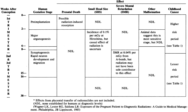
TABLE 3. Upper-Limit Conceptus Doses From Selected X-Ray Examinations*


Examination

Dose

Routine head

<0.5 mGy

Routine thoracic and neck


 Chest

<0.5 mGy

 Mammography

<0.5 mGy

 Cervical spine

<0.5 mGy

 Thoracic spine

<1.0 mGy

Routine extremity


 Upper femur

(?)

 Other (including shoulder and knee arthrography)

<0.5 mGy

Routine pelvic

(?)

Angiography


 Cerebral

<1.0 mGy

 Cardiac catheterization

<5.0 mGy

 Aortography

<1.0 mGy

 Abdominal

(?)

Myelography

(?)

Gastrointestinal

(?)

Urologic

(?)

Computed tomography


 Head (single series of entire head at 1-cm slice intervals)

<0.5 mGy

 Chest (single series of entire chest at 1-cm slice intervals)

<10.0 mGy

 Upper abdominal (20 1-cm contiguous slices more than 2.5 cm from uterus)

<30.0 mGy

 Pelvic

(?)

Conventional tomography


 Head

<1.0 mGy

 Chest

<5.0 mGy


* Assumes patient's pelvis is shielded or outside direct path of x-ray field. Doses can exceed these values if uterus is directly exposed or if x-ray field is not confined to anatomy of interest. A (?) means that an upper-limit estimate is not practical.
(Wagner LK, Lester RG, Saldana LR: Exposure of the Pregnant Patient of Diagnostic Radiations: A Guide to Medical Management. Philadelphia JB Lippincott, 1985)

If it is necessary to obtain an accurate dose evaluation, medical physicists should be consulted. For patients whose abdominal area is to be exposed, it is important not to refer to look-up tables to ascertain the dose because these tables could be off by a factor of 2 to 10, depending on the size of the patient, the extent of the study, the equipment used, and the location of the conceptus relative to the surface of the patient.44,45

Prior to referral of the pregnant patient for diagnostic evaluation it is important to counsel the patient on the potential risks and benefits of the study. Risk factors such as a family history of birth defects, other maternal conditions, or the use of potential teratogenic agents (drugs, cigarettes, alcohol) should be discussed. The normal incidence of birth defects is between 3% and 6%, and this should be brought to the attention of the patient.

The referring physician should consult with the radiologist before the procedure is done. Wagner and co-workers45 point out that there are several issues to consider:

  An alternative study that uses less or no ionizing radiation might be used.
  It may be possible to limit the study to a less-than-standard procedure with a lower dose to the conceptus.
  The radiologist might shield the uterus or otherwise avoid inadvertent exposure to the conceptus.
  The most efficient equipment can be selected for the study.
  If fluoroscopy is needed, dose reduction techniques such as removing the grid might be possible.
  For radionuclide studies, the amount of radioactivity might be reduced from that of a standard procedure or a lower-dose radiopharmaceutical might be chosen.
  When considering the future of the pregnancy of a patient who has been exposed to diagnostic radiation and is only later discovered to have been pregnant at the time, it is important to examine both the gestational age at the time of exposure and the level of radiation received by the conceptus. In critical circumstances, a medical physicist should be consulted to perform a dose calculation.
  When considering the possibility of termination of a pregnancy because of potential health effects from radiation exposure, several recommendations have been proposed.45 The NCRP recommends:
  The risk is considered to be negligible at 5 rad [50 mGy] or less when compared to other risks of pregnancy, and the risk of malformations is significantly increased above control levels only at doses above 15 rad [150 mGy]. Therefore, exposure of the fetus to radiation arising from diagnostic procedures would very rarely be cause, by itself, for terminating a pregnancy.40

Back to Top
REFERENCES

1. Kneale GW, Stewart AM: Mantel-Haenszel analysis of Oxford data: I. Independent effects of several birth factors including fetal irradiation. J Natl Cancer Inst 56: 879, 1976

2. Kneale GW, Stewart AM: Mantel-Haenszel analysis of Oxford data: II. Independent effects of fetal irradiation subfactors. J Natl Cancer Inst 57: 1009, 1976

3. Miller RW, Blot NJ: Small head size after in-utero exposure to atomic radiation. Lancet 2: 784, 1972

4. Miller RW, Mulvihill JJ: Small head size after atomic irradiation. Teratology 14: 355, 1976

5. Otake M, Schull WJ: In utero exposure to A-bomb radiation and mental retardation: A reassessment. Br J Radiol 57: 409, 1984

6. Schull WJ, Otake M: The central nervous system and in-utero exposure to ionizing radiation: The Hiroshima and Nagasaki experiences. In Castellani A (ed): Epidemiology and Quantitation of Environmental Risk in Humans from Radiation and Other Agents, p 515. New York, Plenum Press, 1985

7. Developmental Effects of Irradiation on the Brain of the Embryo and Fetus, International Commission on Radiological Protection Publication 49. Oxford, Pergamon Press, 1986

8. Brent RL: Radiation teratogenesis. Teratology 21: 281, 1980

9. Russell LB, Russell WL: An analysis of the changing radiation response of the developing mouse embryo. J Cell Physiol (Suppl 1)43:103, 1954

10. Genetic and Somatic Effects of Ionizing Radiation, United Nations Scientific Committee on the Effects of Atomic Radiation. New York, United Nations, 1986

11. Roux C, Horvath C, Dupuis R: Effects of preimplantation low-dose radiation on rat embryos. Health Phys 45: 993, 1983

12. MacMahon B: Prenatal x-ray exposure and childhood cancer. J Natl Cancer Inst 28: 1173, 1962

13. Diamond EL, Schmerler H, Lilienfeld AM: The relationship of intrauterine radiation to subsequent mortality in development of leukemia in children. Am J Epidemiol 97: 283, 1973

14. Graham S, Levin ML, Lilienfeld AM et al: Preconception, intrauterine and postnatal irradiation as related to leukemia. Natl Cancer Inst Monogr 19: 347, 1966

15. Monson RR, MacMahon B: Prenatal x-ray exposure and cancer in children. In Boice JD, Fraumeni JF (eds): Radiation Carcinogenesis: Epidemiology and Biological Significance, pp 97–105. New York, Raven Press, 1984

16. Harvey EB, Boice JD, Honeyman M et al: Prenatal x-ray exposure in childhood cancer in twins. N Engl J Med 5: 541, 1985

17. Court-Brown WM, Doll R, Hill AB: Incidence of leukemia after exposure to diagnostic radiation in-utero. Br Med J 2: 1539, 1960

18. Jablon S, Kato H: Childhood cancer in relation to prenatal exposure in atomic bomb radiation. Lancet 2: 1000, 1970

19. The Effects on Populations of Exposure to Low Levels of Ionizing Radiation: 1980, Committee on the Biological Effects of Ionizing Radiation. Washington, DC, Academic Press, 1980

20. Milton RC, Shohoji T: Tentative 1965 Radiation Dose Estimation for Atomic Bomb Survivors. Hiroshima and Nagasaki Atomic Bomb Casualty Commission Technical Report, pp 1–68, 1968

21. Hashizume T, Maruyama T, Nishizawa K et al: Dose estimation of human fetus exposed in utero to radiations from atomic bombs in Hiroshima and Nagasaki. J Radiat Res 14: 346, 1973

22. Kerr GD: Organ dose estimates for the Japanese atomic bomb survivors. Health Phys 37:487. 1979

23. Kerr GD, Pace JV, Scott WH: Tissue kerma vs. distance relationships for initial nuclear radiation from the atomic bombs at Hiroshima and Nagasaki. U.S.-Japan Joint Workshop for Reassessment of Atomic Bomb Dosimetry at Hiroshima and Nagasaki, pp 57–98. Hiroshima, Radiation Effects Research Foundation, 1983

24. Wood JW, Johnson KG, Omori Y et al: Mental retardation in children exposed in-utero to the atomic bomb at Hiroshima/Nagasaki. Am J Public Health 57: 1381, 1967

25. Jacobsen L, Mellengaard L: Anomalies of the eyes in descendents of women irradiated with small x-ray dose during age of fertility. Acta Ophthalmol 46: 352, 1968

26. Hammer-Jacobsen E: Therapeutic abortion on account of x-ray examination during pregnancy. Dan Med Bull 6: 113, 1959

27. Tabuchi A: Fetal disorder due to ionizing radiation. Hiroshima J Med Sci 13: 125, 1964

28. Tabuchi A, Nakagawa S, Hirai T et al: Fetal hazards due to x-ray diagnosis during pregnancy. Hiroshima J Med Sci 16: 49, 1967

29. Villumsen AL: Environmental Factors in Congenital Malformation, pp 130–142. Copenhagen. F.A.D.L.s Forlag, 1970

30. Nokkentved K: Effects of Diagnostic Radiation Upon the Human Fetus, p 228. Copenhagen, Ejnar Munksgaards Forlag, 1968

31. Kinlen LJ, Acheson ED: Diagnostic irradiation, congenital malformations and spontaneous abortion. Br J Radiol 41: 648, 1968

32. Granroth G: Defects of the central nervous system in Finland: IV. Associations with diagnostic x-ray examinations. Am J Obstet Gynecol 133: 191, 1979

33. Choi NW, Klaponski FA: On neural-tube defects: An epidemiological elicitation of etiological factors. Neurology 20: 399, 1970

34. Dyer NC, Brill B: Fetal radiation dose from internally administered 59Fe and 131I. In Sikov MR, Mahoum DD (eds): Radiation Biology of the Fetal and Juvenile Mammal, pp 73–88. Oakridge, TN, USAEC Division of Technical Information, 1969

35. Hicks SP, D'Amato CJ: Effects of radiation on development, especially of the nervous system. Am J Forensic Med Pathol 1: 309, 1980

36. D'Amato CJ, Hicks SP: Effects of low levels of ionizing radiation on the developing cerebral cortex of the rat. Neurology 15: 1104, 1965

37. Wanner RA, Edwards MJ: Comparison of the effects of radiation in hyperthermia on prenatal retardation of brain-growth of guinea pigs. Br J Radiol 56: 33, 1983

38. Edmonds DK, Lindsy KS, Miller JF et al: Early embryonic mortality in women. Fertil Steril 38: 447, 1982

39. Miller JF, Williamson E, Glue J et al: Fetal loss following implantation: A prospective study. Lancet 1: 554, 1980

40. Medical Radiation Exposure of Pregnant and Potentially Pregnant Women. NCRP report No. 54. Washington, DC, National Council on Radiation Protection and Measurements, 1977

41. Nuclear Medicine: Factors Influencing the Choice and Use of Radionuclides in Diagnosis and Therapy. NCRP report No. 70. Washington, DC, National Council on Radiation Protection and Measurements, 1982

42. Protection of the Patient in X-ray Diagnosis, International Commission on Radiation Protection. Oxford, Pergamon Press, 1970

43. International Commission on Radiation Protection, Statement from 1983 Washington meeting. Brit J Radiol 57:415, 1984

44. Wagner LK, Zeck OF, Archer BR: Conceptus dose from two state-of-the-art CT scanners. Radiology 159: 787, 1986

45. Wagner LK, Lester RG, Saldana LR: Exposure of the Pregnant Patient to Diagnostic Radiations: A Guide to Medical Management. Philadelphia, JB Lippincott, 1985

Back to Top