Chapter 81
Direct Fetal Transfusion
Bernd Schumacher and Kenneth J. Moise
Main Menu   Table Of Contents

Search

Bernd Schumacher, MD
Senior Fellow, Division of Maternal—Fetal Medicine, Department of Obstetrics and Gynecology, Baylor College of Medicine, Houston, Texas (Vol 3, Chap 81)

Kenneth J. Moise, Jr, MD
Associate Professor, Obstetrics and Gynecology and Director, Division of Maternal-Fetal Medicine, Baylor College of Medicine, Houston, Texas (Vol 3, Chap 81)

INTRODUCTION
HISTORY
HOW MUCH TO TRANSFUSE?
DYNAMICS OF TRANSFUSED RED BLOOD CELLS IN FETAL CIRCULATION
USE OF MATERNAL BLOOD
ULTRASOUND TO PREDICT FETAL HEMATOCRIT
FETAL PARALYSIS
MONITORING WITH COLOR FLOW DOPPLER
PRACTICAL CONSIDERATIONS
TIMING OF DELIVERY
FETAL PHYSIOLOGIC EFFECTS
COMPLICATIONS
OUTCOME
NEED FOR NEONATAL TOP-UP TRANSFUSION
FETOMATERNAL HEMORRHAGE
PARVOVIRUS INFECTION
PLATELET ALLOIMMUNIZATION
THE FUTURE OF INTRAUTERINE TRANSFUSION
CONCLUSIONS
REFERENCES

INTRODUCTION

Present-day guidelines for the administration of Rhesus (Rh) immune globulin (RhIG) to every Rho(D)-negative women at 28 weeks' gestation and postpartum have greatly reduced the incidence of red blood cell alloimmunization.1 Before the use of RhIG, nearly 1% of all Rho(D)-negative pregnant women were sensitized.2 Nonetheless, in 1986 the Centers for Disease Control reported an incidence of Rh hemolytic disease of 10.6 per 10,000 total births in the United States.3 For every 100 affected patients, 9 fetuses require intrauterine transfusion (IUT).4,5,6

IUT was the first effort at in utero treatment of the human fetus. During the last 30 years, it has become the treatment of choice for patients alloimmunized to red blood cell antigens in whom severe fetal anemia develops remote from term.4,5,6 In these cases, IUT generally is indicated if the fetal hemoglobin concentration is less than the 5th percentile for gestational age7 or if the amniotic fluid ΔOD450 value reaches the 80th percentile of zone 2 on the Liley curve.8 At Baylor College of Medicine in Houston, Texas, a fetal hematocrit less than 30% is used as a threshold value to transfuse a fetus in utero.4,5,6

We refer the reader elsewhere for a description of the complete diagnostic evaluation of the pregnancy affected by Rh hemolytic disease.2,4,5,6,7,8 Instead, this chapter stresses the technical aspects of IUT, including the use of ultrasound, the physiologic effects of IUT on the fetus, complications, neonatal “top-up” transfusions, and pregnancy outcome. The use of IUT in the treatment of fetomaternal hemorrhage, fetal parvovirus infection, and platelet alloimmunization is described and the future of IUT briefly evaluated.

Back to Top
HISTORY

In 1963, Liley9,10,11,12 initiated the in utero therapy of Rh hemolytic disease by developing the technique of fetal intraperitoneal transfusion (IPT). During an amniocentesis, Liley inadvertently placed his needle into the fetal abdomen. After returning from Africa, one of Liley's research associates described how IPT was commonly used there to transfuse children with sickle cell anemia. Liley concluded that this could be a feasible route for IUT if the fetal peritoneal cavity could be entered intentionally.13,14 After instilling radiopaque dye into the amniotic cavity by amniocentesis, Liley made use of the fetal ability to concentrate this contrast material in its lower gastrointestinal tract, thereby providing a radiographic target for needle placement. He then employed paper clips fastened with adhesive tape to mark the maternal skin surface. During the next step of the procedure, Liley inserted a 16-gauge Tuohy needle into the fetal peritoneal cavity and advanced an epidural catheter through it to aspirate 200 mL of fetal ascitic fluid and replace it with 100 mL of packed red blood cells. After three fetal deaths, Liley reported his first success in September 1963.11 Virtually simultaneous, though more aggressive attempts to access the fetal circulation directly included hysterotomy-facilitated catheter placement into chorionic plate vessels or into the fetal femoral artery, saphenous vein, or internal jugular vein.15,16,17,18

Before real-time ultrasound was available, IPT involved blindly injecting radiopaque dye into the amniotic cavity. The fetus would then swallow this contrast material, allowing for identification of its peritoneal cavity at fluoroscopy. Often a “chicken-wire” grid molded to the contour of the maternal abdomen was used to ensure accurate needle placement under fluoroscopic visualization.19,20 This technique was later modified to include the direct injection of radiopaque dye into the fetal abdomen.21 The use of static gray-scale ultrasound imaging for placement of the transfusion needle was first reported in 1975, significantly reducing fetal radiation exposure by eliminating the need for pretransfusion amniography.22,23 Real-time ultrasonography to guide the transfusion needle during IPT was first described in 1977.24,25,26 Only a short time later, fluoroscopy was no longer used. Saline, which produces small bubbles on real-time ultrasound when briskly shaken and injected under pressure, was used instead to observe the transfusion needle successfully entering the fetal abdominal cavity.24,25 Bowman and associates27 stressed that the diameter of the epidural catheter is important to the success of the IPT. To avoid side holes with small diameters causing hemolysis during red blood cell infusion, they recommended that the end of the epidural catheter be removed before being used for IPT. They estimated the appropriate transfusion volume for IPT using the following calculation:

IPT remained the only technique for in utero transfusion therapy until 1981, when Rodeck and co-workers28 performed the first direct fetal intravascular transfusion (IVT) by placing a needle into chorionic plate vessels under fetoscopic visualization. One year later, Bang and colleagues29 successfully transfused a fetus by inserting a needle into the umbilical vein under ultrasound guidance. Seven different techniques for IUT have been reported since then (Table 1). The use of fetoscopy in guiding the IUT needle is described in several studies. This method failed to achieve great popularity.30,31,32,33 Both the direct fetal intravascular approach and fetal intravascular exchange transfusion were evaluated by investigators in the United States during the following 4 years.34,35,36,37 Proponents of exchange transfusion postulated that with this technique the fetus was less likely to become volume-overloaded while more fetal cells were removed from circulation. Advocates of direct IVT believed that the procedure time was shorter in comparison with fetal-exchange IVT. They also asserted that the placental vascular bed would absorb the increased circulatory volume without negatively affecting the fetus. The direct intravascular technique became more widely adopted as experience with both techniques grew. Ultrasound imaging resolution continues to improve, and simple direct IVT has become the procedure of choice at most centers in the United States; IPT alone is performed rarely unless direct access to the umbilical cord is not technically feasible. Transfusion of red blood cells directly into the fetal circulation seems especially important in the treatment of the hydropic fetus whose peritoneal absorption of red blood cells is not inhibited, but it is thought to require several weeks.38,39,40 Comparing neonatal outcome after IVT to that after IPT using historic controls, Harman and co-workers41 clearly demonstrated that IVT significantly improves the chance for survival in the hydropic fetus.

TABLE 1. Techniques for Intrauterine Transfusion

  1. Intraperitoneal
  2. Intravascular by cordocentesis
    1. Exchange transfusion
    2. Simple direct transfusion

  3. Intravascular by intrahepatic venous puncture
    1. Simple direct transfusion

  4. Intracardiac by fetal cardiocentesis
    1. Simple direct transfusion

  5. Combined approaches
    1. Exchange intravascular transfusion followed by intraperitoneal transfusion
    2. Simple direct intravascular transfusion followed by intraperitoneal transfusion

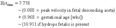
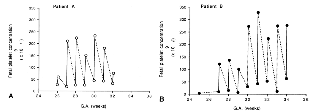
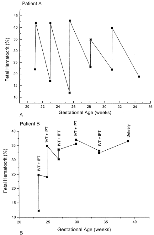
Because complications unique to IVT had been reported, the need for IVT in the severely anemic but nonhydropic fetus remained controversial.42,43,44 Moreover, with the use of direct IVT alone, fetal hematocrits between procedures varied widely. The combination of IVT and IPT therefore was tested at Baylor College of Medicine.45 First, packed red blood cells with a hematocrit of 80% or higher were infused by IVT to increase the fetal hematocrit to approximately 40%. Next, a standard IPT was performed to create a red blood cell reservoir in the fetus for later absorption between IUTs.This combined approach resulted in a more stable fetal hematocrit and longer intervals between procedures (Fig. 1).35,45 As a result of the combined IVT/IPT approach, the average daily decline of the fetal hematocrit was only 0.01% compared with 1.14% when IVT alone was used. Other centers have validated these findings and also use the combined IUT technique.46,47

Fig. 1 . Fetal hematocrit before and after direct intravascular transfusion in patient A ( A) and before and after combined direct intravascular/intraperitoneal transfusion in patient B ( B ).(Patient A data from Berkowitz RL, Chitkara U, Wilkins I et al: Technical aspects of intravascular intrauterine transfusions: Lessons learned from thirty-three procedures. Am J Obstet Gynecol 157:4, 1987)

As an alternate site for IUT, the intrahepatic umbilical vein has been used when access is impossible at the placental cord insertion.48,49,50 Nicolini and co-workers49,50 transfused 72 severely anemic fetuses intrahepatically with a 90% success rate. There were few complications, which consisted mainly of fetal bradycardia and intraperitoneal bleeding. Fetal bradycardia during IUT often is related to inadvertent umbilical arterial puncture and vasospasm.7,51 During intrahepatic transfusion, the incidence of fetal bradycardia was low because of the absence of the umbilical artery at the needle insertion site. Only occasional fetal intraperitoneal bleeding occurred in this series. The extravasated blood was absorbed from the peritoneal cavity in all cases, and there were no adverse fetal effects.49,50

Westgren and co-workers52 described direct fetal intracardiac transfusion (ICT) in patients with severe Rh hemolytic disease. Six patients with evidence of severe erythroblastosis fetalis underwent the procedure at 19- to 31 weeks' gestation. A total of 25 ICTs were completed, with a complication rate of 20%. According to these authors, ICT offers an alternative if direct IVT into the umbilical cord is impossible.52 Using ICT, Harman7 described the resuscitation of five fetuses from exsanguinating hemorrhage after umbilical cord puncture. After the initial ICT was successful, serial IVTs were performed subsequently with good outcome.

Back to Top
HOW MUCH TO TRANSFUSE?

Target fetal hematocrits at the conclusion of IUT vary among treatment centers.53 A final fetal hematocrit of 50% to 60% is usually the goal when IVT alone is performed. A rise in fetal whole-blood viscosity during transfusion can be minimized by restricting the posttransfusion fetal hematocrit to approximately 50% to 55%.54 The final target hematocrit in the combined IVT/IPT approach is usually approximately 40%. This is more physiologic because the fetal hematocrit normally ranges from 33% at 17 weeks' to 47% at 40 weeks' gestation.55 To estimate the volume of red blood cells needed for an IVT, the fetoplacental volume is determined first.56 The fetoplacental volume relative to fetal weight estimated by ultrasound is rather constant throughout the course of pregnancy.57 This value in milliliters is equal to 1.046 plus the ultrasound-estimated fetal weight in grams multiplied by 0.14. The volume to be transfused during IVT is then computed:

This calculation is based on a simple dilution formula and can be computerized. Repeated hematocrit determinations during the IVT can be avoided in this manner.56 Alternatively, a table can be used to determine the volume of blood to be transfused to obtain a desired increase in fetal hematocrit at a given gestational age (Table 2).53

TABLE 2. Volume of Blood (Hematocrit 85%) to be Transfused to Obtain Desired Increase in Fetal Hematocrit


Gestational

Blood Volume (mL

Age (Weeks)

10%

15%

20%

25%

30%

21

13.1

14.2

15.2

16.3

17.3

22

13.7

15.8

17.9

19.9

22.0

23

14.8

17.9

21.1

24.2

27.3

24

16.5

20.6

24.8

30.0

33.1

25

18.7

23.9

29.1

34.3

39.5

26

21.4

27.7

33.9

40.2

46.4

27

24.7

32.0

39.3

46.6

53.9

28

28.6

36.9

45.3

53.6

61.9

29

33.0

42.4

51.7

61.1

70.5

30

37.9

48.4

58.8

69.2

79.6

31

43.4

54.9

66.4

77.8

89.3

32

49.5

62.0

74.5

87.0

99.5

33

56.0

69.6

83.2

96.7

110.3

34

63.2

77.8

92.4

107.0

121.6

(Please DV, Chitkara U, Berkowitz GS et al: Intrauterine intravascular transfusion for severe erythroblastosis fetalis: how much to transfuse? Obstet Gynecol 75:965–969, 1990. Reprinted with permission from The American College of Obstetricians and Gynecologists.)

Most fetuses undergoing IVT with a normal umbilical venous pressure have a pressure of less than 10 mmHg at the end of the transfusion.58 Monitoring fetal umbilical venous pressure is still useful for assessing the fetal response to IUT59 because pressure increases of more than 10 mmHg have predicted fetal death within 24 hours after IVT with a sensitivity of 80%.60 Severely anemic and hydropic fetuses experience particularly high loss rates. Radunovic and co-workers60 therefore recommended that the posttransfusion fetal hematocrit not exceed 25% or a fourfold increase from the pretransfusion value. Selbing and associates61 noted that if the fetus is given more than 20 mL of fluid for each kilogram of its ultrasound-estimated weight, its chance of survival is reduced significantly. These authors strongly suggested an upper transfusion limit of 20 mL/kg, which corresponds to approximately one fifth of the fetoplacental blood volume.

Timing of subsequent fetal transfusions varies among institutions. At Baylor College of Medicine, anemic fetuses are transfused every 2 weeks for the first two transfusions; then the interval between procedures is lengthened to 3 to 4 weeks. Severely anemic fetuses diagnosed early in the second trimester are an exception to this guideline. Such fetuses are transfused at first to achieve a final hematocrit of approximately 25%. In a subsequent IUT approximately 48 hours later, a posttransfusion hematocrit of 35% is the goal. Equations to predict fetal hematocrit at the beginning of subsequent IUTs have been developed, and timing of repeat transfusions can thus be estimated optimally.7,62,63

Back to Top
DYNAMICS OF TRANSFUSED RED BLOOD CELLS IN FETAL CIRCULATION

Initial studies of the effects of IUT focused on the rate of absorption of transfused blood from the peritoneal cavity by the fetus.38,64 At first, there was great concern that hydropic fetuses were unable to absorb red blood cells after IPT.38 One case report showed that 2 hours after a 60-mL IPT, only 18% of circulating red blood cells in the fetus were adult-type cells.65 Using chromium-labeled packed red blood cells during IPT, it was later demonstrated that, though over several weeks' time, even the hydropic fetus can absorb them from its peritoneal cavity.39,40

The survival of transfused red blood cells in the fetal circulation also has been investigated. Maternal donor lymphocytes were shown to persist for more than 2 years in the peripheral blood of four infants transfused in utero.66 A case report by Jones and co-workers67 suggested that the half-life of the donor adult erythrocyte is only 30 days in a second-trimester fetus, compared with approximately 60 days if transfused into an adult. Pattison and Roberts,68 however, studied 27 anemic fetuses and found that adult erythrocyte survival in the fetus was similar to that in the adult circulation. These authors also noted that red blood cell survival was independent of the route of transfusion, gestational age, or presence of hydrops. The fraction of transfused red blood cells surviving at 29 days after IVT was approximately 65%, compared with 8% in the above-cited case report.67

Back to Top
USE OF MATERNAL BLOOD

There has been considerable change in the source of donor red blood cells for IUT. Virtually all centers use fresh, O-negative cells for IUT; several centers use maternal blood as an alternate source. This source of fresh red blood cells is readily available, the risk of maternal alloimmunization to new red blood cell antigens due to any fetomaternal hemorrhage is theoretically reduced, and transmission of viral infections to the fetus is likely to be decreased. However, Vietor and colleagues69 studied 91 women treated with IUT for severe red blood cell alloimmunization and tested them for the development of additional alloantibodies. These investigators detected new alloantibodies against red blood cell antigens in 24 women (26%). These were usually directed against fetal rather than donor antigens. The use of maternal red blood cells during IUT in these cases would not have prevented further alloantibody formation. Finally, the use of maternal red blood cells for IUT has a psychologic benefit in that any guilt over fetal rejection would be alleviated by providing mothers with a practical way to help their fetuses.

Up to 6 U of maternal blood can be safely harvested for fetal transfusion during a gestation.70 Regular intake of prenatal vitamins, folate, and ferrous sulfate prevents significant maternal donor anemia. All mothers are screened routinely for syphilis, HIV, human T-cell lymphotropic virus type 1 (HTLV-1), and all known forms of hepatitis before donating for their fetuses. Donated red blood cells are washed to remove the offending antibodies and packed to achieve a hematocrit of approximately 80%. Units then are processed through a leukocyte-poor filter and irradiated with approximately 2500 rad (25 Gy) to prevent any graft-versus-host reaction. Great care must be taken to ensure timely processing of fresh blood to avoid transfusion of blood with high plasma potassium levels, which could cause fetal arrhythmias after IUT.71,72 Mothers with antibodies to cytomegalovirus may still donate because this virus resides in white blood cells removed during the filtration process. If initial cordocentesis reveals an ABO incompatibility between mother and fetus, maternal red blood cells should be used only with great caution because of the risk of fetal sensitization. At Baylor College of Medicine, maternal blood was utilized in two such cases with no adverse effects observed in the fetus or neonate.

Back to Top
ULTRASOUND TO PREDICT FETAL HEMATOCRIT

Ultrasound has played an important role in improving the outcome in pregnancies affected by Rh hemolytic disease. It is used to establish the correct gestational age on which parameters such as normal fetal hematocrit and amniotic bilirubin levels are based. Ultrasound provides invaluable needle guidance during amniocentesis, cordocentesis, and IUT. Unfortunately, its use in the diagnosis of fetal anemia is limited until overt hydrops fetalis is present. Although some investigators have advocated serial ultrasound examinations to detect signs of impending hydrops fetalis, Nicolaides and co-workers73 were unable to correlate fetal hematocrit with increased placental thickness or increased umbilical vein diameter. Because the liver and spleen are sources of extramedullary hematopoiesis in response to fetal anemia, these organs have been studied separately to predict fetal anemia. Two studies74,75 have proposed that an increase in fetal liver dimensions is a good predictor of anemia. Oepkes and co-workers76 measured the fetal splenic perimeter with ultrasound and noted that splenomegaly was present in all nonhydropic, anemic fetuses. Splenomegaly predicted a hemoglobin deficit in excess of five standard deviations from the normal range for a given gestational age, with a sensitivity of 93%. Some hydropic fetuses still had no splenomegaly.

The cardiac output of the anemic fetus has been demonstrated to be increased over controls.77 This would lead to enhanced velocity of blood in various fetal vessels. For this reason, Doppler ultrasound has been studied to see if it could serve as a valuable adjunct in the evaluation of the status of the fetus requiring IUT.78 Pulsed Doppler wave forms from the fetal descending aorta have been used to predict hematocrit before IVT.79,80 Copel and co-workers81 have proposed the following formula to predict fetal hematocrit:

When they applied this formula to 16 fetuses, they achieved a sensitivity of 90% and a specificity of 69%. Copel and co-workers81,82 further concluded that their Doppler technique was useful in predicting fetal hematocrit and the need for an initial IUT, but the timing of subsequent IUTs was not improved. Pulsed Doppler flow-velocity indices in the umbilical artery do not correlate with fetal blood gas values in red blood cell—alloimmunized pregnancies, nor do they predict fetal hematocrit except in very severely affected fetuses clearly requiring IUT.83,84 Study of fetal cerebral vessels, such as the common carotid and middle cerebral arteries, has not proved Doppler findings in these vessels to be any more useful in the prediction of fetal anemia.85,86

A recent study reevaluating the clinical usefulness of pulsed Doppler flow-velocity indices in the management of severe red blood cell alloimmunization confirmed the hyperdynamic circulation produced by the fetal anemia in both arterial and venous vessels.87 An association with the degree of fetal anemia was again found only for the flow-velocity wave forms in the fetal aorta. Steiner and associates88 showed that the clinical usefulness of the peak aortic velocity is hampered by its low predictive value. Bahado-Singh and co-workers89 recently demonstrated that the splenic artery resistance index increases in fetuses with severe anemia. These authors postulated that this occurs as a reflection of vascular congestion from red blood cells trapped in the splenic circulation. Further investigation is necessary to evaluate whether an increased splenic artery resistance index will prove useful, in conjunction with some of the other Doppler findings discussed above, in identifying the nonhydropic, severely anemic fetus. Overall, though, changes in Doppler parameters due to fetal anemia are believed to be too insignificant or irreproducible to be applied clinically.90

Back to Top
FETAL PARALYSIS

The operator has been plagued by fetal movement during IUT since the first description of the procedure by Liley.11 Fetal immobilization was first described by Liggins,91 who performed IPT by an impaling technique using multiple needle punctures of the fetus. Since then, IUT has been made easier by the widespread use of fetal paralytic agents. Fetal paralysis was first introduced in 1985 in Australia.48 In the United States, D-tubocurarine initially was injected into the fetal thigh under ultrasound guidance.92 The intravascular use of pancuronium bromide through cordocentesis was reported subsequently.93,94 The absorption of red blood cells from the fetal peritoneal cavity was found to be markedly reduced after injection of pancuronium bromide in a sheep model.95 Therefore, short-acting agents such as atracurium besylate and vecuronium bromide are currently in use.96,97,98 These two paralytic agents also do not cause the fetal tachycardia and loss of short-term fetal heart rate variability frequently demonstrated with the use of pancuronium bromide.99 A vecuronium bromide dose of 0.1 mg/kg of ultrasound-estimated fetal weight results in almost immediate cessation of fetal movement when injected intravascularly at the beginning of an IUT. This fetal paralytic effect lasts for up to 2 hours, and no untoward effects in neonates have been reported.

Back to Top
MONITORING WITH COLOR FLOW DOPPLER

The use of color flow Doppler ultrasound also has improved the technique of IUT.100 Color flow Doppler has an advantage over gray-scale ultrasound in that it allows for accurate localization of fetal vessels in difficult cases, such as early gestational age, oligohydramnios, or poor fetal positioning over the umbilical cord insertion site. When targeting the fetal hepatic vein for IUT, color flow Doppler helps to identify the vessel. During the actual injection of blood, intravascular turbulence at the tip of the transfusion needle is visible on color flow Doppler imaging.100 In addition, the fetal heart rate can be monitored by observation of the umbilical artery pulsations. Thus, the operator's view of the transfusion needle is not interrupted by the need to observe the fetal heart rate directly on the ultrasound monitor. Finally, during IPT the color flow Doppler can be used to monitor the free flow of fluid into the peritoneal space, thereby confirming proper placement of the transfusion needle.

Back to Top
PRACTICAL CONSIDERATIONS

At Baylor College of Medicine, the following IUT procedure is used.100 The IUT is performed in close proximity to the labor and delivery suite. With the patient under mild sedation and the abdomen prepped and draped in the usual sterile fashion, a 20-gauge needle, 6 inches in length, is directed under local anesthesia and real-time ultrasound guidance into the placental umbilical cord insertion. An initial fetal blood sample is sent for spun hematocrit, complete blood and reticulocyte counts, Kleihauer-Betke stain, and total and direct bilirubin. Next, the fetus is paralyzed with a vecuronium bromide dose of 0.1 mg/kg of ultrasound-estimated fetal weight, which is injected directly into the umbilical vessel. Red blood cells then are transfused at a rate of approximately 5 to 10 mL/min. A final sample of fetal blood is obtained for a hematocrit and a Kleihauer-Betke stain. After the IVT is completed, a standard IPT is undertaken in the combined IVT plus IPT approach performed at our center; we accomplish this simply by redirecting the IVT needle. After the procedure, the fetal heart rate is monitored continuously for at least 2 hours, after which the patient is discharged home. A follow-up ultrasound is performed the next morning.

Back to Top
TIMING OF DELIVERY

The timing of delivery in fetuses undergoing serial IUTs has undergone considerable change. During the era of IPT, fetuses affected by hemolytic disease were delivered routinely at 32 weeks' gestation. Hyaline membrane disease and hyperbilirubinemia necessitating neonatal exchange transfusion were frequent complications. The widespread use of IVT during the past decade has led many centers to perform the final procedure at approximately 35 weeks' gestation with delivery planned approximately 3 weeks later. This change in management has virtually eliminated hyaline membrane disease and the need for neonatal exchange transfusions for elevated bilirubin. Once fetal viability is reached, IUTs should be undertaken close to the labor and delivery suite so that an immediate cesarean section can be performed in cases of fetal distress.

Back to Top
FETAL PHYSIOLOGIC EFFECTS

IVT leads to extensive fetal cardiovascular changes. The procedure greatly stresses the developing fetus with acute changes in intravascular volume and viscosity. Umbilical venous pressure increases between 1.7 and 4.6 mmHg.58,101,102 Fetal cardiac output decreases by as much as 25%,101 begins to return to pretransfusion levels within 2 hours,103 and returns to normal by approximately 24 hours after the procedure.77 To explain fully the fetal hemodynamic response to IVT, it has been suggested that the procedure leads to increased fetal cardiac afterload secondary to an increase in fetal blood viscosity. The fetus responds to this rise in afterload by a decrease in stroke volume, leading to a decrease in cardiac output and fetal heart rate and an increase in right atrial and umbilical venous pressures.7,101 Pulsed Doppler assessment of umbilical and fetal femoral, renal, and cerebral vessels has demonstrated generalized fetal vasodilatation in response to this increase in afterload.104,105 Results of investigations using pulsatility index as an indicator of fetal anemia and its correction by IVT also are indicative of a decrease in fetal vascular impedance immediately after IVT.101,104,105,106

An increase in fetal vasodilator prostaglandins, which allows the human fetus to tolerate large increases in intravascular volume, has been reported after IVT.107,108 An increase in atrial natriuretic peptide, a vasodilator, also contributes to the apparent acute decrease in vascular tone observed in the human fetus immediately after IVT.109,110,111 Interestingly, a paradoxic increase in fetal plasma arginine vasopressin has been observed in association with IVT.112 This finding remains unexplained.

IVT also leads to significant alterations in fetal metabolism. Nicolini and co-workers113 found that the umbilical venous pH and base excess decrease while the PCO2 acutely increases after IVT. In this study, the transfused blood had a mean pH of 6.76. These investigators believed that the fetoplacental circulation was effective in correcting this exogenous source of acidosis. The oxygen dissociation curve of fetal hemoglobin favors the uptake of oxygen in the placenta. Therefore, replacing fetal red blood cells with adult cells could seriously affect oxygen delivery to fetal tissues. Soothill and colleagues114 compared blood gas values of fetuses transfused with adult hemoglobin-containing red blood cells to those of controls. Transfused fetuses had a lower umbilical arterial pH and greater base deficit, while umbilical venous PO2 was higher. These authors postulated that an increase in uteroplacental blood flow is the compensatory mechanism for fetal tissue hypoxia caused by transfused red blood cells carrying only adult hemoglobin.

An important compensatory response to anemia in the adult is an increase in the 2,3-diphosphoglycerate level, which reduces the oxygen affinity of adult hemoglobin and facilitates oxygen delivery to the tissues. Soothill and co-workers115 demonstrated that when adult red blood cells were transfused into the fetal circulation of 34 anemic fetuses, the 2,3-diphosphoglycerate concentration increased in direct correlation with the degree of anemia, allowing for improved fetal tissue oxygenation. Socol and associates116 reported that IVT decreases hemolysis and reduces circulating levels of fetal plasma glutathione. Glutathione, liberated from hemolyzed fetal red blood cells, causes the inhibition of insulin activity in anemic fetuses, leading to a compensatory fetal hyperinsulinemic response.117 IVT therefore may prevent the hyperinsulinemia frequently associated with red blood cell alloimmunization.116

Nasrat and associates118 demonstrated that there is a potential risk for iron overload in Rh-alloimmunized fetuses undergoing IUT. These authors described plasma ferritin levels indicative of iron overload in several fetuses. Severe fetal hemolytic anemia leads to elevated iron stores, which further increase in direct correlation with the volume of red blood cells transfused. Neonatal cholestasis and hepatitis due to severe intrahepatic iron deposition after IUT have been reported recently.119 Therefore iron supplementation should be withheld in newborns with hemolytic disease until serum ferritin levels return to the normal range.

Back to Top
COMPLICATIONS

Before paralytic agents were in use to immobilize the fetus during IUT, fetal movement had the potential to result in visceral injury or umbilical cord trauma during the procedure. One case report even describes an IPT with subsequent fetal omental herniation through the fetal abdominal needle puncture site.120 Anterior placental location increases the risk of damage to a major fetoplacental vessel with subsequent fetal exsanguination.121 Perinatal infectious complications, such as viral hepatitis, also have been reported in the early experience with IPT.122,123

Several reports have described sinusoidal fetal heart rate patterns immediately after IPT.124,125 A transient sinusoidal fetal heart rate after IPT is not necessarily an ominous sign; however, if persistent, it may herald fetal cardiac decompensation. This likely is caused by an increase in intra-abdominal pressure adversely affecting fetal heart rate control as a result of increased vagal stimulation.126 Animal experiments have revealed that IPT causes increased fetal intraperitoneal pressure, which can lead to fetal hypoxia from umbilical vein compression.126 The need for intraperitoneal pressure monitoring during IPT has since been supported by human data as well.102,127

IVT has led to complications not previously described with IPT, even though IVT generally is safe with a procedure-related pregnancy loss rate of approximately 1% to 3%.8,51,128 Entering umbilical vessels during IVT can cause an umbilical cord hematoma with resulting fetal compromise.43 Some authorities have proposed that transient fetal bradycardia results from a perivascular blood collection leading to umbilical arterial vasospasm, with the extravasated fetal blood acting as an irritative focus. If umbilical vein thrombosis occurs in addition to the hematoma, fetal death may be the consequence.44 Thus, visible echogenic turbulence is a sine qua non feature of the successful IVT. Disappearance of this marker signifies loss of proper needle location.129 Furthermore, Moise and colleagues130 noted that increased bilirubin levels due to hemolysis in the fetus of an alloimmunized pregnancy resulted in icteric serum in spun capillary hematocrit tubes. This finding is useful in confirming fetal vascular access in cases of anterior placentation, where visualization of the needle tip with ultrasound may be problematic.

Fetal bradycardia secondary to umbilical arterial vasospasm transiently occurs in approximately 4% of IVTs, especially as the target transfusion volume is being approached.7,51 When the umbilical artery is entered, the incidence of fetal bradycardia approaches 30%.128 Therefore, the umbilical vein should be targeted routinely for IVT. On occasion, however, the umbilical artery is inadvertently punctured; in these cases, infused red blood cells will stream back toward the placenta during ultrasound visualization. Although immediate removal of the transfusion needle frequently is recommended, at our center we proceed with caution and frequently monitor the fetal heart rate using color flow Doppler. Slowing or stopping the transfusion and maternal oxygen administration usually leads to complete resolution of the fetal bradycardia. If these maneuvers do not reverse the bradycardia, the needle should be removed. Maternal repositioning in the left lateral position is undertaken. If the fetus is viable, we observe the bradycardia for up to 10 minutes before proceeding with an emergency cesarean section. Normal function of the atrioventricular valve leaflets in the fetal heart is a good indication that cardiac output is being maintained despite the slow heart rate. Cessation of movement of these valve leaflets on ultrasound examination warrants delivery.

Congenital viral infections can occur when donor blood is used for IVT. An unfortunate case of fatal congenital cytomegalovirus infection acquired through IUT was described previously.131 Rare, but especially concerning, are cases of severe fetal brain injury after IVT.132,133 Dildy and co-workers42 documented an unexplained fetal porencephalic cyst after IVT. An increase in blood viscosity and fetal bradycardia during the IVT may have contributed to this complication by leading to fetal cerebral hypoperfusion. Fortunately, the infant later was discharged home with a normal neurologic status.

Fetomaternal hemorrhage can accelerate fetal hemolytic disease in the pregnant patient previously sensitized to red blood cell antigens. Moise and co-workers134 described a case of poor fetal outcome after first-trimester transcervical chorionic villi sampling in a previously alloimmunized patient. The patient's antibody titers became elevated, resulting in the demise of a hydropic fetus early in the second trimester. These authors proposed that red blood cell alloimmunization is an absolute contraindication for chorionic villi sampling. Similarly, the placenta should be avoided during placement of the IUT needle. Nicolini and associates135 described fetomaternal hemorrhages with an average volume of 2.4 mL in 21 of 32 patients undergoing IVT with an anterior placenta. Maternal alloantibody titers became elevated when the estimated volume of fetomaternal hemorrhage exceeded 1 mL. Hence, any significant fetomaternal hemorrhage may lead to more severe fetal hemolytic disease during a patient's subsequent pregnancies.

Back to Top
OUTCOME

Survival of the fetus after IUT varies with the operator's experience, the institution, and the technique used. Results of studies on the outcome after IPT alone are presented in Table 3.136–165 Of 2483 reported fetuses who underwent IPT between 1963 and 1988, 39% survived. Only 17% of hydropic fetuses survived after IPT alone. Table 4 summarizes the reported experience with both IVT and IVT/IPT. Of 411 fetuses who underwent the procedure, 84% did well166–179; 94% of nonhydropic fetuses and 74% of hydropic fetuses survived. During the past 9 years at Baylor College of Medicine IUT using the combined IVT/IPT approach has resulted in an overall fetal survival rate of approximately 80%.4,5,6

TABLE 3. Summary of Studies Using Fetal Intraperitoneal Transfusion (IPT)


Total

Survivors

Survivors


Technique for


No.

with

Without

Author

Year

IPT

No.of Patients

Survivors

Hydrops

Hydrops

Watts et al136

1988

IPT/ultrasound

35

30

4

26

Bowman and Manning27

1983

IPT/ultrasound

24

22

6

16

Berkowitz and Hobbins137

1981

IPT/ultrasound

17

12

4

8

Clewell et al138

1981

IPT/ultrasound

16

11

NA

NA

Acker et al139

1980

IPT/ultrasound

35

18

5

13

Frigoletto et al22

1981

IPT/x-ray

330

137

5

132

Ellis140

1980

IPT/x-ray

217

81

NA

NA

Hamilton141

1977

IPT/x-ray

82

29

1

28

Bock142

1976

IPT/x-ray

34

19

NA

NA

Holt et al143

1973

IPT/x-ray

93

44

NA

NA

Whitfield et al144

1972

IPT/x-ray

170

72

NA

NA

Bashore et al145

1971

IPT/x-ray

116

34

8

26

Duhring and Zwirek146

1970

IPT/x-ray

25

12

NA

NA

Fong et al147

1970

IPT/x-ray

73

18

2

16

Eyster et al148

1969

IPT/x-ray

7

5

NA

NA

Horger and Hutchinson149

1969

IPT/x-ray

59

19

NA

NA

Mandelbaum150

1969

IPT/x-ray

142

36

NA

NA

Queenan151

1969

IPT/x-ray

591

203

NA

NA

Wade et al152

1969

IPT/x-ray

48

12

NA

NA

Bjerre et al153

1968

IPT/x-ray

11

4

NA

NA

Stenchever and Cibils154

1968

IPT/x-ray

8

2

NA

NA

Bishop et al155

1967

IPT/x-ray

44

22

NA

NA

Cherry and Rosenfield156

1967

IPT/x-ray

11

4

NA

NA

Fairweather et al157

1967

IPT/x-ray

35

16

NA

NA

Friesen et al158

1967

IPT/x-ray

47

15

NA

NA

Hutchinson et al159

1967

IPT/x-ray

28

11

NA

NA

McCutcheon and Little160

1967

IPT/x-ray

26

9

NA

NA

Charles et al161

1966

IPT/x-ray

17

5

NA

NA

Karnicki162

1966

IPT/x-ray

68

33

NA

NA

Work et al163

1966

IPT/x-ray

13

6

NA

NA

Bowes et al164

1965

IPT/x-ray

7

3

NA

NA

Green et al165

1965

IPT/x-ray

30

10

NA

NA

Liley11

1963

IPT/x-ray

1

1

NA

NA

Queenan151

1969

Hysterotomy

23

3

NA

NA


Total = 2483

958

35

265


Hydrops = 210

(39%)

(17%)

(53%)


No Hydrops =


502


NA = not available

TABLE 4. Summary of Studies Using Direct Fetal Intravascular Transfusion


Total

Survivors

Survivors


Technique

No. of

No.

with

Without

Author

Year

for IVT

Patients

Survivors

Hydrops

Hydrops

Moise et al4,5,6

1994

IVT + IPT

20

19

10

9

Rodeck et al166

1991

IVT + IPT

18

16

16

0

Weiner et al167

1991

IVT

48

46

11

35

Harman et al41

1990

IVT

44

40

18

22

Lemery et al168,169

1989

IVT

15

10

2

8

Nicolini et al46

1989

IVT + IPT

30

25

NA

NA

Pattison and Roberts68

1989

IVT

20

18

1

17

Poissonnier et al170

1989

IVT + IPT

107

84

29

55

Ronkin et al171

1989

IVT

8

8

2

6

Barss et al172,173

1988

IVT + IPT

13

11

4

7

Grannum et al174

1988

IVT

26

21

16

5

Orsini et al175

1988

IVT

15

10

4

6

Parer132

1988

IVT

5

4

NA

NA

Socol et al176

1987

IVT

3

3

3

0

Berkowitz et al34,177

1986

IVT

8

6

2

4

Doyle et al178

1986

IVT

8

5

0

5

Nicolaides et al179

1986

IVT

18

17

10

7

de Crespigny et al48

1985

IVT

4

3

1

2

Bang et al29

1982

IVT

1

1

1

0


Total = 411

347

130

188


Hydrops = 175

(84%)

(74%)

(94%)


No Hydrops = 201


IPT = intraperitoneal transfusion; IVT = intravascular transfusion; NA = not available.

Initially it seemed that infants severely affected by red blood cell alloimmunization and transfused in utero had lower birth weights than matched controls,180,181 but fetal catch-up growth after IVT has been observed in more recent studies.182,183 It has been shown clearly that birth weights of infants transfused in utero are comparable to those of matched controls.

Few long-term outcome studies exist assessing infants transfused in utero.184,185,186,187 Follow-up evaluations of fetuses treated with IPT have identified increased numbers of inguinal hernias in males and umbilical hernias in females as compared with matched siblings.188 In assessing overall physical, intellectual, and social maturity in IPT survivors for several years, Turner189 considered only 50% to be completely normal. Other investigators have evaluated survivors of IPT for up to 11 years and found no significant developmental or physical abnormalities.188,190

Follow-up of IVT survivors is limited and must be viewed cautiously considering that the lives of more moribund fetuses have been saved.184,187 Doyle and co-workers184 studied 38 infants who had undergone IVT and found 35 to be developing normally. The three abnormal outcomes were attributed to prematurity.

Back to Top
NEED FOR NEONATAL TOP-UP TRANSFUSION

Lozinska191 demonstrated that IUT suppresses hematopoiesis. Therefore, many infants treated with IUT before birth require “top-up” transfusions during the early months of life. Because their red blood cell population consists mainly of transfused adult erythrocytes, reticulocytes are virtually absent. Neonatal exchange transfusions, however, rarely are necessary. Typically, infants with a history of IVT present with symptomatic anemia at 1 month of age and require a simple red blood cell transfusion. A review of 40 cases at Baylor College of Medicine showed that a top-up transfusion is necessary in approximately 50% of infants at a mean age of 38 days of life.192 Most infants receive only one such transfusion. Infants requiring late neonatal transfusion are characterized by a lower reticulocyte count at their final IUT, higher umbilical cord hemoglobin at delivery, and a greater number of adult red blood cells seen on a cord Kleihauer-Betke smear. The affected infants have exhibited bone marrow erythroid hypoplasia, low serum erythropoietin, and a decreased number of circulating reticulocytes.193,194 The mechanism causing the decreased erythropoietin is poorly understood; however, because passively acquired maternal antibodies remain elevated for at least 6 weeks in these neonates, the reticulocytopenia could be caused by high bone marrow levels of anti—red blood cell antibodies.

Therefore, weekly hematocrit and reticulocyte determinations should be performed for the first 2 months of life in infants with a history of IUT.195 Infants with a hemoglobin of less than 5 to 6 g/dL clearly should be transfused, even if they are symptom-free. Infants showing signs related to severe anemia, such as failure to thrive and lethargy, also are candidates for transfusion. Scaradavou and colleagues196 reported the use of erythropoietin to treat late anemia caused by suppression of erythropoiesis due to IUT for Rh alloimmunization. Subcutaneous injection of 200 U erythropoietin per kilogram of body weight three times per week, combined with ferrous sulfate and folic acid supplementation, was effective in markedly decreasing the need for postnatal transfusion.

Back to Top
FETOMATERNAL HEMORRHAGE

Massive fetomaternal hemorrhage is the cause of fetal death in 1 per 1000 births and results in significant fetal morbidity in at least 1 per 800 deliveries.197 Seven cases of IUT in this situation have been described. Decreased fetal movement was the chief complaint in four of these cases, and subsequent fetal monitoring revealed a sinusoidal fetal heart rate pattern.198,199,200 In the remaining three cases, routine ultrasound showed a hydropic fetus.201,202,203 The maternal Kleihauer-Betke smear was positive in every case, and IUT was performed immediately. A single IUT procedure was needed in three cases, two procedures in another three cases, and five procedures in the remaining case. With this intervention, pregnancy was prolonged in three cases. In the first case, an IPT at 21 weeks' gestation was successful in reversing hydrops fetalis with subsequent delivery of a normal infant at 38 weeks' gestation.201 Thorp and co-workers203 performed two IVTs at 26 and 27 weeks' gestation. Hydrops fetalis was noted to resolve, and a normal infant was born at 39 weeks' gestation. At Baylor College of Medicine, Montgomery and co-workers202 transfused an affected fetus five times beginning at 27 weeks' gestation using the combined IVT/IPT approach. Abdominal delivery of a 1740-g infant at 30 weeks' gestation was required as a result of chorioamnionitis. The infant's initial hematocrit was 57%, and its neonatal course was uneventful. In all other reported cases, fetal bradycardia or the return of decreased fetal movement necessitated delivery by cesarean section. Cordocentesis in three of these fetuses revealed a decreasing hematocrit due to continued fetomaternal hemorrhage. The use of IUT in the treatment of fetomaternal hemorrhage therefore may prove beneficial only in selected cases.

Back to Top
PARVOVIRUS INFECTION

Human parvovirus B19 causes erythema infectiosum (fifth disease) in children. During outbreaks, household contacts as well as school teachers frequently are exposed. Approximately 50% of persons lack immunity, and 20% of these will become infected after exposure.204,205 One third of infected pregnant women will remain asymptomatic and will not manifest the exanthem typical of this disease.206 Reported rates of fetal infection range from 2.5% to 38%.207,208 Parvovirus suppresses the fetal bone marrow and inhibits fetal erythropoiesis with the subsequent development of aplastic anemia, nonimmune hydrops, and fetal death.

Maternal parvovirus infection is often confirmed by detection of a specific IgM antibody that appears 3 to 4 days after the onset of clinical disease and persists for 3 to 4 months.209 Fetal infection usually occurs 4 to 6 weeks after maternal infection, although hydrops fetalis has been reported as late as 12 weeks after the maternal illness. If maternal infection has been confirmed, the fetus should undergo weekly ultrasound examinations for the presence of hydrops fetalis for a total of 12 weeks. If hydrops is noted, donor red blood cells and platelets should be prepared, and cordocentesis performed. Studies of fetal blood usually show a negative direct Coombs' test, severe anemia, occasional thrombocytopenia, an inappropriately low reticulocyte count, normal serum bilirubin, elevated total IgM, and elevated liver enzymes.210 Under the electron microscope, viral particles may be observed in fetal ascitic fluid or blood.211 Using probes specific for human parvovirus B19, viral DNA also can be identified in some fetal body fluids.209 Spontaneous resolution of hydrops fetalis secondary to fetal parvovirus infection has been reported by several authors.212,213 Thus, if the fetal reticulocyte count is elevated before the first IUT, a second procedure is not necessary because recovery of the fetal bone marrow is ongoing.

To date, a total of 11 fetuses have undergone IVT for confirmed parvovirus infection, with a rate of survival of approximately 80%.206,210,214,215,216,217 All had hydrops fetalis. Severe fetal anemia was corrected with serial IUTs, as needed, at short intervals of 1 to 7 days. Nonimmune hydrops due to in utero parvovirus infection may require several weeks to resolve after the fetal hematocrit has returned to normal. Follow-up of infants transfused in utero for parvovirus infection has revealed normal neonatal outcomes. The data are limited, but IVT should be strongly considered as a therapeutic option in cases of fetal parvovirus infection with resultant hydrops fetalis.

Back to Top
PLATELET ALLOIMMUNIZATION

Neonatal alloimmune thrombocytopenia, a disease process analogous to red blood cell alloimmunization, complicates 1 per 1000 to 2000 live births.218 In this situation, transplacental passage of maternal antibodies to fetal platelet antigens of paternal origin results in severe fetal and neonatal thrombocytopenia. In utero intracranial hemorrhage occurs in 10% of cases and has been documented as early as the second trimester.219,220 In 75% of affected fetuses, the platelet antigen involved is HPA-1.221 Unlike Rh alloimmunization, the first fetus may already be severely affected; maternal antibody titers are not predictive of the degree of fetal thrombocytopenia.219,222 The clinician often becomes aware of the disease only after an affected infant is born to the patient or her sister. Subsequent pregnancies are commonly complicated by progressively worsening fetal disease.223

The initial evaluation of an affected neonate should include platelet antigen testing of the newborn, mother, and father. Determining the paternal zygosity with respect to the HPA-1 antigen will assist in the prediction of the fetal antigen status in the vast majority of instances because only 5% of fathers are heterozygous.224 The likelihood of recurrent HPA-1 fetomaternal incompatibility is therefore approximately 94%.224 Recently, Khouzami and associates225 described the use of amniocentesis to determine the fetal platelet antigen status employing allele-specific oligonucleotide probes in uncultured amniocytes. Currently, fetal platelet count and antigen status of any subsequent pregnancy are determined most commonly by cordocentesis at 20 weeks' gestation. Unfortunately, this approach is not without risk. Several cases of fetal death caused by hemorrhage have occurred after cordocentesis in the investigation of platelet alloimmunization.226 Therefore, maternal platelets for immediate transfusion should be available at the time of cordocentesis should excessive streaming of blood occur from the umbilical vessel puncture site. Alternatively, if the fetal platelet count can be assessed rapidly while the cordocentesis needle is still in place, platelet transfusion should be performed for counts less than 50,000/mm3. This threshold is based on neonatal courses complicated by bleeding in infants born to women with autoimmune thrombocytopenia.227 Because approximately 98% of the population is HPA-1 antigen positive, maternal platelets should be used for fetal transfusion.228 The platelets are obtained by apheresis 24 hours before their anticipated use, washed to remove any trace of offending antibodies and resuspended in ABO-compatible plasma.

The volume of platelet concentrate to be used can be calculated with the following formula:

where V represents the volume of platelets to be transfused, VFP is the estimated fetoplacental blood volume for the respective gestational age,57 C1 is the fetal platelet concentration before transfusion, C2 is the concentration of the donor platelets, and C3 is the fetal platelet concentration desired after transfusion.229 The optimal posttransfusion fetal platelet count to prevent bleeding complications has yet to be determined. For this reason, most clinicians will transfuse a quantity of platelets empirically, based on the formula presented here. Although a final fetal platelet count should be obtained at the conclusion of the procedure, it is not necessary to wait for the result before the transfusion needle is removed.

Initially, fetal platelet transfusions in cases of platelet alloimmunization involved serial infusions of maternal platelets by cordocentesis. Four cases have been described.229,230,231 In two of these cases, the median daily decline in fetal platelet count was 23,600/mm3. Because of the short platelet half-life of 4 to 7 days, weekly transfusions are required to maintain an adequate fetal platelet count (Fig. 2).230 Bussel and colleagues232 have argued that this approach is associated with undue risk to the fetus from multiple umbilical cord punctures. Using an alternative approach, Lynch and co-workers233 successfully combined the maternal administration of steroids and weekly intravenous gamma globulin injections to increase the fetal platelet count at delivery to greater than 50,000/mm3 in 11 of 18 cases. No case of intracranial hemorrhage occurred in the treatment group, compared with a 33% incidence in antecedent siblings.233 Therefore, serial in utero platelet transfusions are not a reasonable primary approach to the management of affected fetuses with alloimmune thrombocytopenia.

Fig. 2 . Fetal platelet count before and after platelet transfusions in patient A ( A) and in patient B ( B ).(Nicolini U, Tannirandorn Y, Gonzalez P et al: Continuing controversy in alloimmune thrombocytopenia: Fetal hyperimmunoglobulinemia fails to prevent thrombocytopenia. Am J Obstet Gynecol 163:1145, 1990)

Daffos and co-workers229 proposed cordocentesis at term in fetuses suspected of having thrombocytopenia due to maternal platelet alloimmunization. If fetal thrombocytopenia is detected, transfusion with maternal platelets can then be performed. Subsequent induction of labor with vaginal delivery would be a safe option. Ten cases of fetal transfusion with platelets just before delivery have been reported.222,229,231,234 Initially, investigators performed the fetal platelet transfusion and then proceeded with elective cesarean section, with the reasoning that cesarean delivery alone is not completely protective against intracranial hemorrhage in fetuses affected by alloimmune thrombocytopenia.235 More recently, vaginal delivery was allowed after fetal platelet transfusion in 4 of the 10 cases in this series. There were no adverse neonatal sequelae. Sia and colleagues,235 however, recommended vaginal delivery only with a posttransfusion fetal platelet count greater than 100,000/mm3.

Severe fetal thrombocytopenia has been associated with hydrops fetalis caused by red blood cell alloimmunization and parvovirus infection.210,236,237 In these cases, random-donor platelets also should be available at the time of IUT with red blood cells. If the fetal platelet count is noted to be less than 50,000/mm3, transfusion with platelets may prevent fetal death due to prolonged bleeding from the umbilical vessel puncture site.

Back to Top
THE FUTURE OF INTRAUTERINE TRANSFUSION

Unfortunately, IUT only briefly relieves the symptoms of severe fetal anemia or thrombocytopenia caused by red blood cell or platelet alloimmunization and is intended only to prolong the intrauterine period available to the developing fetus to achieve maturity. Even though gamma globulin and steroids are promising alternatives to serial fetal platelet transfusion in the treatment of alloimmune thrombocytopenia,233 efforts to treat Rh hemolytic disease directly by administration of intravenous gamma globulin, plasmapheresis, oral Rh antigen ingestion, or promethazine have failed to date.238,239,240,241 IVT is well established at many centers throughout the United States, and improvement of fetal survival beyond the 84% presented here is unlikely. To achieve better treatment results, a direct approach to the disease is needed.

The human Rh gene has been cloned, and the exact blood type of the developing embryo can be determined.242,243,244,245 Using in vitro fertilization techniques, preimplantation diagnosis would allow for the exclusive return of Rho(D)-negative embryos to the uterus.246 Such an approach would lead to an unaffected fetus in all successful implantations.

Using a recently developed rabbit model of Rh hemolytic disease by Moise and associates,247 investigation currently is under way at Baylor College of Medicine to evaluate the clinical usefulness of maternal immunotherapy in the treatment of red blood cell alloimmunization.

Back to Top
CONCLUSIONS

Widespread use of RhIG has reduced the incidence of Rh hemolytic disease in the United States to only 1 per 1000 live births.3 Only 9% of affected patients require IUT yielding less than 500 cases in the United States annually.4,5,6 To optimize outcome with IUT as state-of-the-art therapy for severely anemic fetuses remote from term, regionalization of treatment should be considered at centers where a minimum of 10 fetal transfusions per year are performed. After diagnostic testing using ultrasound and amniocentesis performed by local obstetricians has shown a severely affected fetus requiring cordocentesis and possible IUT, the patient would enter the referral network. Concentrating patients with severe hemolytic disease at such centers would also provide the opportunity for continued basic and clinical research.

IUT is a safe procedure8,51,128 and has saved the lives of many fetuses, including several infected with human parvovirus.206,210,214,215 It is the best available therapy until red blood cell alloimmunization can be prevented completely. IUT in the treatment of fetomaternal hemorrhage may prove beneficial only in selected cases.198,199,200,201,202,203 Fetal platelet transfusions in cases of severe platelet alloimmunization are of limited therapeutic value because of the short half-life of this blood component.229,230,231,232

Back to Top
REFERENCES

1. American College of Obstetricians and Gynecologists: Prevention of D Isoimmunization. Technical Bulletin No. 147, ACOG, Washington, DC, October, 1990

2. Bowman JM: Hemolytic disease (erythroblastosis fetalis). In Creasy RK, Resnik R (eds): Maternal-Fetal Medicine: Principles and Practice, 3rd ed, pp 711–743. Philadelphia, WB Saunders, 1994

3. Chavez GF, Mulinare J, Edmonds LD: Epidemiology of Rh hemolytic disease of the newborn in the United States. JAMA 265: 3270, 1991

4. Moise KJ: Changing trends in the management of red blood cell alloimmunization in pregnancy. Arch Pathol Lab Med 118: 421, 1994

5. Moise KJ, Milam JD, Carpenter RJ: Changing trends in the diagnosis and treatment of Rh alloimmunization. Texas Med 83: 27, 1987

6. Moise KJ: Management of red cell alloimmunization in pregnancy. In Sacher RA, Brecher ME (eds): Obstetric Transfusion Practice, pp 21–47. Bethesda, American Association of Blood Banks, 1993

7. Harman CR: Invasive techniques in the management of alloimmune anemia. In Harman CR (ed): Invasive Fetal Testing and Treatment, pp 107–191. Boston, Blackwell Scientific Publications, 1995

8. Moise KJ: Intrauterine transfusion with red cells and platelets. West J Med 159: 318, 1993

9. Green GH: Historic perspective on Liley's “fetal transfusion.” Vox Sang 48: 184, 1985

10. Green GH: William Liley and fetal transfusion: A perspective in fetal medicine. Fetal Diagn Ther 1: 18, 1986

11. Liley AW: Intrauterine transfusion of foetus in haemolytic disease. Br Med J 2: 1107, 1963

12. Liley AW: The development of the idea of fetal transfusion. Am J Obstet Gynecol 111: 303, 1971

13. Halitsky V, Krumholz BA, Schwalb E, Gromisch DS: The current role of intrauterine fetal transfusion in the management of erythroblastosis fetalis. Obstet Gynecol Surv 23: 301, 1968

14. Queenan JT: Fetal transfusion. Fetal Diagn Ther 1: 59, 1986

15. Asensio SH, Figueroa-Longo JG, Pelegrina IA: Intrauterine exchange transfusion. Am J Obstet Gynecol 95: 1129, 1966

16. Asensio SH, Figueroa-Longo JG, Pelegrina IA: Intrauterine exchange transfusion: A new technique. Obstet Gynecol 32: 350, 1968

17. Freda VJ, Adamsons K: Exchange transfusion in utero: Report of a case. Am J Obstet Gynecol 89: 817, 1964

18. Seelen J, Van Kessel H, Eskes T et al: A new method of exchange transfusion in utero: Cannulation of vessels on the fetal side of the human placenta. Am J Obstet Gynecol 95: 872, 1966

19. Cherry SH: An abdominal grid for intrauterine transfusions. Obstet Gynecol 30: 881, 1967

20. Krumholz BA, Halitsky V: A grid and isometric rulers for accurate needle placement during intrauterine transfusion. Obstet Gynecol 32: 706, 1968

21. Bowman JM: The management of Rh-isoimmunization. Obstet Gynecol 52: 1, 1978

22. Frigoletto FD, Umansky I, Birnholz J et al: Intrauterine fetal transfusions in 365 fetuses during fifteen years. Am J Obstet Gynecol 139: 781, 1981

23. Hobbins JC, Davis ED, Webster J: A new technique utilizing ultrasound to aid in IUT. J Clin Ultrasound 4: 135, 1975

24. Cooperberg PL, Carpenter CW: Ultrasound as an aid in intrauterine transfusion. Am J Obstet Gynecol 128: 239, 1977

25. Frigoletto FD, Birnholz JC, Rothchild SB et al: Intrauterine transfusion with the use of phased array ultrasonography: A new technique. Am J Obstet Gynecol 131: 273, 1978

26. Platt LD, Keegan KA, Druzin ML et al: Intrauterine transfusion utilizing linear-array, real-time B-scan: A preliminary report. Am J Obstet Gynecol 135: 1115, 1979

27. Bowman JM, Manning FA: Intrauterine fetal transfusions: Winnipeg 1982. Obstet Gynecol 61: 203, 1983

28. Rodeck CH, Kemp JR, Holman CA et al: Direct intravascular fetal blood transfusion by fetoscopy in severe Rhesus isoimmunisation. Lancet 1: 625, 1981

29. Bang J, Bock JE, Trolle D: Ultrasound-guided fetal intravenous transfusion for severe Rhesus haemolytic disease. Br Med J 284: 373, 1982

30. MacKenzie IZ, Bowell PJ, Ferguson J et al: In-utero intravascular transfusion of the fetus for the management of severe Rhesus isoimmunization—a reappraisal. Br J Obstet Gynecol 94: 1068, 1987

31. MacKenzie IZ, MacLean DA, Fry A, Evans SL: Midtrimester intrauterine exchange transfusion of the fetus. Am J Obstet Gynecol 143: 555, 1982

32. Mandelbaum B, Pontarelli DA, Brushenko A: Amnioscopy for prenatal transfusion. Am J Obstet Gynecol 98: 1140, 1966

33. Rodeck CH, Nicolaides KH, Warsof SL et al: The management of severe Rhesus isoimmunization by fetoscopic intravascular transfusions. Am J Obstet Gynecol 150: 769, 1984

34. Berkowitz RL, Chitkara U, Goldberg JD et al: Intrauterine intravascular transfusions for severe red blood cell isoimmunization: Ultrasound-guided percutaneous approach. Am J Obstet Gynecol 155: 574, 1986

35. Berkowitz RL, Chitkara U, Wilkins I et al: Technical aspects of intravascular intrauterine transfusions: Lessons learned from thirty-three procedures. Am J Obstet Gynecol 157: 4, 1987

36. Christmas JT, Little BB, Johnston WL et al: Nomograms for rapid estimation of intravascular intrauterine exchange transfusion. Obstet Gynecol 75: 887, 1990

37. Grannum PA, Copel JA, Plaxe SC et al: In utero exchange transfusion by direct intravascular injection in severe erythroblastosis fetalis. N Engl J Med 314: 1431, 1986

38. Lewis M, Bowman JM, Pollock J, Lowen B: Absorption of red cells from the peritoneal cavity of an hydropic twin. Transfusion 13: 37, 1973

39. Creasman WT, Duggan ER, Lund CJ: Absorption of transfused chromium-labeled erythrocytes from the fetal peritoneal cavity in hydrops fetalis. Am J Obstet Gynecol 94: 586, 1966

40. Taylor WW, Scott DE, Pritchard JA: Fate of compatible adult erythrocytes in the fetal peritoneal cavity. Obstet Gynecol 28: 175, 1966

41. Harman CR, Bowman JM, Manning FA, Menticoglou SM: Intrauterine transfusion—intraperitoneal versus intravascular approach: A case-control comparison. Am J Obstet Gynecol 162: 1053, 1990

42. Dildy GA, Smith LG, Moise KJ et al: Porencephalic cyst: A complication of fetal intravascular transfusion. Am J Obstet Gynecol 165: 76, 1991

43. Moise KJ, Carpenter RJ, Huhta JC, Deter RL: Umbilical cord hematoma secondary to in utero intravascular transfusion for Rh isoimmunization. Fetal Diagn Ther 2: 65, 1987

44. Seeds JW, Chescheir NC, Bowes WA, Owl-Smith FA: Fetal death as a complication of intrauterine intravascular transfusion. Obstet Gynecol 74: 461, 1989

45. Moise KJ, Carpenter RJ, Kirshon B et al: Comparison of four types of intrauterine transfusion: Effect on fetal hematocrit. Fetal Diagn Ther 4: 126, 1989

46. Nicolini U, Kochenour NK, Greco P et al: When to perform the next intra-uterine transfusion in patients with Rh allo-immunization: Combined intravascular and intraperitoneal transfusion allows longer intervals. Fetal Diagn Ther 4: 14, 1989

47. Santolaya J, Warsof SL: Combined intravascular-intraperitoneal transfusions in hydropic twins due to Rh (D) alloimmunization. Fetal Diagn Ther 5: 70, 1990

48. de Crespigny LC, Robinson HP, Quinn M et al: Ultrasound-guided fetal blood transfusion for severe Rhesus isoimmunization. Obstet Gynecol 66: 529, 1985

49. Nicolini U, Nicolaidis P, Fisk NM et al: Fetal blood sampling from the intrahepatic vein: Analysis of safety and clinical experience with 214 procedures. Obstet Gynecol 76: 47, 1990

50. Nicolini U, Santolaya J, Ojo OE et al: The fetal intrahepatic umbilical vein as an alternative to cord needling for prenatal diagnosis and therapy. Prenat Diagn 8: 665, 1988

51. Benacerraf BR, Barss VA, Saltzman DH et al: Acute fetal distress associated with percutaneous umbilical blood sampling. Am J Obstet Gynecol 156: 1218, 1987

52. Westgren M, Selbing A, Stangenberg M: Fetal intracardiac transfusions in patients with severe Rhesus isoimmunization. Br Med J 296: 885, 1988

53. Plecas DV, Chitkara U, Berkowitz GS et al:Intrauterine intravascular transfusion for severe erythroblastosis fetalis: How much to transfuse? Obstet Gynecol 75: 965, 1990

54. Welch R, Rampling MW, Anwar A et al: Changes in hemorheology with fetal intravascular transfusion. Am J Obstet Gynecol 170: 726, 1994

55. Nicolaides KH, Thilaganathan B, Mibashan RS: Cordocentesis in the investigation of fetal erythropoiesis. Am J Obstet Gynecol 161: 1197, 1989

56. Mandelbrot L, Daffos F, Forestier F et al: Assessment of fetal blood volume for computer-assisted management of in utero transfusion. Fetal Diagn Ther 3: 60, 1988

57. Leduc L, Moise KJ, Carpenter RJ, Cano LE: Fetoplacental blood volume estimation in pregnancies with Rh alloimmunization. Fetal Diagn Ther 5: 138, 1990

58. Weiner CP, Pelzer GD, Heilskov J et al: The effect of intravascular transfusion on umbilical venous pressure in anemic fetuses with and without hydrops. Am J Obstet Gynecol 161: 1498, 1989

59. Hallak M, Moise KJ, Hesketh DE et al: Intravascular transfusion of fetuses with Rhesus incompatibility: Prediction of fetal outcome by changes in umbilical venous pressure. Obstet Gynecol 80: 286, 1992

60. Radunovic N, Lockwood CJ, Alvarez M et al: The severely anemic and hydropic isoimmune fetus: Changes in fetal hematocrit associated with intrauterine death. Obstet Gynecol 79: 390, 1992

61. Selbing A, Stangenberg M, Westgren M, Rahman F: Intrauterine intravascular transfusions in fetal erythroblastosis: The influence of net transfusion volume on fetal survival. Acta Obstet Gynecol Scand 72: 20, 1993

62. Halitsky V, Krumholz BA: Estimation of red cell volume requirements for intrauterine transfusion. Obstet Gynecol 31: 543, 1968

63. MacGregor SN, Socol ML, Pielet BW et al: Prediction of hematocrit decline after intravascular fetal transfusion. Am J Obstet Gynecol 161: 1491, 1989

64. Hashimoto B, Filly RA, Callen PW, Parer JT: Absorption of fetal intraperitoneal blood after intrauterine transfusion. J Ultrasound Med 6: 421, 1987

65. Montague ACW, Holzman GB: The absorption rate of blood from the peritoneal cavity by the fetus. Am J Obstet Gynecol 94: 282, 1966

66. Hutchinson DL, Turner JH, Schlesinger ER: Persistence of donor cells in neonates after fetal and exchange transfusion. Am J Obstet Gynecol 109: 281, 1971

67. Jones HM, Linch DC, Nicolaides K, Rodeck CH: Survival of transfused adult cells in the fetus. Fetal Diagn Ther 1: 193, 1986

68. Pattison N, Roberts A: The management of severe erythroblastosis fetalis by fetal transfusion: Survival of transfused adult erythrocytes in the fetus. Obstet Gynecol 74: 901, 1989

69. Vietor HE, Kanhai HHH, Brand A: Induction of additional red cell alloantibodies after intrauterine transfusions. Transfusion 34: 970, 1994

70. Gonsulin WJ, Moise KJ, Milam JD et al: Serial maternal blood donations for intrauterine transfusion. Obstet Gynecol 75: 158, 1990

71. Galligan BR, Cairns R, Schifano JV et al: Preparation of packed red cells suitable for intravascular transfusion in utero. Transfusion 29: 179, 1989

72. Thorp JA, Plapp FV, Cohen GR et al: Hyperkalemia after irradiation of packed red blood cells: Possible effects with intravascular fetal transfusion. Am J Obstet Gynecol 163: 607, 1990

73. Nicolaides KH, Fontanarosa M, Gabbe SG, Rodeck CH: Failure of ultrasonographic parameters to predict the severity of fetal anemia in Rhesus isoimmunization. Am J Obstet Gynecol 158: 920, 1988

74. Vintzileos AM, Campbell WA, Storlazzi E et al: Fetal liver ultrasound measurements in isoimmunized pregnancies. Obstet Gynecol 68: 162, 1986

75. Roberts AB, Mitchell JM, Pattison NS: Fetal liver length in normal and isoimmunized pregnancies. Am J Obstet Gynecol 161: 42, 1989

76. Oepkes D, Meerman RH, Vandenbussche FP et al: Ultrasonographic fetal spleen measurements in red blood cell alloimmunized pregnancies. Am J Obstet Gynecol 169: 121, 1993

77. Copel JA, Grannum PA, Green JJ et al: Fetal cardiac output in the isoimmunized pregnancy: A pulsed Doppler-echocardiographic study of patients undergoing intravascular intrauterine transfusion. Am J Obstet Gynecol 161: 361, 1989

78. Rotmensch S, Liberati M, Luo J-S, Hobbins JC: Monitoring of intravascular fetal transfusions with Doppler velocimetry. Am J Obstet Gynecol 167: 1314, 1992

79. Nicolaides KH, Bilardo CM, Campbell S: Prediction of fetal anemia by measurement of the mean blood velocity in the fetal aorta. Am J Obstet Gynecol 162: 209, 1990

80. Rightmire DA, Nicolaides KH, Rodeck CH, Campbell S: Fetal blood velocities in Rh isoimmunization: Relationship to gestational age and to fetal hematocrit. Obstet Gynecol 68: 233, 1986

81. Copel JA, Grannum PA, Belanger K et al: Pulsed Doppler flow-velocity waveforms before and after intrauterine intravascular transfusion for severe erythroblastosis fetalis. Am J Obstet Gynecol 158: 768, 1988

82. Copel JA, Grannum PA, Green JJ et al: Pulsed Doppler flow-velocity waveforms in the prediction of fetal hematocrit of the severely isoimmunized pregnancy. Am J Obstet Gynecol 161: 341, 1989

83. Legarth J, Lingman G, Stangenberg M, Rahman F: Umbilical artery doppler flow-velocity waveforms and fetal acid-base balance in Rhesus-isoimmunized pregnancies. J Clin Ultrasound 22: 37, 1994

84. Legarth J, Lingman G, Stangenberg M, Rahman F: Umbilical artery doppler flow-velocity waveforms in Rhesus-isoimmunized fetuses before and after fetal blood sampling or transfusion. J Clin Ultrasound 22: 43, 1994

85. Bilardo CM, Nicolaides KH, Campbell S: Doppler studies in red cell isoimmunization. Clin Obstet Gynecol 32: 719, 1989

86. Vyas S, Nicolaides KH, Campbell S: Doppler examination of the middle cerebral artery in anemic fetuses. Am J Obstet Gynecol 162: 1066, 1990

87. Hecher K, Snijders R, Campbell S, Nicolaides K: Fetal venous, arterial, and intracardiac blood flows in red blood cell isoimmunization. Obstet Gynecol 85: 122, 1995

88. Steiner H, Schaffer H, Spitzer D et al: The relationship between peak velocity in the fetal descending aorta and hematocrit in Rhesus isoimmunization. Obstet Gynecol 85: 659, 1995

89. Bahado-Singh R, Pirhonen J, Rahman F et al: Effect of fetal anemia on splenic artery resistance index in red cell isoimmunization [abstr]. J Soc Gynecol Invest 2: 146, 1995

90. Moise KJ: Red cell alloimmunization. In Copel JA, Reed KL (eds): Doppler Ultrasound in Obstetrics and Gynecology, pp 133–138. New York, Raven Press, 1995

91. Liggins GC: Fetal transfusion by the impaling technique. Obstet Gynecol 27: 617, 1966

92. Moise KJ, Carpenter RJ, Deter RL et al: The use of fetal neuromuscular blockade during intrauterine procedures. Am J Obstet Gynecol 157: 874, 1987

93. Copel JA, Grannum PA, Harrison D, Hobbins JC: The use of intravenous pancuronium bromide to produce fetal paralysis during intravascular transfusion. Am J Obstet Gynecol 158: 170, 1988

94. Moise KJ, Deter RL, Kirshon B et al: Intravenous pancuronium bromide for fetal neuromuscular blockade during intrauterine transfusion for red-cell alloimmunization. Obstet Gynecol 74: 905, 1989

95. Menticoglou SM, Harman CR, Manning FA, Bowman JM: Intraperitoneal fetal transfusion: Paralysis inhibits red cell absorption. Fetal Diagn Ther 2: 154, 1987

96. Bernstein HH, Chitkara U, Plosker H et al: Use of atracurium besylate to arrest fetal activity during intrauterine intravascular transfusions. Obstet Gynecol 72: 813, 1988

97. Daffos F, Forestier F, Mac Alesse J et al: Fetal curarization for prenatal magnetic resonance imaging. Prenat Diagn 8: 311, 1988

98. Leveque C, Murat I, Toubas F et al: Fetal neuromuscular blockade with vecuronium bromide: Studies during intravascular intrauterine transfusion in isoimmunized pregnancies. Anesthesiology 76: 642, 1992

99. Pielet BW, Socol ML, MacGregor SN et al: Fetal heart rate changes after fetal intravascular treatment with pancuronium bromide. Am J Obstet Gynecol 159: 640, 1988

100. Moise KJ, Belfort MA, Saade GR et al: Intrauterine transfusion: A review of current methods. Video J Color Flow Imag 2: 36, 1992

101. Moise KJ, Mari G, Fisher DJ et al: Acute fetal hemodynamic alterations after intrauterine transfusion for treatment of severe red blood cell alloimmunization. Am J Obstet Gynecol 163: 776, 1990

102. Nicolini U, Talbert DG, Fisk NM, Rodeck CH: Pathophysiology of pressure changes during intrauterine transfusion. Am J Obstet Gynecol 160: 1139, 1989

103. Rizzo G, Nicolaides KH, Arduini D, Campbell S: Effects of intravascular fetal blood transfusion on fetal intracardiac Doppler velocity waveforms. Am J Obstet Gynecol 163: 1231, 1990

104. Mari G, Moise KJ, Deter RL, Carpenter RJ: Flow velocity waveforms of the umbilical and cerebral arteries before and after intravascular transfusion. Obstet Gynecol 75: 584, 1990

105. Mari G, Moise KJ, Deter RL et al: Flow velocity waveforms of the vascular system in the anemic fetus before and after intravascular transfusion for severe red blood cell alloimmunization. Am J Obstet Gynecol 162: 1060, 1990

106. Mari G, Moise KJ, Deter RL, Carpenter RJ: Doppler assessment of renal blood flow velocity waveforms in the anemic fetus before and after intravascular transfusion for severe red cell alloimmunization. J Clin Ultrasound 19: 15, 1991

107. Weiner CP, Robillard JE: Effect of acute intravascular volume expansion on human fetal prostaglandin concentrations. Am J Obstet Gynecol 161: 1494, 1989

108. Weiner CP: Nonhematologic effects of intravascular transfusion on the human fetus. Semin Perinatol 13: 338, 1989

109. Kingdom JCP, Ryan G, Whittle MJ et al: Atrial natriuretic peptide: A vasodilator of the fetoplacental circulation? Am J Obstet Gynecol 165: 791, 1991

110. Panos MZ, Nicolaides KH, Anderson JV et al: Plasma atrial natriuretic peptide in human fetus: Response to intravascular blood transfusion. Am J Obstet Gynecol 161: 357, 1989

111. Robillard JE, Weiner C: Atrial natriuretic factor in the human fetus: Effect of volume expansion. J Pediatr 113: 552, 1988

112. Weiner CP, Smith F, Robillard JE: Arginine vasopressin and acute, intravascular volume expansion in the human fetus. Fetal Diagn Ther 4: 69, 1989

113. Nicolini U, Santolaya J, Fisk NM et al: Changes in fetal acid base status during intravascular transfusion. Arch Dis Child 63: 710, 1988

114. Soothill PW, Nicolaides KH, Rodeck CH, Bellingham AJ: The effect of replacing fetal hemoglobin with adult hemoglobin on blood gas and acid-base parameters in human fetuses. Am J Obstet Gynecol 158: 66, 1988

115. Soothill PW, Lestas AN, Nicolaides KH et al: 2,3-Diphosphoglycerate in normal, anaemic and transfused human fetuses. Clin Sci 74: 527, 1988

116. Socol ML, Dooley SL, Ney JA et al: Absence of hyperinsulinemia in isoimmunized fetuses treated with intravascular transfusion. Am J Obstet Gynecol 165: 1737, 1991

117. Steinke J, Gries FA, Driscoll SG: In vitro studies of insulin inactivation with reference to erythroblastosis fetalis. Blood 30: 359, 1967

118. Nasrat HA, Nicolini U, Nicolaidis P et al: The effect of intrauterine intravascular blood transfusion on iron metabolism in fetuses with Rh alloimmunization. Obstet Gynecol 77: 558, 1991

119. Lasker MR, Eddleman K, Toor AH: Neonatal hepatitis and excessive hepatic iron deposition following intrauterine blood transfusion. Am J Perinatol 12: 14, 1995

120. Chaim W, Mares AJ, Leiberman J, Cohen A: Omental herniation: An unusual fetal complication of intrauterine transfusion. Obstet Gynecol 47: 621, 1976

121. Cassady G, Barnett R, Ceballos R: Dangers of fetal transfusion: Importance of placental location. Am J Obstet Gynecol 110: 672, 1971

122. Goodlin RC: Intrauterine transfusion complicated by amnionitis and maternal peritonitis. Obstet Gynecol 26: 803, 1965

123. Mandelbaum B, Brough AJ: Hepatitis following multiple intrauterine transfusions: Report of a case. Obstet Gynecol 30: 188, 1967

124. Lowe TW, Leveno KJ, Quirk JG et al: Sinusoidal fetal heart rate pattern after intrauterine transfusion. Obstet Gynecol 64: 21S, 1984

125. Mueller-Heubach E, Caritis SN, Edelstone DI: Sinusoidal fetal heart rate pattern following intrauterine fetal transfusion. Obstet Gynecol 52: 43S, 1978

126. Crosby WM, Brobmann GF, Chang ACK: Intrauterine transfusion and fetal death: Relationship of intraperitoneal pressure to umbilical vein blood flow. Am J Obstet Gynecol 108: 135, 1970

127. Dunnihoo DR: Intrauterine fetal transfusion: Monitoring intraperitoneal pressures. Am J Obstet Gynecol 142: 241, 1982

128. Weiner CP, Wenstrom KD, Sipes SL, Williamson RA: Risk factors for cordocentesis and fetal intravascular transfusion. Am J Obstet Gynecol 165: 1020, 1991

129. Seeds JW, Bowes WA, Chescheir NC: Echogenic venous turbulence is a critical feature of successful intravascular intrauterine transfusion. Obstet Gynecol 73: 488, 1989

130. Moise KJ, Kirshon B, Carpenter RJ, Deter RL: Icteric serum as an indicator of fetal vascular access during intravascular transfusion for hemolytic disease. Am J Perinatol 7: 279, 1990

131. Evans DGR, Lyon AJ: Fatal congenital cytomegalovirus infection acquired by an intra-uterine transfusion. Eur J Pediatr 150: 780, 1991

132. Parer JT: Severe Rh isoimmunization—current methods of in utero diagnosis and treatment. Am J Obstet Gynecol 158: 1323, 1988

133. Nicolaides KH, Thorpe-Beeston JG, Salvesen DR, Snijders RJM: Fetal blood transfusion by cordocentesis. In Chervenak FA, Isaacson GC, Campbell S (eds): Ultrasound in Obstetrics and Gynecology, Vol 2, pp 1315–1320. Boston, Little, Brown & Co, 1993

134. Moise KJ, Carpenter RJ: Increased severity of fetal hemolytic disease with known Rhesus alloimmunization after first-trimester transcervical chorionic villus biopsy. Fetal Diagn Ther 5: 76, 1990

135. Nicolini U, Kechenour NK, Greco P et al: Consequences of fetomaternal haemorrhage after intrauterine transfusion. Br Med J 297: 1379, 1988

136. Watts DH, Luthy DA, Benedetti TJ et al: Intraperitoneal fetal transfusion under direct ultrasound guidance. Obstet Gynecol 71: 84, 1988

137. Berkowitz RI, Hobbins JC: Intrauterine transfusion utilizing ultrasound. Obstet Gynecol 57: 33, 1981

138. Clewell WH, Dunne MG, Johnson ML, Bowes WA: Fetal transfusion with real-time ultrasound guidance. Obstet Gynecol 57: 516, 1981

139. Acker D, Frigoletto FD, Birnholz JC et al: Ultrasound-facilitated intrauterine transfusions. Am J Obstet Gynecol 138: 1200, 1980

140. Ellis MI: Follow-up study of survivors after intra-uterine transfusion. Develop Med Child Neurol 22: 48, 1980

141. Hamilton EG: Intrauterine transfusion: Safeguard or peril? Obstet Gynecol 50: 255, 1977

142. Bock JE: Intra-uterine transfusion in the management of pregnant women with severe Rhesus isoimmunization: Results and discussion. Acta Obstet Gynecol Scand 53S: 29, 1976

143. Holt EM, Boyd IE, Dewhurst CJ et al: Intrauterine transfusion: 101 consecutive cases treated at Queen Charlotte's maternity hospital. Br Med J 3: 39, 1973

144. Whitfield CR, Thompson W, Armstrong MJ: Intrauterine fetal transfusion for severe Rhesus haemolytic disease. J Obstet Gynaecol Brit Comm 79: 931, 1972

145. Bashore RA, Lecky JW: Intrauterine fetal transfusion in the management of Rh disease. Obstet Gynecol 38: 79, 1971

146. Duhring JL, Zwirek SJ: Intrauterine fetal transfusion: Five years of experience. Am J Obstet Gynecol 110: 670, 1970

147. Fong SW, Margolis AJ, Westberg JA, Johnson P: Intra-uterine transfusion: Fetal outcome and complications. Pediatrics 45: 576, 1970

148. Eyster ME, Queenan JT, Haber JM: Use of maternal blood for intrauterine transfusions. Obstet Gynecol 34: 636, 1969

149. Horger EO, Hutchinson DL: Intrauterine fetal transfusion in the treatment of erythroblastosis fetalis. Am J Obstet Gynecol 103: 959, 1969

150. Mandelbaum B: Fetal transfusions. Int J Gynaecol Obstet 7: 71, 1969

151. Queenan JT: Intrauterine transfusion: A cooperative study. Am J Obstet Gynecol 104: 397, 1969

152. Wade ME, Ogden JA, Anderson GG, Davis CD: Intrauterine fetal transfusions: Experience with 101 transfusions in 48 mothers. Am J Obstet Gynecol 105: 962, 1969

153. Bjerre S, Gold CC, Wilson R, Doran TA: Amniotic fluid spectrophotometry, urinary estrogen estimations, and intrauterine transfusion in severe Rh isoimmunization. Am J Obstet Gynecol 102: 275, 1968

154. Stenchever MA, Cibils LA: Management of the Rh-sensitized patient: A 3 year experience with amniocentesis and intrauterine transfusion. Am J Obstet Gynecol 100: 554, 1968

155. Bishop EH, Weber LL, Israel SL: Intrauterine transfusion: A second and critical look. Am J Obstet Gynecol 99: 615, 1967

156. Cherry SH, Rosenfield RE: Intrauterine fetal transfusions for the management of erythroblastosis. Am J Obstet Gynecol 98: 275, 1967

157. Fairweather DVI, Tacchi D, Coxon A et al: Intrauterine transfusion in Rh-isoimmunization. Br Med J 4: 189, 1967

158. Friesen RF, Bowman JM, Barnes PH et al: Intrauterine fetal transfusions for erythroblastosis: A report on 100 consecutive operations. Am J Obstet Gynecol 97: 343, 1967

159. Hutchinson DL, Maxwell NG, Turner JH: Advantages of use of maternal erythrocytes for fetal transfusion. Am J Obstet Gynecol 99: 702, 1967

160. McCutcheon E, Little B: Amniotic fluid evaluation and the management of erythroblastosis fetalis. Am J Obstet Gynecol 98: 266, 1967

161. Charles AG, Alpern WM, Friedman EA: Intrauterine transfusion. Obstet Gynecol 28: 182, 1966

162. Karnicki J: Intra amniotic blood transfusion. J Int Fed Gynecol Obstet 4: 101, 1966

163. Work B, Jaffe RB, Campbell C, Whitehouse W: A technique of intrauterine transfusion of the fetus. Obstet Gynecol 27: 319, 1966

164. Bowes WA, Drose VE, Bruns PD: Amniocentesis and intrauterine fetal transfusion in erythroblastosis. Am J Obstet Gynecol 93: 822, 1965

165. Green GH, Liley AW, Liggins GC: The case of foetal transfusion in haemolytic disease. Aust N Z J Obstet Gynaecol 5: 53, 1965

166. Rodeck CH, Santolaya J, Nicolini U: The fetus with immune hydrops. In Harrison MR, Golbus MS, Filly RA (eds): The Unborn Patient: Prenatal Diagnosis and Treatment, 2nd ed, pp 215–227. Philadelphia, WB Saunders, 1991

167. Weiner CP, Williamson RA, Wenstrom KD et al: Management of fetal hemolytic disease by cordocentesis: II. Outcome of treatment. Am J Obstet Gynecol 165: 1302, 1991

168. Lemery D, Urbain M-F, Micorek J-C, Jacquetin B: Fetal umbilical cord catheterization under ultrasound guidance. Fetal Diagn Ther 3: 37, 1988

169. Lemery D, Urbain MF, VanLieferinghen P et al: Intra-uterine exchange transfusion under ultrasound guidance. Eur J Obstet Gynecol Reprod Biol 33: 161, 1989

170. Poissonnier MH, Brossard Y, Demedeiros N et al: Two hundred intrauterine exchange transfusions in severe blood incompatibilities. Am J Obstet Gynecol 161: 709, 1989

171. Ronkin S, Chayen B, Wapner RJ et al: Intravascular exchange and bolus transfusion in the severely isoimmunized fetus. Am J Obstet Gynecol 160: 407, 1989

172. Barss VA, Benacerraf BR, Frigoletto FD et al: Management of isoimmunized pregnancy by use of intravascular techniques. Am J Obstet Gynecol 159: 932, 1988

173. Barss VA, Benacerraf BR, Greene MF et al: Use of a small-gauge needle for intrauterine fetal transfusions. Am J Obstet Gynecol 155: 1057, 1986

174. Grannum PAT, Copel JA, Moya FR et al: The reversal of hydrops fetalis by intravascular intrauterine transfusion in severe isoimmune fetal anemia. Am J Obstet Gynecol 158: 914, 1988

175. Orsini LF, Pilu G, Calderoni P et al: Intravascular intrauterine transfusion for severe erythroblastosis fetalis using different techniques. Fetal Diagn Ther 3: 50, 1988

176. Socol ML, MacGregor SN, Pielet BW et al: Percutaneous umbilical transfusion in severe Rhesus isoimmunization: Resolution of fetal hydrops. Am J Obstet Gynecol 157: 1369, 1987

177. Berkowitz RL, Chitkara U, Wilkins IA et al: Intravascular monitoring and management of erythroblastosis fetalis. Am J Obstet Gynecol 158: 783, 1988

178. Doyle LW, Cauchi M, de Crespigny LC et al: Fetal intravascular transfusion for severe erythroblastosis: Effects on haematology and survival. Aust N Z J Obstet Gynaecol 26: 192, 1986

179. Nicolaides KH, Soothill PW, Rodeck CH, Clewell W: Rh disease: Intravascular fetal blood transfusion by cordocentesis. Fetal Diagn Ther 1: 185, 1986

180. Altmann P, Bruce JEF, Karnicki J: The influence of intrauterine transfusion upon birthweight in Rhesus hemolytic disease. Eur J Obstet Gynecol Reprod Biol 5: 247, 1975

181. Binks AS, Lind T, McNay RA: Effects of Rhesus haemolytic disease upon birthweight. J Obstet Gynaecol Br Commonw 80: 301, 1973

182. Roberts A, Grannum P, Belanger K et al: Fetal growth and birthweight in isoimmunized pregnancies after intravenous intrauterine transfusion. Fetal Diagn Ther 8: 407, 1993

183. Utter GO, Socol ML, Dooley SL et al: Is intrauterine transfusion associated with diminished fetal growth? Am J Obstet Gynecol 163: 1781, 1990

184. Doyle LW, Kelly EA, Rickards AL et al: Sensorineural outcome at 2 years for survivors of erythroblastosis treated with fetal intravascular transfusions. Obstet Gynecol 81: 931, 1993

185. Hardyment AF, Salvador HS, Towell ME et al: Follow-up of intrauterine transfused surviving children. Am J Obstet Gynecol 133: 235, 1979

186. Oh W, Arbit J, Blonsky ER, Cassell S: Neurologic and psychometric follow-up study of Rh-erythroblastotic infants requiring intrauterine blood transfusion. Am J Obstet Gynecol 110: 330, 1971

187. Stewart G, Day RE, Del Priore C et al: Developmental outcome after intravascular intrauterine transfusion for Rhesus haemolytic disease. Arch Dis Child 70: F52, 1994

188. White CA, Coplerud CP, Kisker CT et al: Intrauterine fetal transfusion, 1965-1976, with an assessment of the surviving children. Am J Obstet Gynecol 130: 933, 1978

189. Turner JH, Hutchinson DL, Hayashi TT et al: Fetal and maternal risks associated with intrauterine transfusion procedures. Am J Obstet Gynecol 123: 251, 1975

190. Knobbe T, Meier P, Wenar C, Cordero L: Psychological development of children who received intrauterine transfusions. Am J Obstet Gynecol 133: 877, 1979

191. Lozinska D: The impact of intrauterine transfusions on morphological picture of red cells in Rh incompatibility. Arch Immunol Ther Exp 35: 775, 1987

192. Saade GS, Moise KJ, Belfort MA et al: Fetal and neonatal hematologic parameters in red cell alloimmunization: Predicting the need for late neonatal transfusions. Fetal Diagn Ther 8: 161, 1993

193. Thorp JA, O'Connor T, Callenbach J et al: Hypogenerative anemia associated with intrauterine transfusion in Rhesus hemolytic disease. Am J Obstet Gynecol 165: 79, 1991

194. Koenig JM, Ashton RD, DeVore GR, Christensen RD: Late hypogenerative anemia in Rh hemolytic disease. J Pediatr 115: 315, 1989

195. Millard DD, Gidding SS, Socol ML et al: Effects of intravascular, intrauterine transfusion on prenatal and postnatal hemolysis and erythropoiesis in severe fetal isoimmunization. J Pediatr 117: 447, 1990

196. Scaradavou A, Inglis S, Peterson P et al: Suppression of erythroblastosis by intrauterine transfusions in hemolytic disease of the newborn: Use of erythropoietin to treat the late anemia. J Pediatr 123: 279, 1993

197. Renaer M, Van de Putte I, Vermylen C: Massive fetomaternal hemorrhage as a cause of perinatal mortality and morbidity. Eur J Obstet Gynecol Reprod Biol 6: 125, 1976

198. Fisher RL, Kuhlman K, Grover J et al: Chronic, massive fetomaternal hemorrhage treated with repeated fetal intravascular transfusions. Am J Obstet Gynecol 162: 203, 1990

199. Rouse D, Weiner C: Ongoing fetomaternal hemorrhage treated by serial fetal intravascular transfusions. Obstet Gynecol 76: 974, 1990

200. Elliot JP: Massive fetomaternal hemorrhage treated by fetal intravascular transfusion. Obstet Gynecol 78: 520, 1991

201. Cardwell MS: Successful treatment of hydrops fetalis caused by fetomaternal hemorrhage. Am J Obstet Gynecol 158: 131, 1988

202. Montgomery LD, Belfort MA, Adam K: Massive fetomaternal hemorrhage treated with serial combined intravascular and intraperitoneal fetal transfusions. Am J Obstet Gynecol 173: 234, 1995

203. Thorp JA, Cohen GR, Yeast JD et al: Nonimmune hydrops caused by massive fetomaternal hemorrhage and treated by intravascular transfusion. Am J Perinatol 9: 22, 1992

204. Anderson LJ, Hurwitz ES: Human parvovirus B19 and pregnancy. Clin Perinatol 15: 273, 1988

205. Public Health Laboratory Service Working Party on Fifth Disease: Study of human parvovirus (B19) infection in pregnancy. CDR 20:3, 1987

206. Schwarz TF, Roggendorf M, Hottentraeger B et al: Letter: Human parvovirus B19 infection in pregnancy. Lancet 1: 655, 1988

207. Rodis JF, Quinn DL, Gary GW Jr et al:Management and outcomes of pregnancies complicated by human B19 parvovirus infection: A prospective study. Am J Obstet Gynecol 163: 1168, 1990

208. Rodis JF, Hovick TJ, Quinn DL et al: Human parvovirus infection in pregnancy. Obstet Gynecol 72: 733, 1988

209. Anderson LJ, Tsou C, Parker RA et al: Detection of antibodies and antigens of human parvovirus B19 by enzyme-linked immunosorbent assay. J Clin Microbiol 24: 522, 1986

210. Peters MT, Nicolaides KH: Cordocentesis for the diagnosis and treatment of human fetal parvovirus infection. Obstet Gynecol 75: 501, 1990

211. Naides SJ, Weiner CP: Antenatal diagnosis and palliative treatment of non-immune hydrops fetalis secondary to fetal parvovirus B19 infection. Prenat Diagn 9: 105, 1989

212. Humphrey W, Magoon M, O'Shaugnessy R: Severe non-immune hydrops secondary to parvovirus B-19 infection: Spontaneous reversal in utero and survival of a term infant. Obstet Gynecol 78: 900, 1991

213. Pryde PG, Nugent CE, Pridjian G et al: Spontaneous resolution of nonimmune hydrops fetalis secondary to human parvovirus B19 infection. Obstet Gynecol 79: 859, 1992

214. Sahakian V, Weiner CP, Naides SJ et al: Intrauterine transfusion treatment of nonimmune hydrops fetalis secondary to human parvovirus B19 infection. Am J Obstet Gynecol 164: 1090, 1991

215. Soothill P: Letter: Intrauterine blood transfusion for non-immune hydrops fetalis due to parvovirus B19 infection. Lancet 2: 121, 1990

216. Schwartz TF, Nerlich A, Roggendorf M: Parvovirus B19 infection in pregnancy. Behring Inst Mitt 85: 69, 1990

217. Zelop C, Benacerraf BR: The causes and natural history of fetal ascites. Prenat Diagn 14: 941, 1994

218. Blanchette VS, Chen L, de Friedberg ZS et al: Alloimmunization to the PLA1 platelet antigen: Results of a prospective study. Br J Haematol 74: 209, 1990

219. Deaver JE, Leppert PC, Zaroulis CG: Neonatal alloimmune thrombocytopenia purpura. Am J Perinatol 3: 127, 1986

220. Giovangrandi Y, Daffos F, Kaplan C et al: Letter: Very early intracranial haemorrhage in alloimmune fetal thrombocytopenia. Lancet 2: 310, 1990

221. Mueller-Eckhardt C, Kiefel V, Grubert A et al: 348 cases of suspected neonatal alloimmune thrombocytopenia. Lancet 1: 363, 1989

222. Kaplan C, Daffos F, Forestier F et al: Management of alloimmune thrombocytopenia: Antenatal diagnosis and in utero transfusion of maternal platelets. Blood 72: 340, 1988

223. Muller JY, Reznikoff-Etievant MF, Patereau C et al: Thrombopenies neo-natales allo-immunes: Etude clinique et biologique de 84 cas [English abstr]. Presse Med 14: 83, 1985

224. Pillai M: Platelet disorders in pregnancy. In Harman CR (ed): Invasive Fetal Testing and Treatment, pp 193–215. Boston, Blackwell Scientific Publications, 1995

225. Khouzami A, Blakemore K, Maghak B et al: Molecular determination of fetal platelet antigen status using uncultured amniocytes [abstr]. Am J Obstet Gynecol 172: 397, 1995

226. Paidas MJ, Berkowitz RL, Lynch L et al: Alloimmune thrombocytopenia: fetal and neonatal losses related to cordocentesis. Am J Obstet Gynecol 172: 475, 1995

227. Scott JR, Cruikshank DP, Kochenour NK et al: Fetal platelet counts in the obstetric management of immunologic thrombocytopenic purpura. Am J Obstet Gynecol 136: 495, 1980

228. Soulier JP, Patereau C, Drouet J: Platelet indirect radioactive Coombs test—its utilization for PLA1 grouping. Vox Sang 29: 253, 1975

229. Daffos F, Forestier F, Kaplan C, Cox W: Prenatal diagnosis and management of bleeding disorders with fetal blood sampling. Am J Obstet Gynecol 158: 939, 1988

230. Nicolini U, Tannirandorn Y, Gonzalez P et al: Continuing controversy in alloimmune thrombocytopenia: Fetal hyperimmunoglobulinemia fails to prevent thrombocytopenia. Am J Obstet Gynecol 163: 1144, 1990

231. Waters AH, Ireland R, Mibashan RS et al: Fetal platelet transfusions in the management of alloimmune thrombocytopenia. Thromb Haemost 58: 323, 1987

232. Bussel J, Berkowitz R, McFarland J et al: Letter: In-utero platelet transfusion for alloimmune thrombocytopenia. Lancet 2: 1307, 1988

233. Lynch L, Bussel JB, McFarland JG et al: Antenatal treatment of alloimmune thrombocytopenia. Obstet Gynecol 80: 67, 1992

234. Daffos F, Forestier F, Muller JY et al: Letter: Prenatal treatment of alloimmune thrombocytopenia. Lancet 2: 632, 1984

235. Sia CG, Amigo NC, Harper RG et al: Failure of cesarean section to prevent intracranial hemorrhage in siblings with isoimmune thrombocytopenia. Am J Obstet Gynecol 153: 79, 1985

236. Koenig JM, Christensen RD: Neutropenia and thrombocytopenia in infants with Rh hemolytic disease. J Pediatr 114: 625, 1989

237. Saade GR, Moise KJ, Copel JA et al: Fetal platelet counts correlate with the severity of the anemia in red-cell alloimmunization. Obstet Gynecol 82: 987, 1993

238. Chitkara U, Bussel J, Alvarez M et al: High-dose intravenous gamma globulin: Does it have a role in the treatment of severe erythroblastosis fetalis? Obstet Gynecol 76: 703, 1990

239. Gold WR, Queenan JT, Woody J, Sacher RA: Oral desensitization in Rh disease. Am J Obstet Gynecol 146: 980, 1983

240. Graham-Pole J, Barr W, Willoughby MLN: Continuous-flow plasmapheresis in management of severe Rhesus disease. Br Med J 1: 1185, 1977

241. Gudson JP, Witherow C: Possible ameliorating effects of erythroblastosis by promethazine hydrochloride. Am J Obstet Gynecol 117: 1101, 1973

242. Jackson GM, Cowley FS, Gale WL et al: Assessment protocols for Rh(D) isoimmunized pregnancy: A decision analysis [abstr]. Am J Obstet Gynecol 172: 277, 1995

243. Kim C, Mouro I, Cherif-Zahar B et al: Molecular cloning and primary structure of the human blood group RhD polypeptide. Proc Natl Acad Sci USA 89: 10925, 1992

244. Sepulveda W, Lighten A, Overton T et al: Accuracy of prenatal determination of RhD type status by polymerase chain reaction (PCR) using fetal cells obtained at midtrimester amniocentesis in RhD-negative women [abstr]. Am J Obstet Gynecol 172: 277, 1995

245. Spence WC, Maddalena A, Demers DB, Bick DP: Molecular analysis of the RhD genotype in fetuses at risk for RhD hemolytic disease. Obstet Gynecol 85: 296, 1995

246. Van den Veyver IB, Chong SS, Cota J et al: Single-cell analysis of the RhD blood type for use in preimplantation diagnosis in the prevention of severe hemolytic disease of the newborn. Am J Obstet Gynecol 172: 533, 1995

247. Moise KJ, Rodkey LS, Saade G et al: An animal model for hemolytic disease of the fetus and newborn. Am J Obstet Gynecol 173: 51, 1995

Back to Top