Chapter 113
Antenatal Diagnosis of Chromosomal Abnormalities
Marion S. Verp
Main Menu   Table Of Contents

Search

Marion S. Verp, MD
Associate Professor, Department of Obstetrics and Gynecology, University of Chicago Lying-in Hospital, Chicago, Illinois (Vol 3, Chaps 111, 113; Vol 5, Chap 88)

 
TISSUES FOR PRENATAL CYTOGENETIC DIAGNOSIS
ACCEPTED INDICATIONS FOR ANTENATAL CYTOGENETIC STUDIES
POTENTIAL INDICATIONS FOR CYTOGENETIC STUDIES
FUTURE DIRECTIONS
REFERENCES

Antenatal diagnosis is an accepted component of prenatal care for women at increased risk for chromosomally abnormal offspring. The purpose of this chapter, which updates previous communications1,2 is to review currently accepted indications and techniques for antenatal cytogenetic (chromosome) diagnosis, to consider additional indications that may prove valid, and to discuss the development of new techniques for detecting fetal chromosomal abnormalities.

Back to Top
TISSUES FOR PRENATAL CYTOGENETIC DIAGNOSIS

Chromosome analysis can be performed on any of a variety of fetal tissues. Consideration of risks, technical expertise, and desire for rapid diagnosis figure in choice of method.

Amniotic Fluid Cells

Details of amniotic fluid cytology and cell culture methodology have been published elsewhere,3 and the technique of amniocentesis is described in detail by Verp and Gerbie4 and elsewhere in these volumes. Therefore, features important to the clinician are emphasized in this chapter.

In early gestation, amniotic fluid resembles a dialysate of maternal serum; solutes are present in concentrations similar to those found in maternal serum. In addition, amniotic fluid contains fetal proteins (e.g., a-fetoprotein) and desquamated cells presumably derived from fetal skin, from gastrointestinal, genitourinary, and respiratory tracts, and from amnion. Cell concentration in amniotic fluid increases with gestation; however, not all of these cells are viable (35% at 15 to 17 weeks' gestation).5 Cytogenetic analysis requires that cells be in mitosis; therefore, viable amniotic fluid cells must be cultivated in tissue culture before analysis is possible. With recent advances in culture media, laboratories routinely complete tissue culture and analysis of amniotic fluid cells in 2 weeks or less.

Because the interval from sampling to diagnosis is disconcerting to many patients, investigators have sought ways to shorten this period. An approach that uses interphase rather than mitotic cells offers significant advantages in this respect. Chromosome-specific DNA probes hybridize to chromosomes irrespective of cell cycle phase. Incubation of amniotic fluid cells with a labeled chromosome-specific DNA probe results in visual hybridization signals equal in number to the number of copies of that chromosome in the cell (e.g., fluorescence in situ hybridization [FISH]). Not only can the number of copies of one particular chromosome be detected but also cells can be probed simultaneously for several chromosomes (e.g., chromosomes 21, 18, 13, X, and Y), thereby detecting all the common aneuploidies seen in newborns. Amniotic fluid cells, chorionic villus cells, fetal lymphocytes, or nucleated erythrocytes all are feasible targets for hybridization.6 This technique already has proved useful for analysis of fetal cells detected in the maternal circulation. A few prospective studies of FISH analysis of amniotic fluid cells have been reported, with good predictive values and results available in 1 day.7 However, not all samples are informative, and not all chromosome abnormalities are detectable with this approach.8,9 Therefore, standard cytogenetic analysis must be performed in addition to FISH.10

Chorionic Villi

Because results from amniotic fluid cultures usually are not available until the middle of the second trimester, if an abnormality is diagnosed and pregnancy termination performed, maternal risk, psychological stress, and expense are considerably greater than they would have been for a first-trimester pregnancy termination. Such considerations led to the development of chorionic villus sampling (CVS), a technique in which a biopsy is performed on the placenta in the first trimester. Because early trophoblastic tissue contains many spontaneously dividing cells, results are usually available sooner than with cultured amniotic fluid cells. A full discussion of the methods and risks associated with CVS is given elsewhere in these volumes.

Fetal Lymphocytes

It also is possible to perform chromosome analysis on fetal lymphocytes historically obtained by fetoscopically directed aspiration or by placentesis. Results can be obtained from such cultures in less than 1 week. However, with fetoscopy and placentesis, there was a 5% risk of fetal loss; therefore, such procedures rarely were indicated for routine cytogenetic studies.

Presently, fetal lymphocytes are aspirated in the second and third trimesters by ultrasound-directed umbilical cord blood sampling.11,12 This approach allows rapid diagnosis, particularly helpful in the case of late referral. Because only a narrow-gauge needle is inserted into the uterus, there is considerably less morbidity than that following fetoscopy.

Other Tissues

Because cytogenetic analysis and FISH studies can be performed on most types of nucleated fetal cells, occasionally it is expedient to sample a different fetal tissue. For example, both cystic hygroma fluid and fetal urine (from a distended bladder) are amenable to cytogenetic evaluation.13,14 Either of these fluids, if accessible, can be substituted for amniotic fluid.

Potential Problems in Interpretation

In most well-established laboratories, the success rate for amniotic fluid cultures is high, although variation naturally exists as a function of the experience and techniques of a given laboratory. Nonetheless, there are several potential sources of error or confusion. First, cells may not grow, or poor growth may provide insufficient cells for proper analysis. Some culture failures may be the result of insufficient numbers of viable cells in the original sample. Direct preparation of spontaneously dividing CVS cells usually yields some metaphases. These metaphases may, however, be of poorer quality than those obtained from cultures. A second source of error is that maternal rather than fetal cells may be cultured. Benn and Hsu15 have estimated that this phenom-enon occurs in .3% of amniocenteses. Almost all cases of 46,XX/46,XY mosaicism in amniotic fluid cultures are caused by maternal cell contamination of a sample from a normal male fetus. The incidence of maternal cell contamination can be minimized by not using the first few drops of aspirated amniotic fluid for cell culture. Analysis of maternal rather than fetal cells is a particular concern in CVS because the sample obtained is usually a mixture of maternal decidua and chorionic villi. Careful attention must be given to separation of these tissues. Fetal rather than maternal origin of cultured cells can be verified by comparing chromosome or DNA polymorphisms in maternal and paternal blood samples to those in the presumptive fetal specimen. However, because of the rarity of maternal cell contamination, this analysis is not cost-effective in routine cases. Problems caused by cross-contamination of amniotic fluid in twin gestations are rare. My colleagues and I have cultured cells from both sacs of a large number of unlike sexed twins and have never observed cross-contamination. Cross-contamination is potentially a greater problem in CVS, unless completely separate placentas can be seen on ultrasound.


Table 1. Hypermodality in 1000 Amniotic Fluid Specimens at Northwestern University


Finding No. of Specimens
Hypermodal cells absent 941(94.1%)
Hypermodal cells present 59(5.9%)
True fetal mosaicism 0
Pseudomosaicism 59(5.9%)
Structurally abnormal chromosomes 30(3%)
Acentric or centric fragments 13(1.3%)
Broken chromosomes* 17(1.7%)
Structurally normal chromosomes 29(2.9%)

*A single chromosome broken into two to yield a spurious hypermodal count. (Simpson JL, Martin AO, Verp MS, et al: Hypermodal cells in amniotic fluid cultures: Frequency, interpretation, and clinical significance. Am J Obstet Gynecol 143:250, 1982)


A third source of error is in vitro origin of aberrations. In vitro aberrations arise in all culture systems and should be suspected if many different aberrations are detected in the same specimen or if an abnormality is detected in only one of several cultures initiated from the same specimen. The problems of in vitro aberration are considered in more detail elsewhere,16 but briefly, cells containing an extra chromosome (N = 47) occur in approximately 5% of amniotic fluid specimens (Table 1). If the aberrant cells are confined to a single clone (in situ technique) or culture (flask technique), and multiple other clones or cultures do not contain cells with the identical aberration, the finding is termed pseudomosaicism and is without clinical significance in almost all cases. Detection of a single cell with a chromosomal trisomy associated with live birth (e.g., trisomy 21, polysomy X) should, however, be of greater concern than the finding of a cell with trisomy 2, an aberration that has never been observed in a liveborn infant.17 Tetraploidy typically occurs in human amnion and should not cause concern.

In contrast to pseudomosaicism, true fetal mosaicism is likely to be present if cells with the same abnormal complement are detected in more than one flask or clone. True fetal mosaicism, defined by the presence of consistent abnormalities in multiple flasks, occurred in 0 of 1000 amniotic fluid specimens that our group analyzed16 and in only .25% of a large collaborative study.18 When consistent abnormalities are present, the neonate is subsequently confirmed to be a mosaic in 67% of cases. Autosomal mosaicism is much more frequently associated with phenotypic anomalies at birth or abortion (29%) than is sex chromosome mosaicism (12%).19 The finding of mosaic chromosomal abnormalities in chorionic villi is more common than in amniotic fluid cultures and occurs in approximately 1% of cases. Most of the time, the abnormality proves to be limited to villi and is not present in the fetus. Amniocentesis usually is recommended for clarification of fetal chromosome status in such cases, with uniparental disomy studies if the additional chromosome is one known to be clinically relevant.20,21,22 True mosaicism may not be detected prenatally if the minority cell line is limited to tissues not sampled or if the line is of low frequency. Detection of mosaicism always is a concern in cytogenetic analysis; however, the problem is of particular relevance to antenatal diagnosis because relatively few cells are of sufficient quality for reliable analysis. Despite potential for error, however, accuracy in cytogenetic diagnosis is 99% or greater.

In addition to errors, dilemmas in interpretation arise from time to time. For example, as mentioned previously, prediction of the phenotype is difficult when chromosomal mosaicism is diagnosed. The same is true when an apparently balanced translocation, an inversion, or a small supernumerary (marker) chromosome is detected. In such cases, parental chromosomes should be analyzed immediately. If a phenotypically normal parent has the identical translocation, inversion, or marker chromosome, the fetus also can be expected to be phenotypically normal, although in rare cases, inheritance of a parental translocation involving chromosomes 14 or 15 has resulted in a child with uniparental disomy.23 Conversely, if the translocation, inversion, or marker chromosome has arisen de novo in the fetus, pooled data indicate that 5% to 15% of such fetuses will be phenotypically abnormal.24 In the case of a marker chromosome, additional analysis with FISH and unique sequence chromosome-specific DNA probes, and special banding studies, usually allows the origin of the marker to be identified, aiding prognostication.25 Comparative genomic hybridization (CGH) is a quantitative method of evaluating additional (or missing) chromosomal material. A digital image system analyses fluorescently labeled aberrant DNA, comparing it to the genomic DNA found in a normal cell. This approach can be used to further clarify the origin of supernumerary marker chromosomes.26

The finding of a sex chromosome abnormality also creates a quandary. Although abnormal phenotype and slow development are associated with 45,X, 47,XXX, 47,XXY, and 47,XYY, most individuals with these complements are neither severely retarded nor grossly malformed.27,28,29,30 Parents may have great difficulty in deciding whether to terminate such a pregnancy. At Northwestern University, we found that only 41% of pregnancies with sex chromosome abnormalities diagnosed at amniocentesis were terminated, in contrast to 88% of those with autosomal trisomies and none with de novo balanced structural abnormalities.31

Back to Top
ACCEPTED INDICATIONS FOR ANTENATAL CYTOGENETIC STUDIES

Cytogenetic studies can be performed readily from amniotic fluid cells, chorionic villus cells, or fetal lymphocytes. Thus, virtually all chromosomal disorders are potentially detectable in utero. Although technically feasible, however, it is not appropriate to determine the complement of every fetus because for many couples, the risks of prenatal diagnosis outweigh the potential benefits. Amniocentesis is considered to increase the risk of spontaneous abortion by approximately .5% over the background (discussed elsewhere in this volume.) CVS increases the pregnancy loss rate by approximately 1%; the risk associated with umbilical cord blood sampling probably is similar.11,12 In this section, unequivocal indications for cytogenetic studies are considered.

Advanced Maternal Age

One of the most common indications for antenatal cytogenetic studies is increased maternal age.32 Although there still is no unequivocal explanation for the relationship between aneuploidy and advanced maternal age, one factor could be that with advancing maternal age, there is decreasing maternal selection against chromosomally abnormal conceptuses.33 Given that approximately 7% of conceptuses but only .5% of liveborn infants are chromosomally abnormal, it is not unreasonable to suggest that maternal selection may be a factor in elimination of abnormal embryos. Another, more widely accepted, hypothesis is that chiasmata between homologous chromosomes decrease in aging oocytes, leading to nondisjunction and chromosomally abnormal ova. A more recent theory is that it is the decline in the oocyte pool, or in the number of maturing oocytes per cycle, that accounts for the increase in trisomies with advancing maternal age.34,35 In any case, in contrast to the overall incidence of trisomy 21 (1:800 live births in the United States),36 the likelihood of a 35-year-old mother having a child with trisomy 21 is 1:385; at 39 years of age, the risk is 1:137, and at 45 years of age, the risk is 1:30 (Table 2).37


Table 2. Risk of Having a Liveborn Child With Down Syndrome or Other Chromosomal Abnormality


Maternal Age (y) Risk of Down Syndrome Total Risk for All Chromosomal Abnormalities
20 1/1667 1/526
21 1/1667 1/526
22 1/1429 1/500
23 1/1429 1/500
24 1/1250 1/476
25 1/1250 1/476
26 1/1176 1/476
27 1/1111 1/455
28 1/1053 1/435
29 1/1000 1/417
30 1/952 1/384
31 1/909 1/384
32 1/769 1/322
33 1/625 1/317
34 1/500 1/260
35 1/385 1/204
36 1/294 1/164
37 1/227 1/130
38 1/175 1/103
39 1/137 1/82
40 1/106 1/65
41 1/82 1/51
42 1/64 1/40
43 1/50 1/32
44 1/38 1/25
45 1/30 1/20
46 1/23 1/15
47 1/18 1/12
48 1/14 1/10
49 1/11 1/7

Because sample size for some intervals is relatively small, 95% confidence limits are sometimes relatively large. Nonetheless, these figures are suitable for genetic counseling.
*47,XXX excluded for ages 20–32 (data not available). (Data from Hook EB: Rates of chromosome abnormalities at different maternal ages. Obstet Gynecol 58:282, 1981; and Hook EB, Cross PK, Schreinemachers DM: Chromosomal abnormality rates at amniocentesis and in live-born infants. JAMA 249:2034, 1983)


Trisomy 21 is not the only chromosomal abnormality that increases with maternal age. Trisomy 13, trisomy 18, 47,XXX, and 47,XXY also show an increased mean maternal age.28 Based on these data, most United States authorities believe that prenatal diagnosis should be offered to all women who will be 35 years of age or older when their infant is born. However, the choice of a particular age is largely arbitrary because the risk for a chromosomally abnormal child increases steadily year to year, even among younger women. Therefore, flexibility is desirable when confronted by an inquiry from a woman younger than 35 years of age. Some women younger that 35 years of age may be relatively less concerned about the risk of abortion than the risk of a chromosomally abnormal liveborn infant and may wish to have a diagnostic procedure, despite the ostensibly unfavorable risk-to-benefit ratio.

Finally, it is worth emphasizing that the aforementioned risk figures are based on detection in liveborn infants. In fact, the incidence of abnormalities in antenatal studies at 16 to 18 weeks' gestation is approximately 50% higher than that in liveborn infants,37,38 and the incidence in first-trimester CVS studies is even greater.39 The discrepancies between the frequencies in liveborn infants and in first-trimester and second-trimester fetuses are accounted for by the disproportionate number of chromosomally abnormal fetuses that abort spontaneously before live birth.37,38,39,40

Maternal Serum Screening

Maternal serum alpha-fetoprotein (MSAFP) screening initially was developed for detection of fetal neural tube defects, which are associated with elevated values of MSAFP. However, in 1984, Merkatz and associates observed that fetal autosomal trisomy was associated with low MSAFP values.41 Why MSAFP is decreased in such pregnancies still is uncertain but probably relates to decreased fetal production of alpha-fetoprotein. Complicating screening, however, is that the median MSAFP level is decreased only slightly in pregnant women carrying fetuses with Down syndrome. Fortunately, other fetal–placental products have also proved useful for prenatal screening for Down syndrome in both the first and second trimesters (see also chapter 114). Initially studied in second trimester pregnancies, Bogart and colleagues showed that maternal serum human chorionic gonadotropin levels were significantly higher in pregnancies complicated by chromosome abnormalities.42 In fact, human chorionic gonadotropin was superior to MSAFP as a screening tool for chromosomal abnormalities. Canick and colleagues reported decreased levels of maternal serum unconjugated estriol in the second trimester in women with Down syndrome pregnancies.43 The authors suggested that decreased unconjugated estriol was related to immaturity of the fetal adrenal cortex, fetal liver, and placenta.

This suggested that screening would be most efficient if based on the aggregated values of maternal age, MSAFP, unconjugated estriol, and human chorionic gonadotropin (hCG). Using this combination of maternal serum markers and maternal age, Wald and colleagues retrospectively identified 60% of 77 Down syndrome pregnancies with a projected amniocentesis rate of 5%.44 In the first prospective trial of this approach in the United States, Haddow and associates reported screening more than 25,000 women.45 Patient-specific risk estimates were based on maternal age and serum screening, and patients were offered amniocentesis when the calculated risk for fetal Down syndrome was greater than or equal to 1 in 190 and gestational age was verified (3.8% of the population). Two thirds of known cases of Down syndrome were detected. Assuming that the prevalence of Down syndrome in the second trimester actually was higher but that some undetected abnormal fetuses were aborted spontaneously before live birth, the authors calculated a detection rate for Down syndrome of 58%. In a similar fashion, Phillips and colleagues have reported a prospective trial restricted to women younger than 35 years old.46 In this study, the risk cut-off was 1 in 274, with identification of 57% (4 of 7) of known cases of Down syndrome and an amniocentesis rate of 3.2%. A third prospective trial by Burton and colleagues used a cut-off of 1 in 270.47 With a 10.4% initial positive rate and an offered amniocentesis rate of 5.9%, 10 of 12 known cases of Down syndrome were identified. Increased risk for trisomy 18 also was identified with a different combination of cut-off values for the three serum markers (decreased MSAFP, unconjugated estriol and hCG). One abnormality was detected for every 33 amniocenteses performed, including two cases of trisomy 18 and one case of triploidy. In addition, three of the five cases of 45,X in the screened population were detected.

More recently, addition of inhibin A to the Down syndrome serum screening panel has been shown to increase detection efficiency.48 Further, urinary analytes, e.g., hyperglycosylated hCG, are being examined for usefulness in Down syndrome screening.49 In the first trimester, median free beta-hCG is elevated and pregnancy-associated plasma protein A (PAPP-A) is decreased in women with Down syndrome fetuses.50 These markers, combined with measurement of fetal nuchal translucency, are being studied in Great Britain and the United States to determine if first trimester screening is superior to screening in the second trimester.51,52

Second trimester serum screening has proved very useful for providing an individual, patient-specific risk for women younger than 35 years of age who typically would not be offered prenatal diagnosis. Whether maternal serum screening should be substituted for amniocentesis or CVS for women 35 years of age and older is a controversial issue.53,54,55 If invasive testing is offered only to women with abnormal serum screens rather than to all women older than 34 years of age, the detection rate of fetal Down syndrome is lower (89%), but only 25% of this population requires amniocentesis. Conversely, if all older women are offered and accept amniocentesis or CVS, the detection rate is 100%. Additionally, multiple marker screening is not highly sensitive in detecting chromosome abnormalities other than trisomy 21 and trisomy 18, which also increase with advancing maternal age. Thus, a significant number of aneuploidies are missed when using serum screening alone rather than invasive testing. Awaiting the results of second trimester multiple marker screening before offering invasive testing also frequently results in delay in the diagnosis of chromosome abnormalities until approximately 20 weeks' gestation, rather than detection in the late first or early second trimester after CVS or amniocentesis. Finally, justly or not, obstetricians are concerned about their legal liability should an older woman have a child with Down syndrome after a normal serum screen. For these reasons, the advantages and limitations of using serum screening as an alternative to invasive testing for cytogenetic abnormalities should be discussed carefully with patients before a choice is made.

Previous Child With Chromosomal Abnormality

After the birth of one child with either an autosomal trisomy or a sex chromosome abnormality, the likelihood that subsequent progeny will have a chromosomal abnormality traditionally has been considered increased, even if parental chromosome complements are normal. However, the risk for a second offspring with Down syndrome or another chromosomal abnormality appears to be increased only for mothers 29 years of age or younger at the time of the birth of the proband with Down syndrome (Table 3).56 Nonetheless, parental anxiety dictates that antenatal chromosomal studies at least be offered to all couples who have previously had a child with Down syndrome.


Table 3. Relationship of Recurrence Risk to Maternal Age at Birth of Proband With Down Syndrome


Maternal Age (y) at Birth of Proband No. of Expected Cases Based on Maternal Age No. of Observed Cases Observed vs Expected p Value
≤24 1.19 8 0.0004
25–29 1.59 6 0.0060
30–34 1.26 2 0.36*
35–39 2 4 0.16*
≥40 0.75 0  

*Not significant. (Data from Mikkelsen M: Down syndrome: Current stage of cytogenic epidemiology. In Bonne-Tamir B, Cohen T, Goodman RM, (eds): Human Genetics, Part B: Medical Aspects, pp 297–309. New York, Alan R. Liss, 1982)


Information concerning recurrence risk after the birth of a child with a chromosomal abnormality other than trisomy 21 is very limited, but data from five collaborative studies indicate that the risk is 1% to 2% for either the same or a different chromosomal abnormality (Table 4).57,58,59,60,61 Thus, antenatal studies also should be offered to such couples.


Table 4. Recurrence Risk for a Chromosomal Abnormality After Birth of a Child With a Chromosomal Abnormality Other Than Trisomy 21


Proband No. of Abnormals/Total (at Amniocentesis or CVS)
Trisomy 13 4/596 2: +18; 1:+21; 1: t(Y;22)
Trisomy 18 20/1132 7: +21; 6: +13; 3:+18; 1: +9; 1: +12; 1:+15; 1: inv[18]
Other autosomal abnormalities 4/256 2:XXY; 1: +21; 1: mos t[B;G]
Sex chromosomal abnormalities 3/142 1: 45,X; 1: XYY; 1: +13
Total 2126 31(1.5%)

CVS, chorionic villus sampling
(Data from Mikkelsen M; Stene J: Previous child with Down syndrome and other chromosome aberrations. In Murken JD, Stengel-Rutdowski S, Schwinger E, [eds]: Prenatal Diagnosis: Proceedings of the Third European Conference on Prenatal Diagnosis of Genetic Disorders, pp 22–23. Stuttgart, F Enke, 1979; Simoni G, Fraccaro M, Arslanian A, et al: Cytogenetic findings in 4952 prenatal diagnoses: An Italian collaborative study. Hum Genet 60:63, 1982; Stene J, Stene E, Mikkelsen M: Risk for chromosome abnormality at amniocentesis following a child with a noninherited chromosome aberration. Prenat Diagn 4 [special issue]: 81, 1984; Mikkelsen M: In Jackson L [ed]: CVS Newsletter, No. 19, pp 7–10. December 1, 1986; Jewell AF, Keene WE, Ferre MM, et al: Analysis of the recurrence risks for trisomy 13 and 18. Am J Hum Genet 59:A121, 1996


Parental Translocation, Inversion, or Aneuploidy

TRANSLOCATION

A third, less common, cytogenetic indication for antenatal diagnosis is the presence of a balanced translocation in a parent. The rare detection of an inversion or a numerical chromosomal abnormality (aneuploidy) warrants similar attention. The significance of a translocation can be illustrated by considering the most common type of translocation Down syndrome, a Robertsonian translocation between chromosomes 14 and 21 (Robertsonian translocations involve the acrocentric chromosomes: 13, 14, 15, 21, 22). If a child has Down syndrome resulting from such a translocation (e.g., 46,XX-14, + t[14q;21q]), the rearrangement originates de novo in 50% to 75% of cases (e.g., it is present in neither parent). The likelihood of Down syndrome recurring in the progeny of parents whose previous offspring had a de novo translocation probably is minimal, although recurrence of apparently de novo translocations (21q;21q) has been reported.62 Conversely, in 25% to 50% of subjects who have Down syndrome as a result of a translocation, one parent has the same translocation chromosome in a balanced state (e.g., 45,XX-14,−21, + t[14q;21q]). The theoretical risk that a parent carrying a t(14q;21q) chromosome will have a child with Down syndrome is 33%. However, empirical risks are considerably less. If the father carries the translocation, the risk is approximately 3%, whereas if the mother carries the translocation, the risk is approximately 10% to 15%. This sex-specific difference has been found in cases ascertained through chromosomally abnormal liveborn infants,63 as well as in collaborative reports of amniotic fluid studies64 and CVS60 (Table 5). Risks are considered similar for other Robertsonian translocations involving chromosome 21 (e.g., t[13q;21q], t[15q;21q], t[21q;22q]), but Robertsonian translocations that do not include chromosome 21 apparently carry much lower risks for unbalanced offspring. In fact, t(13q;14q), the most common Robertsonian translocation found in normal persons, apparently confers 1% to 2% risk (see Table 5).64 Liveborn offspring of individuals with balanced homologous translocations (e.g., 21q;21q or 13q;13q) will virtually all be trisomic for the involved chromosome.


Table 5. Risk of an Unbalanced Rearrangement in a Second Trimester Fetus If a Parent Has a Balanced Rearrangement (Carrier)


Fetus
Rearrangement Sex of Carrier Normal Carrier Unbalanced
t(14q; 21q)* Female 35 55 14 (13.5%)
  Male 17 12 1 (3.3%)
t(13q; 14q) Female 39 96 1 (0.7%)
  Male 12 54 2 (2.9%)
Reciprocal translocations (pooled) Female 235 365 76 (11.2%)
  Male 169 265 47 (9.8%)
Inversions (pooled) Female 9 79 3 (3.3%)
  Male 8 74 0

(Data from Mikkelsen M: In Jackson L (ed): CVS Newsletter, No. 19, pp 7–10. December 1, 1986 and Daniel A, Hook EB, Wulf G: Risks of unbalanced pregnancy at amniocentesis to carriers of chromosomal rearrangements: Data from United States and Canadian laboratories. Am J Med Genet 31:14, 1989)


Reciprocal translocations do not involve centromeric fusion and, hence, usually do not involve acrocentric chromosomes. Unfortunately, because of their individual rarity, specific empirical data for most translocations are not available, and generalizations must be made on the basis of pooled data derived from many different translocations. Knowledge of the length of the translocated segment provides some additional guidance in predicting risk of a fetus with an unbalanced translocation, in other words, a longer translocation segment is associated with a lower risk.64 However overall, theoretical risks for abnormal (unbalanced) offspring are greater than empirical risks, which are approximately 10% for either maternal or paternal carriers (see Table 5).64

INVERSIONS

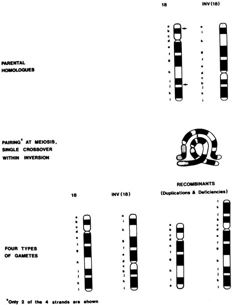
In a chromosomal inversion, the normal sequence of genes on the chromosome is altered. Subjects with such inversions are phenotypically normal; however, they may produce unbalanced gametes if, during meiosis I, crossing over (recombination) occurs within the inverted sequence. Thus, certain genes would be duplicated and others would be deficient in the unbalanced gamete (see Fig. 1). Pericentric inversions and inversions involving long segments are more likely to be associated with anomalous offspring than are paracentric or short inversion segments.65 Empirical data are not available for specific inversions, but pooled data for all inversions indicate approximately a 3% risk for abnormal progeny, with maternal carriers again at greater risk than paternal carriers39 (see Table 5). An exception is inv(9), which is a common variant and is thought to be without clinical significance.

Fig. 1. Recombination in an inv(18) heterozygote. The effects of crossing over within a pericentric inversion and the potential products. (Simpson JL: Pregnancies in women with chromosomal abnormalities. In Schulman JD, Simpson JL [eds]: Genetic Diseases in Pregnancy, pp 440–471. New York, Academic Press, 1981.)

ANEUPLOIDY

If a parent has a numerical chromosomal abnormality (aneuploidy), the risk to offspring is increased. For example, approximately 35% (but not 50%) of offspring of females with 47,XX, + 21 (Down syndrome) are aneuploid;66 therefore, antenatal chromosomal studies are indicated in a pregnant female with Down syndrome. Males with Down syndrome are sterile. If a parent is mosaic for trisomy 21, antenatal diagnosis is again in order.67,68 Although risk figures are plainly biased by the method of ascertainment, approximately 20% of offspring of fertile 45,X; 45,X/46,XX; and 45,X/46,XX/47,XXX subjects are said to show abnormalities.41 Women with 47,XXX or 46,XX/47,XXX also have produced children with chromosomal abnormalities, although most of the offspring are normal. Theoretically, 47,XYY men are also at increased risk for chromosomally abnormal offspring, and several abnormal offspring have been reported. Men with 47,XXY (Klinefelter syndrome) are sterile, but those with mosaicism (46,XY/47,XXY) may be fertile. Antenatal diagnosis should be offered to all aneuploid parents.

RELATIONSHIP OF ASCERTAINMENT TO EMPIRICAL RISK

Mode of ascertainment is a significant determinant of empirical risk for an unbalanced liveborn infant. Thus, when a family with a translocation is ascertained through a balanced proband, the risk for an unbalanced liveborn infant is very low. In contrast, if ascertainment is through an unbalanced individual, the risk for unbalanced offspring is significant.69,70 Presumably, then, with some translocations, unbalanced gametes do not arise during meiosis, or, alternatively, unbalanced products are selected against at the gametic or embryonic level. If a rearrangement has been ascertained during an evaluation for repetitive abortions, the risk for an unbalanced liveborn infant is lower than the risk expected after ascertainment through an anomalous liveborn infant but still is substantial.70

Fetuses Manifesting Intrauterine Growth Retardation or Anomalies on Ultrasound Examination

The potential indications considered previously are based on the premise that abnormal fetal outcome can be predicted on the basis of certain parental characteristics. However, trisomic fetuses, especially fetuses with trisomy 13 or 18, often show intrauterine growth retardation, which may be clinically evident during the second trimester. Clinical suspicion of intrauterine growth retardation can be followed with ultrasound monitoring to confirm intrauterine growth retardation. Gross anomalies also frequently can be visualized in fetuses with chromosomal abnormalities (Table 6).71–91 Antenatal chromosomal studies are appropriate if an abnormal fetus is detected on ultrasound examination. In addition to routine cytogenetic studies, particular defects suggest the need for more specific studies. For example, conotruncal heart defects are frequently associated with deletion of a small portion of chromosome 22 (del22q.11.2). FISH with specific probes will be diagnostic of this microdeletion that also implies the presence of other defects, for example, absent thymus and parathyroids (DiGeorge/velocardiofacial syndrome).92,93 Even if chromosomal studies cannot be obtained sufficiently early to permit pregnancy termination or termination is not desired, cesarean section for fetal distress in a fetus with a lethal abnormality might be avoided.


Table 6. Chromosomal Abnormalities in Pregnancies With Anomalies Detected by Ultrasonography


Ultrasound Finding* No. of Abnormal/Total (% Abnormal) Chromosome Results
Diaphragmatic hernia 20/173 (11.6) 12: + 18; 2: +21; 1: +21 +21; 1: triploid; 4: unbalanced autosomes
Duodenal atresia 35/118 (29.6) 30: + 21; 1: + 18; 1: + 13; 1: triploid; 1: unbalanced autosome; 1: XXX
Gastroschisis 5/84 (6.0) 2:+18; 1: + 13; 1: 45,X; 1: unbalanced autosome
Omphalocele 78/319 (24.5) 64: + 18; 6: +13 1: XXY; 1: triploid; 1: 45,X; 1: not specified; 4: unbalanced autosomes
Genitourinary 43/443 (9.7) 10: + 18; 8: +13; 7: + 21; 2: triploid; 1:45, X; 1 +9; 1: +8; 1: XYY; 8: unbalanced autosomes; 4: not specified
Cardiac 129/473 (27.3) 39: + 18; 25: + 21; 11: 45,X; 10: + 13; 1: + 9; 1: + 17; 1: triploid; 2: XXY; 4: unbalanced autosomes; 35: unspecified trisomy
Hydrocephalus 55/402 (13.7) 10: +21;10: + 18, 6: triploid, 6: + 13; 2: 45,X 1:XXX; 1:XXY: 16: unbalanced autosomes; 3: not specified
Choroid plexus cyst with no other anomalies seen 28/723 (3.9) 14: +18; 4: +21; 1:XXX; 1:XXY; 3:mosaic 45,X; 5: unbalanced autosomes
Holoprosencephaly 21/44 (47.7) 15: + 13; 1: + 18; 1: triploid; 4: unbalanced autosomes
Growth retardation and/or oligohydramnios 112/780 (14.4) 30: + 18; 23: triploid; 12: + 21; 9: + 13; 1 45,X; 34: unbalanced autosomes; 3: not specified
Polyhydramnios 50/718 (7.0) 15: +21; 18: + 18; 4: + 13; 1: triploid; 2: 45,X 1:XXX; 1: 46,X,i(Xq); 5: unbalanced autosomes; 3: not specified
Cystic hygroma 183/296 (61.8) 131: 45,X; 24: + 21; 13: + 18; 4: unbalanced autosomes; 2: +13; 1: triploid; 2: XXY; 6: not specified
Nonimmune hydrops without cystic hygroma 86/254 (33.9) 17: + 21; 5: + 18; 45: 45,X; 3: polyploid; 2: + 13; 5: unbalanced autosomes; 9: not specified

*Listed under primary defect indicated by the investigator. In some cases, multiple abnormalities were present.


Several authors have reported that certain biometric findings (e.g., short femur, short humerus, pyelectasis, thickened nuchal fold, nuchal translucency, echogenic bowel, absent nasal bone) are indicative of an increased risk of fetal Down syndrome.94,95,96,97 The positive predictive value of the ultrasound findings depends on the patient's a priori risk based on maternal age or biochemical serum screening. Different recommendations have been made as to how best to estimate the absolute risk of Down syndrome.98 Antenatal diagnosis should be offered when the risk estimate is greater than the procedure-associated risk of pregnancy loss.

Mendelian Disorders Associated With Chromosome Breakage

Several inherited disorders are characterized by chromosome breakage in vivo and in vitro. Persons with these disorders often show increased propensity for neoplasia, growth retardation, and various somatic anomalies. Bloom's syndrome, ataxia-telangiectasia, and Fanconi's anemia are examples of such disorders. In some of these disorders, when the precise molecular defect is not known in individual families, distinctive cytogenetic features may permit antenatal diagnosis. For example, Voss and colleagues diagnosed Fanconi's anemia in a second-trimester fetus on the basis of high frequencies of spontaneous and clastogen-induced chromosome breakage in amniotic fluid cells.99 Similar studies have been performed with chorionic villus tissue100 and fetal blood.101 Parallel cultures of cells from other family members are required to distinguish affected fetuses from heterozygotes.102

Knowledge that patients with ataxia-telangiectasia (A-T) have an increased rate of spontaneous chromosome breakage has historically facilitated the diagnosis in the second trimester.103,104 However, localization of the A-T gene now allows more reliable molecular genetic testing.105 In Bloom's syndrome, the rate of sister chromatid exchanges is increased in peripheral lymphocytes, fibroblasts, and bone marrow cells, making prenatal diagnosis feasible even in families in which the gene mutation is unidentified. All these disorders are inherited in autosomal recessive fashion; therefore, couples who have had an affected child have a 25% recurrence risk in each pregnancy. Antenatal diagnosis should be offered to such families.

Fragile X Syndrome and Other X-linked Recessive Disorders

The fragile X syndrome is an X-linked disorder characterized in males by moderate mental retardation, macroorchidism, and a long face with a prominent jaw. Approximately one third of female carriers (heterozygotes) are mildly retarded, and the others have a normal phenotype. This syndrome accounts for a significant proportion of cases of familial X-linked mental retardation. The gene responsible for the condition is linked to a fragile site on the long arm of the X chromosome, visible as a break, or gap, in the chromosome structure. The fragile site is seen only when cells are grown in special medium deficient in folic acid and thymidine or when an antimetabolite such as 5-fluorodeoxyuridine or methotrexate is added to the culture medium.

Prenatal diagnosis has been accomplished by visualization of the fragile site in amniotic fluid cells, chorionic villus tissue, and fetal blood.106,107,108,109,110 Unfortunately, both false-negative and false-positive results have occurred in amniotic fluid and CVS samples.108,109,110 Molecular (DNA) methods also have been used to diagnose fragile X syndrome and have proven the more reliable approach.111,112

In other X-linked recessive, or male-limited autosomal dominant traits, only males are affected. It is possible to distinguish affected from unaffected male fetuses in some but not all of the sex-limited disorders. In others, affected infants can be avoided consistently only by terminating all pregnancies in which the fetus is male. In these cases, antenatal chromosomal studies to determine fetal sex may be indicated.

Back to Top
POTENTIAL INDICATIONS FOR CYTOGENETIC STUDIES

Even if the use of antenatal cytogenetic studies increased greatly, the incidence of liveborn infants with chromosomal abnormalities would not be decreased greatly as long as these studies are performed only for the aforementioned indications. Offering antenatal diagnosis only to women 35 years of age and older decreases the frequency of trisomy 21 by less than 20%.113 Even maternal serum screening does not detect the majority of chromosome abnormalities. Monitoring on the basis of the other cytogenetic indications also results in detection of only a minority of fetuses with chromosomal abnormalities. Conversely, offering antenatal diagnosis to all women does not seem justified because of the small, yet finite, risk of invasive procedures. Therefore, one would hope to identify categories of younger women whose risk of having a chromosomally abnormal fetus justifies the risk of prenatal diagnosis. That is, such women would constitute a special high-risk group based on factors such as previous reproductive or medical history or fetal characteristics. Following are potential indications for antenatal studies.

Advanced Paternal Age

Although the relationship between aneuploidy and increased maternal age is better recognized and more established, Down syndrome also has been associated in some studies with advanced paternal age. Stene and colleagues114 and Matsunaga and associates115 found that the risk of siring offspring with trisomy 21 increased by paternal age 55 years and perhaps as early as 41 years of age.116 Other investigators, however, have found a much smaller or no effect.117,118,119,120 Therefore, advanced paternal age alone is not a sufficient indication for prenatal cytogenetic studies.

Previous Stillborn or Spontaneous Abortions

Couples experiencing repetitive abortions should undergo cytogenetic studies to exclude the presence of a parental translocation or inversion, either of which clearly justifies antenatal chromosomal studies. By virtue of the following argument, antenatal diagnosis also might be considered for couples who have had one or more spontaneous abortions or stillborn infants but who have not been found to have a parental chromosomal rearrangement.

Approximately 50% to 60% of all first-trimester abortuses show chromosomal abnormalities, and 5% of stillborn infants do. Of abortuses with chromosomal abnormalities, 50% are trisomic; thus, 25% of all abortuses are trisomic.

If women who have chromosomally abnormal abortuses have an increased risk for a subsequent trisomic conceptus, it would be reasonable to offer antenatal cytogenetic studies on the presumption that the couple's next trisomic conceptus might not abort, but rather continue to the liveborn stage. Indeed, pooled results of several small studies, albeit not corrected for maternal age, suggest that couples with a trisomic abortus have approximately a 1% risk for an aneuploid live birth (Table 7).121,122,123,124,125 Other investigators disagree, however, on the validity of such studies and on whether aneuploidy recurs more often than would be expected based on maternal age alone.126,127,128,129 In practice, chromosomal studies rarely are performed on abortuses or stillborn infants. Therefore, one usually cannot say in a particular case whether a couple with several abortuses or stillborn fetuses experienced recurrent aneuploid conceptions and, hence, might benefit from antenatal diagnosis. To be considered is whether the theoretical risk for an aneuploid livebirth in such circumstances outweighs the empirical risk of an invasive diagnostic procedure.


Table 7. Risk of Trisomic or Live-Born Fetus Preceding or Following a Trisomie Abortus


Studies Frequency of Aneuploid Liveborns
Boué and Boué121 1/25
Boué and coworkers122 0/117
Alberman123 1/73
Alberman and coworkers124 5/244
Warburton and coworkers125 1/343
Total 8/802 (1%)


Exposure to Irradiation or Chemotherapeutic Agents

Retrospective case-control studies by several independent groups have found that women whose pregnancies terminated in liveborn offspring with Down syndrome received significantly more X-irradiation before conception than had controls. The irradiation occurred 2 to 10 years before conception, and doses as small as 2 rads appeared to predispose to aneuploidy.130,131 These studies are highly suggestive; however, other studies revealed no such correlation.132,133,134 Although additional data clearly are necessary, it seems inappropriate to recommend antenatal diagnosis solely on the basis of diagnostic X-irradiation before or especially during gestation. Therapeutic radiation, however, is a more complex issue. Theoretical risk for gametic chromosomal and genetic damage is a serious consideration, and men treated with testicular radiation doses of .4 to 5 grays (40 to 500 rads) have shown increased chromosomal abnormalities in sperm.135 However, empirically, no increase in congenital anomalies has been found in the offspring of persons treated with X-irradiation for Hodgkin's disease or other cancers.136,137,138,139,140,141 Japanese women exposed to X-irradiation through proximity to the atomic bomb explosions also did not show an increased prevalence of Down syndrome offspring.142 Therefore, antenatal cytogenetic studies might be discussed but not encouraged for women (or men) who have undergone radiation therapy. Couples electing to undergo antenatal diagnostic studies must realize that only numerical and structural chromosomal abnormalities can be assessed. There is no possibility of monitoring for gene mutations, which also would be predicted to increase after irradiation.

Similar reasoning also might apply to men and women who have received chemotherapeutic agents because many agents used to treat neoplasia produce in vitro chromosomal damage and induce mutations. A person who previously has received such agents should thus theoretically be at increased risk for chromosomally abnormal progeny. Again, and analogous to irradiation data, no increase in the actual rate of anomalies has been observed in liveborn infants of such couples.137,138,139,140,141,142,143,144 Thus, antenatal cytogenetic studies are not necessarily indicated in this situation, although the issues may be worthy of discussion.

Parental Metabolic Derangements

Parental, especially maternal, metabolic derangements could predispose to aneuploidy. Although once considered suspect, diabetes mellitus,145 infectious hepatitis, and other infectious diseases146 do not seem to have this effect. Whether parental a1-antitrypsin (a1-protease inhibitor) phenotype is a significant risk factor is still controversial.147,148,149 Conversely, data showing a correlation between the presence of maternal antithyroid antibodies or hyperthyroidism and offspring with Down syndrome are more convincing.150,151,152 No conclusive studies, however, have been published; therefore, the presence of antithyroid antibodies or hyperthyroidism alone should not be considered an obligatory indication for antenatal cytogenetic studies. In a similar vein, based on the hypothesis that both Alzheimer's disease and chromosomal nondisjunction may be caused by failure of microtubular organization, Heston purported to demonstrate an excessive incidence of trisomy 21 in relatives of probands with Alzheimer's disease.153 However, the expected incidence was not adjusted for maternal age, nor were karyotypes performed on all subjects with “trisomy 21” to exclude the possibility of a familial translocation. Therefore, further verification is required before these findings are accepted.

Parental Chromosomal Variants

Chromosomal variants are structural polymorphisms believed to be without phenotypic effect. However, it has been proposed that such variants or other in vitro findings (e.g., increased satellite association) may predispose to chromosomal nondisjunction and thereby aneuploid gametes. There is evidence both for and against this position;154,155,156,157 biases of ascertainment and publication make evaluation of the available data difficult. An association between the presence of a particular variant, a double nucleolar organizing region on an acrocentric chromosome, and predisposition to aneuploid gametes has been claimed158 but not uniformly corroborated.159,160,161 Therefore, routine studies on all couples to detect such variants or in vitro aberrations are not indicated.

Periconceptive Events and Assisted Reproduction

Certain periconceptive events might predispose a couple to liveborn infants with chromosomal abnormalities. For example, in a questionnaire survey of pregnancy outcome after artificial insemination by donor, Forse and colleagues found three aneuploid liveborn infants (2 with trisomy 21 and 1 with trisomy 13) in a population of 400 term offspring (p < .05).162 In contrast, neither the experience of my colleagues and I nor that of other investigators reveals an increase in autosomal trisomy in artificial insemination by donor pregnancies.163,164 However, some men with severe oligospermia and nonobstructive azoospermia have microdeletions of the Y chromosome. This will be transmitted to their male offspring if conception occurs, either naturally or with the assistance of in vitro fertilization/intracytoplasmic sperm insemination (IVF/ICSI). Presumably couples undergoing IVF/ICSI are willing to accept potential infertility in their sons, but if not, prenatal chromosome analysis is possible. A more serious concern is the finding that children born after IVF or ICSI have an increased rate of autosomal and sex chromosome anomalies (1% to 3%).165,166 Couples should be counseled accordingly and prenatal diagnosis considered.

Aging gametes and asynchrony between oocyte and sperm have also been associated with chromosome abnormalities. An example is that in animals fertilization of an aged oocyte results in polyploidy and sometimes aneuploidy.167 In humans, intrafollicular (preovulatory) delays may contribute to polyploidy but not to aneuploidy.168 Asynchrony between an oocyte and its fertilizing sperm also might occur in pregnancies associated with ovulation induction (clomiphene or human menopausal gonadotropins), intercourse after a period of abstinence, or intercourse occurring more than 1 day before or after ovulation.169 Boué and Boué found a higher frequency of chromosomal abnormalities in abortuses recovered from pregnancies associated with ovulation induction than in abortuses recovered from pregnancies in which ovulation was not induced.170 Other investigators have reported an increased incidence of liveborn infants with Down syndrome after pregnancies associated with ovulation induction171; however, these findings have not been verified.

Simpson and associates have examined data from women who conceived while using natural family planning methods and recording coital events.172 No association was found between conception outside the optimal period immediately surrounding ovulation and aneuploidy (Down syndrome) in the resultant offspring.

Conversely, in a recent study, women with a reduced ovarian complement based on previous unilateral oophorectomy or congenital absence of one ovary, were significantly more likely to have a child with Down syndrome than were the controls who did not have absence of an ovary.35 This finding is consistent with Warburton's hypothesis that a suboptimally developed oocyte may become the dominant follicle in situations in which only a small number of oocytes are available (e.g., in older women).173 This hypothesis is further supported by the finding that age of menopause is 1 year earlier in women with trisomic losses compared to women with euploid losses or euploid livebirths.34 This implies that women with trisomic conceptions have accelerated oocyte atresia or a smaller number of oocytes. If the findings of Freeman and associates35 are confirmed, discussion of prenatal diagnosis may be indicated in women with unilateral ovaries.

Back to Top
FUTURE DIRECTIONS

Preimplantation Diagnosis

Diagnosis of a chromosome disorder before implantation of the zygote in the uterus offers the ability to select and transfer only normal conceptuses. For some families who are at very high risk (e.g., a parental chromosome translocation), avoiding the need for repeated pregnancy terminations of affected fetuses is a significant advantage. Details of preimplantation diagnosis are found elsewhere in these volumes.

Fetal Cells in the Maternal Circulation

For many years, obstetricians have wished for a method of prenatal diagnosis that is risk-free, in other words, noninvasive. In 1975, Schroder and associates showed that fetal lymphocytes are present in the maternal circulation during pregnancy.174 However, practical exploitation of this knowledge required the ability to separate fetal from maternal lymphocytes, thereby enriching the sample for fetal cells. A number of enrichment schemes were tried; however, poor specificity and the difficulty in inducing mitosis in fetal lymphocytes isolated in this fashion made this approach impractical.

More recently, however, with new developments in cell sorting techniques, in situ hybridization with chromosome-specific probes, and polymerase chain reaction for amplification of small amounts of DNA, several groups have been successful in isolating and analyzing fetal cells and fetal DNA from maternal blood samples. Fetal trophoblasts, lymphocytes, granulocytes, and erythroblasts (nucleated erythrocytes) all have been potential target cells. In one example, Lo and colleagues obtained blood samples from 27 women at 6 to 41 weeks' gestation and, without sorting fetal from maternal cells, performed polymerase chain reaction with primers for a single-copy sequence on the Y chromosome.175 Of 17 women carrying a male fetus, 13 showed a signal for Y DNA. Of 10 pregnancies with female fetuses, eight were negative for male (Y) DNA. Other investigators have used monoclonal antibodies to separate fetal from maternal cells, followed by polymerase chain reaction to detect Y sequences. In one study, fetal sex prediction was correct in 11 of 12 women studied, with one female fetus misdiagnosed as a male.176

Using techniques such as these, several groups have reported diagnosis of fetal aneuploidy and Mendelian traits from maternal blood samples obtained before or after amniocentesis or CVS.177,178 Chromosome diagnosis takes advantage of the presumption that virtually all pregnant women have a 46,XX karyotype. Any other chromosomal complement in a maternal blood sample can be ascribed to a conceptus, either from the current or from a previous pregnancy.

In a demonstration of the possibilities of this approach, Price and associates described the detection of fetal chromosome abnormalities in maternal blood samples.177 The authors first flow-sorted nucleated erythrocytes from maternal samples on the basis of cell size, granularity, and presence of transferrin receptors and glycophorin A cell surface antigens. In a series of samples from first- and second-trimester pregnancies, approximately 10% of the final sorted samples were fetal cells (nuclei).2–20,103 The authors used both polymerase chain reaction with single copy Y-specific sequences and in situ hybridization with a Y-specific probe to confirm that they were enriching for fetal cells.

In one of two cases with fetal aneuploidy, blood was drawn from the mother 1 week after CVS was performed. After enrichment, the sample was analyzed by in situ hybridization with a chromosome 21-specific probe. Thirty-nine percent of the cells showed three signals, indicative of trisomy 21, and consistent with the findings in villi and in abortus tissue.

In the second case, maternal blood was drawn before CVS, enriched, and analyzed with probes for chromosomes X, Y, 18 and 21. Nine percent of cells showed hybridization with the Y probe, indicating that male fetal cells constituted approximately 9% of the sample. The same percentage of cells in the sample showed three hybridization signals with the probe for chromosome 18, thereby diagnosing a male fetus with trisomy 18. This diagnosis was confirmed with abortus tissue.

Subsequently, a collaborative study of 69 maternal blood samples, obtained before or after an invasive procedure and analyzed with chromosome-specific probes, showed that it was possible to detect both sex chromosome polysomy, as well as autosomal trisomy.179 Other investigators have shown that the number of fetal cells and fetal DNA in the maternal circulation is increased in aneuploid pregnancies.180,181 This finding may prove useful as an initial screen.

Thus, noninvasive prenatal diagnosis with maternal blood samples appears feasible; however, the sensitivity and specificity still are not adequate for clinical application. The National Institute of Child Health and Human Development (NICHD) currently is funding a multicenter clinical trial of this technology (NIFTY). A recent analysis of 5 years of data showed correct diagnosis of fetal sex in 41% of women carrying male fetuses and 89% of women carrying female fetuses. One or more aneuploid cells were detected in 74% of cases in which aneuploidy was truly present, with a false-positive rate of .6% to 4%.182

If difficulties can be worked out, screening for chromosome disorders early in gestation with maternal blood samples might be offered to all pregnant women, similar to the screening now performed with biochemical analytes. Initially, the test might be viewed as a screening test only, which is followed by CVS or amniocentesis for definitive diagnosis if abnormal cells are detected. Eventually, however, if maternal blood screening proves sensitive and specific enough, it could be the hoped for noninvasive diagnostic test.

Back to Top
REFERENCES

1. Verp MS, Simpson JL: Amniocentesis for prenatal genetic diagnosis. In: Filkins K, Russo JF (eds): Human Prenatal Diagnosis. pp 305-355, 2nd ed. New York, Marcel Dekker, 1990

2. Verp MS, Simpson JL, Ober C: Prenatal diagnosis of genetic disorders. In: Lin C-C, Verp MS, Sabbagha RE (eds): The High-Risk Fetus. pp 172-202, New York, Springer-Verlag, 1993

3. Martin AO: Characteristics of amniotic fluid cells in vitro and attempts to improve culture techniques. Clin Obstet Gynecol 7:143, 1980

4. Verp MS, Gerbie AB: Amniocentesis for prenatal diagnosis. Clin Obstet Gynecol 24:1007, 1981

5. Jenks JD, Martin AO, Simpson JL: Relationship of gestational age to concentration and viability of amniotic fluid cells, p 66. Abstract from the 29th Annual Meeting of the Society on Gynecologic Investigation, 1982

6. Evans MI, Klinger KW, Isada NB et al: Rapid prenatal diagnosis by fluorescent in situ hybridization of chorionic villi: An adjunct to long-term culture and karyotype. Am J Obstet Gynecol 167:1522, 1992

7. Klinger K, Laudes G, Shook D et al: Rapid detection of chromosome aneuploidies in uncultured amniocytes by using fluorescence in situ hybridization (FISH). Am J Hum Genet 51:55, 1992

8. D'Alton ME, Malone FD, Chelmow D et al: Defining the role of fluorescence in situ hybridization on uncultured amniocytes for prenatal diagnosis of aneuploidies. Am J Obstet Gynecol 176:769, 1997

9. Bryndorf T, Christensen B, Vad M et al: Prenatal detection of chromosome aneuploidies by fluorescence in situ hybridization: Experience with 2000 uncultured amniotic fluid samples in a prospective preclinical trial. Prenat Diagn 17:333, 1997

10. American College of Medical Genetics: Prenatal interphase fluorescence in situ hybridization (FISH) policy statement. Am J Hum Genet 53:526, 1993

11. Daffos F, Capella-Pavlovsky M, Forestier F: Fetal blood sampling during pregnancy with use of a needle guided by ultrasound. Am J Obstet Gynecol 153:655, 1985

12. Weiner CP: Percutaneous umbilical blood sampling (cordocentesis). In: Lin C-C, Verp MS, Sabbagha RE (eds): The High-Risk Fetus. pp 352-359, New York, Springer-Verlag, 1993

13. Donnenfeld AE, Lockwood D, Lamb AN: Prenatal diagnosis from cystic hygroma fluid: The value of fluorescence in situ hybridization. Am J Obstet Gynecol 185:1004, 2001

14. Donnenfeld AE, Lockwood D, Custer T et al: Prenatal diagnosis from fetal urine in bladder outlet obstruction: Success rates for traditional cytogenetic evaluation and interphase fluorescence in situ hybridization. Genet Med 4:444, 2002

15. Benn PA, Hsu LYF: Maternal cell contamination of amniotic fluid cell cultures: Results of a U.S. nationwide survey Am J Med Genet 15:297, 1983

16. Simpson JL, Martin AO, Verp MS et al: Hypermodal cells in amniotic fluid cultures: Frequency, interpretation, and clinical significance. Am J Obstet Gynecol 143:250, 1982

17. Najafzadeh TM, Cahill TC, Dumars KW: Prenatal detection of chromosomal mosaicism. Prenat Diagn 2:7, 1982

18. Hsu LYF, Perlis TE: United States survey on chromosome mosaicism and pseudomosaicism in prenatal diagnosis. Prenat Diagn 4:97, 1984

19. Hsu LYF: Prenatal diagnosis of chromosomal abnormalities through amniocentesis. In: Milunksy A (ed): Genetic Disorders and the Fetus. pp 222-223, 4th ed.. New York, Johns Hopkins University Press, 1998

20. Black SH, Dorfmann A, Colyer C et al: Amniocentesis after transcervical chorionic villus sampling (CVS): Mosaicism and elevated serum alpha-fetoprotein (MSAFP) concentrations. Am J Hum Genet 41:A267, 1987

21. Shaffer LG, Agan N, Goldberg JD et al: American College of Medical Genetics statement on diagnostic testing for uniparental disomy. Genet Med 3:206, 2001

22. Kotzot D: Review and meta-analysis of systemic searches for uniparental disomy (UPD) other than UPD 15. Am J Med Genet 111:366, 2002

23. Berend SA, Horwitz J, McCaskill C et al: Identification of uniparental disomy following prenatal detection of Robertsonian translocations and isochromosomes. Am J Hum Genet 66:1787, 2000

24. Warburton D: De novo balanced chromosome rearrangements and extra marker chromosomes identified at prenatal diagnosis: Clinical significance and distribution of breakpoints. Am J Hum Genet 49:995, 1991

25. Brondum-Nielsen K, Mikkelsen M: A 10-year survey, 1980-1990, of prenatally diagnosed small supernumerary marker chromosomes, identified by FISH analysis. Outcome and follow-up of 14 cases diagnosed in a series of 12699 prenatal samples Prenat Diagn 15:615, 1995

26. Daniely M, Aviram-Goldring A, Barkai G et al: Detection of chromosomal aberration in fetuses arising from recurrent spontaneous abortion by comparative genomic hybridization. Hum Reprod 13:805, 1998

27. Simpson JL: Klinefelter syndrome; Polysomy X in females and polysomy Y in males. In: Simpson JL (ed): Disorders of Sexual Differentiation: Etiology and Clinical Differentiation. pp 304-322, 361-370, New York, Academic Press, 1976

28. Robinson A, Lubs HA, Bergsma D: Sex chromosome aneuploidy: Prospective studies on children. Birth Defects 15:(1):1979

29. Nielsen J, Sorensen AM, Sorensen K: Mental development of unselected children with sex chromosome abnormalities. Hum Genet 59:324, 1981

30. Evans JA, Hamerton JL, Robinson A: Children and young adults with sex chromosome aneuploidy. Birth Defects 26:(1):1990

31. Verp MS, Bombard AT, Simpson JL et al: Parental decision following prenatal diagnosis of fetal chromosome abnormality. Am J Med Genet 29:613, 1988

32. Simpson JL, Elias S, Gatlin MJ et al: Genetic counseling and genetic services in obstetrics and gynecology: Implications for educational goals and clinical practice. Am J Obstet Gynecol 140:70, 1981

33. Ayme S, Lippman-Hand A: Maternal-age effect in aneuploidy: Does altered embryonic selection play a role? Am J Hum Genet 34:558, 1982

34. Kline J, Kinney A, Levin B, Warburton D: Trisomic pregnancy and earlier age at menopause. Am J Hum Genet 67:395, 2000

35. Freeman SB, Yang Q, Allran K et al: Women with a reduced ovarian complement may have an increased risk for a child with Down syndrome. Am J Hum Genet 66:1680, 2000

36. Hook EB, Hamerton JL: The frequency of chromosome abnormalities detected in consecutive newborn studies: Differences between studies: Results by sex and by severity of phenotypic involvement. In: Hook EB, Porter IH (eds): Population Cytogenetics. pp 63-79, New York, Academic Press, 1978

37. Hook EB, Cross PK, Schreinemachers DM: Chromosomal abnormality rates at amniocentesis and in live-born infants. JAMA 249:2034, 1983

38. Ferguson-Smith MA: Maternal age specific incidence of chromosome aberrations at amniocentesis. In: Murken JD, Stengel-Rutkowski S, Schwinger E (eds): Prenatal Diagnosis: Proceedings of the Third European Conference on Prenatal Diagnosis of Genetic Disorders. pp 1-14, Stuttgart, F Enke, 1979

39. Hook EB, Cross PK, Jackson L et al: Maternal age-specific rates of 47, +21 and other cytogenetic abnormalities diagnosed in the first trimester of pregnancy in chorionic villus biopsy specimens: Comparison with rates expected from observations at amniocentesis. Am J Hum Genet 42:797, 1988

40. Hook EB: Spontaneous deaths of fetuses with chromosomal abnormalities diagnosed prenatally. N Engl J Med 299:1036, 1978

41. Merkatz IR, Nitowsky HM, Macri JN et al: An association between low maternal serum alpha-fetoprotein and fetal chromosomal abnormalities. Am J Obstet Gynecol 148:886, 1984

42. Bogart MH, Pandian MR, Jones OW: Abnormal maternal serum chorionic gonadotropin levels in pregnancies with fetal chromosome abnormalities. Prenat Diagn 7:623, 1987

43. Canick JA, Knight GJ, Palomaki GE et al: Low second trimester maternal serum unconjugated oestriol in pregnancies with Down's syndrome. Br J Obstet Gynaecol 95:330, 1988

44. Wald NJ, Cuckle HS, Densem JW et al: Maternal serum screening for Down's syndrome in early pregnancy. Br Med J 297:883, 1988

45. Haddow JE, Palomaki GE, Knight GJ et al: Prenatal screening for Down's syndrome with use of maternal serum markers. N Engl J Med 327:588, 1992

46. Phillips OP, Elias S, Shulman LP et al: Maternal serum screening for fetal Down syndrome in women less than 35 years of age using alpha-fetoprotein, hCG, and unconjugated estriol: A prospective 2-year study. Obstet Gynecol 80:353, 1992

47. Burton BK, Prins GS, Verp MS: A prospective trial of prenatal screening for Down syndrome by means of maternal serum alpha-fetoprotein, human chorionic gonadotropin, and unconjugated estriol. Am J Obstet Gynecol 169:526, 1993

48. Wenstrom KD, Owen J, Chu DC et al: Prospective evaluation of free beta-subunit of human chorionic gonadotropin and dimeric inhibin A for aneuploidy detection. Am J Obstet Gynecol 181:887, 1999

49. Cole LA, Shahabi S, Oz UA et al: Urinary screening tests for fetal Down syndrome: II. Hyperglycosylated hCG Prenat Diagn 19:351, 1999

50. Krantz DA, Larsen JW, Buchanan PD et al: First-trimester Down syndrome screening: Free beta-human chorionic gonadotropin and pregnancy-associated plasma protein A. Am J Obstet Gynecol 174:612, 1996

51. Bindra R, Heath V, Liao A et al: One-stop clinic for assessment of risk for trisomy 21 at 11-14 weeks: A prospective study of 15030 pregnancies. Ultrasound Obstet Gynecol 20:219, 2002

52. Simpson JL et al: Noninvasive screening for aneuploidy: who, when, and why? Cont OB/GYN 76, 2000

53. Haddow JE, Palomaki GE, Knight GJ: Reducing the need for amniocentesis in women 35 years of age or older with serum markers for screening. N Engl J Med 330:1114, 1994

54. American College of Medical Genetics: ACMG position statement on multiple marker screening in women 35 and older. ACMG Newsletter 2:1, 1994

55. ACOG Practice Bulletin: Prenatal diagnosis of fetal chromosomal abnormalities. May 27, 2001

56. Mikkelsen M: Down syndrome: Current stage of cytogenic epidemiology. In: Bonne-Tamir B, Cohen T, Goodman RM (eds): Human Genetics, Part B: Medical Aspects. pp 297-309, New York, Alan R. Liss, 1982

57. Mikkelsen M, Stene J: Previous child with Down's syndrome and other chromosome aberrations. In: Murken JD, Stengel-Rutkowski S, Schwinger E (eds): Prenatal Diagnosis: Proceedings of the Third European Conference on Prenatal Diagnosis of Genetic Disorders. pp 22-23, Stuttgart, F Enke, 1979

58. Simoni G, Fraccaro M, Arslanian A et al: Cytogenetic findings in 4952 prenatal diagnoses: An Italian collaborative study. Hum Genet 60:63, 1982

59. Stene J, Stene E, Mikkelsen M: Risk for chromosome abnormality at amniocentesis following a child with a non-inherited chromosome aberration. Prenat Diagn 4:(special issue):81, 1984

60. Mikkelsen M: In: Jackson L (ed): CVS Newsletter. 19:7, 1986

61. Jewell AF, Keene WE, Ferre MM et al: Analysis of the recurrence risks for trisomies 13 and 18. Am J Hum Genet 59:A121, 1996

62. Garver KL, Marchese SG, Steele MW et al: Recurrence risks in 21q/21q translocation Down syndrome. J Pediatr 100:243, 1982

63. Mikkelsen M: Down's syndrome: Current stage of cytogenetic research. Hum Genet 12:1, 1971

64. Daniel A, Hook EB, Wulf G: Risks of unbalanced progeny at amniocentesis to carriers of chromosomal rearrangements: Data from United States and Canadian laboratories. Am J Med Genet 31:14, 1989

65. Simpson JL: Pregnancies in women with chromosomal abnormalities. In: Schulman JD, Simpson JL (eds): Genetic Diseases in Pregnancy. pp 440-471, New York, Academic Press, 1981

66. Verp MS: Chromosomal disorders in pregnancy. In: Gleicher N (ed): Principles of Medical Therapy in Pregnancy. pp 1223-1229, New York, Plenum Publishing, 1985

67. Harris DJ, Begleiter ML, Chamberlin J et al: Parental trisomy 21 mosaicism. Am J Hum Genet 34:125, 1982

68. Werner W, Herrmann FH, John B: Cytogenetic studies of a family with trisomy 21 mosaicism in two successive generations as the cause of Down's syndrome. Hum Genet 60:202, 1982

69. Jacobs PA, Aitken J, Frackiewicz A et al: The inheritance of translocations in man: Data from families ascertained through a balanced heterozygote. Ann Hum Genet 34:119, 1970

70. Daniel A, Boue A, Gallano P: Prospective risk in reciprocal translocation heterozygotes at amniocentesis as determined by potential chromosome imbalance sizes: Data of the European Collaborative Prenatal Diagnosis Centres. Prenat Diagn 5:315, 1986

71. Chervenak FA, Isaacson F, Blakemore KJ et al: Fetal cystic hygroma: Cause and natural history. N Engl J Med 309:822, 1983

72. Marchese CA, Carozzi F, Mosso R et al: Fetal karyotype in malformations detected by ultrasound. Am J Hum Genet 37:A223, 1985

73. Morris K, Hesser J, Michalskik, et al: High chromosome abnormality rate from obstetrically indicated late amniocentesis. Am J Hum Genet 37:A223, 1985

74. Nielsen LB, Bang J, Norgaard-Pedersen B: Prenatal diagnosis of omphalocele and gastroschisis by ultrasonography. Prenat Diagn 5:381, 1985

75. Waldimiroff JW, Stewart PA, Sachs ES et al: Prenatal diagnosis and management of congenital heart defect: Significance of associated fetal anomalies and prenatal chromosome studies. Am J Med Genet 21:285, 1985

76. Garden AS, Benzie RJ, Miskin M et al: Fetal cystic hygroma colli: Antenatal diagnosis, significance, and management. Am J Obstet Gynecol 154:221, 1986

77. Nicolaides KH, Rodeck CH, Gosden CM: Rapid karyotyping in non-lethal fetal malformations. Lancet 1:283, 1986

78. Gilbert WM, Nicolaides KH: Fetal omphalocele: Associated malformations and chromosomal defects. Obstet Gynecol 70:633, 1987

79. Landy HJ, Isada NB, Larsen JW Jr: Genetic implications of idiopathic hydramnios. Am J Obstet Gynecol 157:114, 1987

80. Palmer CG, Miles JH, Howard-Peebles PN et al: Fetal karyotype following ascertainment of fetal anomalies by ultrasound. Prenat Diagn 7:551, 1987

81. Pearce JM, Griffin D, Campbell S: Cystic hygromata in trisomy 18 and 21. Prenat Diagn 4:371, 1984

82. Redford DHA, McNay MB, Ferguson-Smith ME et al: Aneuploidy and cystic hygroma detectable by ultrasound. Prenat Diagn 4:377, 1984

83. DeVore GR, Platt LD, Siassi B et al: The incidence of chromosomal defects associated with the prenatal diagnosis of congenital heart disease. Society of Perinatal Obstetricians Ninth Annual Meeting178:1989

84. Brumfield CG, Davis RO, Cosper P et al: Chromosomal abnormalities associated with sonographically detected fetal anomalies. Society of Perinatal Obstetricians Ninth Annual Meeting176:1989

85. Eydoux P, Choiset A, LePorrier N et al: Chromosomal prenatal diagnosis: Study of 936 cases of intrauterine abnormalities after ultrasound assessment. Prenat Diagn 9:255, 1989

86. Wilson RD, Chitayat D, McGillivray BC: Fetal ultrasound abnormalities: Correlation with fetal karyotype, autopsy findings, and postnatal outcome: Five-year prospective study. Am J Med Genet 44:586, 1992

87. Brady K, Polzin WJ, Kopelman JN et al: Risk of chromosomal abnormalities in patients with idiopathic polyhydramnios. Obstet Gynecol 79:234, 1992

88. Gross SJ, Shulman LP, Tolley EA et al: Isolated fetal choroid plexus cysts and trisomy 18: A review and metaanalysis. Am J Hum Genet 53:A97, 1993

89. Ranzine AC, Blakemore K, Corson L et al: Aneuploidy at 15–20 weeks gestation in fetuses with isolated choroid plexus cysts. Am J Hum Genet 53:A1450, 1993

90. Halliday J, Lumley J, Bankier A: Karyotype abnormalities in fetuses diagnosed as abnormal on ultrasound before 20 weeks' gestational age. Prenat Diagn 14:689, 1994

91. Hanna JS, Neu RL, Lockwood DH: Prenatal cytogenetic results from cases referred for 44 different types of abnormal ultrasound findings. Prenat Diagn 16:109, 1996

92. Raymond FL, Simpson JM, Sharland GK et al: Fetal echocardiography as a predictor of chromosomal abnormality. Lancet 350:30, 1997

93. Johnson MC, Hing A, Wood MK et al: Chromosome abnormalities in congenital heart disease. Am J Med Genet 70:292, 1997

94. Benacerraf BR: The second-trimester fetus with Down syndrome: Detection using sonographic features. Ultrasound Obstet Gynecol 7:147, 1996

95. Taipale P, Hiilesmaa V, Salonen R et al: Increased nuchal translucency as a marker for fetal chromosomal defects. N Engl J Med 337:1654, 1997

96. Vintzileos AM, Egan JFX: Adjusting the risk for trisomy 21 on the basis of second-trimester ultrasonography. Am J Obstet Gynecol 172:837, 1995

97. Cicero S, Curcio P, Papageorghiou A et al: Absence of nasal bone in fetuses with trisomy 21 at 11–14 weeks of gestation: an observational study. Lancet 358:1665, 2001

98. Shipp TD, Benacerraf B: Second trimester ultrasound screening for chromosomal abnormalities. Prenat Diagn 22:296, 2002

99. Voss R, Kohn G, Shaham M et al: Prenatal diagnosis of Fanconi anemia. Clin Genet 20:185, 1981

100. Auerbach AD, Min Z, Ghosh R et al: Clastogen-induced chromosomal breakage as a marker for first trimester prenatal diagnosis of Fanconi anemia. Hum Genet 73:86, 1986

101. Shipley J, Rodeck CH, Garrett C et al: Mitomycin C induced chromosome damage in fetal blood cultures and prenatal diagnosis of Fanconi's anemia. Prenat Diagn 4:217, 1984

102. Cohen MM, Simpson SJ, Honig GR et al: The identification of Fanconi anemia genotypes of clastogenic stress. Am J Hum Genet 34:794, 1982

103. Shaham M, Voss R, Becker Y et al: Prenatal diagnosis of ataxia telangiectasia. J Pediatr 100:134, 1982

104. Schwartz S, Flannery DB, Cohen MM: Tests appropriate for the prenatal diagnosis of ataxia telangiectasia. Prenat Diagn 5:9, 1985

105. Gatti RA, Peterson KL, Novak J et al: Prenatal genotyping of ataxia-telangiectasia. Lancet 342:376, 1993

106. Jenkins EC, Brown WT, Duncan CJ et al: Feasibility of fragile X chromosome prenatal diagnosis demonstrated. Lancet 2:1292, 1981

107. Shapiro LR, Wilmot PL, Brenholz P et al: Prenatal diagnosis of fragile X chromosome. Lancet 1:99, 1982

108. Jenkins EC, Shapiro LR, Brown WT: Prenatal diagnosis of the fragile X syndrome. In: Milunsky A (ed): Genetic Disorders and the Fetus. pp 241-255, 3rd ed.. New York, Johns Hopkins University Press, 1992

109. Webb TP, Rodeck CH, Nicolaides KH et al: Prenatal diagnosis of the fragile X syndrome using fetal blood and amniotic fluid. Prenat Diagn 7:203, 1987

110. Shapiro LR, Wilmot PL, Murphy PD et al: Multiple approaches to the prenatal diagnosis of the fragile X syndrome. Am J Hum Genet 41:A285, 1987

111. Sutherland GR, Gedeon A, Kornman L et al: Prenatal diagnosis of fragile X syndrome by direct detection of the unstable DNA sequence. N Engl J Med 352:1720, 1991

112. Wang Q, Green E, Barnicoat A et al: Cytogenetic versus DNA diagnosis in routine referrals for fragile X syndrome. Lancet 342:1025, 1993

113. Adams MM, Erickson JD, Layde PM et al: Down's syndrome: Recent trends in the United States. JAMA 246:758, 1981

114. Stene J, Fischer G, Stene E et al: Paternal age effect in Down syndrome. Ann Hum Genet 40:299, 1977

115. Matsunaga E, Tonomura A, Oishi H et al: Re-examination of paternal age effect in Down syndrome. Hum Genet 40:259, 1978

116. Stene J, Stene E, Stengel-Rutkowski S et al: Paternal age and Down's syndrome: Data from prenatal diagnosis (DFG). Hum Genet 59:119, 1981

117. Hook EB, Cross PK: Paternal age and Down's syndrome genotypes diagnosed prenatally: No association in New York data. Hum Genet 62:167, 1982

118. Roth M-P, Feingold J, Baumgarten A et al: Re-examination of paternal age effect in Down's syndrome. Hum Genet 63:149, 1983

119. Roth M-P, Stoll C, Taillemite JL et al: Paternal age and Down's syndrome diagnosed prenatally: No association in French data. Prenat Diagn 3:327, 1983

120. Hook EB, Regal RR: A search for a paternal-age effect upon cases of 47, +21 in which the extra chromosome is of paternal origin. Am J Hum Genet 36:413, 1984

121. Boué J, Boué A: Chromosomal analysis of two consecutive abortuses in each of 43 women. Humangenetik 19:275, 1973

122. Boué J, Boué A, Lazar P: Retrospective and prospective epidemiological studies of 1500 karyotyped spontaneous human abortuses. Teratology 12:11, 1975

123. Alberman ED: The abortus as a prediction of future trisomy 21 pregnancies. In: de la Cruz FF, Gerald PS (eds): Trisomy 21 (Down Syndrome). pp 69-76, Baltimore, University Park Press, 1981

124. Alberman E, Elliott M, Creasy M et al: Previous reproductive history in mothers presenting with spontaneous abortions. Br J Obstet Gynaecol 82:366, 1975

125. Warburton D, Hutzler M, Kline J et al: Recurrence risks for trisomy following a trisomic spontaneous abortion. Presented at the 1982 Annual Meeting of the American Society of Human Genetics, Detroit, September 28–October 2, 1982

126. Hook EB, Cross PK: Spontaneous abortion and subsequent Down syndrome live birth. Hum Genet 64:267, 1983

127. Warburton D, Kline J, Stein Z et al: Does the karyotype of a spontaneous abortion predict the karyotype of a subsequent abortion? Evidence from 273 women with two karyotyped spontaneous abortions Am J Hum Genet 41:465, 1987

128. Morton NE, Chiu D, Holland C et al: Chromosome anomalies as predictors of recurrence risk for spontaneous abortion. Am J Med Genet 28:353, 1987

129. Robinson WP, McFadden DE, Stephenson MD: The origin of abnormalities in recurrent aneuploidy/polyploidy. Am J Hum Genet 69:1245, 2001

130. Uchida IA, Holunga R, Lawler C: Maternal radiation and chromosome aberrations. Lancet 2:1045, 1968

131. Alberman E, Polani PE, Fraser Roberts JA et al: Parental exposure to X-irradiation and Down's syndrome. Ann Hum Genet 36:195, 1972

132. Carter CO, Evans KA, Stewart AM: Maternal radiation and Down's syndrome (Mongolism). Lancet 2:1042, 1962

133. Marmol JG, Scriggins AL, Vollman RF: Mothers of Mongoloid infants in the collaborative project. Am J Obstet Gynecol 104:533, 1969

134. Cohen BH, Lilienfeld AM, Kramer S et al: Parental factors in Down's syndrome: Results of the second Baltimore case-control study. In: Hook EB, Porter IH (eds): Population Cytogenetics. pp 301-352, New York, Academic Press, 1977

135. Martin RH: Chromosomal abnormalities in human sperm. In: Dellarco VL, Voytek PE, Hollaender A (eds): Aneuploidy: Etiology and Mechanisms. pp 91-102, New York, Plenum Publishing, 1985

136. Le Floch O, Donaldson S, Kaplan HS: Pregnancy following oophoropexy and total nodal irradiation in women with Hodgkin's disease. Cancer 41:1317, 1978

137. Holmes GE, Holmes FF: Pregnancy outcome of patients treated for Hodgkin's disease. Cancer 41:1317, 1978

138. Horning SJ, Hoppe RT, Kaplan HS et al: Female reproductive potential after treatment for Hodgkin's disease. N Engl J Med 304:1377, 1982

139. Mulvihill JJ, Byrne J, Steinhorn SA et al: Genetic disease in offspring of survivors of cancer in the young. Am J Hum Genet 39:A72, 1986

140. Mulvihill JJ, McKeen EA, Rosner F et al: Pregnancy outcome in cancer patients: Experience in a large cooperative group. Cancer 60:1143, 1987

141. Meistrich ML, Byrne J: Genetic disease in offspring of long-term survivors of childhood and adolescent cancer treated with potentially mutagenic therapies. Am J Hum Genet 70:1069, 2002

142. Schull WJ, Need JV: Maternal radiation and mongolism. Lancet 1:537, 1962

143. Ross GT: Congenital anomalies among children born of mothers receiving chemotherapy for gestational trophoblastic neoplasms. Cancer 37:1043, 1976

144. Walden PAM, Bagshawe KD: Pregnancies after chemotherapy for gestational trophoblastic tumors. Lancet 2:1241, 1979

145. Simpson JL, Elias S, Martin AO et al: Diabetes in pregnancy, Northwestern University Series (1977–1981): I. Prospective study of anomalies in offspring of mothers with diabetes Am J Obstet Gynecol 146:263, 1983

146. Baird PA: Etiology of meiotic nondisjunction: Possible causative factor: Infectious agents. Am J Hum Genet 33:A10, 1981

147. Breg WR, Fineman RM, Johnson AM et al: The current status of alpha1-antitrypsin and other factors in Down syndrome. In: de la Cruz FF, Gerald PS (eds): Trisomy 21 (Down Syndrome). pp 205-214, Baltimore, University Park Press, 1981

148. Jongbloet PH, Frants RR, Hamers AJ: Parental Alpha1-antitrypsin (PI) types and meiotic nondisjunction in the etiology of Down syndrome. Clin Genet 20:304, 1981

149. Bufton L, Magenis RE, Lovrien EW: Alpha-1-antitrypsin protease inhibitor (Pi) phenotype in Down's syndrome patients and their parents. Clin Genet 21:14, 1982

150. Harris M: Early diagnosis of human genetic defects, Fogarty International Center, Proceedings No. 6, HEW #72–25 (NIH), Bethesda, MD 1970

151. Fialkow PJ, Thuline HC, Hecht F et al: Familial predisposition to thyroid disease in Down's syndrome: Controlled immunoclinical studies. Am J Hum Genet 23:67, 1971

152. Flannery DB, Jackson-Cook C, Bright GM: Thyroid antibodies are associated with non-disjunction of chromosome 21. Am J Hum Genet 36:50S, 1984

153. Heston LL: Alzheimer's disease, trisomy 21, and myeloproliferative disorders: Associations suggesting a genetic diathesis. Science 196:322, 1977

154. Carothers AD, Buckton KE, Collyer S et al: The effect of variant chromosomes on reproductive fitness in man. Clin Genet 21:280, 1982

155. Miller OJ: Role of the nucleolus organizer in the etiology of Down syndrome. In: de la Cruz FF, Gerald PS (eds): Trisomy 21 (Down Syndrome). pp 153-176, Baltimore, University Park Press, 1981

156. Taysi K: Satellite association: Giemsa banding studies in parents of Down's syndrome patients. Clin Genet 8:313, 1975

157. Jacobs PA, Mayer M: The origin of human trisomy: A study of heteromorphisms and satellite associations. Ann Hum Genet 45:357, 1981

158. Jackson-Cook CK, Flannery DB, Corey LA et al: Nucleolar organizer region variants as a risk factor for Down syndrome. Am J Hum Genet 37:1049, 1985

159. Spinner NB, Eunpu DL, Schmickel RD et al: The role of cytologic and molecular NOR variants in trisomy 21. Am J Hum Genet 39:A133, 1986

160. Hassold T, Jacobs PA, Pettay D: Analysis of nucleolar organizing regions in parents of trisomic spontaneous abortions. Hum Genet 76:381, 1987

161. Kopita J, Rutherford T, Tayel A et al: Lack of correlation between d(NORs) and Down syndrome. Am J Hum Genet 41:A127, 1987

162. Forse RA, Ackman CFD, Fraser FC: Possible teratogenic effects of artificial insemination by donor. Clin Genet 28:23, 1985

163. Verp MS, Cohen MR, Simpson JL: Necessity of formal genetic screening in artificial insemination by donor. Obstet Gynecol 62:474, 1983

164. Amuzu B, Laxova R, Shapiro SS: Pregnancy outcome, health of children, and family adjustment after donor insemination. Obstet Gynecol 75:899, 1990

165. Aboulghar H, Aboulghar M, Mansour R et al: A prospective controlled study of karyotyping for 430 consecutive babies conceived through intracytoplasmic sperm injection. Fertil Steril 76:249, 2001

166. Hansen M, Kurinczuk JJ, Bower C et al: The risk of major birth defects after intracytoplasmic sperm injection and in vitro fertilization. N Engl J Med 346:725, 2002

167. Simpson JL: Genetic consequences of aging sperm or aging ova: Animal studies and relevance to humans. In: Sciarra JJ, Zatuchni GI, Speidel JJ (eds): Risks, Benefits, and Controversies in Fertility Control. pp 506-519, Hagerstown, MD, Harper & Row, 1978

168. Boué J, Boué A, Lazar P: The epidemiology of human spontaneous abortions with chromosomal anomalies. In: Blandau RG (ed): Aging Gametes: Their Biology and Physiology. pp 330-348, Basel, S Karger, 1975

169. Juberg RC: Origin of chromosomal abnormalities: Evidence for delayed fertilization in meiotic nondisjunction. Hum Genet 64:122, 1983

170. Boué JG, Boué A: Increased frequency of chromosomal anomalies in abortions after induced ovulation. Lancet 1:679, 1973

171. Oakley GP, Flynt JW: Increased prevalence of Down's syndrome (mongolism) among the offspring of women treated with ovulation-inducing agents. Teratology 5:264, 1972

172. Simpson JL, Gray R, Perez A et al: Fertilization involving ageing gametes, major birth defects, and Down's syndrome. Lancet 359:1670, 2002

173. Warburton D: The effect of maternal age on the frequency of trisomy: change in meiosis or in uteo selection? In:. Hassold TJ, Epstein CJ (eds): Molecular and Cytogenetic Studies of Nondisjunction. pp 165-181, New York, Liss, 1989

174. Shroeder J: Transplacental passage of blood cells. J Med Genet 12:230, 1975

175. Lo Y-M D, Patel P, Sampietro M et al: Detection of single-copy fetal DNA sequence from maternal blood. Lancet 2:1463, 1990

176. Mueller UW, Hawes CS, Wright AE et al: Isolation of fetal trophoblast cells from peripheral blood of pregnant women. Lancet 336:197, 1990

177. Price JO, Elias S, Wachtel SS et al: Prenatal diagnosis with fetal cells isolated from maternal blood by multiparameter flow cytometry. Am J Obstet Gynecol 165:1731, 1991

178. Camaschella C, Alfarno A, Gattardi E et al: Prenatal diagnosis of fetal hemoglobin Lepore-Boston disease on maternal peripheral blood. Blood 75:2102, 1990

179. Simpson JL, Elias S: Isolating fetal cells from maternal blood: Advances in prenatal diagnosis through molecular technology. JAMA 270:2357, 1993

180. Bianchi DW, Williams JM, Sullivan LM et al: PCR quantitation of fetal cells in maternal blood in normal and aneuploid pregnancies. Am J Hum Genet 61:822, 1997

181. Zhong XY, Burk MR, Troeger C et al: Fetal DNA in maternal plasma is elevated in pregnancies with aneuploid fetuses. Prenat Diagn 20:795, 2000

182. Bianchi DW, Simpson JL, Jackson LG et al: Fetal gender and aneuploidy detection using fetal cells in maternal blood: analysis of NIFTY I data. Prenat Diagn 22:609, 2002

Back to Top