Chapter 116
Alpha-Fetoprotein and Neural Tube Defects
Nancy C. Rose and Michael T. Mennuti
Main Menu   Table Of Contents

Search

Nancy C. Rose, MD
Clinical Instructor, Divisions of Reproductive Genetics and Maternal—Fetal Medicine, Department of Obstetrics and Gynecology, University of Pennsylvania Medical Center, Philadelphia, Pennsylvania (Vol 3, Chap 116)

Michael T. Mennuti, MD
Professor and Chairman, Department of Obstetrics and Gynecology, University of Pennsylvania Medical Center, Philadelphia, Pennsylvania (Vol 3, Chap 116)

ALPHA-FETOPROTEIN
NEURAL TUBE DEFECTS
MANAGEMENT
CONCLUSIONS
REFERENCES

ALPHA-FETOPROTEIN

Since its introduction into obstetric practice, maternal serum alpha-fetoprotein (MSAFP) screening has become the earliest noninvasive biochemical test to provide information regarding the fetus. Its utility has expanded from identifying pregnancies at a high risk of neural tube defects to detecting other structural and chromosomal abnormalities of the fetus. MSAFP screening decreases morbidity and mortality by promoting access to earlier diagnosis, enabling families to make informed reproductive choices, and designing appropriate strategies for prenatal care and delivery.

Biologic Properties

Normal production of alpha-fetoprotein (AFP) is unique to fetal development, making it an ideal marker for early fetal evaluation. AFP was first isolated in 1956 by Bergstrand and Czar.1 Its name reflects its location on protein electrophoresis (in the α1 region between albumin and α1-globulin) and its fetal origin. It is structurally and functionally related to albumin. Genes for both proteins originate on chromosome 4,2 and both proteins have a molecular mass of 69,000 daltons.

Several functions have been postulated for AFP. Like albumin, it may be an intravascular transport protein and may play a role in maintaining oncotic pressure. An immunosuppressive effect of AFP has also been suggested as a mechanism for protecting paternally derived antigens in the fetus against maternal antibodies. However, because there are reported cases of congenital deficiency of AFP resulting in normal newborns,3 the actual function of AFP remains speculative.

AFP is produced sequentially by the fetal yolk sac, gastrointestinal tract, and liver. It reaches a peak concentration in fetal serum of approximately 300 mg/dl by the end of the first trimester.4 The fetal liver produces a constant amount of AFP through the 30th week of gestation, although levels in the fetal blood decrease as the pregnancy advances. This is best explained by a dilutional effect in the enlarging fetal intravascular compartment. After 30 weeks' gestation, fetal AFP production declines precipitously.

AFP is also found in high concentrations in amniotic fluid. The decrease in amniotic fluid AFP throughout the second and third trimester closely parallels the decrease in AFP in the fetal blood. A small proportion of AFP enters the amniotic fluid after filtration of the fetal blood through the kidney. As the fetus swallows amniotic fluid, AFP is destroyed by gastrointestinal proteolytic enzymes. AFP concentration in amniotic fluid is approximately 150 times less than in fetal serum.

In the maternal circulation, AFP levels rise until the 30th gestational week. Thereafter, levels decline until term and drop precipitously after delivery. During the second trimester, maternal serum AFP levels increases while fetal serum levels decline. This paradox is not completely understood, but it may result from the enlarging placenta allowing a greater capacity for diffusion of AFP or changes in the permeability of the placenta to AFP. The mechanism for transfer of AFP to the maternal circulation is transplacental (two thirds) and transamniotic (one third).5 A comparison of AFP levels in the maternal and fetal compartments is shown in Figure 1.

Fig. 1. Alpha-fetoprotein values in different compartments. A. Fetal serum. B. Amniotic fluid. C. Maternal serum. Notice the various laboratory units for each graph.(Habib A: Maternal serum alpha-fetoprotein: Its value in antenatal diagnosis of genetic disease and obstreticical-gynecologic care. Acta Obstet Gynecol Scand 6[Suppl]:14, 1977.)

Understanding Multiples of the Median

An AFP test measurement is typically reported as a multiple of the median (MoM). This statistical convention was introduced by the First U.K. Collaborative Study on AFP7 as a method for participating laboratories to compare individual test results. Measurements of AFP can be affected by laboratory technique resulting in difficulty comparing absolute results between centers. Standard deviations are influenced by data spread. Because MoMs are a reflection of an individual patient's value compared with the median, it is not influenced by outlying values. Each laboratory should develop reference data, with a median MSAFP value from unaffected pregnancies calculated for each week of gestation. The AFP calculation for an individual patient is adjusted by other variables that affect the interpretation of the result; these factors include maternal weight, race, multiple gestation, and insulindependent diabetes mellitus.

The adjusted AFP result is expressed as a MoM by dividing the AFP concentration by the median value for the appropriate week of gestation. The log gaussian distribution of maternal serum AFP levels in unaffected pregnancies and in those with open spina bifida and Down syndrome is shown in Figure 2. The median MSAFP value for each week of gestation is designated as 1.0 MoM. Most commonly, MSAFP in open spina bifida has a median MoM of 3.3 to 3.8; anencephaly, 7.7 MoM; gastroschisis, 7.8 MoM; and omphalocele, 4.5 MoM.8 MSAFP is not used as a diagnostic test, but rather as a screening test. Screening differs from diagnostic testing in that a positive MSAFP result does not mean that the patient has an affected fetus but that the patient is in a category of sufficient risk to warrant further studies such as ultrasound or amniocentesis.

Fig. 2. Log gaussian distribution of alpha-fetoprotein levels in maternal serum between 16 and 18 weeks in singleton pregnancies.

Back to Top
NEURAL TUBE DEFECTS

Embryology

Neural tube defects (NTDs) are a heterogeneous group of malformations resulting from failure of neural tube closure between the third and fourth week of embryologic development. Approximately 18 days after conception, the neural plate folds inward to form a central neural groove and bilateral neural folds. The neural folds then fuse in the midline to begin formation of the neural tube. Fusion continues toward the cranial and caudal ends simultaneously, with closure of the anterior neuropore by day 24 of development and closure of the posterior neuropore by day 26. The cranial end of the neural tube becomes the forebrain, midbrain, and hindbrain, and a failure of closure results in anencephaly. The caudal end of the neural tube becomes the spinal cord, and a failure of posterior neuropore closure results in spina bifida. This classic description of the mechanism of spinal cord closure has been challenged by Van Allen and colleagues.9 Using reviews of previous published clinical reports, this group has shown evidence of simultaneous multisite neural tube closure based on five common sites for NTD lesions.

Anencephaly, encephalocele, and spina bifida are the three most common forms of NTDs. Anencephaly is the most severe of these lesions. With failure of brain development, the cranium does not form (called acrania), and the remaining neural elements are covered by a thin membrane. Encephaloceles (Fig. 3) are much less common than anencephaly or spina bifida. They are cystic extensions of the brain through an overlying scalp and skull defect, somewhat analogous to a spina bifida.

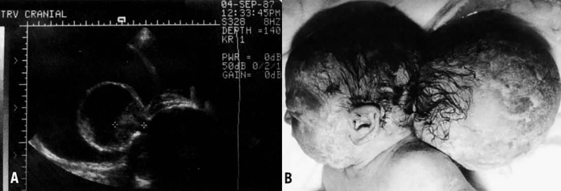
Fig. 3. A. An encephalocele diagnosed by ultrasound. Notice the cursors identifying the small amount of extruded brain tissue through the cranial defect. B. The infant after delivery.

A disruption of the vertebral arches often accompanied by underlying spinal cord defects is collectively called spinal dysraphism or spina bifida (Fig. 4). They are classified as spina bifida occulta if the disruption involves only bony structures and spina bifida cystica if there is a saccular defect involving neural elements. Meningomyeloceles constitute 90% of spina bifida and are composed of neural tissue covered by meninges that extrude through the vertebral column. Alternatively, a fluid filled sac (not containing neural elements) covered by meninges that protrudes through the bony defect is called a meningocele. Although spinal dysraphism can occur at any region of the vertebral column, the most common site for these defects is the lumbosacral area.

Fig. 4. Spina bifida by ultrasound (A) and after delivery (B).

Spina bifida is frequently accompanied by the Arnold-Chiari malformation. This anomaly results from a downward displacement of the medulla, fourth ventricle, and cerebellum through the foramen magna into the region of the cervical spine. This downward displacement of the hindbrain can hinder the egress of cerebrospinal fluid from the brain, causing an enlargement of the ventricles. This accounts for the 70% to 90% incidence of hydrocephalus associated with spina bifida.10

Other, less common defects include exencephaly (i.e. exteriorization of an abnormally formed brain) and iniencephaly (i.e. defect of the skull base, cervical spine, and underlying neural tissue). Myeloschisis, usually seen as an early fetal defect, describes an open flat neural plate that may be extensive.11 Myelodysplasia, or occult spinal dysraphism, describes less obvious malformations of the cord resulting from maldevelopment of the caudal region of the neural tube. These defects are often associated with lipomas or cutaneous changes overlying the region such as dimpling, sinus tracts, or hairy patches. These probably result from an embryologic mechanism similar to NTDs and may be associated with neurologic or orthopedic disabilities. Schut and coworkers12 have extensively reviewed the clinical aspects and variations of these defects.

Etiology, Incidence, and Recurrence Risks

Eighty-five percent of NTDs occur by multifactorial inheritance, a genetic predisposition from an interplay between various genes and environmental factors. The etiologic heterogeneity of NTDs was best illustrated by Holmes and coworkers, who reported that, of 106 liveborn or stillborn infants with an NTD, about 12% had identifiable causes.13 A small proportion of NTDs occur because of single-gene disorders, chromosomal aneuploidy, and teratogen exposure. Meckel syndrome is the most common of the single-gene disorders associated with a NTD. This autosomal recessive syndrome includes posterior encephalocele, polydactyly, cleft palate, and cystic dysplasia of the kidneys. Because the recurrence risk for such a defect is 25%, it underscores the need for careful evaluation of all infant with NTDs, because recurrence risks depend on the cause of the malformation.

Chromosomal aneuploidy also accounts for a small percentage of NTDs. Of the various types of NTDs, encephaloceles and spina bifida are more likely to be associated with triploidy; with trisomies 13, 18, and 21; and with various unbalanced translocations. The recurrence risk for these disorders varies with the mechanism responsible for the aneuploidy. For example, the recurrence risk for a trisomy is approximately 1%,, and triploidy is thought to be a sporadic event with a negligible recurrence risk. Recurrence estimates for translocations depend on the specific nature of the translocation and whether they are maternally or paternally transmitted.

Several teratogens have been implicated in the cause of NTDs. Two anticonvulsant medications in current use, carbamazepine and valproic acid, have been demonstrated to cause these defects. Robert and Guibaud14 originally reported an association between valproic acid and NTDs, noting a 1% risk for NTDs in patients taking this medication. This observation has been substantiated in several animal models.15–17 Carbamazepine also is associated with a 1% risk of spina bifida.18 Children of mothers with insulin-dependent diabetes mellitus have a 1% to 2% risk of NTD and a twofold to threefold (4% to 9%) increased incidence of congenital malformations compared with the general population.19 Although glycemic control may not be the sole etiologic factor in malformations in infants of diabetic women, careful preconceptional control is believed to decrease the prevalence of NTDs and other anomalies in these patients. The causes of NTDs are listed in Table 1.

TABLE 1. Causes of Neural Tube Defects

  Multifactorial inheritance
  Single-gene (autosomal recessive) disorders

  Meckel syndrome (most common)
  Robert syndrome
  Jarcho-Levin syndrome
  Median facial cleft syndrome
  HARDE (Walker-Warburg) syndrome
  Oculo-auriculo-vertebral (Goldenhar) syndrome


  Chromosomal aneuploidy

  Trisomy 18
  Trisomy 13
  Trisomy 21
  Triploidy
  Unbalanced translocations, markers, ring chromosomes


  Teratogens

  Valproic acid
  Carbamazepine
  Aminopterin
  Thalidomide


  Amniotic band sequence
  Cloacal extrophy
  Sacrococcygeal teratoma
  Maternal insulin-dependent diabetes mellitus

NTDs are the second most common fetal malformation in the United States, surpassed only by congenital heart defects. The incidence of NTDs varies with race, geographic location, and various other predisposing factors. In the United States, the incidence is approximately 1 to 2 cases per 1000 live births, whereas the incidence in Britain is about four times greater. Families who have had a child with an NTD have a 10-fold increase in their recurrence risk. In the United States, a family with an affected child has a 2% recurrence risk of another child with an NTD. If the defect in the first affected pregnancy was anencephaly, the family has a higher risk for recurrence of anencephaly than for recurrence of spina bifida. The risks for other affected U.S. populations are listed in Table 2. Between 90% and 95% of NTDs occur in families without a prior family history of an NTD.

TABLE 2. Estimated Incidence of Neural Tube Defects Based on Specific Risk Factors in the United States


Population

Incidence/1000 Live Births

Mother as reference


 General incidence

1.4–1.6

 Women undergoing amniocentesis for advanced maternal age

1.5–3.0

 Women with diabetes mellitus

20

 Women on valproic acid in first trimester

10–20

Fetus as reference


 One sibling with NTD

15–30

 Two siblings with NTD*

57

 Parent with NTD

11

 Half sibling with NTD

8

 First cousin (mother's sister's child)

10

 Other first cousins

3

 Sibling with severe scoliosis secondary to multiple vertebral defects

15–30

 Sibling with occult spinal dysraphism

15–30

 Sibling with sacrococcygeal teratoma or hamartoma

15–30


NTD, neural tube defect.
*Risk is higher in British studies. Risk increases further for three or more siblings or combinations of other close relatives.
Main DM, Mennuti MT: Neural tube defects: Issues in prenatal diagnosis and counseling. Obstet Gynecology 67:1–15, 1986.

Screening and Diagnostic Tests

ALPHA-FETOPROTEIN LEVELS.

In 1972, Brock and Sutcliffe measured AFP in the amniotic fluid of 31 pregnancies with anencephaly and 6 pregnancies with spina bifida, hydrocephaly, or both conditions.21 All of the cases of anencephaly and most of the spina bifida cases before 30 weeks' gestation demonstrated amniotic fluid AFP levels that were markedly elevated during pregnancy. When the fetus has an open (not skin covered) NTD, AFP leaks from the fetal circulation into the amniotic fluid. In 1974, Wald and coworkers performed a case-controlled study comparing maternal serum AFP levels in seven pregnancies with open NTDs with 14 control pregnancies matched for maternal age, parity, and gestational age.21 Maternal serum AFP levels in the affected pregnancies were significantly higher than those of the control population. This led to the hypothesis that there would be a role for measuring MSAFP in screening for NTDs. The U.K. Collaborative studydemonstrated the utility of this test for prospective open NTD screening in 1977.7

In anencephaly, the malformed skull is not completely covered by overlying skin, and it is therefore the lesion most accurately detected with MSAFP screening. More than 90% of anencephaly cases can be detected by MSAFP screening, and 99% can be detected by ultrasound examination. Approximately 99% of anencephaly cases can also be detected by amniotic fluid AFP and acetylcholinesterase (AChE) testing. In contrast, most encephaloceles are skin covered and therefore are less likely to be identified by MSAFP screening or amniocentesis and are most often detected by ultrasound.

Spina bifida and anencephaly occur with equal frequency. Approximately 80% of spinal cord defects are open—the tissue overlying the defect is not skin covered. The remainder of spinal cord defects are covered by skin or by a thick membrane and are not detectable by screening. In general, MSAFP screening programs detect approximately 85% of open fetal NTDs: 80% of open spina bifida and 90% of anencephaly. Almost all of these open lesions can then be diagnosed by amniotic fluid testing. The object of any screening program is to maximize detection at an acceptable false-positive rate. A screening test cutoff point is a balance between these two factors. The correct MoM value for MSAFP can only be calculated after all the appropriate information regarding the patient is taken into account. This includes weight (at the time the blood sample was obtained), gestational age, and race and considers whether the patient has insulin-dependent diabetes mellitus. An MSAFP level is considered elevated if the value is greater than 2.0 or 2.5 times the median value (2.0 or 2.5 MoMs) for normal controls at the same week of gestation.

MSAFP screening is most accurate when performed between 16 and 18 weeks' gestation, but testing can be performed between 15 and 22 weeks. Screening earlier or later than the optimal gestational age decreases the sensitivity of the test. Screening should be voluntary and should be performed after the patient has been fully informed regarding the benefits and limitations of the test. The patient should understand that a normal MSAFP result does not ensure a child without an abnormality (including an NTD), and that an elevated MSAFP level does not specifically diagnose an abnormality. Instead, an elevated value places the patient in a high-risk group that necessitates further evaluation. The most common causes of false-positive and false-negative MSAFP results are listed in Table 3.

TABLE 3. Common Causes of False-Positive and False-Negative Maternal Serum Alpha-Fetoprotein Levels

  False-Positive Levels
  Inaccurate gestational dating (patient has a more advanced gestation than estimated)
  Multiple gestation
  Race (black patients have higher levels than white patients)
  Underweight patients (less than 90 pounds)
  Spontaneous fetal to maternal bleeding
  False-Negative Levels
  Inaccurate gestational dating (patient has less advanced gestation than estimated)
  Maternal insulin-dependent diabetes mellitus
  Obesity

Consider the hypothetical example given with the protocol in Figure 5. A cohort of 10,000 consecutive women present for MSAFP screening with a level of risk comparable to the U.S. population. About 10 to 15 of these pregnancies would be affected with an NTD. MSAFP screening would detect 8 to 12 of these defects. At a cutoff point of 2.0 MoMs, the false-positive rate for this test is 4%, but a screen positive test result would only imply a 3% risk of having a fetus affected with an open NTD. A positive screening test result increases these patients' risks from 1.5 per 1000 to 3 per 100. Conversely, 97% of pregnancies with a positive screening test result are unaffected. If a screening cutoff point of 2.5 MoM is used, the false-positive rate is approximately 2%.

Fig. 5. Anticipated results for MSAFP screening of 10,000 prenatal patients.(Adapted from Haddow JE: Screening for spinal defects. Hosp Pract 17:128–138, 1982.)

AMNIOTIC FLUID ALPHA-FETOPROTEIN AND ACETYLCHOLINESTERASE.

Amniocentesis is often used to differentiate the disorders responsible for a maternal serum AFP elevation. If there is an amniotic fluid AFP elevation, a secondary test for the presence or absence of the AChE enzyme by gel electrophoresis is performed on the fluid. AChE is not normally identified in amniotic fluid. Tissues containing AChE are red blood cells, muscle, and neural tissue. Concentrations of AChE are much higher in fetal cerebrospinal fluid than in fetal serum. If the fetus has an open NTD, amniotic fluid AFP and AChE are usually both elevated and the high concentration of AChE in cerebrospinal fluid transudates across the defect into the amniotic fluid. AChE is a sensitive test for confirming an open NTD.

Fetal blood contamination is the most common source of falsely elevated AFP levels in amniotic fluid, and the amniocentesis performed to obtain the sample is the most common cause of fetal blood in the fluid. In such cases, amniotic fluid AFP is usually in the 3 to 5 standard deviation range. AChE is not detected in 90% of cases because of the relatively low AChE concentrations in the fetal blood. In congenital (Finnish) nephrosis, a rare autosomal recessive disorder, amniotic fluid AFP levels may be very high, and AChE is not identified.

At the time of amniocentesis for elevated MSAFP, karyotype analysis should also be performed regardless of the amniotic fluid AFP result. Omphaloceles and NTDs are both associated with chromosomal aneuploidy. Even when the amniotic fluid AFP level is normal, the addition of chromosome analysis allows more informative counseling regarding perinatal outcome.

ULTRASONOGRAPHIC DETECTION.

Ultrasound is an integral part of the management of patients with an elevated MSAFP level. It should be used as part of the initial fetal evaluation to exclude improper gestational dating, multiple gestation, and fetal demise. It can also identify other reasons for an elevated MSAFP value (Table 4). Although amniocentesis is usually performed to explain elevated MSAFP levels, a number of investigators have suggested that high-resolution ultrasound alone is an acceptable alternative for diagnostic evaluation of elevated MSAFP levels, particularly those in the range of 2.0 to 3.0 MoM.24,25 However, the accuracy of NTD diagnosis may be limited by the location or extent of the lesion, fetal position, quality of the images, and experience of the sonologist.

TABLE 4. Other Abnormalities Identified by the Alpha-Fetoprotein Screening Process

  Ventral wall defects

  Omphalocele
  Gastroschisis


  Triploidy
  Trisomies: 18, 13, 21
  Unbalanced translocations
  Amniotic band sequence
  Pentalogy of Cantrell: omphalocele, lower sternal defect, deficiency of diaphragmatic pericardium, intracardiac abnormality, anterior diaphragm defect
  Renal agenesis
  Fetal demise
  Multiple gestation
  Congenital nephrosis (Finnish type)
  Sacrococcygeal teratoma
  Dermatologic disorders
  Epidermolysis bullosa
  Congenital icthyosiform erythroderma
  Chorioangioma
  Maternal hepatoma
  Maternal ovarian teratoma

The use of ultrasound to detect spina bifida was addressed by Platt and colleagues through the California Maternal Serum Alpha-Fetoprotein Screening Program.26 Through this program, all patients with an elevated MSAFP level were offered an ultrasound evaluation with an experienced sonographer at one of 20 specialized prenatal diagnosis centers in the state of California. Of the 161 cases of spina bifida identified, 148 (91.9%) were detected by ultrasound. Because this detection rate at experienced prenatal diagnosis centers is well below 100%, in the absence of an ultrasonographic finding, amniocentesis for measurement of amniotic fluid AFP is recommended.

Three routine transverse views of the cranium are recommended by Nyberg.27 The biparietal diameter involves obtaining a transverse view of the fetal skull at the level of the thalami; this view includes the cavum septum pellucidi and the frontal horns of the lateral ventricles. The transventricular view, above the plane of the biparietal diameters (BPD), contains the lateral ventricles and choroid plexus. Both views are essential in imaging the ventriculomegaly that is found in 80% of NTD cases. The transcerebellar view, obtained by angling through the posterior fossa, demonstrates the cisterna magna and the cerebellum. The fetal spine should be imaged in sagittal, transverse, and coronal axes to detect the presence of an NTD and any disruption in the overlying soft tissues.

Several cranial ultrasound markers are useful in identifying NTDs. Nicolaides and coworkers28 first described the “banana” and “lemon” signs in a retrospective study of 70 patients with open spina bifida between 16 and 24 weeks' gestation. In 100% of cases, the lemon sign was present, whereas in cases for which the posterior fossa could be evaluated, 95% had an absent or banana-shaped cerebellum. The lemon sign (Fig. 6) is caused by a scalloping of the frontal bones secondary to decreased intracranial pressure from extrusion of the NTD. The cerebellar or banana sign is a consequence of the Arnold-Chiari malformation. It is attributed to a downward displacement of the cerebellum that is caused by protrusion of the neural contents through the foramen magnum, which results from the NTD. With advancing gestation there can be a further herniation of the brain through the cisterna magna with an inability to image the cerebellum.

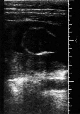
Fig. 6. Biparietal diameter with the lemon sign at 15 weeks' gestation.

Other investigators have suggested that the presence of the lemon sign is related to gestational age.29 Among 50 cases with open spina bifida, the lemon sign was found in 24 (89%) of 27 fetuses examined before 24 weeks, in 8 (50%) of 16 fetuses between 24 and 34 weeks, and in none of the fetuses at 35 weeks' gestation or more. Van den Hof and colleagues30 prospectively evaluated 130 fetuses with open NTDs. The lemon sign was present in 98% of fetuses at less than 24 weeks' gestation but found only in 13% of fetuses at more than 24 weeks' gestation. Cerebellar abnormalities were present in 95% of fetuses irrespective of gestational age. Growth retardation and ventriculomegaly significantly worsened with gestation, but the head circumference remained disproportionately small throughout pregnancy. Loss of the lemon sign may occur because of compensatory bony changes as gestation increases or because of cerebral ventriculomegaly that may compensate for the loss of brain through the cisterna magna.

Fetuses with open spina bifida have smaller BPDs by approximately 2 weeks' gestation than expected for their gestational age. This finding increases the detection rate for NTDs. If the gestational age is based on BPD alone (not a composite of BPD and femur length), the MSAFP MoM is increased when interpreted 2 weeks earlier than the correct gestational age.31,32 Between 16 and 18 weeks, the detection of an open NTD increases to 90% at a screening cutoff of 2.5 MoMs when gestational age as determined by BPD measurement is used to interpret the result.

Back to Top
MANAGEMENT

Counseling About Morbidity and Mortality

Thorough counseling of patients who have an NTD identified in an ongoing pregnancy is essential. Because anencephaly is uniformly fatal, the most important aspect of counseling is identifying the cause for the purpose of accurate recurrence risk counseling and preparation of the parents for the loss of their child at or shortly after birth. In a review of 181 liveborn infants with anencephaly, 40% were alive at 24 hours of age, and 5% lived to 1 week of age.33 Encephalocele, although a closed lesion, is a serious condition with a mortality rate of 60% to 75% during the first year.

The disabilities among survivors with spina bifida are accounted for by the location and extent of the lesion and the presence or absence of hydrocephaly. In general, because neural function is interrupted distal to the lesion, the higher the lesion, the greater is the neurologic deficit. If a patient survives with an NTD, the major morbidities include developmental delay and the ability to ambulate and maintain continence. Eventual outcome for the child varies with perinatal management and availability of support services.

Althouse and Wald evaluated an unselected series of 213 patients born in Britain with spina bifida (including encephaloceles) between 1965 and 1972.34 Their data reflect the natural history of these lesions before the advent of MSAFP screening. The 5-year survival rate for all patients was 36% for those with open lesions, 60% for those with closed lesions, and 18% for those with lesions that could not be classified. Closed cranial lesions (i.e. occipital meningomyelocele or encephalocele) were more commonly associated with severe handicap (75%) than were closed spinal lesions (23%).

Bamforth and Baird performed a population based study of patients with both spina bifida and hydrocephalus and compared life expectancy between these cases, which was ascertained from 1962 to 1970 and compared with the group from 1970 to 1986.35 At least 60% of patients had serious disabilities. These included a cerebrospinal fluid shunt (28%), neurogenic bladder (23%), congenital hip dislocation (23%), talipes equinovarus (23%), spasticity (15%), urinary obstruction (5%), and scoliosis (3%), developmental delay (6%), seizures (2%), and blindness (1%). The group of patients ascertained between 1970 and 1986 had a dramatic improvement in the probability of survival to the first birthday. There was no difference in survival the two cohorts between the ages of 7 and 16 years.

The best outcomes were reported by Hunt and coworkers.36 Between 1963 and 1971, 117 consecutive infants with open spina bifida were followed to their 16th birthday. All had surgical repair within 48 hours of life. The overall survival rate was 60%. Fifty percent of patients could ambulate more than 50 yards, 25% of patients were continent, and 70% of patients had an IQ of more than 80. If the lesion was at L3 or below, 75% of patients survived. Of the survivors, 90% of patients could ambulate more than 50 yards, 45% were continent, and 80% of patients had an IQ higher than 80.

Obstetric Management

Patients who have had prenatal diagnosis of an NTD can have more accurate genetic counseling after a fetal karyotype and a detailed ultrasound, including fetal echocardiography, are performed to look for other structural defects. If the parents elect to continue the pregnancy, serial ultrasound examinations to look for the presence or exacerbation of hydrocephalus are indicated. Prenatal diagnosis also informs the neurosurgeons and pediatricians who will care for the child after delivery. Information regarding support groups may also be initiated during pregnancy.

The mode of delivery for patients with NTDs has been addressed by Luthy and coworkers.37 In their retrospective analysis, infants who were born before the onset of labor had better motor function than those undergoing labor before cesarean section or spontaneous vaginal delivery. The infants delivered by cesarean section without labor retained greater neurologic function at an average of 3.3 spinal segments above those born after labor, regardless of the ultimate type of delivery. Labor did not affect intellectual performance. Concerns with this study include ascertainment bias of lesions identified antenatally that might have resulted in altered obstetric management because the study was not prospectively randomized.

Preventative Therapy

It has been observed that lower socioeconomic classes have a predisposition for NTDs, leading to the theory that nutritional deficiency may be a causative factor. In 1976, Smithells and coworkers38 found decreased levels of folate, ascorbate, and riboflavin in lower socioeconomic groups. In 1981, the same investigators recruited women who had given birth to infants with an NTD to begin vitamin supplementation before conception and to continue it for 8 weeks after conception. Because their original proposal for a double-blind, randomized control trial was rejected, they modified their study to include women who were fully or partially supplemented with prenatal vitamins and a group of unsupplemented women who presented at the end of the first trimester. The unsupplemented mothers had a 4% recurrence of NTDs, but the partial and fully supplemented study group had a 0.4% to 0.5% recurrence risk, respectively.39 This experience was verified by their continuing work in 1983.40

In 1981, the first randomized trial of folic acid supplementation (4 mg/day) was reported and yielded inconclusive results when analyzed according to randomly allocated treatment group.41 In 1991, a landmark study substantiating the prevention of NTDs by folate supplementation was reported by the Medical Research Council Vitamin Study Research Group.42 This prospective, randomized, double-blind study demonstrated that women who had prior pregnancies with isolated NTDs had a 72% reduction of a recurrence of the NTD when supplemented with 4 mg of folate per day at least 4 weeks before conception through the 12th week of gestation.

After it had been determined that folic acid supplementation could decrease the recurrence risk, attention was then directed toward evaluating women to prevent the first occurrence of an NTD. In 1992, Czeizel and colleagues conducted a randomized, controlled trial in Hungary in which 7540 women were given a multivitamin tablet containing 0.8 mg of folic acid or a placebo containing trace elements before conception.43 Of these, 4753 achieved a pregnancy, with outcomes available for for 2104 women who received vitamin supplementation and 2052 women who received the placebo. There were six affected pregnancies in the placebo group and none in the supplemented group (p = 0.029), demonstrating the protective effect of folic acid in the prevention of first occurrence of NTDs.

The United States Public Health Service has recommended that all women of childbearing age should be supplemented with 0.4 mg/day of folic acid.44 This may be achieved through diet, ingestion of foods fortified with folic acid (e.g. fortified breakfast cereals), or by ingesting a vitamin supplement. Along with the American College of Obstetricians and Gynecologists,45 they also recommend that women previously diagnosed with a fetus affected with an NTD be supplemented with 4 mg of folic acid per day. Supplementation should begin at least 1 month before conception and should be continued for the first 3 months of pregnancy. At the University of Pennsylvania, our preferred supplementation regimen for high-risk patients consists of one prenatal vitamin with a 1-mg tablet of folic acid in the morning and two 1-mg folic acid tablets in the afternoon or evening. They are advised to avoid taking four prenatal vitamins per day, because that may cause an overdosing of vitamin A, which can be teratogenic.

Women carrying affected fetuses have lower plasma levels of folic acid and vitamin B12.46 Folic acid appears to prevent NTDs by helping the metabolism of the amino acid homocysteine. Methionine synthetase is the only known enzymatic pathway that uses the cofactors of B12 and folate to metabolize homocysteine to methionine. Mills and coworkers obtained blood from 81 pregnant women carrying affected fetuses and 323 women with normal-appearing gestations and assayed for homocysteine levels, B12, and plasma and red cell folate levels.47 The women carrying affected pregnancies had significantly higher homocysteine levels than matched controls. This metabolic mechanism appears to account for a large percentage of NTDs and can be overcome by supplementation with B12 and folate.

Questions remain regarding the lowest effective dosage of folate supplementation in the occurrence and recurrence populations and the optimal duration of supplementation before and after conception. Patients need to be counseled regarding vitamin supplementation but also need to be aware that NTDs have a multifactorial cause and can occur despite vitamin therapy. Patients with a previous affected offspring still need genetic counseling, ultrasound examination, and MSAFP screening to evaluate a new pregnancy for a recurrence.

Other Benefits of Screening

The association between elevated level of MSAFP and NTDs is not specific for NTDs but is observed in a variety of other malformations and clinical conditions (Fig. 2). The potential for MSAFP screening to reduce fetal morbidity and mortality is substantial. Ventral wall defects are the second major group of open fetal anomalies identifiable by MSAFP screening (Table 2). Omphaloceles and gastroschises are the two major types of ventral wall defects. They have different causes and different risks for morbidity and mortality.

Omphaloceles are midline defects with the umbilical cord insertion at the center of the lesion. They result from an abnormality in body folding between days 22 and 28 of fetal development. Up to 60% of fetuses with omphaloceles have additional malformations. About 12% of them have karyotypic abnormalities, and most of these are trisomies (i.e. 13, 18, and 21) or triploidy. Omphaloceles are also associated with several single-gene disorders such as Meckel syndrome and Beckwith-Wiedemann syndrome. Because these lesions are usually covered by peritoneum, they are less often detected by MSAFP screening than gastroschisis. Intraabdominal organs other than bowel can herniate into the omphalocele and can affect outcome. Hepatic herniation has the poorest prognosis, often leading to cardiopulmonary insufficiency. Because other malformation syndromes are associated with this defect, targeted ultrasound, fetal echocardiography, and chromosome analysis are recommended when an omphalocele is detected.

Gastroschisis occurs between days 28 and 32 of development and is thought to be caused by a vascular disruption sequence. It has been suggested that the vessel responsible is the omphalomesenteric artery or the right umbilical vein. These lesions occur sporadically without a clearly defined cause. They are usually found to the right of the midline with free floating bowel in the amniotic fluid. Morbidity and mortality is substantially lower with gastroschisis than with omphalocele. Gastrointestinal atresia and “short gut” syndrome is the most serious complication of these defects. In many cases, these lesions can be readily classified by an experienced sonographer. Although gastroschisis usually is not associated with chromosomal abnormalities, rupture of the omphalocele sac can mimic gastroschisis. Amniocentesis and fetal echocardiography are indicated when gastroschisis is suspected.

Amniotic fluid AFP levels can be elevated in cases of ventral wall defects, and AChE testing also detects about 75% of ventral wall defects. AChE is present in nearly all cases of gastroschisis and about 60% of omphaloceles. The exposed splanchnic bed has been suggested as the source of amniotic fluid AChE in ventral wall defects. The diagnosis of ventral wall defects can be further elucidated by a ratio of AChE to pseudocholinesterase (PChE). PChE, another cholinesterase enzyme, is normally present in high concentrations in the fetal serum and in lesser amounts in the amniotic fluid. The abdominal wall defect allows more PChE to transudate into the fluid. This enzyme is also detected by gel electrophoresis and appears as a slower moving band than AChE. The ratio of AChE to PChE detects nearly all abdominal wall defects, but it cannot distinguish false-positive results due to intraamniotic fetal bleeding, because the ratio is elevated with any type of fetal bleeding. Other causes of elevated AChE and AFP values in amniotic fluid include excess fetal bleeding, fetal demise (singleton or twin), and placental or umbilical cord hemangiomas.

Cystic hygromas are caused by jugular lymphatic obstruction resulting in nuchal cyst development and are not detected by MSAFP screening because they are often closed lesions. These structures are usually located in the occipital-cervical region and are often septated. Elevated amniotic fluid AFP values found in these lesions are caused by spontaneous rupture of the cyst or inadvertent puncturing of the cystic hygroma during amniocentesis. Most of these lesions carry a poor prognosis and are associated with karyotypic abnormalities such as 45,X or trisomy 21, but they can also be found in disorders such as pterygium colli or Noonan syndrome. An amniocentesis for fetal karyotype and genetic counseling is recommended when fetal cystic hygroma is detected.

In 1984, Merkatz and coworkers reported that MSAFP levels were about 25% lower in Down syndrome pregnancies than in normal controls.48 This was subsequently confirmed by Cuckle and associates, who suggested that MSAFP could be used as screening test for Down syndrome.49 Several prospective studies have validated this work.50,51 Although the incidence of fetal Down syndrome increases with advancing maternal age, only about 20% of Down syndrome pregnancies occur in women who are older than 35 years of age. Because the association of low MSAFP values and Down syndrome are independent of maternal age, these two factors can be used to modify a patient's risk for Down syndrome. The median MoM for a woman carrying a Down syndrome pregnancy is about 0.8 MoM compared with control pregnancies for each week of gestation (Fig. 2). There is much overlap between MSAFP distribution for the affected and unaffected pregnancies at each week of gestation. It is not practical to establish a cutoff point to discriminate two separate populations, as is done for NTD screening. Instead, the patient's risk is calculated using maternal age and AFP level and is usually interpreted as screen positive when the patient's risk reaches that of a 35-year-old woman (i.e. a 1:270 mid-trimester risk). This approach identifies approximately 20% of Down syndrome fetuses in the population of pregnant women younger than 35 years, with a positive (or amniocentesis) rate of about 5% to 6%. Although it is common practice to repeat the assessment for elevated MSAFP values, low values should not cause repeat examinations when gestational age is correct, because they tend to regress to the mean, which leads to a falsely reassuring reduction of risk. Because MSAFP increases as pregnancy advances, the second value should be higher than the first and therefore may be less accurate.

Given the association of low MSAFP levels and Down syndrome, other serum markers have been investigated to develop better screening tests for fetal aneuploidy. Using the hypothesis that decreased secretion or synthesis of AFP by the fetal liver accounts for the lower levels in Down syndrome pregnancies, other serum markers associated with fetal liver function have been studied. Unconjugated serum estriol is a steroid product of the fetal-maternal unit that requires participation of the fetal liver for production. Canick and coworkers reported that serum estriol levels in 22 women carrying Down syndrome pregnancies were reduced compared with controls.52 Subsequently, Wald and coworkers studied 77 Down syndrome pregnancies and confirmed that unconjugated serum estriol was reduced by 27% compared with controls. This reduction was independent of AFP level and maternal age and therefore could be incorporated into a screening test for Down syndrome with MSAFP.53

In 1984, Chard and coworkers suggested a relation between human chorionic gonadotropin and biochemical screening for Down syndrome.54 Subsequently, Bogart and colleagues reported that human chorionic gonadotropin levels were significantly higher in aneuploid pregnancies than in unaffected pregnancies.55 Human chorionic gonadotropin combined with AFP, unconjugated estriol, and maternal age are being evaluated as maternal serum screening tests for Down syndrome. The combination of these tests detects about 60% of Down syndrome cases, with an amniocentesis rate of about 5%.56 These serum markers detect this high percentage of Down syndrome cases and about 40% to 60% of trisomy 18 cases, with a false-positive rate of about 0.4%.57 This combined biochemical screening program tests for trisomy 21 and 18 while detecting 85% of NTDs because AFP is one component of the screening test.

Although no randomized clinical trials have been performed to demonstrate their validity, many other serum markers are being investigated to increase the detection rate for fetal Down syndrome. However, the only serum marker effective in the detection of open fetal defects is AFP, and no other markers have been identified that achieve a detection rate of NTDS comparable to AFP.

Back to Top
CONCLUSIONS

MSAFP screening is a major advance in the prenatal detection of many fetal anomalies. The use of other markers in conjunction with AFP screening is being investigated. However, MSAFP remains the primary test for population screening of fetal anomalies. Implementation of MSAFP screening in an obstetric practice must be undertaken in conjunction with capable support services that include vigilant laboratory quality control, competent counseling of abnormal test results, and adequate confirmatory procedures such as targeted fetal ultrasound, amniocentesis, and cytogenetic analysis. Patient participation in an MSAFP screening program should be voluntary. Because MSAFP is an imperfect test with false-negative and false-positive results, a thorough understanding of the principles and pitfalls of MSAFP is essential.

Back to Top
REFERENCES

1. Bergstrand CG, Czar B: Demonstration of a new protein fraction in serum from the human fetus. Scan J Clin Lab Invest 8: 174, 1956

2. Harper ME, Dugaiczyk A: Linkage of evolutionary-related serum albumin and alpha-fetoprotein genes within 911-22 of human chromosome 4. Am J Hum Genet 35: 565, 1983

3. Faucett WA, Greenberg F, Rose E et al: Congenital deficiency of alpha-fetoprotein (abstract). Am J Hum Genet 45: 1019A, 1989

4. Haddow JE, Palomaki G, Knight GE et al: Prenatal screening for major fetal disorders.

5. Los FJ, Debruijn HWA, Van Beek Calkoen-Carpay T et al: AFP transport across the fetal membranes in the human. Prenat Diagn 5: 277, 1985

6. Habib A: Maternal serum alpha-fetoprotein: Its value in antenatal diagnosis of genetic disease and in obstetrical-gynecologic care. Acta Obstet Gynecol Scand Suppl 61: 14, 1977

7. UK Collaborative Study on Alpha Fetoprotein in Relation to Neural Tube Defects: Maternal serum alpha-fetoprotein measurement in antenatal screening for anencephaly and spina bifida in early pregnancy. Lancet 2:1323–1332, 1977

8. Haddow JE, Palomaki G, Knight GJ et al: Prenatal screening for major fetal disorders. In Haddow JE (ed): Fetal Disorders Associated With Elevated MSAFP Levels, vol 1, p 4. Scarborough, ME: Foundation for Blood Research, 1990

9. Van Allen MI, Kalousek DK, Chernoff GF et al: Evidence for multisite closure of the neural tube in humans. Am J Med Genet 47; 723– 743, 1993

10. Leonard CO, Freeman JM: Spina bifida: A new disease. Pediatrics 68: 136, 1981

11. Osaka K, Tanimura T, Hirayama A et al: Meningomyelocele before birth. J Neurosurg 49: 711, 1978

12. Schut L, Pizza EJ, Bruce DA: Occult spinal dysraphism. In McLaurin RL (ed): Myocele. New York: Grune & Stratton, 1977

13. Holmes LB, Driscoll SG, Atkins L: Etiologic heterogeneity of neural tube defects. N Engl J Med 294: 365, 1976

14. Robert E, Guibaud P: Maternal valproic acid and congenital neural tube defects. Lancet 2: 937, 1982

15. Bruckner A, Lee YJ, O'Shea KS, Henneberry RC: Teratogenic effects of valproic acid and diphenylhydantoin on mouse embryos in culture. Teratology 27: 29– 42, 1983

16. Kao J, Brown NA, Schmid B et al: Teratogenicity of valproic acid: In vivo and in vitro investigations. Teratog Carcinog Mutagen 1: 367– 376, 1981

17. Fabro S, Schull G, Brown NA: The relative teratogenic index and teratogenic potency: Proposed components of developmental toxicity risk assessment. Teratog Carcinog Mutagen 2: 61– 76, 1982

18. Rosa FW: Spina bifida in infants of women treated with carbamazepine during pregnancy. N Engl J Med 324: 675– 677, 1991

19. Mills JL, Knopp RH, Simpson JL et al: Lack of relation of increased malformation rates in infants of diabetic mothers to glycemic control during organogenesis. N Engl J Med 318: 671– 676, 1988

20. Main DM, Mennuti MT: Neural tube defects: Issues in prenatal diagnosis and counselling. Obstet Gynecol 1: 67, 1986

21. Brock DJH and Sutcliffe RG: Alpha fetoprotein in the antenatal diagnosis of anencephaly and spina bifida. Lancet 2: 197, 1972

22. Wald NJ, Brock DJH, Bonnar J: Prenatal diagnosis of spina bifida and anencephaly by maternal serum alpha-fetoprotein measurement. Lancet 1: 767, 1974

23. Haddow JE: Screening for spinal defects. Hosp Pract 17: 128– 138, 1982

24. Gray D, Crane J, Johnson C: Ultrasound versus amniocentesis for evaluation of pregnancies at risk for neural tube defects. Am J Hum Genet 49 (Suppl 4): 1172A, 1991

25. Nadel AS, Green JK, Holmes LB et al: Absence of need for amniocentesis in patients with elevated levels of maternal serum alpha-fetoprotein and normal ultrasonographic examinations. N Engl J Med 323: 557– 561, 1990

26. Platt LD, Feuchtbaum L, Filly R et al: The California Maternal Serum Alpha-Fetoprotein Screening Program: The role of sonography in the detection of spina bifida. Am J Obstet Gynecol 1992:166:1328–1329

27. Nyberg DA: Recommendations for obstetric sonography in the evaluation of the fetal cranium. Radiology 172: 309, 1989

28. Nicolaides K, Campbell S, Gabbe S, Guidetti R: Ultrasound screening for spina bifida: Cranial and cerebellar signs. Lancet 2: 71– 74, 1986

29. Nyberg D, Mack L, Hirsh J, Mahony B: Abnormalities of the fetal cranial contour in sonographic detection of spina bifida: Evaluation of the “lemon” sign. Radiology 167: 387– 392, 1988

30. Van den Hof MC, Nicolaides KH, Campbell J, Campbell S: Evaluation of the lemon and banana signs in one hundred thirty fetuses with open spina bifida. Am J Obstet Gynecol 162: 322– 327, 1990

31. Wald NJ, Cuckle HS, Boreham J: Small biparietal diameter of fetuses with open spina bifida: Implications for antenatal screening. Br J Obstet Gynecol 87: 219, 1980

32. Wald NJ, Cuckle HS, Boreham J: Effect of estimating gestational age by ultrasound cephalometry on the specificity of alpha-fetoprotein screening for open neural tube defects. Br J Obstet Gynecol 89: 1050, 1982

33. Baird PA, Sadovnick AD: Survival in infants with anencephaly. Clin Pediatr 23: 268, 1984

34. Althouse R, Wald N: Survival and handicap of infants with spina bifida. Arch Dis Child 55: 845– 850, 1980

35. Bamforth SJ, Baird PA: Spina bifida and hydrocephalus: A population study over a 35 year period. Am J Hum Genet 44: 225– 232, 1989

36. Hunt GM: Open spina bifida: Outcome for a complete cohort treated unselectively and followed into adulthood. Dev Med Child Neurol 32: 108– 118, 1990

37. Luthy DA, Wardinsky T, Shurtleff et al: Cesarean section before the onset of labor and subsequent motor function in infants with meningomyelocele diagnosed antenatally. N Engl J Med 324: 662– 666, 1991

38. Smithells RW, Sheppard S, Schorah CJ: Vitamin deficiencies and neural tube defects. Arch Dis Child 51: 944– 950, 1976

39. Smithells RW, Sheppard S, Schorah CJ et al: Apparent prevention of neural tube defects by periconceptional vitamin supplementation. Arch Dis Child 56: 911– 918, 1981

40. Smithells RW, Seller MJ, Harris R et al: Further experience of vitamin supplementation for prevention of neural tube defect recurrences. Lancet 1: 1027– 1031, 1983

41. Laurence KM, James N, Miller MH et al: Double blind randomized controlled trial of folate treatment before conception to prevent recurrence of neural tube defects. Br Med J 282: 1509– 1511, 1981

42. Prevention of neural tube defects: Results of the medical research council vitamin study. Lancet 338:131–137, 1991

43. Czeizel AE, Dudas I: Prevention of the first occurrence of neural tube defects by periconceptional vitamin supplementation. N Engl J Med 327: 1832– 1835, 1992

44. Centers for Disease Control and Prevention: Recommendations for the use of folic acid to reduce number of cases of spina bifida and other neural tube defects. MMWR Morb Mortal Wkly Rep 41(RR14):1–7, 1992

45. American College of Obstetricians and Gynecologists. Folic acid for the prevention of neural tube defects. Committee opinion #20. Washington, DC: American College of Obstetricians and Gynecologists, 1993

46. Kirke PN, Molloy AM, Daly LE et al: Maternal plasma folate and vitamin B12 are independent risk factors for neural tube defects. Q J Med 1993;703–708

47. Mills JL, McPartlin JM, Kirke PN et al: Homocysteine metabolism in pregnancies complicated by neural tube defects. Lancet 345: 149– 151, 1995

48. Merkatz IR, Nitowsky HM, Macri JN et al: An association between low maternal serum alpha-fetoprotein and fetal chromosome abnormalities. Am J Obstet Gynecol 148: 886– 894, 1984

49. Cuckle HS and Wald NJ: Maternal serum alpha-fetoprotein measurement: A screening test for Down syndrome. Lancet 1: 926– 929, 1984.

50. Baumgarten A, Schoenfeld M, Mahoney M et al: Prospective screening for Down syndrome using maternal serum AFP. Lancet 1: 1280– 1281, 1985

51. New England Regional Genetics Group Prenatal Collaborative Study on Down Syndrome Screening: Combing maternal serum alpha-fetoprotein measurements and age to screen for Down syndrome in pregnant women under age 35. Am J Obstet Gynecol 160:575–581, 1989

52. Canick JA, Knight GJ, Palomaki GE et al: Low second trimester maternal serum unconjugated oestriol in pregnancies with Down syndrome. Br J Obstet Gynecol 95: 330, 1987

53. Wald NJ, Cuckle HS, Densen JW et al: Maternal serum unconjugated oestriol as an antenatal screening test for Down syndrome. Br J Obstet Gynecol 95: 334, 1987

54. Chard T, Lowings C, Kitau MJ: Alpha-fetoprotein and chorionic gonadotropin levels in relation to Down's syndrome. Lancet 2: 750, 1984

55. Bogart MH, Pandian MR, Jones OW: Abnormal maternal serum chorionic gonadotropin levels in pregnancies with fetal chromosome abnormalities. Prenat Diagn 7: 623– 630, 1987

56. Wald NJ, Cuckle HS, Densem JW et al: Maternal serum screening for Down's syndrome in early pregnancy. BMJ 297: 883– 887, 1988

57. Canick JA, Palomaki GE, Osathanondh R: Prenatal screening for trisomy 18 in the second trimester. Prenat Diagn 10: 546– 548, 1990

Back to Top