Chapter 5
Microinvasive Carcinoma of the Cervix
Edward W. Savage Jr, Groesbeck P. Parham and Michael L. Hicks
Main Menu   Table Of Contents

Search

Edward W. Savage, Jr, MD
Medical Director, and Professor and Chief, Division of Gynecology, King/Drew Medical Center, Los Angeles, California (Vol 4, Chap 5)

Groesbeck P. Parham, MD
Associate Professor and Chief, Division of Gynecologic Oncology, University of Arkansas for Medical Sciences, Little Rock, Arkansas (Vol 4, Chap 5)

Michael L. Hicks, MD
Assistant Professor, Division of Gynecologic Oncology, Henry Ford Hospital, Detroit, Michigan (Vol 4, Chap 5)

INTRODUCTION
CARCINOGENESIS
HISTOGENESIS
CYTOLOGY
COLPOSCOPY
PATHOLOGY
DIAGNOSIS
TREATMENT
CONCLUSION
REFERENCES

INTRODUCTION

Microinvasive carcinoma of the uterine cervix represents a stage in the continuum of cervical neoplasia that begins with intraepithelial neoplasia and proceeds to frankly invasive cancer. Because microinvasive carcinoma has not been well-defined, the management remains controversial. Part of the controversy stems from confusion of terms and a lack of detail in the description of the lesion. The need for standardization of nomenclature becomes obvious when one considers the many terms used interchangeably to describe microinvasive carcinoma:

  Microinvasive carcinoma
  Microcarcinoma
  Covert invasive carcinoma
  Incipient invasion
  Early invasive carcinoma
  Microscopic foci of stromal invasion
  Very small carcinoma
  Superficial invasion
  Minimal invasion
  Intraepithelial carcinoma with microinvasive foci
  Early invasive preclinical carcinoma
  Clinical stage IA1 or IA2

Definitions also vary considerably.1,2,3,4,5,6,7,8,9,10 The term was originally defined by Mestwerdt11 as a cancer with up to 5-mm penetration. While some authors such as Averette and associates12 have restricted their definition to invasion less than 1 mm below the basement membrane with no lymphatic or vascular involvement, Larson and colleagues13 are willing to tolerate a depth of invasion up to 8 mm. The Society of Gynecologic Oncologists proposed the following in 1974: "The microinvasive lesion should be defined as one in which neoplastic epithelium invades the stroma in one or more places to a depth of 3 mm or less below the base of the epithelium and in which lymphatic or blood vascular involvement is not demonstrated.”

No statement was made regarding confluence of the invasive foci, although several authors consider this to be an important diagnostic factor.1,3,14,15,16,17 The International Federation of Gynecology and Obstetrics (FIGO) has changed the definition of microinvasive squamous carcinoma of the cervix six times since 1961. In 1976, FIGO defined stage IA as "microinvasive carcinoma (early stromal invasion).”

Neither depth of invasion, confluence, nor lymphatic or vascular space involvement was included in this definition. The omission by FIGO of strict quantitative criteria permitted subjective interpretation and led to confusion as to the proper evaluation and treatment of this disease entity. Consequently, at the FIGO Cancer Committee Meeting in 1985, new proposals were finalized, published in 1987,18 and agreed on at the 1988 meeting of the Cancer Committee. Those modifications are as follows:

  Stage IA—Preclinical carcinomas of the cervix (i.e., those diagnosed only by microscopy)
  Stage IA1—Minimal microscopically evident stromal invasion
  Stage IA2—Lesions detected microscopically that can be measured. The upper limit of the measurement should not show a depth of invasion of more than 5 mm taken from the base of the epithelium, either surface or glandular, from which it originates. A second dimension, the horizontal spread, must not exceed 7 mm. Larger lesions should be staged as IB.

The choice of 5 mm and 7 mm as the maximum depth and lateral spread, respectively, was based on the following: (1) a three dimensional volumetric analysis of cervical carcinomas by Burghardt19 revealed that tumor volumes of less than 400 mm3 were unassociated with lymph node metastasis; and (2) the third dimension (length), although not measured, usually does not exceed the lateral diameter (width) by more than 50%. Thus, for a lesion that is 5 mm deep and 7 mm wide, the assumed maximum length would be 10.5 mm. The volume would thus equal 5 mm x 7 mm x 10.5 mm = 367.5 mm3.

Lesions with invasion up to 5 mm occur with various degrees of lateral spread. There is a direct correlation between the volume of tumor and its potential to metastasize. Consequently, a thoughtful definition of microinvasive carcinoma should consider other dimensions of the lesion besides its depth of invasion. Reports which support this view include the study by Simon and co-workers20 in which 125 patients with squamous cell carcinoma invading to no more than 5 mm displayed lateral spread of up to 20 mm, the median width being 4.8 mm. Similarly, Sedlis and associates21 found that 22% of 133 specimens with less than 4 mm of invasion had evidence of lateral spread ranging from 8 mm to 22 mm. In spite of FIGO's precise quantitative bidimensional definition, most pathologists only report the depth of invasion and many only render a descriptive analysis such as "early invasion” or "superficial invasion.”

Back to Top
CARCINOGENESIS

Many factors have been implicated in human cervical carcinogenesis, including smegma, hormones, viruses, social status, hygiene, infection, and early age at first sexual contact; the latter is compounded by numerous partners. Despite the accumulated epidemiologic evidence, the ultimate cause of cervical cancer remains unknown. Exogenous factors are more frequently implicated than endogenous factors, although it is probable that a particular relationship between the two is necessary for the development of cervical carcinoma.

In the past, the herpes simplex virus type 2 was thought to be the primary agent in cervical carcinogenesis.22,23,24,25 Since the 1980s, there has been a virtual explosion of information concerning the human papillomavirus as the etiologic agent of cervical cancer. Human papillomavirus (HPV) types 16, 18, 31, 33, and 35 are usually associated with invasive cervical cancer, as well as the more aneuploid dysplasias, while types 6, 11, 42, and 44 occur primarily in the lesser dysplasias and benign condylomas that have a polypoid DNA distribution. Since aneuploidy confers a substantial risk of progression and since polypoidy is associated with regression, it appears that the oncogenic risk of HPV infection is, among other things, dependent on the infecting viral strain as supported by the studies of Fu and colleagues,26 Crum and co-workers,27 Crum and Levine,28 Rastkar and associates,29 Stanbridge and Butler,30 Reid and colleagues,31 and Winkler and co-workers.32 That there is an association between HPV infection and the development of squamous carcinoma of the cervix is supported by recent findings that introduction of expression vectors for HPV E6 and E7 proteins in cultured mammalian cells can result in cell transformation.33,34 Possibly the mechanism of malignant transformation by the E6 and E7 proteins is explained by the fact that these two HPV viral proteins bind to the p53 tumor-suppressor gene.35,36 The loss of p53 tumor suppressor function is attributed to accelerated degeneration of the normal protein elicited by its binding to the viral E6 gene product of HPV types 16 and 18.37 In the rare instances in which both HPV DNA and a p53 mutation in the cellular genome are present, cervical cancers are particularly aggressive. Although p53 mutations are rare in cervical cancer, their occurrence has a negative effect on survival.38,39

Back to Top
HISTOGENESIS

Cervical intraepithelial neoplasia (CIN) is the term now used to encompass all epithelial abnormalities of the cervix. The epithelial cells are malignant but confined to the epithelium. Older terminology used dysplasia and carcinoma in situ to connote a two-tier disease process that, in the past, impacted on therapy. It was felt that if only dysplasia was present, no or limited treatment was needed. If carcinoma was diagnosed, in many instances a hysterectomy was recommended. This concept is now felt to be inappropriate, particularly since the cervical epithelium may be no thicker than 0.25 mm. Prospective studies strongly suggest that CIN is a continuum of changes from the earliest recognizable abnormality in the deep layers of the stratified squamous epithelium (CIN I or mild dysplasia) that, if all conditions are met, will gradually progress to involve the middle (CIN II) and superficial (CIN III) layers before invading the underlying stroma.

Loss of epithelial basement membrane is the sine qua non of an invasive neoplasm. Invasion is facilitated by adhesion, proteolysis, and migration.40 Adherence to basement membrane and subsequent release and activation of proteolytic enzymes permit tumor cells to invade into the underlying stroma and create sites for entry into and exit from lymphatics and blood vessels. The actual process of translocation across the basement membrane (tumor cell locomotion) may be due to tumor cell recognition and response to basement membrane proteins, chemotactic cytokines, and autocrine motility factors.40,41 Many physiologic cellular functions are necessary for cells to successfully traverse the barriers to invasion. These range from the autonomous ability to stimulate normal endothelium to form capillaries to feed and provide transportation for the tumor cells to the ability to enter that vascular access. Attachment to specific glycoproteins of the extracellular matrix, such as fibronectin, collagen, and laminin is mediated through tumor cell receptors.42,43,44,45 In an ultrastructural and immunohistochemical study of infiltration in microinvasive carcinoma of the uterine cervix, Kudo and associates46 used transmission electron microscopy to examine features of locally infiltrating lesions. Many pseudopod-like cytoplasmic protrusions of the cancer cells and abundant microfilaments parallel to the direction of the protrusion were seen. Concomitant with the disappearance of part of the basal lamina, many vesicles 70 nm to 90 nm in diameter were observed, suggesting a role for these vesicles in cancer infiltration. With the immunoperoxidase method, the distribution of fibronectin around the microinvasive lesions was noted. Fibronectin is a component of extracellular matrices and, presumably, in view of its action on cell adhesion, is a resistant factor against cancer cell infiltration. Fibronectin decreased in the transitional area between the cancer nest and the stroma during the stage of microinvasion.

In its earliest manifestations, invasion of the connective tissue stroma may be characterized by isolated cells, small circumscribed groups of cells, or irregularly shaped, fingerlike processes. At this time, the invasive epithelial masses are limited in their depth of penetration and are also discrete. Many cell types may be found in invasive pegs, sometimes with keratinization. Manifestations of degeneration or retrogressive changes are encountered in some of the transected invasive cell masses. Nuclear pyknosis or chromatolysis is frequently observed along with a shrinking of the nuclear membrane. Loss of the affinity of the cytoplasmic dyes and pronounced vacuolization of the cytoplasm may be observed. Occasionally, calcification is seen.

Back to Top
CYTOLOGY

Microinvasive carcinoma is a histologic diagnosis and depends on the extent of stromal invasion. The cytologist has no way to judge from the Papanicolaou smear the extent of stromal invasion. Therefore, the diagnosis of microinvasive carcinoma cannot be made cytologically. However, several studies in the past have suggested that a cytologic diagnosis of microinvasive squamous cancer of the cervix can be made with a high degree of accuracy. Ng47 noted that 65 of 66 microinvasive cancers had abnormal surface reactions including cellular and nuclear pleomorphism, disorganized cellular polarity, circumscribed foci of differentiation, presence of nucleoli, and keratinization. All of the changes were more conspicuous in the epithelium overlying the site of microinvasion and were more pronounced with infiltration. Surface ulceration was uncommon and limited infiltrations were more conspicuous as the depth of penetration increased. Cellular features found in microinvasive cervical carcinoma were as follows:

  Distribution (isolated sheets, synctia)
  Cellular configuration (polyhedral, round, oval, irregular)
  Chromatin pattern (uniformly finely granular, irregularly finely granular, irregularly coarsely granular, pyknotic)
  Nucleolus (total number, micronucleoli, macronucleoli, single or multiple).

When cellular findings were considered in relation to the extent of the epithelial infiltration, certain distinctive patterns were evident. The cellular features of lesions with an epithelial infiltration of 2.1 mm to 3 mm were the most distinctive. On the basis of cellular features, Ng was able to diagnose 27 of 31 (87.1%) cases of microinvasive cancers correctly. Of the four that were incorrectly diagnosed, two were microinvasive cancers with a depth of less than 3 mm, erroneously classified as severely dysplastic lesions. The other two were microinvasive carcinomas incorrectly classified as carcinomas with invasive depths greater than3 mm.

Rubio,48 in a retrospective review, was unable to confirm Ng's data. Instead, he found the numbers of abnormal cells and smears and the analysis of the various nuclei and cytoplasmic characteristics were essentially the same for 103 cases of microinvasive carcinoma and for 256 cases of severe dysplasia. Nguyen,49 using Ng's criteria, could identify three of five microinvasive carcinomas with depth of invasion between 1 mm and 1.2 mm and 15 of 20 lesions with invasion from 2.1 mm to 3 mm.

The cytologic concept of microinvasive carcinoma perhaps may be valid in some laboratories under strictly controlled conditions. However, it has limited validity in the day-to-day practice of diagnostic cytology. Consequently, any serious attempt by cytopathologists to make this diagnosis is both impractical and unnecessary.

Back to Top
COLPOSCOPY

The use of colposcopy as a tool for the diagnosis of early cervical neoplasia has increased markedly in popularity since the late 1960s. Its greatest value lies in directing the site of biopsy when a smear is abnormal. In addition, it can be used to identify extension of intraepithelial neoplasia or microinvasion onto the vagina.

Hinselmann50 first focused on vascular patterns in cervical cancer in 1940. These changes were elaborated upon by Johannisson and co-workers.51 Colposcopic diagnoses are based on the following features: vascular pattern, intercapillary distance, color tone relative to adjacent tissue, surface pattern, and borderline between two adjacent areas.

The distances between the normal "hairpin” vessels and networks vary between 50 μ and 250 μ, averaging about 150 μ. These distances increase progressively in intraepithelial neoplasia and early invasion. With advancing grades of the lesion, the vessels become larger and more bizarre in their configuration. In addition, the surface pattern becomes more uneven, granulated, papillomatous, or nodular.

The first colposcopic indications of early stromal invasion may be observed as areas of irregular mosaic or punctation. The most superficial parts of the basketlike mosaic vessels proliferate into the mosaic field. Similarly, the tops of the loops of coarse punctation vessels can be found running parallel to the surface covered only by a few cell layers. These horizontal vessels are so superficial that they are easily seen. By further proliferation of these horizontal vessels, a typical area of mosaic and punctation changes into an area with definitely atypical vessels. Initially, the intercapillary distance may be reduced, but as the process continues, relatively large vascular areas are formed. These atypical branched vessels are not found in intraepithelial neoplasia, only in early and frankly invasive carcinoma.

Thus, the irregularity of the vascular pattern, the degree of increase in intercapillary distance, and the character of the surface help to distinguish the severity of epithelial neoplasia. Johannisson and co-workers,51 in reviewing the literature, found that the number of false-negative colposcopic findings (without biopsy) varied from 10% to 30% in cases of CIN III. In their own series, the figure was 6.5%. Similar false-negative figures for microinvasion are hard to find, but even if the error is assumed to be less, the number is still too high to use the colposcope as a single diagnostic tool. The instrument, however, is invaluable as an aid for directing the biopsy, although the diagnosis must be established histologically.

Back to Top
PATHOLOGY

A generally accepted definition of microinvasive carcinoma is a lesion that is predominantly intraepithelial with a focus of invasion of microscopic dimensions confined to the superficial stroma. Gland involvement does not remove the lesion from the category of intraepithelial neoplasia.

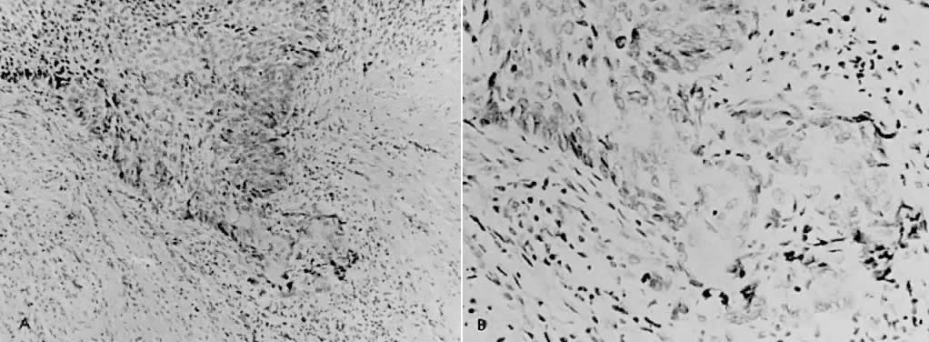
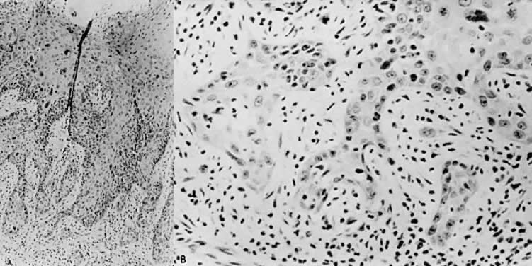
There is general agreement that disruption of the basement membrane is the criterion by which microinvasive carcinoma can usually be identified. This basement "membrane” has been demonstrated by Luiebel and colleagues52 using electron microscopy. He noted it to be complete in CIN III but straighter than in normal squamous epithelium. In invasive carcinoma, it was usually absent or present in an imperfect form. Where the membrane was absent, small cytoplasmic protrusions of carcinoma in the adjacent stroma could be seen, apparently representing invasion by malignant neoplastic cells. Ashworth and co-workers53 confirmed these electron microscopic findings and discussed histochemical procedures including the periodic acid-Schiff reaction alone or in combination with other techniques. He added that invasive cell groups are also found partially or completely invested by a basement membrane that appears to be newly formed by those cells. The typical histologic picture of early stromal invasion below CIN III may show some differentiation of the invasive peg. The cells are rich in cytoplasm and the nuclei larger and clearer than those of the intraepithelial portion of the lesion. There may be cells in various stages of degeneration with some leukocytic infiltration. In hematoxylin-eosin preparations, these pegs may appear eosinophilic and stand out well from the basophilic matrix (Fig. 1) with a dense round cell infiltration surrounding the invasive peg. Tapering or branching small tongues of epithelial cells, as well as the formation of narrow cell columns or the appearance of cell groups that seem to be invading the stroma may be considered criteria of invasion (Fig. 2). On the other hand, the formation of very large epithelial pegs can only be regarded as suspicious of early invasion.54

Fig. 1. A. Early stromal invasion showing some differentiation of the invasive peg. A moderate leukocytic infiltrate is evident. (x100) B. Higher magnification demonstrating intracellular and extracellular keratinization. The basement membrane is absent at the tip of the advancing peg.

Fig. 2. A.Tapering and branching tongues of epithelial cells are seen with areas of coalescence and separate cell groups that seem to invade the stroma. (x100) B. Higher magnification showing pleomorphism and keratinization. (x450)

Disagreement occurs in the specific definition of microinvasive carcinoma regarding the exact depth of invasion, presence or absence of confluence, or vascular space involvement. There is fairly general agreement that the invasion should be 5 mm or less if the lesion is to be considered microinvasive. Averette and colleagues12 used penetration beneath the basement membrane of less than 1 mm as the criterion. Some authors consider confluence1,3,14,16,17 and vascular involvement55,56 as important factors that necessitate removing patients from the microinvasive category.

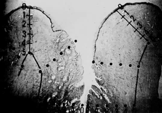
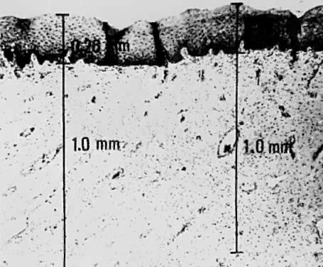
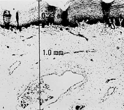
Using a hysterectomy specimen and a micrometer, Averette12 demonstrated the variable distances into the stroma from the basement membrane of ectocervix, squamocolumnar junction, and distant endocervix (Fig. 3). He showed the variable thickness of normal epithelium, which may cause variations in the relative stromal depth when a 1-mm point is measured from the basement membrane (Fig. 4). In addition, vascular channels are shown less than 1 mm beneath the epithelium (Fig. 5). These figures suggest the following:

Fig. 3. Section of cervix showing various depths into stroma, as measured by the micrometer. (x20) The ectocervix, squamocolumnar junction, and distal endocervix are shown. (Averette HE et al: Diagnosis and management of microinvasive [stage IA] carcinoma of the uterine cervix. Cancer 38:414, 1976)

Fig. 4. Section of cervical squamous mucosa demonstrating variable thicknesses of normal squamous mucosa and a 1-mm depth into submucosa, as measured by the micrometer. (x85) (Averette HE et al: Diagnosis and management of microinvasive [stage IA] carcinoma of the uterine cervix. Cancer 38:414, 1976)

Fig. 5. A 1-mm submucosa of cervix and its relationship to vascular channels. (x85) (Averette HE et al: Diagnosis and management of microinvasive [stage IA] carcinoma of the uterine cervix. Cancer 38:414, 1976)

  The depth into the stroma may vary depending on the point of measurement.
  Five-millimeter depth is significant in terms of total cervical thickness.
  There may be a relative difference in stromal depth when 1 mm is measured from two separate points.
  Vascular channels are present within 1 mm of the stroma and are potentially a conduit for spread even in very early invasive lesions.

Duncan and Walker57 found vascular involvement in only 2 of 76 patients with invasion of less than 3 mm, whereas vascular involvement was apparent in 8 of 15 patients who had stromal invasion in the 3 mm to 5 mm depth range. Only 2 of 91 patients had metastatic disease, both with vascular involvement. Vascular involvement was present in only 6 of 135 patients reported by Hasumi and colleagues58: there was no vascular involvement in patients with invasion up to 1 mm but 3 of 45 (6.7%) with invasion of 1.1 mm to 3 mm and 3 of 29 (11.1%) with invasion of 3.1 mm to 5 mm had vascular involvement. None of the six cases with vascular invasion had nodal metastasis. Sedlis and colleagues21 reported vascular space invasion by tumor in 31 of 132 patients (23%); this increased with depth of penetration. There was a strong correlation between both residual tumor in the hysterectomy specimen and vascular space involvement with deep stromal penetration and extensive lateral spread. No positive lymph nodes were observed in the 74 patients treated by radical hysterectomy, and recurrence was noted in two patients with extensive vascular involvement.

Simon20 and Creasman59 reviewed the recent literature in regard to lymph vascular space and lymph node involvement. In the study by Simon,20 54 (14.3%) of 378 women subjected to radical surgery and pelvic lymphadenectomy had lymph vascular space involvement and only 1 of the 54 (1.9%) had a node metastasis. None of the 213 women with long-term follow-up had either local recurrence or died from tumor. In Creasman's59 analysis of 267 women treated in a similar surgical fashion and with microinvasive tumors invading below the basement membrane to 3 mm or less, 39 had lymph vascular space involvement and 1 (without lymph vascular involvement) had lymph node metastasis. Of 105 women with tumors invading between 3.1 and 5 mm, 49 (47%) had lymph vascular permeation, 1 of which had lymph node metastasis. Of the 56 patients without lymph vascular involvement, 6 (11%) had positive nodes. Thus, it appears that in relationship to microinvasive carcinoma of the cervix, the presence of tumor cells in lymph vascular spaces does not support the theory that lymph node metastases are imminent. Additionally, there is disagreement as to what constitutes a lymph vascular space.

Confluence appears to increase with increasing depth of penetration.21,58,60 However, several authors believe this factor to be nonprognostic.21,60

Burghardt19 calculated tumor volume by measuring the depth, width, and length of the tumor. There were no lymph node metastases in 12 patients with an area of up to 99 mm2, which corresponded to 1000 mm3. In larger tumors, the frequency of metastasis increased in relation to tumor size. The smallest tumor showing metastasis had an area of 120 mm2.

Back to Top
DIAGNOSIS

Clinical signs and symptoms of microinvasive carcinoma are either nonexistent or nonspecific and, therefore, offer no assistance in making the diagnosis. The diagnosis of carcinoma requires a specimen for histopathologic examination. In the absence of a gross lesion, the histologic material is usually obtained because of an abnormal Papanicolaou smear. The diagnosis is best made by colposcopically directed biopsy or cervical cone biopsy. There is still some controversy as to which of these should be the primary approach. A random punch biopsy is inadequate for diagnosis.1,61

It is now well-established that colposcopy is a safe and reliable method for the diagnosis of CIN. One study, which included a review of the literature, found a significant difference in results obtained by the two methods (colposcopically directed biopsy versus conization) in only 4% of patients.62 This study included a series of 516 consecutive patients in whom no invasive lesions were missed by colposcopically directed biopsy. Proper patient selection and physician experience are essential for the safety of this technique. When a cone biopsy is necessary, colposcopic direction is also of value in tailoring the operation to fit the lesion.

Conization is necessary following initial diagnosis by a colposcopically directed biopsy to accurately define the extent of microscopic penetration into the stroma. A carefully handled cone specimen gives the pathologist the best material for diagnosis. In order that the greatest amount of information be obtained from the cone, it is essential that the entire specimen be cut into blocks and step-serial sections be done on each block. A cone, properly performed, should include a portion of the portio vaginalis, the external cervical os, and the endocervical canal as high up as is feasible, preferably to the internal os. The entire transformation zone should be incorporated. The inclusion of all abnormal epithelium may be facilitated by the use of the colposcope or, if that is unavailable, Schiller's solution. Theoretically, if an endocervical curettage is also done, all cervical lesions can be detected by proper examination of the tissue.

A preferred method of histologic evaluation of the cone uses 15 blocks from the cone and involves removal of 8 to 10 sections at intervals through each block.63 Another approach would be to dismantle the cone in wedge-shaped blocks and take two sections from each block. If any of the sections reveal frankly invasive carcinoma, no further study is necessary. If no malignant lesion or only CIN III is found, each block should be step-sectioned so that 10 additional slides are obtained from each block, totalling 100 to 150 sections.14 Other methods necessitate 100 to 200 serial sections.51,64,65

More recently, the loop electrosurgical excision procedure has been used in the United States to perform cervical conizations on an outpatient basis. In general, cervical conization using an electrical current, which flows through a fine wire loop, has been shown to be a safe, effective, and well-tolerated method. The procedure is performed effectively under local anesthesia and in most situations histologic specimens are excellent as demonstrated by the study of Mor-Yosef and associates.66 In a study67 of 1000 women referred over 20 months with abnormal smears and in whom the entire transformation zone could be seen, the aim was to test the feasibility of colposcopic assessment and then treatment with the loop electrosurgical excision procedure at one visit to the clinic. Eight hundred ninety-seven women needed only one visit. This was achieved by using a modification of the large loop diathermy excision technique and by careful attention of the timing of the clinical visit. One hundred three patients required further visits, for the following reasons: incomplete excision on histology and/or subsequent abnormal smear; secondary hemorrhage;6 microinvasion or invasive carcinoma;9 or biopsy specimens unusable.3 Further treatment was given in 4.1% of the cervical dysplasias. All patients treated in a single visit preferred this approach to the alternative, which was colposcopic assessment and biopsy followed by local ablation by laser, excision by loop biopsy technique, or cone biopsy, following histological examination. Side effects of this procedure are mainly secondary hemorrhage, which is found to be in the range of 1% to 4% as experience is gained.67,68,69 Cautery artifact at the conization specimen margins still remains a troublesome entity, particularly when evidence of microinvasive cancer exists at the edge of the cauterized portion of the specimen. Significant data does not yet exist in the published literature to determine whether the best approach in this situation is to repeat the conization using the loop electrosurgical excision procedure; to repeat the conization using a cold knife; or to assume that the disease is frankly invasive and proceed with a more radical procedure.

Back to Top
TREATMENT

There have been many methods of treatment of microinvasive carcinoma of the cervix ranging from cervical conization to radical hysterectomy with pelvic lymph node dissection and from local radium treatment to full pelvic irradiation. Recently, Querleu and colleagues 70 recommended diagnostic pelvic lymphadenectomy in the staging of early carcinoma of the cervix as a less morbid method to determine regional lymph node status. Laparoscopic radical hysterectomy with paraaortic and pelvic lymph node dissection has recently been reported as therapy for microinvasive carcinoma of the cervix.71

Averette,12 reported a multi-center study of microinvasive carcinoma of the cervix. The criteria used was penetration of invasive carcinoma beneath the basement membrane of less than 1 mm, determined by calibrated optics. Additionally, the literature was reviewed for criteria of diagnosis of microinvasive cancer, as well as methods of management. In the literature, when depth of penetration of up to 5 mm was used as the criterion, the incidence of nodal metastasis was as high as 3.5%. In the institutions involved in this study, patients were treated by radical hysterectomy and pelvic lymph node dissection. There were no recurrences and no positive pelvic nodes. The conclusion was that simple hysterectomy was not adequate therapy for lesions with stromal invasion to a depth of 5 mm.

Bohm and colleagues,72 Seski and co-workers,60 Duncan and Walker,57 and Boronow,73 in different studies, reported a combined total of 165 radical hysterectomies with pelvic lymph node dissections in which there were four patients with positive pelvic nodes. The limits of invasion used by the first three authors was 3 mm; however, Boronow used the latest FIGO stage IA without defining the limits of the lesion. All four patients with positive nodes were reported by Bohm.72 Two of those four patients had lymphatic or blood vessel invasion; two patients died of cancer. Roche and Norris74 found no positive nodes in 30 patients with stromal penetration up to 5 mm. Of 111 patients with microinvasive carcinoma who were studied over a 21-year period by Christopherson and Parker,1 the sole pathologic criteria for inclusion was equivocal invasion to a depth of no more than 5 mm. Eighty-four patients were treated by simple hysterectomy or less extensive surgery, 14 by conization and irradiation, 3 by hysterectomy and irradiation, and 10 by radical hysterectomy. Ninety-one patients were followed for 5 years or until death, and 80 patients were followed for 10 years or until death; 1 patient was lost to follow-up at 5.5 years. Two deaths officially attributed to cervical cancer were doubtful. Both had been previously treated by cervical conization and irradiation. Eighty-four patients had been treated by either simple hysterectomy, excision of the cervical stump, or cervical conization only. There were no recurrences in this group. It was suggested that simple hysterectomy should be the maximal treatment indicated.

Hasumi and co-workers58 reported on 135 patients with stage I epidermoid carcinoma of the uterine cervix invading less than 5 mm below the basement membrane. These cases were studies to determine the biological behavior of early invasive carcinoma and to establish diagnostic criteria for microinvasive carcinoma of the cervix. None of the 135 patients had metastasis to the parametrial tissue in the final surgical specimen. One (0.9%) of 106 patients with invasion up to 3 mm had lymph nod e metastasis, while 4 (13.9%) of 29 patients with invasion of 3.1 mm to 5 mm had nodal metastasis. Of the 106 patients with invasion up to 3 mm, 25 had confluent invasion. None of the 25 patients had lymph node metastasis. In view of these results, it was felt that carcinomas with invasion less than 3 mm may be regarded as a separate diagnostic group because of their limited metastatic potential and may be treated by conservative methods used for CIN III, even if there was a confluent pattern. For carcinomas with invasion of 3.1 mm to 5 mm, more extensive procedures as used for frankly invasive cancers were thought to probably be necessary. Van Nagell75 reported on 177 patients with squamous cell carcinoma that invaded the cervical stroma to a depth of 5 mm or less. In 52 patients with lesions that invaded the cervical stroma to a depth of 3 mm or less, none contained metastatic tumor in their lymph nodes. Conversely, lymph node metastases were present in 10% of patients with lesions that had stromal invasion of 3.1 mm to 5 mm. Among 145 patients with lesions that invaded the stroma to a depth of 3 mm or less, only 2 developed recurrences, both of which were intraepithelial. Among the 32 cases of carcinoma that invaded the stroma 3.1 mm to 5 mm, there were 3 invasive recurrences and 2 deaths. Creasman59 analyzed 114 patients with stage IA carcinoma of the cervix retrospectively in regard to depth of invasion, capillary-like space involvement, stromal reaction, status of conization margins, and the incidence of lymph node metastasis. There were no lymph node metastases or recurrences in the group of patients whose depth of stromal invasion was less than 3 mm. Maiman and associates,76 reporting on a series of 117 women with histologically defined superficially invasive (1 mm–5 mm) squamous cell carcinoma of the cervix, revealed an overall incidence of pelvic lymph node metastasis of 5%. The incidence of metastasis in those patients with invasion 3 mm or less was 2% and in those with invasion 3.1 mm to 5 mm was 13%. Microscopic lymph vascular invasion and degree of lateral spread of tumor were also associated with lymph node metastasis, whereas tumor grade was not. Most recently, Sevin and colleagues77 attempted to assess the risk of lymph node metastasis and treatment failures of microinvasive carcinoma according to the latest FIGO and Society of Gynecologic Oncologists (SGO) definitions by retrospectively reviewing the histopathologic material on 370 patients with microinvasive carcinoma of the cervix who were treated by radical hysterectomy and pelvic-aortic lymph node dissection. Histopathologic analysis of tumors was based on a uniform format including measurement of the maximum depth of invasion, the width and length of the horizontal tumor spread, invasive growth pattern, cell type, tumor grade, and lymphatic or vascular space involvement. Of 370 patients, 110 had a depth of invasion of 5 mm or less. Of these, 54 patients fulfilled the SGO definition of microinvasive carcinoma; 42, the new FIGO stage IA2 definition; and 27, both definitions. None of the patients with microinvasive carcinoma, as defined by either the SGO or the new FIGO stage IA2 had lymph node metastases or tumor recurrence. These data support the conclusion that microinvasive carcinomas, defined by either the SGO or FIGO definitions, have a low risk for metastasizing to regional lymph nodes or recurring. He did note, however, that a review of the literature indicated a recurrence rate for stage IA2 of 4.2%.

Roman and Latour78 treated 84 cases of stage IA carcinoma by simple hysterectomy alone, with an overall 5-year survival of 97.5%. Boyes and co-workers79 treat microinvasive carcinoma by cervical conization. The specimen was then examined by step-serial sections. If the lesion appeared to have been removed completely, no further treatment was instituted. If the cancer had not been completely removed by the cone biopsy, either a second cone biopsy or hysterectomy with an adequate vaginal cuff was recommended, depending on the lesion and other clinical considerations. If lymphatic involvement was seen in the biopsy, the patient was treated by irradiation or radical hysterectomy. Way and co-workers80 treated microinvasive lesions by modified radical hysterectomy including at least 3 cm of the upper vagina. He stated: (1) node dissection is unnecessary; (2) total hysterectomy is inadequate; and (3) conservation should be reserved only for those who desire further pregnancies. Nelson81 described 355 such operations with one fistula and no operative deaths.

Cervical cone biopsy has been used by several authors with good results under carefully selected circumstances. Of 113 patients added from different series in the literature,19,57,82,83 there was one recurrence of CIN III in a group of patients that were followed for an adequate length of time. Mestwerdt11 thought that conization could be considered adequate therapy but only for young women who wanted to bear children, since this method has a high relapse quotient. Sedlis and colleagues21 reviewed 100 cases in which they had both conization and subsequent hysterectomy specimens. Residual invasive tumor on the hysterectomy specimen was deeper than in the cone in nine cases. Residual invasive tumor was less than that in the cone in six cases. Residual dysplasia was present in 31 cases, 27 of those harboring CIN III. No residual tumor was found in only 54 cases. The conization specimen of the nine cases with invasion greater in the hysterectomy specimen than in the cone had the following characteristics: depth of invasion greater than 2 mm in 77.5% (7 of 9); lateral extension greater than 4 mm in 90% (8 of 9); vascular space invasion present in 90% (8 of 9); and tumor involvement of the surgical margins present in 90% (8 of 9). Residual invasive carcinoma of the hysterectomy specimen increased from 0 in cases with invasion less than 1 mm on the cone specimen to 61% if invasion on the cone was deeper than 3 mm. Of 15 patients in whom the invasion did not exceed the 5-mm limit in the hysterectomy specimen and the surgical margins of the cone were involved, residual tumor was present in 12 (80%). In eight of these patients, the invasion was more extensive in the uterus than in the cone. One of the patients with recurrence of carcinoma had tumor involvement of the surgical margin on the cone specimen. In Seski's60 study of 54 patients, 78% had residual CIN III or microinvasive carcinoma in the hysterectomy specimen following cone biopsy.

Greer and co-workers84 reported on 50 patients with early invasive squamous cell carcinoma of the cervix treated with a cervical conization followed by a radical hysterectomy and pelvic lymph node dissection. He found histologically positive margins at the time of cone biopsy in 66% of patients. Negative margins at the time of the cone biopsy were identified in 34%. Residual invasive disease at the time of radical hysterectomy was found in 24% of patients with negative margins. Four percent had positive lymph nodes. Three patients had recurrent metastatic disease. This study of stageIA2 patients demonstrated that a preoperative diagnosis of that stage is difficult to establish and creates a therapeutic dilemma regarding treatment.

Back to Top
CONCLUSION

It is difficult to draw therapeutic conclusions though considerable information is now available from the literature. It appears that lesions that have no lymphatic or vascular involvement and are less than 3 mm in depth may be treated conservatively, but only when adequate tissue is submitted for pathologic examination by the gynecologist and the tissue is examined adequately by the pathologist. The extent of early invasion must be carefully ascertained, usually by cervical cone biopsy. The pathologist should properly orient and cut the cone into 15 to 18 blocks, depending on the size of the specimen. Each block should be step-serial sectioned in a manner to assess the exact extent of invasion, using a micrometer. This procedure may entail more than 100 slides. If this plan is rigidly followed, it appears that cervical conization is adequate treatment when the lesion is less than 1 mm in depth. Total hysterectomy or conization is probably safe if the lesion is less than 3 mm. For lesions that are greater than 3 mm in depth, radical surgery or radiation is indicated. Unless elaborate measures are taken to establish the extent of involvement, the modified radical operation is the minimal safe surgical procedure and conforms to the operative principle of wide excision of the lesion.

The significance of confluence and lymphatic vascular space involvement has not been clearly established to date. It does appear that both phenomena increase as the depth of penetration increases and that neither occurs with regularity if the depth is less than 1 mm and neither occurs commonly up to a depth of 3 mm. Therefore, it seems that the depth should be used as a primary guide, but the other factors may be indicators of tumor volume and, therefore, should modify a decision regarding conservative treatment.

Conization as therapy should primarily be reserved for the patient who desires fertility. If this method is chosen, a careful explanation of the risks should be given. Additionally, the pathologic specimen must be rigorously evaluated, the cone margins should be negative for invasion or CIN, there should be no evidence of lymphatic or vascular invasion, the lesion should be of squamous type, and the patient must be compliant.

The point at which the biologic nature of microinvasive carcinoma of the cervix changes and it begins to behave as a truly invasive lesion is not well-defined. The crux of the problem lies in the fact that investigators continue to use light microscopy to define what is in essence a molecular problem.

Back to Top
REFERENCES

1. Christopherson WM, Parker JE: Microinvasive carcinoma of the uterine cervix. Cancer 17: 1125, 1964

2. Enterline HT: Management of microinvasive carcinoma. JAMA 193: 220, 1965

3. Fidler HK, Boyd JR: Occult invasive squamous carcinoma of the cervix. Cancer 13: 764, 1960

4. Frick HC II, Janovski NA, Gusberg SB et al: Early invasive cancer of the cervix. Am J Obstet Gynecol 85: 926, 1963

5. Koss LG: Diagnostic Cytology and Its Histopathologic Bases, 2nd ed. Philadelphia, JB Lippincott, 1968

6. Marcuse PM: Incipient microinvasive carcinoma of the uterine cervix. Obstet Gynecol 37: 360, 1971

7. Schreiner WE, Wyss HI: Symposium on cervical lesions: May one treat the early invasive carcinoma (microcarcinoma) less radically than the more extensive carcinoma? Acta Cytol 6: 173, 1962

8. Song J, Nettles JB: Circulating tumor cells in patients with carcinoma in situ of the cervix uteri. Am J Obstet Gynecol 104: 713, 1969

9. Stolz H: Symposium on cervical lesions: May one treat the early invasive carcinoma (microcarcinoma) less radically than the more extensive carcinoma? Acta Cytol 6: 173, 1962

10. Mattingly RF, Thompson JD: Te Linde's Operative Gynecology, 6th ed. Philadelphia, JB Lippincott, 1985

11. Mestwerdt G: Fruhdiagnose des Kollumkarzinoms. Zentralbl Gynakol 69: 326, 1947

12. Averette HE, Nelson JH, Ng ABP et al: Diagnosis and management of microinvasive (Stage 1A) carcinoma of the uterine cervix. Cancer 38: 414, 1976

13. Larson G, Alm P, Gullberg B, Grundsell H: Prognostic factors in early invasive carcinoma of the uterine cervix. Am J Obstet Gynecol 146: 145, 1983

14. Bryans FE, Boyes DA, Boyd JR et al: The cytology program in British Columbia. Can Med Assoc J 90: 62, 1964

15. Carter B: Results in the management of preclinical carcinoma of the cervix. Am J Obstet Gynecol 81: 511, 1961

16. Ng ABP, Reagan JW: Microinvasive carcinoma of the uterine cervix. Am J Clin Pathol 52: 511, 1969

17. Ullery JC, Boutselis JG, Botschner AC: Microinvasive carcinoma of the cervix. Obstet Gynecol 26: 866, 1965

18. FIGO News International: J Gynecol Obstet 25:87, 1987

19. Burghardt E: Microinvasive carcinoma. Obstet Gynecol Surv 34: 836, 1979

20. Simon NL, Gore H, Shingleton HM et al: Study of superfically invasive carcinoma of the cervix. Obstet Gynecol 68: 19, 1986

21. Sedlis A, Sall S, Tsukada Y: Microinvasive carcinoma of the uterine cervix. A clinical-pathologic study. Am J Obstet Gynecol 133: 64, 1979

22. Wentz WB, Reagan JW: Experimental carcinogenesis of the cervix with inactivated Herpes virus. Read before the Ninth Annual Meeting of the Society of Gynecologic Oncologists, Key Biscayne, FL, January 1978

23. Naib ZM, Nahmias AJ, Josey WE et al: Genital herpetic infection associated with cervical dysplasia and carcinoma. Cancer 23: 940, 1969

24. Kolstad P, Stafl A: Atlas of Colposcopy, p. 51. Baltimore, University Park Press, 1972

25. Averette HE: Indications for radical hysterectomy. Med Opinion Rev 7: 73, 1971

26. Fu YS, Reagan JW, Townsend DE: Nuclear DNA study of vulvar intraepithelial and invasive squamous neoplasms. Obstet Gynecol 57: 643, 1981

27. Crum CP, Braun LA, Shah KV: Correlation of nuclear DNA content and the presence of a human papillomavirus (HPV) structural antigen. Cancer 49: 468, 1982

28. Crum CP, Levine RU: Human papillomavirus infection and cervical neoplasia: New perspectives. Int J Gynecol Pathol 3: 376, 1984

29. Rastkar G, Okagaki T, Twiggs LB, Clark BA: Early invasive and in-situ warty carcinoma of the vulva: Clinical, histologic, and electron microscopic study with particular reference to viral association. Am J Obstet Gynecol 143: 814, 1982

30. Stanbridge C, Butler EB: Human papillomavirus infection of the lower female genital tract: Association with multicentric neoplasia. Int J Gynecol Pathol 2: 264, 1983

31. Reid R, Fu YS, Herschman BR: Genital warts and cervical. VI. The relationship between aneuploid and polypoid cervical lesions. Am J Obstet Gynecol 150:189,

32. Winkler B, Crum CP, Fujii T: Koilocytotic lesions of the cervix: The relationship of mitotic abnormalities to the presence of papillomavirus antigens and nuclear DNA content. Cancer 53: 1081, 1984

33. DiPaolo JA, Woodworth CD, Popescu NC: Induction of human cervical squamous cell carcinoma by sequential transfection with human papillomavirus 16 DNA and viral Harvey RAS. Oncogene 4: 395, 1989

34. Sedman SA, Barbosa MS, Vass WC: The full-length E6 protein of human papillomavirus type 16 has transforming and transactivating activities and cooperates with E7 to immortalize keratinocytes in cultures. J Virol 65: 4860, 1991

35. Howley PM: Role of the human papillomaviruses in human cancer. Cancer Res 51: 5019, 1991

36. Jones RE, Wergyn RJ, Patrick DR: Identification of HPV-16 E7 peptides that are potent antagonists of E7 binding to the retinoblastoma suppressor protein. J Biol Chem 265: 2782, 1990

37. Scheffner M, Werness BA, Huibregtse JM et al: The E6 oncoprotein encoded by human papillomavirus types 16 and 18 promotes the degradation of p53. Cell 63: 1129, 1990

38. Crook T, Vousden KH: Properties of p53 mutations detected in primary and secondary cervical cancers suggest mechanisms of metastasis and involvement of environmental carcinogens. EMBO J 11: 3935, 1992

39. Borresen AL, Helland A, Nesland J et al: Papillomavirus and cervical cancer. Lancet 339: 1350, 1992

40. Liotta LA: Tumor invasion: Role of the extracellular matrix. Cancer Res 46: 1, 1986

41. McCarthy JB, Hager SJ, Furcht LT: Human fibronectin contains distinct adhesion- and motility-promoting domains for metastatic melanoma cells. J Cell Biol 102: 179, 1986

42. Hynes RO: Integrins: Versatility, modulation, and signaling in cell adhesion. Cell 69: 11, 1992

43. Chen W-T, Wang J, Hasegawa T: Regulation of fibronectin receptor distribution by transformation, exogenous fibronectin, and synthetic peptides. J Cell Biol 103: 1649, 1986

44. Takeichi M: Cadherins: A molecular family important in selective cell-cell adhesion. Annu Rev Biochem 59: 237, 1990

45. Wewer UM, Liotta LA, Jaye M: Altered levels of laminin receptor mRNA in various human carcinoma cells that have different abilities to bind laminin. Proc Natl Acad Sci U S A 83: 7137, 1986

46. Kudo R, Sato T, Mizuuchi H: Ultrastructural and immunohistochemical study of infiltration in microinvasive carcinoma of the uterine cervix. Gynecol Oncol 36: 23, 1990

47. Ng ABP: Third tutorial on clinical cytology: Microinvasive carcinoma of the uterine cervix. Read before the Symposium of the International Academy of Cytology at the University of Chicago, January 8, 1971

48. Rubio CA: Cytologic studies in cases with carcinoma in-situ and microinvasive carcinoma of the cervix. Acta Pathol Microbiol Scand A82:161, 1974

49. Nguyen GK: Exfoliative cytology of microinvasive squamous-cell carcinoma of the uterine cervix. A retrospective study of 42 cases. Acta Cytol 28: 457, 1984

50. Hinselmann H: Zur Kenntnis der praekanzerösen Veränderungen der Portio. Zentralbl Gynakol 51: 901, 1927

51. Johannisson E, Kolstad P, Soderberg G: Cytologic, vascular and histologic patterns of dysplasia, carcinoma in situ and early invasive carcinoma of the cervix. Acta Radiol Suppl 258, 1966

52. Luiebel FJ, Sanders E, Ashworth CT: Electron microscopic study of carcinoma in situ and invasive carcinoma of the cervix uteri. Cancer Res 20: 357, 1960

53. Ashworth CT, Stembridge VA, Luibel FJ: A study of basement membranes of normal epithelium, carcinoma in situ, and invasive carcinoma of the uterine cervix utilizing electron microscopy and histochemical methods. Acta Cytol 5: 369, 1961

54. Burghardt E, Holzer E: Diagnosis and treatment of microinvasive carcinoma of the cervix uteri. J Obstet Gynecol 49: 641, 1977

55. Margulis RR, Ely CW Jr, Ng ABP: Diagnosis and management of microinvasive (Stage 1A) carcinoma of the uterine cervix. Cancer 38: 414, 1976

56. Moore JG, Morton DG, Applegate JW: Management of early carcinoma. Am J Obstet Gyencol 81: 1175, 1961

57. Duncan ID, Walker J: Microinvasive squamous carcinoma of the cervix in the Tayside region of Scotland. Br J Obstet Gynecol 84: 67, 1977

58. Hasumi K, Atsuhiko S, Sugano H: Microinvasive carcinoma of the uterine cervix. Cancer 45: 928, 1980

59. Creasman WT, Fetter BF, Clarke-Pearson DL et al: Management of Stage 1A carcinoma of the cervix. Am J Obstet Gynecol 153: 164, 1985

60. Seski JC, Abell MR, Morley GW: Microinvasive squamous carcinoma of the cervix: Definition, histologic analysis, late results of treatment. Obstet Gynecol 50: 4, 1977

61. Bickenbach W, Soost HJ, Campos J: What percentage of cervices show early invasion in serial histological sections in uteri which were removed under the biopsy diagnosis of "carcinoma in situ.” Acta Cytol 5: 340, 1961

62. Savage EW: Correlation of colposcopically directed biopsy and conization with histologic diagnosis of cervical lesions. J Reprod Med 15: 211, 1975

63. Fidler Boyes DA, Lock DR: Intraepithelial carcinoma of the cervix: 214 cases with emphasis on investigation by cytology and cone biopsy. Can Med Assoc J 77: 79, 1957

64. Foushee JHS, Greiss FC, Lock FR: Stage 1A squamous cell carcinoma of the uterine cervix. Am J Obstet Gynecol 105: 46, 1969

65. Parker RT, Cuyler WK, Kauffman LA et al: Intraepithelial (Stage O) cancer of the cervix. Am J Obstet Gynecol 80: 693, 1960

66. Mor-Yosef S, Lopes A, Pearson S, Monaghan JM: Loop diathermy cone biopsy. Obstet Gynecol 75: 884, 1990

67. Bigrigg MA, Codling BW, Pearson P: Colposcopic diagnosis and treatment of cervical dysplasia at a single clinic visit. Lancet 336: 229, 1990

68. Murdoch JB, Gishane RN, Monaghan JM: Loop diathermy excision of the abnormal cervical transformation zone. Int J Gynecol Canc 1: 1105, 1991

69. Luesky DM, Cullimore J, Redman CWE: Loop diathermy excision of the cervical transformation zone in patients with abnormal cervical smears. Br Med J 300: 1090, 1990

70. Querleu D, Leblanc E, Castelain B: Laparoscopic pelvic lymphadenectomy in the staging of early carcinoma of the cervix. Am J Obstet Gynecol 164: 579, 1991

71. Nezhat CR, Burrell MO, Nezhat FR et al: Laparoscopic radical hysterectomy with paraaortic and pelvic node dissection.

72. Bohm JW, Krupp PJ, Lee FYL: Lymph node metastasis in microinvasive epidermoid cancer of the cervix. Obstet Gynecol 48: 65, 1976

73. Boronow RC: Stage I cervix cancer and pelvic node metastasis: Special reference to the implications of the new and the recently replaced FIGO classification on Stage 1A. Am J Obstet Gynecol 127: 2, 1977

74. Roche WD, Norris HJ: Microinvasive carcinoma of the cervix: The significance of lymphatic invasion and confluent patterns of stromal growth. Cancer 36: 180, 1975

75. van Nagell Jr, Greewell N, Powel DF et al: Microinvasive carcinoma of the cervix. Am J Obstet Gynecol 145:981, 1983

76. Maiman MA, Fruchter RG, DiMaio TM, Boyce JG: Superficially invasive squamous cell carcinoma of the cervix. Obstet Gynecol 72: 339, 1988

77. Sevin BU, Nadji M, Averette HE et al: Microinvasive carcinoma of the cervix. Cancer 70: 2121, 1992

78. Roman TN, Latour JPA: The effect of early diagnosis on survival statistics in carcinoma of the uterine cervix. Am J Obstet Gynecol 97: 739, 1967

79. Boyes DA, Worth AJ, Fidler HK: The result of treatment of 4389 cases of preclinical cervical squamous carcinoma. J Obstet Gynecol Br Common 7: 769, 1970

80. Way S, Hennigan M, Wright VC: Some experiences with preinvasive and microinvasive carcinoma of the cervix. J Obstet Gynecol Br Common 75: 593, 1968

81. Nelson JH: Atlas of Radical Pelvic Surgery. New York, Appleton-Century-Crofts, 1969

82. Wilkinson E, Komorowski R: Borderline microinvasive carcinoma of the cervix. Obstet Gynecol 51: 472, 1978

83. Popkin D, Pilgorge R, Latour J: The treatment of microinvasive squamous cell carcinoma of the uterine cervix. Gynecol Oncol 8: 84, 1979

84. Greer BE, Figge DC, Tamimi HK et al: Stage IA2 squamous carcinoma of the cervix: Difficult diagnosis and therapeutic dilemma. Am J Obstet Gynecol 162: 1406, 1990

Back to Top