Chapter 6
Pathology of Cervical Carcinoma
Yao S. Fu
Main Menu   Table Of Contents

Search

Yao S. Fu, MD
Department of Pathology, St. Joseph Medical Center, Burbank, California (Vol 4, Chap 6)

INTRODUCTION
PROPER HANDLING AND PROCESSING OF CERVICAL SPECIMENS
MORPHOGENESIS OF CERVICAL CANCER
CLINICAL PRESENTATION AND NATURAL HISTORY OF CERVICAL CANCER
SQUAMOUS CARCINOMA AND ITS VARIANTS
MALIGNANT GLANDULAR NEOPLASMS
OTHER MALIGNANT NEOPLASMS
PROGNOSTIC FACTORS
TUMOR RECURRENCE
STUDIES USING SPECIAL TECHNIQUES
CONCLUSION
REFERENCES

INTRODUCTION

In the last three decades the epidemiology of cervical cancer has undergone some important changes. In America, it is currently the third most common gynecologic cancer following those of endometrium and ovary. However, the number of women dead of cervical cancer is exceeded only by that of ovarian cancer.1 Squamous cell carcinoma, which comprised more than 90% of primary cervical cancers before 1960, has been reduced steadily in large part to effective cytologic detection and subsequent eradication of its precursors. In addition, the use of endocervical sampling devices, such as Cytobrush (International Cytobrush, Inc., Hollywood, FL, USA) and Cervex (Rovers B.D., Oss, The Netherlands), has enhanced the detection cervical glandular dysplasia and neoplasms. Consequently, there is a relative increase in cervical adenocarcinoma comprising 13% to 34% of all cervical cancers.2–4 Whether the incidence of cervical adenocarcinoma has actually increased remains to be clarified.

In addition to squamous and glandular neoplasms, neuroendocrine, mesenchymal, and metastatic tumors also involve the cervix. New techniques in diagnosis have helped to modify histologic classifications. Specifically, the use of immunohistochemistry in the last decade has improved diagnostic accuracy. This chapter deals primarily with cervical malignancies, which are International Federation of Gynecology and Obstetrics (FIGO) stage IB or higher. The emphasis is on proper handling and reporting of surgical specimens, pathologic classification and criteria, and important pathologic parameters, which have impact on prognosis and management of patient. Finally, investigative findings are described to provide additional insights on the current development.

Back to Top
PROPER HANDLING AND PROCESSING OF CERVICAL SPECIMENS

The accuracy of histologic interpretation and diagnosis is strongly governed by the quality of tissue provided, and proper handling and processing of the specimen. In cervical biopsy specimens, many factors lead to unsatisfactory specimens. Distortion and crush artifacts usually result from the use of dull or small instruments.5 Damage and denudation of the mucosa occur readily even with minor trauma to the cervix. Poor orientation with tangential sections not only precludes accurate interpretation and measurement of stromal invasion, but also contributes to erroneous diagnoses. After the tissue is obtained, the specimen should be placed on a piece of paper towel with the mucosal surface upward and the base of the tissue downward. After the blood and mucus at the base of the tissue become adherent to the paper towel, the specimen and the paper towel are placed in buffered formalin and submitted to the laboratory accompanied by adequate demographic and clinical information.

The single most common cause of an inadequate biopsy specimen is the failure to provide abnormal tissue of sufficient amount and depth. Without the underlying stroma, an invasive neoplasm is likely to be interpreted as in situ cancer. Verrucous squamous carcinoma and papillary, exophytic neoplasms are particularly prone to be underdiagnosed as a benign proliferation, when the specimen contains only the superficial layers. Unless the specimen includes the base of tumor and its underlying stroma, a correct diagnosis may not be made. In mixed tumors (e.g. adenosquamous carcinoma or carcinosarcoma), an inadequate biopsy specimen may not contain both cellular elements, again resulting in misdiagnosis.

For the diagnosis and treatment of cervical intraepithelial neoplasia (CIN), cervical tissue removed by loop electrosurgical excision procedure (LEEP) has become widely accepted. The pathologist usually receives tissue from the anterior lip and the posterior lip, respectively. A third disc-shaped tissue fragment from the base of excision or endocervical margin is received. Diagnostic problems of these specimens are most often caused by a lack of orientation and thermal damage. The specimen should be oriented by a suture or ink to indicate 6 and 12 o'clock. This allows sectioning along the axis of cervical canal. Without orientation, the specimen may be cut erroneously and the lesion may be missed entirely. Excessive maneuvers by the surgeon or pathologist lead to extensive denudation and loss of the cervical mucosa and the lesion. Prolonged contact between the loop and the tissue results in broad zones of thermal damage, coagulative necrosis, and tissue distortion that preclude an accurate diagnosis of the lesion and the status of excision margins. In one study, 20% of ectocervical margin and 44% of endocervical margin removed by LEEP were unsatisfactory for evaluation.6 With proper surgical technique, orientation of the specimen, and marking of the margins by the surgeon, most LEEP specimens are reported to be adequate for interpretation. The status of excision margins correlates well with the subsequent recurrence of CIN.7

Cervical conization by cold knife is most often performed for large, high-grade CIN and CIN suspected with coexisting invasive carcinoma. If cervical biopsy specimen or endocervical curettage specimen suggests the possibility of severe glandular dysplasia or adenocarcinoma in situ, cervical conization is used to confirm the diagnosis and to exclude coexisting invasive adenocarcinoma. A deep cone is appropriate for young women with adenocarcinoma in situ, who desire to retain fertility.

Cervical conization specimens should be oriented by placing a suture at 12 o'clock, and should be submitted to the laboratory in the fresh state. After opening at 12 o'clock along the cervical canal, the specimen is pinned out on a cork board, immersed in fixative, and subsequently blocked around the clock. This method of sectioning provides vertical cuts through the mucosa and wall allowing for accurate determination of the disease process, and its extent, depth, dimension and relation to the surgical margins.5 If the specimen is already fixed, it is too rigid to be spread open.

In radical hysterectomy specimens, representative sections should include the most advanced area of tumor to determine the maximal stromal invasion. All surgical margins should be carefully identified and marked with India ink. The parametrium needs special attention, as involvement of this area by the tumor has important clinical implications.

Pelvic lymph nodes should be properly labeled as to their anatomic sites. Benign germinal inclusion cysts (endosalpingiosis) and decidual cells are known to occur in the pelvic wall, peritoneum, and pelvic and paraaortic lymph nodes. Without knowledge of a pregnancy history, decidual cells in lymph nodes and pelvis may be misclassified as metastatic carcinoma cells on frozen sections. Similarly, tissue reactions to prior surgery, radiation and complications, such as intestinal perforation, can be difficult to separate from malignant tumor, especially on frozen sections. Thus, an accurate pathologic interpretation requires a close collaboration, communication and understanding between the clinician and the pathologist.

In radically resected uteri, the pathology report should include the following:

  The local extent of tumor expressed by the depth of stromal invasion in mm and percent of cervical wall involvement (alternatively upper third, middle third, deeper third, upper half or deeper half), and the relation to parametrium
  The presence or absence of lymphatic-vascular space invasion
  The status of surgical margins: vaginal, pericervical, and parametrial

When the tumor is deeply invasive, its closeness to the parametrium and parametrial margin should be expressed by measurement in millimeters. Involvement of endometrium should be noted, although its presence does not alter the FIGO stage. All pelvic and paraaortic lymph nodes received should be embedded and carefully studied, sometimes by multiple levels as indicated by the institutional guidelines.

Back to Top
MORPHOGENESIS OF CERVICAL CANCER

The ectocervix is covered by mature squamous mucosa, whereas the endocervix is lined by mucus secreting endocervical epithelium. The latter undergoes squamous metaplasia through reserve cell hyperplasia. In addition, endometrial cells, meta-plastic glandular cells (ciliated tubal, serous, and oxyphilic cells), and mesonephric remnants occur in the endocervix. Rare neuroectodermal cells (argyrophilic, neuroendocrine, melanocytic cells) also exist in the normal cervix. These epithelial and neuroectodermal cells are potential progenitors or components of cervical carcinoma. Cervical stromal cells may rarely become neoplastic presenting as a pure mesodermal tumor or mixed with an epithelial neoplasm.

Most cervical squamous carcinomas originate from the metaplastic squamous epithelium located between the original and new squamocolumnar junctions, the transformation zone. Adenocarcinomas occur within the cervical canal, and 25% to 50% of these contain squamous elements. Undifferentiated carcinoma may contain malignant neuroectodermal cells, squamous cells, and glandular cells.

Clinical and pathologic data support the concept that most invasive carcinomas develop through the stages of intraepithelial neoplasia and microinvasive carcinoma. Although the cause of cervical cancer remains to be determined, human papillomavirus (HPV) DNAs, particularly high-risk types 16, 18, 31, 33, 35, 39, 45, 51, 52, 56, and 58, have been identified in squamous, glandular, neuroendocrine, and undifferentiated neoplasms of the cervix by molecular techniques.8 HPV infection is believed to play an important role at least as an initiator of cervical neoplasia.

Back to Top
CLINICAL PRESENTATION AND NATURAL HISTORY OF CERVICAL CANCER

Carcinoma of the cervix may have a variety of clinical presentations. It may be discovered on routine Papanicolaou (Pap) smear in an asymptomatic woman; patients may present with irregular vaginal bleeding; or, in late stages, patients may present with symptoms of a mass lesion or metastatic disease. The cervix with early carcinoma has a poorly circumscribed granular or eroded appearance and bleeds easily on contact. At later stages, nodular, ulcerated lesions or an exophytic mass appears. Endophytic growth occurs in the cervical canal with direct infiltration into the wall causing diffuse enlargement and hardening of the cervix. The mucosal surface may be covered by normal epithelium, and the underlying malignant cells may escape detection by cytologic smear. Some cervical carcinomas are located in the cervical canal and grow endophytically without causing gross abnormality. When the cervix is diffusely enlarged, bulky, and larger than 6 cm in size, it is referred to as a barrel-shaped cervix. This gross appearance can be seen in any tumor type, although it is most commonly associated with adenocarcinoma.

Local extension of cervical carcinoma proceeds in a predictable manner to involve the endometrium superiorly and the upper vagina inferiorly. Parametrial involvement results from extension through the cervical stroma. From the parametrium the tumor may extend laterally to the pelvic sidewall, anteriorly to the bladder base or posteriorly to the rectum. An unusual pattern of local spread is in the form of squamous cell carcinoma in situ into the endometrium, fallopian tubes, and ovaries.9,10 Vascular and lymphatic invasion occurs early leading to pelvic lymph nodal metastasis.

FIGO updated their clinical staging system in 1995 to utilizes the findings of pelvic examination, radiology, and endoscopy.11 These criteria are summarized in Table 1.

TABLE 1. Definition of FIGO Clinical Staging for Cervical Cancer


Stage

Definition

0

Carcinoma in situ, intraepithelial carcinoma

I

Carcinoma strictly confined to the cervix (extension to the corpus should be disregarded)

 IA

Invasive cancer identified only microscopically; invasion limited to measured stromal invasion with maximum depth of 5 mm and no wider than 7 mm (the depth of invasion should not be more than 5 mm taken from the base of the epithelium, either surface or glandular, from which it originates; vascular space involvement, either venous or lymphatic, should not alter the staging)

  IA1

Measured invasion of stroma no greater than 3 mm in depth and no wider than 7 mm

  IA2

Measured invasion of stroma greater than 3 mm and no greater than 5 mm and no wider than 7 mm

 IB

Clinical lesions confined to the cervix or preclinical lesions greater than stage IA

  IB1

Clinical lesions no greater than 4 cm in size

  IB2

Clinical lesions greater than 4 cm in size

II

The carcinoma extends beyond the cervix but has not extended to the pelvic wall; the carcinoma involves the vagina but not as far as the lower third

 IIA

No obvious parametrial involvement

 IIB

Obvious parametrial involvement

III

The carcinoma has extended to the pelvic wall; on rectal examination, there is no cancer-free space between the tumor and the pelvic wall; the tumor involves the lower third of the vagina; all cases with a hydronephrosis or nonfunctioning kidney are included unless they are known to be due to other causes

 IIIA

No extension to the pelvic wall

 IIIB

Extension to the pelvic wall and/or hydronephrosis or nonfunctioning kidney

IV

The carcinoma has extended beyond the true pelvis or has clinically involved the mucosa of the bladder or rectum; a bullous edema as such does not permit a case to be allotted to stage IV

 IVA

Spread of the growth to adjacent organs

 IVB

Spread to distant organs


Adapted from New gynecologic cancer staging: FIGO Cancer Committee. Gynecol Oncol 58:157–158, 1995
Back to Top
SQUAMOUS CARCINOMA AND ITS VARIANTS

Cell Type

In 1958 Wentz and Reagan12 divided cervical squamous carcinomas into three cell types: large cell keratinizing, large cell nonkeratinizing, and small cell. With the advent of electron microscopy and immunohistochemistry, it became apparent that what had been termed small cell carcinoma really represents a heterogeneous group of tumors. These include small cell squamous carcinoma, small cell anaplastic carcinoma, and small cell neuroendocrine carcinoma. Furthermore, a newly proposed classification has expanded the spectrum of neuroendocrine carcinoma to include those poorly differentiated large cell carcinomas demonstrating neuroendocrine differentiation by immunohisto-chem-istry.13

Large cell keratinizing squamous carcinoma is characterized by sheets and nests of cells with abundant cytoplasm, large pleomorphic nuclei and inconspicuous nucleoli. Keratin pearls and intercellular bridges are evident (Fig. 1). Mitotic figures are noted occasionally, and the growth pattern is largely infiltrative.14

Fig. 1. Squamous cell carcinoma, large cell keratinizing type. Malignant squamous cells form irregular nests invade the stroma. In the center of the nest, laminated keratin pearl is present. Individual cells have abundant eosinophilic keratinized cytoplasm.(Hematoxylin-eosin stain, original magnification 200.)

Large cell nonkeratinizing squamous carcinoma has large cells of similar size and shape. The cytoplasm is moderate in amount, eosinophilic to amphophilic, some having individual cell keratinization with distinct cell borders (Fig. 2). By definition keratin pearl formation should be absent. Nucleoli are prominent and mitotic figures are common. The invasive edge is often smooth.14

Fig. 2. Squamous cell carcinoma, large cell nonkeratinizing type. Tumor cells have abundant eosinophilic cytoplasm and distinct cell borders to suggest individual cell keratinization. The irregular, large nuclei contain multiple nucleoli.(Hematoxylin-eosin stain, original magnification 400.)

Small cell squamous carcinoma is characterized by loosely cohesive nests and sheets of small to medium sized cells with hyperchromatic nuclei, scant cytoplasm and small nucleoli. Keratinization is minimal or absent, and mitotic figures are abundant (Fig. 3). The nuclear chromatin is finely to coarsely granular, and small nucleoli are often evident (Fig. 3).15 Crush artifact and nuclear smudging are not prominent. The nuclear cytoplasmic ratio is lower than small cell anaplastic carcinoma. The cell borders are also more distinct. Rare cytoplasmic keratinization also belies the squamous nature of the lesion.15

Fig. 3. Squamous cell carcinoma, small cell nonkeratinizing type. The tumor cells have small round-to-oval nuclei, finely granular chromatin, and small nucleoli. Most of the tumor cells contain a small amount of eosinophilic cytoplasm. Mitotic figures are abundant.(Hematoxylin-eosin stain, original magnification 400.)

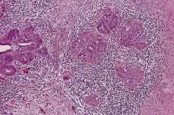
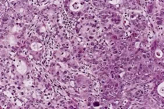
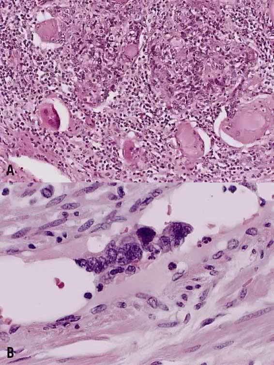
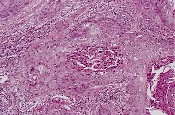
The category of small cell anaplastic carcinoma should be reserved for those cervical tumors that resemble small cell or intermediate cell anaplastic carcinomas of the lung.16–20 Small cell anaplastic carcinoma is made up of sheets and cords of cells diffusely infiltrating a delicate fibrovascular stroma, simulating lymphoma (Fig. 4A). The tumor cells have round, oval to elongated, small to intermediate sized nuclei. The nuclear chromatin ranges from coarsely granular to dark and sometimes smudged (Fig. 4B). Nucleoli are inconspicuous and mitotic figures are frequent. The cytoplasm is scant resulting in nuclear molding and high nuclear cytoplasmic ratios. Necrosis and crush artifact are common. Desmoplastic reaction of the stroma is minimal or absent.

Fig. 4. Small cell anaplastic carcinoma. ( A) A diffuse infiltrative pattern by sheets and cords of malignant cells. The crush artifact with smudged nuclei is also characteristic of this neoplasm. ( B) The small nuclei are hyperchromatic and have coarsely granular compact chromatin. The nucleoli are not visible nor inconspicuous. The cytoplasm is scant, resulting in nuclear molding. Mitotic activity is high.(Hematoxylin-eosin stain, original magnification A: 100, B: 400.)

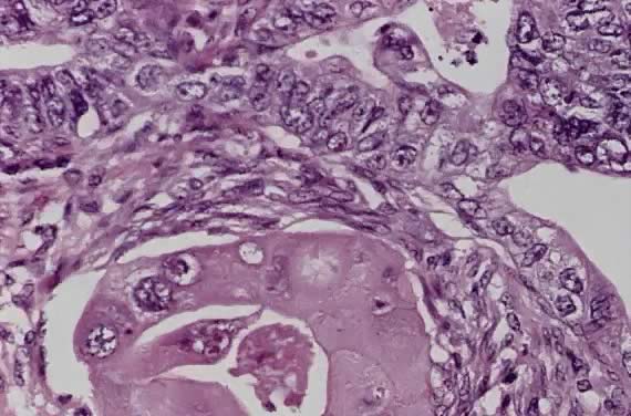
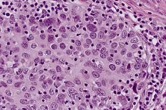
In recent years, it has become clear that neuroendocrine carcinomas are not limited to the small sized tumor cells. Poorly differentiated large cell carcinomas may express neuroendocrine differentiation by immunohistochemistry. These large tumor cells have the nuclear characteristics common to neuroendocrine carcinoma, namely coarsely granular chromatin (Fig. 5A). However, the nucleoli vary from indistinct to medium size. In addition to diffuse infiltrative pattern, tumor cells are also arranged in trabeculae, ribbons and rosettes (Fig. 5B), and the cytoplasm varies from scant to moderate in amount. In a recently proposed classification, neuroendocrine carcinomas are divided into small and large cell types.13

Fig. 5. Large cell neuroendocrine carcinoma. ( A) Tumor cells have large oval of elongated hyperchromatic nuclei which contain coarsely granular chromatin. The nucleoli are small or absent. There is a small amount of eosinophilic cytoplasm. ( B) Tumor cells are arranged in cords and ribbons. ( C) Same tumor as in A. Diffuse cytoplasmic deposit of chromogranin indicative of neuroendocrine differentiation. ( D) Neuroendocrine carcinoma ( right lower field) coexists with adenocarcinoma ( left upper field ).(Hematoxylin-eosin stain, original magnification A: 400, B: 200, D: 100; C: immunohistochemical stain for chromogranin, original magnification 400.)

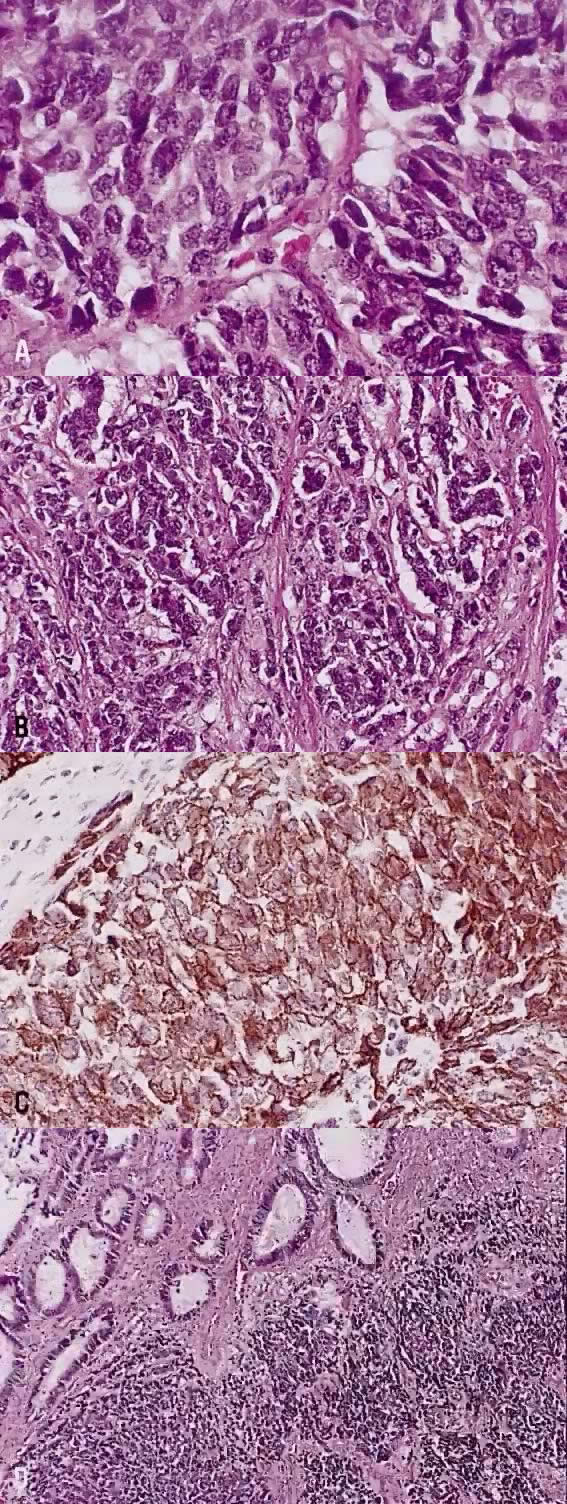
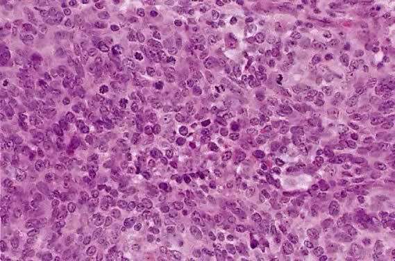
All poorly differentiated small cell and large cell carcinomas with the above morphologic features should be studied by immunohistochemistry for neuroendocrine markers (neuron-specific enolase, chromogranin, synaptophysin; Fig. 5C). By immunohistochemical stains, 20% to 100% of small cell anaplastic carcinomas were reported to express neuroendocrine differentiation.13,18 This subset is classified as small cell neuroendocrine carcinoma. Neuroendocrine carcinomas sometimes coexist with adenocarcinoma or squamous carcinoma (Fig. 5D).

Small cell anaplastic carcinomas are believed to derive from multipotential cell or argyrophilic cells in the basal cell layer of the endocervical mucosa.21 This tumor has propensity to occur in young women (mean age, 36 years vs 50 years in patients with small cell squamous carcinoma),22 strong association with HPV type 18,23 and aggressive behavior.22,24 When treated with radical hysterectomy and pelvic nodal dissection, 4 of 6 (67%) patients with small cell anaplastic carcinoma had pelvic nodal metastasis and 5 of 8 (63%) patients developed tumor recurrence, compared with 18% nodal metastasis and 9% tumor recurrence in women with small cell squamous carcinoma.22 Spread beyond the uterus and pelvis is common even for stage I tumor. It is recognized as one of the most aggressive types of cervical cancer with a 5-year survival rate of 14% in one study.24

Sevin and colleagues25 reported 12 women with small cell anaplastic carcinoma, including one FIGO stage IA, 10 stage IB, and one stage II. When compared with stage IB and II cervical squamous carcinoma and adenocarcinoma combined, small cell anaplastic carcinoma had a higher frequency of vascular lymphatic space invasion (82% vs 62%), more frequent lymph node metastasis (45.5% vs 18.9%), and lower 5-year survival rate (36.4% vs 71.6%). Only 42% (5 of 12) of patients were disease free at the time of report. In view of these findings, a combined therapy of surgery, radiotherapy, and cytotoxic chemotherapy is recommended.25

The value of separating squamous carcinoma by cell type was evaluated using the data of the Gynecologic Oncology Group (GOG). Among women with stage I squamous carcinoma treated surgically, the cell type was not predictive of pelvic nodal metastasis and outcome.26,27 The percentages of patients progression-free at 5 years were 84% for large cell keratinizing and 74% for large cell nonkeratinizing (p = not significant), and 75% for grade 1, 82% for grade 2, and 78% for grade 3 (p = not significant).26

The consistency and reproducibility among pathologists in separating large cell keratinizing and nonkeratinizing tumors based on cervical biopsy specimens cause problems in interpretation. In the GOG study of women with IIB to IVA squamous carcinoma treated by radiation therapy, when the histologic criteria were modified to include all tumors with individual cell keratinization in the large cell keratinizing category, this group had a significantly higher recurrence/death rate than the large cell nonkeratinizing group (65.8% vs 53.5%, p = .0074).28

Histologic Grade

The histologic grade reflects the degree of differentiation of the tumor cells. The most commonly used grading system for squamous carcinoma is a modification of the original Broders' system consisting of three grades based on the amount of keratin, the degree of nuclear atypia, and the mitotic activity. Grade 1, well-differentiated lesions exhibit abundant intercellular bridging, cytoplasmic keratinization, and keratin pearls. The cells are relatively uniform with minimal nuclear pleomorphism. The mitotic rate should be less than 2 per high-power field.15 Grade 2, moderately differentiated lesions show primarily individual cell keratinization, moderate nuclear pleomorphism, and up to four mitotic figures per high-power field.15 Grade 3, poorly differentiated lesions show little evidence of squamous differentiation. The tumor cells are immature, with marked nuclear pleomorphism, scant cytoplasm, and more than four mitotic figures per high-power field.15

In surgically treated patients with stage I and II squamous carcinomas, some studies have found histologic grade to influence prognosis and pelvic nodal metastasis.27 In the study of stage I squamous carcinoma from GOG, the histologic grade was correlated with the frequency of pelvic nodal metastasis. Pelvic nodal metastasis occurred in 9.7%, 13.9%, and 21.8% of grade 1, grade 2, and grade 3 tumors, respective.27 A comparable results were reported by Fuller and colleagues29 (Table 2). Some investigators of stage IB and IIA carcinomas have found that grade 3 tumors were larger and had a higher incidence of lymph node metastasis than lower-grade lesions.29 Fuller and colleagues29 found that 25% of grade 3 tumors had pelvic lymph node metastases, compared with 9% and 16% in grades 1 and 2 tumors, respectively.

TABLE 2. Frequency of Lymph Node Metastasis in Surgically Treated Cases


Delgado et al27 (745 stage I squamous carcinoma >3 mm in depth)

Fuller et al29 (431 IB and IIA squamous carcinoma, adenocarcinoma, and others)

FIGO stage

I

15.5%

IB

15%


IIA

22%

Stromal invasion

3–5 mm

3.4%

<1/3

0%


6–10 mm

15.1%

2/3

12%


11–15 mm

22.2%

>2/3

24%


16–20 mm

38.8%


21 mm

22.6%


LVSI

Absent

8.2%

Absent

14%


Present

25.4%

Lymphatic

36%


Parametrial disease

Absent

13.5%

Not evaluated


Present

25%


Tumor size

Occult

8.9%


Gross

20.9%


Histologic grade

Grade 1

9.7%

Grade 1

9%


Grade 2

13.9%

Grade 2

16%


Grade 3

21.8%

Grade 3

25%

Not significant


Cell type

LCK

17.2%

Squamous carcinoma

18%


LCNK

17.2%

Adenocarcinoma

18%


Small cell and others

17.6%

Adenosquamous carcinoma

17%


LCK, large cell keratinizing; LCNK, large cell non-keratinizing.

Among 445 patients with stage IIB through IVA squamous carcinoma treated by radiation therapy following GOG protocols, the histologic grade had no impact on prognosis.28 Similar findings were reported by others.30,31

In the early 1980s, a malignancy grading system (MGS) was proposed as an alternative grading system.32 In the MGS system eight morphologic parameters of a tumor, consisting of four characteristics of the tumor cell population (structure [i.e. papillary vs solid], degree of cell differentiation, nuclear pleomorphism, and mitotic activity] and four characteristics of the tumor-host relation (mode of invasion, stage of invasion, extent of vascular invasion, degree of host inflammatory response [lymphoplasmacytic]) are evaluated and scored on a one- to three-point scale with minimum score of 8 points and a maximum score of 24 points.32 When this grading system was applied to 445 IIB to IVA squamous carcinoma patients treated by radiation therapy, the recurrence/death rates were 32.9% for women whose tumors scored up to 12 points, 57.3% for tumors scoring 14 to 16 points, and 64.8% for neoplasms with scores of more than 18 points (p = .004).28

Variants of Squamous Carcinoma

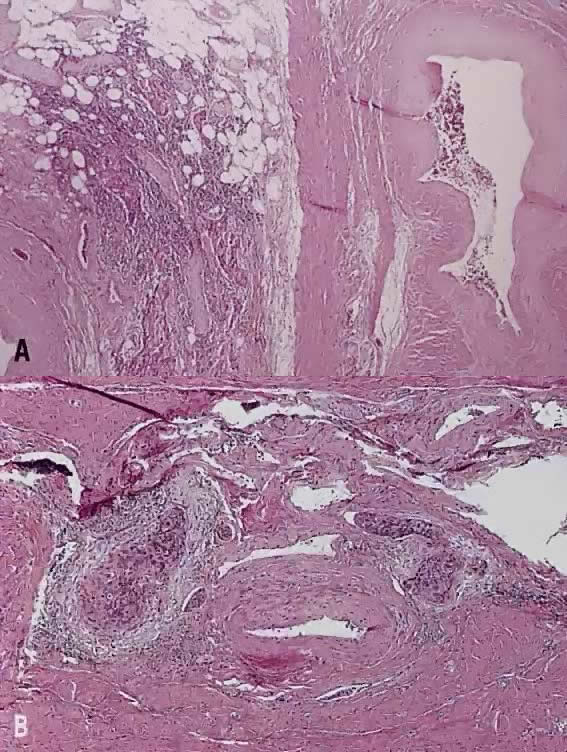
Verrucous squamous carcinoma of the cervix, like that of other sites, represents a special variant of well differentiated squamous carcinoma. Grossly, these tumors appear exophytic and warty, and may simulate a condyloma acuminatum. Histologically, the cells in this variant show orderly maturation and lack cytologic atypia. The tumor grows by expansion with smooth pushing margins, as opposed to the infiltrating pattern of conventional squamous carcinoma.

To differentiate verrucous squamous carcinoma from either condyloma, pseudoepitheliomatous hyperplasia or typical squamous carcinoma, full-thickness biopsy specimens are necessary. Some squamous carcinomas have a verrucous appearance, but show severe nuclear atypia and foci of invasion by nests or single cells. These tumors behave like conventional squamous carcinoma and should be identified as such. Condyloma acuminatum has prominent koilocytosis and delicate fibrovascular cores, as opposed to the compressed cores and confluent epithelial growth pattern seen in verrucous squamous carcinoma. Condylomas also lack the expansile, endophytic extension into the stroma seen in verrucous squamous carcinomas.

None of at least 18 cases of verrucous carcinoma reported in the literature had lymph node metastasis. Direct extension into the vagina occurred in 6 women and the endometrium in 3 cases. About 40% to 50% had local recurrence, due to either incomplete excision or resistance to radiation therapy.33

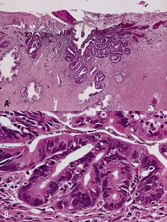
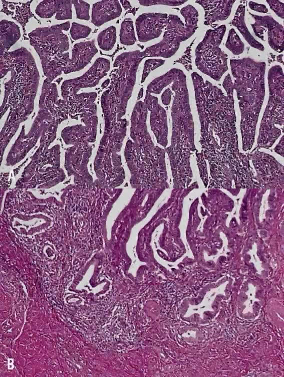
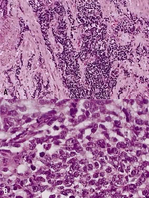
Papillary squamous carcinoma of cervix is characterized by highly dysplastic squamous cells forming papillary fronds with thin fibrovascular cores (Fig. 6A). Not surprisingly, the gross appearance of this lesion may be warty or fungating as in verrucous squamous carcinoma. In recent years, terms such as papillary squamotransitional cell carcinoma and transitional cell carcinoma were used.34,35 Although there is some morphologic resemblance to the poorly differentiated transition cell carcinoma of the urinary bladder, by immunostains, only 2 of 21 (9.5%) tumors expressed a distinct marker of transitional cells, cytokeratin 20. Thus these tumors are squamous cell type.34

Fig. 6. Papillary squamous carcinoma. ( A) Multiple papillary fronds are supported by delicate fibrovascular cores. ( B) Irregular nests of invasive squamous cells at the base of papillary structures. Desmoplastic reaction occurs around the tumor cells.(Hematoxylin-eosin stain, original magnification A: 40, B: 40.)

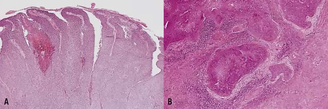
In a series of 32 women, their age varied from 22 to 93 years (mean, 50 years).34 They presented with abnormal bleeding or abnormal cervical smears. The tumor size ranged from 0.7 cm to 6 cm, mean 3.0 cm. The diagnostic problems are indicated by the fact that only 20 of 32 (63%) specimens were considered to be adequate. The remaining 37% were too superficial to determine in situ or invasive carcinoma. Of those with suitable specimens, 90% (18 of 20) had stromal invasion.34 Full-thickness biopsy specimens are necessary to distinguish in situ from invasive lesions. This is because invasion is evident only in the stroma beneath the papillary surface components (Fig. 6B).

In a series of 9 cases, 1 tumor was an in situ lesion, whereas the remaining 8 cases ranged from stage I to stage IV invasive carcinoma.36 In another study, the FIGO stage was known in 11 women, including 2 stage 0, 3 stage IA, 2 stage IB, 1 stage IIA, 1 stage IIB, and 2 stage IIIB.34 In this series, only 12 women had follow-up information, 3 patients died all stage IIB or higher, mean survival 13 months after diagnosis.34 One patient developed ovarian metastasis, 1 patient had vaginal recurrence 12 years later, and in 1 patient, adenosquamous carcinoma of the endometrium occurred. Five women were alive and well, and 3 died of other causes. Late recurrence and metastasis were known to occur.36

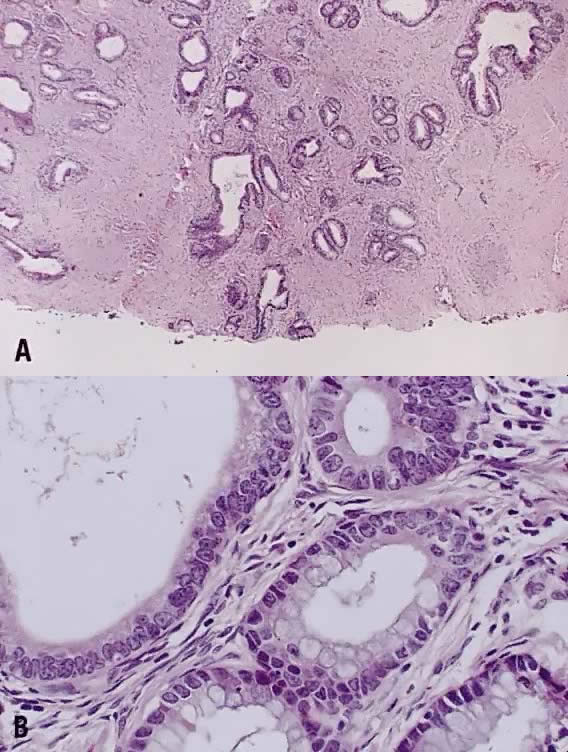
Circumscribed carcinoma of the uterine cervix was described in 1977 by Hasumi and associates37 In their study, the tumors were characterized by solid cords of cells with neither squamous nor glandular differentiation, surrounded by a dense lymphocytic infiltrate, which occasionally contained a considerable number of eosinophils and plasma cells. The tumor cells were fairly monomorphic, with large nuclei, one or more nucleoli, and clear to eosinophilic, granular cytoplasm. Many mitotic figures were seen. All 39 cases reported measured larger than 5 mm in depth, but only 2 (5%) had lymph node metastases at the time of surgery, compared with 18% of ordinary squamous carcinomas of comparable stage. Improved 5 year survival was also seen in these cases (97% vs 79%, p < .05).37

Studies of similar, if not identical, tumors use the term lymphoepithelioma to indicate the histologic likeness to the lymphoepithelioma of the nasopharynx and the malignant lymphoepithelial lesions of the salivary glands.38,39 In a study by Tseng and associates,40 15 such tumors were compared with the conventional squamous carcinomas by polymerase chain reaction (PCR). Epstein-Barr viral gene sequences were found in 11 of 15 tumors (73%), compared with 4 of 15 (27%) of the usual squamous carcinomas (p = .001). Interestingly, HPV 16 and 18 types were detected in 20% (3 of 15) of these tumors, compared with 80% (12 of 15) of the usual squamous carcinomas (p = .001).40

Although the total number of cases in the literature is too small for complete understanding of these neoplasms, they appear to have a better prognosis than the conventional squamous carcinoma. After radical hysterectomy all 15 patients were alive and well.40

Spindle cell squamous carcinoma is a rare variant of poorly differentiated carcinoma that may be confused with either melanoma or sarcoma.5,41 This tumor is composed of cells with large, spindle shaped, or oblong nuclei arranged in fascicles (Fig. 7). Keratin formation and the nesting pattern typical of epithelial tumors may be absent. Stromal changes such as heavy collagen deposition may give the appearance of a fibrosarcoma or osteosarcoma. When confronted with such a lesion electron microscopic examination or immunohistochemistry is often required to identify the epithelial nature. A positive immunohistochemical stain for cytokeratin and the demonstration of desmosomes and tonofilaments by electron microscopy distinguish these lesions from mesenchymal tumors.

Fig. 7. Squamous carcinoma, spindle cell type. Elongated tumor cells are arranged in bundles simulating spindle cell sarcoma. Immunohistochemical stain for cytokeratin is positive to confirm carcinoma (not shown).(Hematoxylin-eosin stain, original magnification 200.)

One may also rarely see abnormal spindle cells in the cervical stroma adjacent to typical squamous carcinoma. This so called pseudosarcoma was reported by Watty and colleagues42 who described individual atypical stromal cells with elongated, pleomorphic nuclei and frequent multinucleation. Rare abnormal mitotic figures were seen. The authors felt that this change represented a response to the nearby tumor and noted that similar lesions have been reported in squamous carcinomas of the head and neck region.

Back to Top
MALIGNANT GLANDULAR NEOPLASMS

Cervical carcinomas with glandular differentiation are of heterogeneous cell types, diverse growth patterns and variable differentiation. The majority of glandular neoplasms are pure adenocarcinomas. As many as 25% to 50% of cervical adenocarcinomas contain malignant squamous elements.5 Of these, adenoid cystic carcinoma and adenoid basal carcinoma are sufficiently distinct to be recognized as separate entities. A simplified histologic classification of in situ and invasive glandular neoplasms according to the cell type is listed below.

  Adenocarcinoma in situ
  Adenosquamous carcinoma in situ
  Microinvasive adenocarcinoma
  Invasive adenocarcinoma

  1. Endocervical type
    1. Usual type
    2. Minimal deviation adenocarcinoma (adenoma malignum)
    3. Well-differentiated villoglandular adenocarcinoma

  2. Endometrioid type
  3. Clear cell type
  4. Papillary serous type
  5. Intestinal type
  6. Mesonephric type
  7. Mixed type

  Adenosquamous carcinoma
  1. Mature type
  2. Signet-ring type (mucoepidermoid carcinoma)
  3. Glassy cell type

  Adenoid cystic carcinoma
  Adenoid basal carcinoma

Adenocarcinoma in Situ

Morphologic changes less severe than adenocarcinoma in situ have been described and designated as glandular atypia or glandular dysplasia.5,43 These lesions are detected in specimens removed for adenocarcinoma in situ or cytologic diagnosis of atypical glandular cells of undetermined significance. In the endocervical curettage specimens and cervical biopsy specimens, atypical endocervical cells present with nuclear enlargement, irregularity, and multinucleation in a background of chronic cervicitis. The adjacent squamous mucosa may have condyloma or low-grade dysplasia.5,43 These changes are probably reactive atypia and do not have malignant potential. On the other hand, dysplastic glands lined by endocervical cells with moderate or more severe degree of nuclear atypia, enlarged hyperchromatic nuclei, two to three stratified layers and increased mitotic activity are potential precursor of adenocarcinoma. Dysplastic change also occurs in glands lined by ciliated tubal metaplastic cells. The morphologic criteria and biologic potential of glandular dysplasia remain to be defined.

The entity of cervical adenocarcinoma in situ has been widely accepted and recognized as the precursors of invasive adenocarcinoma. Endometrioid43 and clear cell types44 of adenocarcinoma in situ have also been described. More than 50% of adenocarcinomas in situ were found to have coexisting squamous dysplasia or carcinoma in situ. When squamous carcinoma in situ contains mucus secreting cells, Steiner and Friedell45 designated the lesion as adenosquamous carcinoma in situ.

The majority of adenocarcinomas in situ are detected initially in the cervical smears of asymptomatic women or incidentally in hysterectomy specimens removed for benign conditions. Some are found in cervical biopsy specimens, endocervical curettage specimens, or cone specimens removed for squamous neoplasia. Colposcopic findings are nonspecific, such as patchy acetowhite lesion in the cervical canal, fused and papillary columnar villi, and abnormal vessels.

Most adenocarcinomas in situ begin in the region of the squamocolumnar junction and spread proximally (Fig. 8A). With few exceptions, both the endocervical mucosal surface and the underlying glands are involved. The affected surface may be flat, papillary or villous in appearance. The neoplastic glands form budding and crowded back to back glands. Excessive proliferation within large glands sometimes results in a cribriform pattern. The most characteristic features are the preservation of normal branching patterns, the smooth configuration of the glandular profiles and normal fibromuscular stroma without desmoplasia (Fig. 8A).

Fig. 8. Adenocarcinoma in situ, endocervical type. ( A) The neoplastic glands retains the branching and budding pattern of normal endocervical glands. These glands have smooth borders and are surrounded by normal fibromuscular stroma without desmoplastic reaction. At the base of tumor, malignant cells replace normal endocervical cells. ( B) Higher magnification to reveal tall columnar neoplastic cells with nuclear stratification, hyperchromasia, elongation, and irregularity.(Hematoxylin-eosin stain, original magnification A: 40, B: 100.)

Over 80% of cervical adenocarcinomas in situ are made up exclusively or predominantly of mucus-secreting endocervical cells.5 Endometrial cells, intestinal cells, and clear cells occur in the remaining 20%. Normally, endocervical cells are arranged in a single layer and have basally located small nuclei. Nucleoli and mitotic figures are absent or rarely observed. Neoplastic endocervical cells demonstrate nuclear pseudostratification, enlargement, hyperchromasia and elongation (Fig. 8B). Nucleoli are multiple and mitotic figures are easily identified. The cytoplasm appears basophilic, clear or vacuolated. In the deeper portion of the endocervical glands, a sharp transition between the normal and neoplastic cells is apparent.

In the endometrioid adenocarcinoma in situ the cells have densely eosinophilic cytoplasm, which is less abundant than in the endocervical type. The intestinal type consists of absorptive cells with brush borders and goblet cells with discrete vacuoles.

Squamous cell dysplasias and carcinomas in situ coexist with 35% to 71% of adenocarcinomas in situ.46–48 Rarely, microinvasive squamous carcinoma has been recorded.47 The squamous and glandular elements often merge into one lesion.

Although the diagnosis of adenocarcinoma in situ may be made on the basis of cytology and biopsy specimens, it is difficult to separate in situ from well-differentiated invasive adenocarcinoma. As both lesions may occur concurrently, cervical conization is usually performed for a definitive diagnosis.

Estimates based on conization and hysterectomy specimens have found adenocarcinoma in situ extending as deep as 3 to 5 mm from the mucosal surface. The linear extent along the cervical canal varies from 0.5 to 25 mm with a mean of 12 mm.49 If measured from the external os, these lesions may reach up to 30 mm.50 This implies the need for deep conization to encompass the entire lesion. Young women, who prefer to retain the uterus, may be treated by conization alone and followed regularly by endocervical aspiration or curettage.49 However, even if the surgical margins of the conization specimen appear uninvolved, residual tumor may exist. A possible explanation for this is a multifocal disease, which is estimated to be 15%.49

The importance of assessing surgical margins of conization specimens is emphasized in the study of Wolf and associates.48 They studied 61 women (mean age, 35.9 years) with adenocarcinoma in situ. Of the 55 women who underwent cone biopsies, 50 had known status of cone margins, and 44 had subsequent hysterectomy. The conization margins were positive in 23 of 50 (46%) women, and 19 of these had hysterectomy. A residual tumor was found in 10 of 19 (53%) hysterectomy specimens, including adenocarcinoma in situ in 4, invasive adenocarcinoma in 5, and glandular atypia in 1.48 Three of 4 women, who received no further treatment, were well for 2 to 9 years. The fourth woman was lost to follow-up.48

The conization margins were negative in 27 of 50 patients (54%), and 21 had had a hysterectomy. A residual tumor was found in 7 of 21 (33%) cases, including invasive adenocarcinoma in 3 (2 neoplasms 2 mm or less, 1 tumor 5 mm), adenocarcinoma in situ in 3, and glandular atypia in 1.48 Of 6 women who refused hysterectomy after cone biopsy, 2 were lost to follow-up, 1 developed 5-mm-deep invasive adenocarcinoma 2 years after the cone biopsy, 1 developed glandular atypia which was treated by cone biopsy, and the remaining 2 have no evidence of disease.48 These findings provide sufficient evidence of the malignant nature and the multifocality of adenocarcinoma in situ. When treated by conization alone, a close follow-up with endocervical samples is recommended.

Adenosquamous Carcinoma in Situ

The entity of adenosquamous carcinoma in situ described by Steiner and Friedell45 closely resembles squamous cell carcinoma in situ. Intermixed with the dysplastic squamous cells, however, are cells with vacuolated or basophilic cytoplasm. Mucicarmine and periodic acid-Schiff (PAS) stains reveal mucin production in these cells. Similar change sometimes occurs in the vicinity of adenosquamous carcinomas.

Microinvasive Adenocarcinoma

Several investigators have attempted to define the morphologic criteria for microinvasive adenocarcinoma. Teshima and associates51 defined early adenocarcinoma as less than 5 mm of stromal invasion measuring from the mucosal surface. All 30 patients in this study were treated by hysterectomy and 1 woman developed tumor recurrence. This patient had a tumor, which was 3 mm in depth, did not involve the capillary-lymphatic spaces or pelvic lymph node, but recurred in the vaginal cuff. Buscema and Woodruff52 also reported a 3 mm deep, adenosquamous carcinoma that metastasized widely. Among women with stage I and II cervical adenocarcinoma up to 5 mm in depth, Berek and colleagues53 found that 2 (8%) of 24 women had pelvic lymph node metastasis and the overall 5-year survival rate was 92%. Thus, adenocarcinomas as superficial as 5 mm have a small risk of pelvic nodal metastasis.

In a recent study of 77 early invasive adenocarcinomas by Ostor and associates,54 microinvasive adenocarcinoma was defined as less than 5 mm in depth. The invasive tumor was measured from the mucosal surface to the deepest point of tumor. This type of measurement is referred to as tumor thickness by other authors. The tumor thickness was 0.1 to 1.1 mm in 12 cases, 1.1 to 2.0 mm in 12 cases, 2.1 to 3.0 mm in 19 cases, 3.1 to 4.0 mm in 22 cases, and 4.1 to 5.0 mm in 12 cases. The histologic type of these tumors is heterogeneous, including adenocarcinoma in situ with early stromal invasion, well-differentiated adenocarcinoma, and papillary villoglandular adenocarcinoma. Seven (9%) tumors had vascular lymphatic space invasion, and 21 (27%) neoplasms had multifocal lesion involving both cervical lips. Other findings included 66% of women had abnormal glandular cells in the Pap smear, 38% had abnormal colposcopic findings, and 53% had used oral contraceptive pills.54

Treatment modalities in the study of Ostor and associates54 included cold-knife conization and simple to radical hysterectomy. There was no parametrial involvement in any of the 26 radical hysterectomy specimens. Pelvic lymph nodes in 48 women undergoing nodal sampling were all negative. Twenty-three women had one or both ovaries removed, and all were free of tumor.54 Two women had tumor recurrence. The first, a 62-year-old woman whose original tumor was 3.2 mm in depth, 21 mm in length, and 670 cubic mm in volume had total abdominal hysterectomy, bilateral salpingo-oophorectomy, and external pelvic irradiation. Five years later, she developed recurrent tumor in the vaginal vault, which was excised. Four years later, tumor recurred in the vault and treated by upper vaginectomy. This patient died postoperatively. In the second woman, the original tumor was 5 mm in depth, 10 mm in length, and 450 cubic mm in volume. Nine years following total abdominal hysterectomy, squamous carcinoma was found in the vaginal vault.54

The study of Ostor and associates54 provides an alternative approach to express the extent of stromal invasion by measuring from the surface of the lesion to the deepest point of tumor in the form of tumor thickness. However, not all early invasive adenocarcinomas arise from the mucosal surface. Isolated foci of invasion sometimes take place from the periphery of endocervical glands involved by adenocarcinoma in situ (Fig. 9). These glands may be superficial or deep. How to measure these tumors remain to be clarified. In addition, glandular tumors less than 5 mm in depth, but have potential to metastasize, such as poorly differentiated adenocarcinoma, serous carcinoma, and adenosquamous carcinoma, are best excluded from the microinvasive category.

Fig. 9. Microinvasive adenocarcinoma forms multiple, irregular tongue-like protrusions from the periphery of endocervical glands involved by adenocarcinoma in situ. These protrusions are associated with fibrotic stroma and chronic inflammation ( right two thirds ). In contrast, adenocarcinoma in situ retains smooth borders ( left one third ).(Hematoxylin-eosin stain, original magnification 200.)

Invasive Adenocarcinoma

The clinical presentation and gross appearance of cervical adenocarcinoma are basically similar to those of squamous carcinoma. In a series of 55 women with IB adenocarcinoma by Greer and colleagues,55 44% of women had no symptoms and 58% (32 of 55) had no gross lesions. The diagnosis was suspected on the basis of abnormal cervical smears in 16 women (29%). Of these, 15 required conization for diagnosis.55

Cervical Adenocarcinoma, Endocervical Type

Depending on the degree of differentiation, the predominant growth pattern may be glandular, mucinous (colloid), papillary, solid or mixed.56 In endocervical adenocarcinoma of the usual type, well-differentiated tumors present with small, back to back glands. The lining cells have eosinophilic or finely vacuolated pale staining cytoplasm resembling normal endocervical cells. In poorly differentiated neoplasms, anaplastic cells are arranged in solid nests and some have a signet-ring appearance. Moderately differentiated tumors have a mixture of glands and solid nests (Fig. 10). Tumors with abundant extracellular mucin are referred to as colloid type. The tumor cells resemble endocervical or intestinal type.

Fig. 10. Adenocarcinoma, endocervical type, moderately differentiated. Tumor cells form small irregular glands and solid nests. The cytoplasm is vacuolated and mucinous in appearance. Nuclear atypia is apparent.(Hematoxylin-eosin stain, original magnification 100.)

An extremely well-differentiated adenocarcinoma closely resembling normal endocervical cells has been designated as adenoma malignum,57 or minimal deviation adenocarcinoma.58 This variant deserves special recognition, because of its difficulty to diagnose. The tumor retains the branching pattern of normal endocervical glands, presents with minimal nuclear atypia and causes minimal stromal responses (Fig. 11A, B). Although similar characteristics have also been observed in well-differentiated endometrioid, clear cell and mesonephric carcinomas,59 most authors apply the term adenoma malignum57 only to the endocervical type.

Fig. 11. Minimal deviation adenocarcinoma, endocervical type. ( A) Neoplastic cells form branching, budding glands resembling normal endocervical glands. Tumor cells extend into the deep margin of conization specimen. ( B) Individual cells also closely mimic normal endocervical cells, having tall columnar configuration and abundant mucinous cytoplasm and basally located, small nuclei. On closer examination, nuclear irregularity and small nucleoli become evident.(Hematoxylin-eosin stain, original magnification A: 40, B: 400.)

The possibility of reaching a correct diagnosis is enhanced by the knowledge of cervical abnormality and a deep biopsy. The latter may provide clues as to the subtle irregularity in the size and shape of neoplastic glands. Encroachment of the blood vessels and nerve fibers adds further support for stromal invasion. Careful survey of individual lining cells reveals mild nuclear atypia and uneven chromatin. Nucleoli may be evident (Fig. 11B).

Patients with minimal deviation adenocarcinoma are reported to have a dismal outcome by some,57,60 but favorable survival rates by others.58,59 There is no question that the prognosis is strongly governed by the extent of local disease and pelvic nodal status. Patients with stage I or II neoplasm without pelvic nodal metastases are expected to have a favorable outcome following radical hysterectomy. The presence of pelvic nodal metastasis or advanced local disease adversely affects survival. Delay in correct diagnosis may account for some of the advanced cases.

Equally important, benign conditions, such as tunnel clusters of endocervical glands, hyperplastic mesonephric ducts, and endometriosis, should not be confused with minimal deviation adenocarcinoma. Immunoperoxidase stain for carcinoembryonic antigen can be helpful, because this tumor marker is negative in benign changes and positive in minimal deviation adenocarcinoma.61

Young and Scully62 reported a series of 13 villoglandular papillary adenocarcinomas of the uterine cervix. This tumor occurred mostly in young women (mean age, 33 years). The lesions were described clinically as polypoid, condylomatous, eroded, nodular, white, friable or fungating. Microscopically, the superficial portion of tumor consists of complex papillae lined by well-differentiated endocervical cells (Fig. 12A). In the deeper portion of the tumors, tumor cells form branching tubular glands pushing into fibrous stroma (Fig. 12B). In 6 women, the tumor was confined to the superficial one third of cervical wall, whereas deep invasion occurred in 2 women. After hysterectomy, no tumor recurrence was noted in 10 women observed for 2 to 14 years.62

Fig. 12. Villoglandular adenocarcinoma, endocervical type. ( A) On the surface are multiple papillary projections consisting of columnar cells and fibrovascular cores. ( B) The base of tumor has smooth borders without infiltrative pattern.(Hematoxylin-eosin stain, original magnification A: 100, B: 100.)

In a subsequent study of 24 cases by Jones and associates,63 the mean age was 37 years (27–54 years), and 50% were in their third decade of life. There was history of oral contraceptive use in 63% of women, this is in contrast to 28% among women having other types of cervical adenocarcinoma (p = .02). Grossly, the tumor had an exophytic polypoid appearance. Microscopically, the predominant cell type was endocervical in 50%, endometrioid in 33%, and intestinal cell in 17%. 33% had coexisting squamous dysplasia or carcinoma in situ. Five (21%) tumors were entirely in situ without stromal invasion; 75% (18 tumors) were superficial, confined to inner third of cervical wall; and 4% (1 tumor) invaded 75% of cervical wall.63

Treatment modalities included local excision or cone biopsy in 5 women, simple hysterectomy in 4 women, and radical hysterectomy in 15 women.63 All tumors were confined to the cervix without pelvic lymph node metastasis. All women were alive and well 7 to 77 months (mean, 36 months) later. In view of the distinct clinical and pathologic features, these tumors are separated from other cervical adenocarcinomas. A conservative treatment is considered to be justified, especially in young women who wants to retain fertility.63

It should be emphasized that the term villoglandular papillary adenocarcinomas should be reserved only for those tumors which meet the stringent morphologic criteria. The degree of nuclear atypia should be no worse than moderate in degree. The tumor borders should be smooth (Fig. 12B), and tumors made up of clear cells and serous cells are excluded. In the cervical biopsy specimens and endocervical curettage specimens, there are fragments of tumor that have villous pattern. However, in the subsequently excised specimens, some of these tumors prove to have poorly differentiated elements or have infiltrative borders. Thus, the diagnosis of well-differentiated villoglandular should be made on completely excised specimens.

Distinguishing endocervical and endometrial adenocarcinomas can be difficult on routinely stained sections. PAS and mucicarmine stains and immunohistochemical stains are helpful to identify the cell type. Mucinous secretion is evident in endocervical cells by mucicarmine and PAS stains. By immunohistochemisty, these cells frequently express carcinoembryonic antigen, but are negative for vimentin. Neoplastic endometrioid cells, on the other hand, are negative for cytoplasmic mucin and carcinoembryonic antigen, but are positive for vimentin.64,65 These stains, however, cannot be used to determine the anatomic site of origin. This is because 15% of endocervical adenocarcinomas belong to the endometrioid cell type, and endocervical mucinous metaplasia occurs in as many as 40% of primary endometrial carcinomas.5 Thus, the distinction between endocervical and endometrial tumors often requires additional clinical findings and tissue samples.5

Cervical Adenocarcinoma, Endometrioid Type

This group of tumors has the appearance of grade I or grade II adenocarcinoma of the endometrium. The predominant growth pattern is glandular or less commonly papillary. The lining cells are tall, columnar, and have densely basophilic or eosinophilic cytoplasm. The diagnosis is justified only if the endometrium is normal after careful sampling for histologic examination. Sometimes, mature metaplastic squamous cells occur within the neoplastic glands. These tumor are designated as adenoacanthomas.5,56

Cervical Adenocarcinoma, Clear Cell Type

Cervical clear cell adenocarcinoma affects women with or without exposure to diethylstilbestrol in utero. The age distribution has a bimodal peak, one around 20 years of age and the other in the fifth and sixth decades of life.66 The most common complaint is vaginal bleeding and, on examination, a polypoid, exophytic or fungating tumor is visible.66,67 About two thirds are FIGO stage IB and the remaining are stage II or higher.67

In the DES exposed progeny, the cervical clear cell carcinomas are located mainly in the ectocervix and rarely in the endocervix.68

Under the microscope, the tumor cells have distinct clear, empty appearing cytoplasm and enlarged, hyperchromatic nuclei, which project into the apical cytoplasm, the so called hobnail appearance. The clear cytoplasm is attributed to the accumulation of abundant glycogen similar in appearance to that seen in secretory endometrial cells. The cells grow predominantly in tubulocystic, papillary or solid pattern. This growth pattern is important prognostically. The most favorable outcome is associated with the tubulocystic pattern, followed by the papillary and solid patterns.

Depth of stromal invasion, FIGO stage and pelvic nodal status are key prognostic indicators.68 Five-year survival rates are better than 90% for stage I cases. Most patients had tumor recurred in 3 years. Rare case of recurrence occurred 20 years after therapy. Metastases to the lung and supraclavicular lymph nodes were more common than squamous carcinoma.68

Cervical Adenocarcinoma, Papillary Serous Type

A few cervical adenocarcinomas indistinguishable from papillary serous carcinoma of the ovary have been reported.69,70 An aggressive behavior was noted.70

Cervical Adenocarcinoma, Mesonephric Type

Most of the mesonephric duct carcinomas reported earlier belong to the clear cell adenocarcinoma category by current classification. Of the few true mesonephric adenocarcinomas, the tumor cells form glands, tubules or microcysts. One important diagnostic feature is the presence of eosinophilic hyalin material in the lumen, which is positive with PAS stain and negative with mucicarmine stain. It should be noted that benign adenomatous hyperplasia of mesonephric duct can be found deep in the cervical stroma, mimicking adenoma malignum and mesonephric adenocarcinoma.71

Clement and associates72 reported on a series of 8 women 34 to 71 years of age (mean, 54.5 years) who complained of vaginal bleeding. These tumors presented with a variety of growth patterns, including ductal, tubular, retiform, solid, and sex cord-like structures. Rarely, spindle cells resembled endometrial stromal sarcoma and contained osteoid and chondroid metaplasias.72 All tumors were stage IB. Two of 5 women underwent pelvic nodal dissections had micrometastases.72 Follow-up data indicated that 3 women were alive and well, 2 experienced recurrences, and 1 died of unrelated clear cell carcinoma of the ovary.72

Adenosquamous Carcinoma

Glucksmann and Cherry73 subdivided adenosquamous carcinomas into mature, well-differentiated (35%) and immature, poorly differentiated (65%) types. The latter of which is further divided into signet-ring (44%) and glassy cell (21%) types. Glassy cell carcinoma is estimated to comprise 2% of all cervical cancer and 13% of glandular neoplasms.73

Adenosquamous Carcinoma, Mature Type

In the mature type, both the glandular and squamous carcinomas are well to moderately well differentiated to be readily recognized without special stains. In most cases, the squamous components have the appearance of large cell nonkeratinizing type (Fig. 13). Keratinizing and small cell types are less common.5,56 The cells contain abundant eosinophilic cytoplasm and evidence of individual cell keratinization. Sometimes, the squamous cells have clear cytoplasm with abundant glycogen. Glandular formation is evident in most of the adenocarcinomas.

Fig. 13. Adenosquamous carcinoma, mature, well-differentiated type. Large malignant squamous cell carcinoma with abundant eosinophilic cytoplasm ( right ); neoplastic columnar cells form glandular lumens ( left ).(Hematoxylin-eosin stain, original magnification 200.)

Adenosquamous Carcinoma, Signet-Ring Type

This tumor consists of solid nests and sheets of tumor cells resembling nonkeratinizing squamous cell carcinoma (Fig. 14A). On closer examination, there are additional cells, which have basophilic, vacuolated or clear cytoplasm and compressed nuclei, resembling signet-ring cells (Fig. 14B).5,56 With the mucicarmine stain, the presence of mucinous product can be confirmed. Some of the tumors reported as “mucoepidermoid carcinoma”74 and “squamous cell carcinoma with mucin secretion” by Benda and associates75 and Ireland and colleagues76 fall into this category.

Fig. 14. Adenosquamous carcinoma, signet-ring type. ( A) Neoplastic squamous cells have abundant eosinophilic or clear cytoplasm and distinct cell borders. In addition, there are cells with basophilic cytoplasm. ( B) Higher magnification reveals signet-ring cells with basophilic vacuolated cytoplasm.(Hematoxylin-eosin stain, original magnification A: 100, B: 400.)

In the study by Benda and associates75 69 consecutive IB cervical cancers, including 55 (80%) squamous carcinomas, 12 (17%) adenocarcinomas, and 2 (3%) undifferentiated carcinomas, were stained by mucicarmine and PAS stains. Eighteen (33%) of 55 squamous carcinomas contained mucin in the cytoplasm. If these are accepted as mixed carcinoma, only 54% (37) of tumors remain in the squamous category (35% keratinizing, 16% nonkeratinizing, 3% small cell) and the frequency of mixed carcinoma is increased to 26%. This distinction is important as there is a difference in the risk of pelvic nodal metastasis. Six (33%) of 18 squamous carcinomas with mucin production had pelvic lymph node metastasis, compared with only 2 (5%) of 37 squamous carcinomas without mucin (p = .01).75 In a separate series of 23 squamous cell carcinomas studied by PAS and Alcian blue stains for mucin, only 2 (25%) of 8 women whose tumors demonstrated cytoplasmic mucin were alive after 3 years. In contrast, 13 (87%) of 15 women whose tumors lacked mucin survived (p < .01).76 These findings suggest that squamous cell carcinoma with mucin production has a more aggressive behavior than conventional squamous cell carcinoma.

Adenosquamous Carcinoma, Glassy Cell Type

Tumor cells of this type have abundant, eosinophilic, granular and ground-glass cytoplasm, sharp cell borders, uniformly round to oval nuclei, and giant nucleoli. Mitotic figures are abundant. Many eosinophils and plasma cells are noted in the stroma. In the original description of this entity,73 tumor cells had PAS positive material but no mucinous substance. However, in the study by Maier and Norris77 7 of 8 tumors studied by the mucicarmine stain were positive. Three of 8 neoplasms contained glandular lumens, and 3 others had squamous differentiation. One tumor had both glandular and squamous foci. By electron microscopic study, the glassy appearance corresponded to abundant polyribosomes and rough endoplasmic reticulum. The tonofilaments were scant.78 Although no intracytoplasmic lumens were identified, there were intercellular spaces lined by microvilli to suggest glandular differentiation. Some tumor cells also contained mucinous material in the cytoplasm.78 These histochemical and ultrastructural findings support the poorly differentiated nature of these neoplasms and the presence of glandular differentiation in some of the tumor cells.78

Adenoid Cystic Carcinoma

Adenoid cystic carcinoma of the female lower genital tract occurs most commonly in the Bartholin gland. This is followed by the uterine cervix, affecting primarily postmenopausal women in their seventh decade of life, about 20 years later than squamous carcinoma. The histologic features are similar to those occurring in the salivary gland. It is suggested that multipotential reserve cells in the endocervical glands acquire myoepithelial differentiation, which is not normally seen in the cervix.

Histologically, basaloid cells are typically arranged in cribriform glands with hyaline or mucinous material in the microcystic spaces. Tubules and solid nests are less common. Tumors with predominantly solid growth pattern may metastasize early. The individual cells have scanty cytoplasm and small, uniform, hyperchromatic nuclei. Mitotic figures are variable depending on the degree of differentiation.79

In about 50% of cases, squamous differentiation is apparent. In such cases, squamous cells replace the glandular lumens partially or completely. When squamous elements predominate, adenoid cystic carcinoma may not be recognized in a small biopsy specimen. Adenoid cystic carcinoma may also be associated with squamous carcinoma in situ, because of this some women are detected initially by abnormal cervical smears. Other types of adenocarcinoma, undifferentiated carcinoma, or sarcoma may sometimes coexist with adenoid cystic carcinoma.79,80

In one study, 3- to 5-year survival rates for patients with stage I adenoid cystic carcinoma were 56% compared with 27% for stage II. None of the patients with stage III or IV disease survived.81 Most of the treatment failures were caused by distant and/or local pelvic recurrence. Metastases occurred most frequently in the lung (44%) and less commonly in the bone, liver and brain. With only 32% of patients free of disease at last follow-up, the overall prognosis of adenoid cystic carcinoma is worse than that of squamous carcinoma or pure adenocarcinoma of the cervix.81 In a recent review, bulky early stage tumors are best treated with combined surgery and postoperative radiation therapy and chemotherapy with cisplatin.82

Adenoid Basal Carcinoma

This rare cervical tumor closely mimics basal cell carcinoma of the skin in its histologic appearance. Most patients were postmenopausal and asymptomatic. The cervix was grossly normal and lesions were detected initially by abnormal cervical smears. Microscopically, basaloid cells with scanty cytoplasm and uniformly small, round nuclei are arranged in solid nests. These nests are grouped together in a lobular pattern. Cells in the outermost layer have a distinct palisaded nuclear arrangement. Tubules and cribriform glands with mucinous material are prominent in some cases. Squamous metaplasia may occur. Mitotic activity is absent to less than one mitotic figure per nest.79 Vascular lymphatic invasion was not seen in any of the cases reported.79

Most adenoid basal carcinomas have coexisting squamous severe dysplasia or squamous carcinoma in situ. Microinvasive squamous carcinoma may evolve from the periphery of solid nests replaced by neoplastic squamous cells. All reported cases were FIGO stage IA or IB. The prognosis is excellent following hysterectomy. In a review of 26 cases reported in the literature, 85% of patients were alive without tumor recurrence. Three women (12%) died of other causes. Only 1 case is known to have died of disease with metastasis in the lung.83

The differential diagnosis includes adenoid basal hyperplasia, which occurs in the superficial endocervical glands. Adenoid cystic carcinoma presents with infiltrative cribriform glands associated with extracellular mucinous material or hyaline cylinders. Cytologic atypia is evident in the tumor cells. In the case of rare basaloid squamous carcinoma, the tumor cells are cytologically malignant and mitotically active. The infiltrative tumor nests are surrounded by desmoplastic stroma sometimes hyalinized to simulate hyaline cylinders.

In a study of 5 cases of adenoid basal carcinoma by Jones and colleagues,84 HPV DNA type 16 was detected in all tumors studied by PCR. No K-ras-2 point mutation was detected. One tumor had a point mutation of P53, and 4 others had weak reaction of P53 and 2 of these expressed WAF1 (an effector protein induced by wild-type P53). Grayson and associates85 studied 9 tumors by nonisotopic in situ hybridization (NISH) and PCR using E6 consensus primers to HPV. HPV DNA type 16 was detected in 4 tumors (44%) and HPV 33 in 2 cases (27%) by in situ method, and three tumors were positive by PCR. HPV DNA was thought to be integrated based on punctate and combined punctate and diffuse signals. It is postulated that HPV integration leads to E2 disruption and loss of suppressive effects on E6 and E7, which are related to P53 and RB gene product.85

Back to Top
OTHER MALIGNANT NEOPLASMS

Carcinoid Tumor and Neuroendocrine Carcinoma

Neuroendocrine neoplasms of the cervix have a wide morphologic spectrum. On the one end are well-differentiated carcinoid tumors that resemble similar tumor of the lung and gastrointestinal tract. On the other end of the spectrum are poorly differentiated carcinomas that are indistinguishable from small cell anaplastic carcinoma and large cell carcinoma of the lung. These morphologic features are discussed in the earlier squamous carcinoma section. Occasionally, squamous carcinoma and adenocarcinoma coexist with neuroendocrine carcinoma.

The prognosis of this group of tumors is related to the extent of disease and the degree of differentiation. Poorly differentiated tumors are highly aggressive with a propensity for local and distal spread.86,87

Miscellaneous Nonepithelial Neoplasms

Other less common malignant neoplasms include endocervical stromal sarcoma, rhabdomyosarcoma, leiomyosarcoma, Mullerian adenosarcoma, mixed mesodermal sarcoma, lymphoma, leukemia, malignant melanoma, choriocarcinoma, yolk sac tumor, Wilms' tumor and others. These are detailed elsewhere.5,20

Metastatic Secondary Tumors

Finally, secondary metastatic tumors to the uterine cervix occur most commonly from direct extension of adjacent malignancy, such as endometrium and vagina.5,20 Less commonly, tumor of the breast, ovary, colon, and stomach spread to the cervix by embolic phenomenon.5,20

Back to Top
PROGNOSTIC FACTORS

In recent years several large series have applied statistical models to identify important prognostic parameter of cervical carcinoma among women treated by radical surgery and radiation therapy. In the study of Sevin and associates,88 370 women with stage I or II carcinoma were treated by radical hysterectomy and pelvic lymphadenectomy. By univariate analysis, the disease free survival rates were closely related to the depth of stromal invasion, tumor size, presence or absence of lymphatic vascular space invasion, pelvic lymph node status, tumor volume, and clinical stage.88

Kamura and colleagues89 studied 211 women with stage IB and II cervical carcinoma treated by radical hysterectomy and pelvic lymphadenectomy. By univariate analysis, 5-year survival rates were correlated to the lymph node status, cell type (squamous carcinoma vs adenocarcinoma), tumor dimension, depth of cervical wall invasion measured by mm and expressed by inner third, middle third, and outer third; lymphatic vascular space invasion and parametrial invasion. The histologic grade was not evaluated in this study.89

Fuller and colleagues29 studied 431 women with stage IB or IIA carcinoma treated by radical hysterectomy, including 85% squamous carcinomas, 9% adenocarcinomas, 3% adenosquamous carcinomas, 2% small cell carcinomas and 1% clear cell carcinomas. A decreased survival was strongly related to the presence of pelvic lymph node metastasis.29 Among lymph node negative cases, adenocarcinoma cell type, increasing tumor size, deeper stromal invasion, and poor histologic grade were associated with decreased survival.29 Increasing tumor size and depth of invasion, and histologic grade were covariable and predictors of both lymph node metastasis and recurrence.29 In this study, poorly differentiated tumors had larger tumor size.29

Thus most authors have agreed that for surgically treated stage IB and II cervical carcinomas, the depth of stromal invasion, tumor dimension, the presence or absence of lymphatic vascular space invasion, and pelvic lymph node status are important prognosticators. The value of classification by cell type and histologic grade continues to be controversial.

By a multivariate stepwise regression model, it is possible to use several parameters to subdivide tumors into different risk groups. Kamura and associates89 developed such a scheme based on lymph node status, cell type, and tumor dimension. Sevin and colleagues88 identified three risk groups using depth of stromal invasion, lymphatic vascular space invasion, age, and lymph node status.

When treated by radiation therapy, somewhat different prognostic factors are identified. Barillot and colleagues90 reviewed 1875 women with cervical carcinoma treated by radiation therapy, including stage IA/IB (25.5%), IIA (12%), IIb (29%), IIIA (5%), IIIB (25%), and IV (3.5%). By univariate analysis of stage I/II cases: FIGO stage, tumor diameter, and lymph nodal status were significant parameters. The 5-year survival rates were 83.5% for stage IB, 81% for stage IIA, 71% for stage IIB, 65% for stage IIIA, and 59% for stage IIIB.90 For tumors smaller than 3 cm the 5-year survival rates were 86%, for those with 3- to 5-cm tumors the 5-year survival rates were 76%, and for those with tumors larger than 5 cm the 5-years survival rates were 61.5%. In the case of negative lymphangiogram the survival rates were 90%, compared with 65% if positive. Women younger than 30 years had a better survival rates than older women (91% vs 75%). Women with adenocarcinoma had 10% lower 5-year survival rates than those with squamous cell carcinoma.90

By multivariate analysis, FIGO stage and nodal involvement remained significant for all stages. For stage I/II tumors, tumor larger than 5 cm and adenocarcinoma were poor prognosticators.90

Cell Type

The differences in prognosis of women with cervical squamous carcinoma, adenocarcinoma, and adenosquamous carcinoma have been controversial for sometime. Some investigators have found no difference in survival for the three tumor types when compared stage by stage.91 In a study by Yazigi and associates,92 women with stage IB adenosquamous carcinoma 3 cm or smaller in size have similar rates for pelvic nodal metastasis and 5-year survival to women with comparable squamous carcinomas. Others have noted worse survival for women with adenocarcinoma and adenosquamous carcinoma compared with squamous carcinoma89,93 (Table 3).

TABLE 3. Five-Year Survival Rates in Surgically Treated Cases


Parameter

Kamura et al89

Sevin et al88

No. cases

211 IB and II


370 I and II

Cell type

Squamous carcinoma

91.1%

Not significant


Adenocarcinoma

70.1%


Tumor size

<2 cm

97.6%

<1 cm

93%


2–4 cm

85.6%

1.1–2.0

76%


>4 cm

80.1%

2.1–3.0

64%


Stromal invasion

<3 mm

95.7%


3–5 mm

91.7%


5–10 mm

83%


>10 mm

81.8%


<1/3

94.8%


Middle third

88.1%


>2/3

79.9%


LVSI

Absent

91.5%

Absent

85%


Present

82.4%

Present

62%


Parametrial disease

Absent

90.3%


Present

77.3%


Lymph node metastasis

Negative

90.8%

Negative

77%


1+

96.7%

1–2+

55%


2+

44.3%

>2+

39%


3+

36%


Histologic grade

Not evaluated


Not significant

The data collected from the Patient Care and Evaluation Study on Cancer (PCE) of American College of Surgeons from 1984 to 1990 were complex.4 Women with squamous carcinoma had higher survival rates than those adenocarcinoma for all stages, but statistically significant only for stage II cases. For stage IB tumors, patients with squamous carcinoma and adenocarcinoma treated by surgery had better survival (93.1% and 94.6%, respectively) than radiation alone or combined surgery and radiation.4 In contrast, women with adenosquamous carcinoma the 5-year survival rates were 87.3% following combined therapy, 69.2% by surgery alone, and 79.2% by radiation therapy alone.4

In the study of Costa and associates,70 which included 35 pure adenocarcinoma, 26 adenosquamous carcinoma (4 glassy cell carcinoma), and 4 villoglandular papillary adenocarcinoma, 21 (38%) patients had recurrence (3 local, 10 distant, and 8 both). Thirty-five (62%) women were disease free. Women with adenosquamous and serous differentiation were the only histological types developed tumor recurrence.70 Other risk factors for recurrence included vascular space invasion, deeper invasion, high nuclear grade, large tumor on clinical or pathologic examination, and lymph node metastasis at surgery. There was no difference in prognosis among mucinous, endometrioid or clear cell tumors. All patients with villoglandular adenocarcinoma were alive and well.70

As for glassy cell carcinoma, studies have shown a high frequency of pelvic and extrapelvic spread, poor response to surgery and/or radiotherapy, and 5-year survival rates in the range of 31% to 33%.94 This poor outcome is thought in part to be related to radioresistance and understaging of the tumor.94

Degree of Differentiation

The histologic grade of stage I and II cervical carcinomas (including squamous carcinoma and adenocarcinoma) did not affect survival rates in the study by Sevin and associates.88 Others have found poorly differentiated tumor to have a higher pelvic nodal metastasis27,29 (Table 2). The degree of differentiation in adenocarcinoma is closely related to prognosis and pelvic nodal metastasis. In stage I and II adenocarcinomas, pelvic nodal metastases were found in 5% of grade I, 11% of grade II, and 50% of grade III tumors.53

Tumor Dimension and Depth of Stromal Invasion

Careful assessment of tumor dimension by clinical or pathologic method provides useful prognostic information (Table 3). The depth of stromal invasion adds critical information about the survival rates and the risk for pelvic nodal metastasis and tumor recurrence (Tables 2 and 3). The depth of stromal invasion is measured from the basement membrane of the overlying abnormal epithelium to the deepest tumor and expressed in mm. In ulcerated tumor, the adjacent intact mucosal base is used as reference point for invasion. Stromal invasion is also expressed by dividing the cervical wall into thirds. In a study of stages IB, IIA, or IIB squamous carcinomas, pelvic lymph node metastasis was found in none of 62 tumors involving the inner one third, in 16 of 130 (12%) tumors invading up to the middle third, and 55 of 226 (24%) tumors extending into outer one third.29

Looked at from a different perspective, Kishi and colleagues95 measured the thickness of uninvolved cervical stroma from the deepest tumor to the external cervical wall in 287 of stage IB, IIA, or IIB cervical squamous cancer. The pelvic nodal metastatic and 5-year cancer death rates were 7% and 8%, respectively, when the uninvolved stroma measured greater than 3 mm. Corresponding figures were 37% and 26% when the uninvolved stroma measured less than 3 mm. The authors feel that the uninvolved stroma acts as a barrier to cancer spread, and its width is therefore a more important measurement than the depth of tumor invasion.95

Parametrial spread carries important prognostic information. Its presence or absence should be noted in all pathology reports. In surgically treated stage IB and II cervical cancers, the 5-year survival rates were 90.3% and 77.3% without and with parametrial involvement, respectively89 (Table 3). Tumor extension to this highly vascular site occurs by contiguous spread, and less often from lymphatic invasion (Fig. 15A). When present, parametrial invasion is associated with a higher incidence of vascular invasion (Fig. 15B), positive lymph nodes, recurrence, and death27,96 (Table 2).

Fig. 15. ( A) Tumor extension into the parametrial fibroadipose tissue ( left field ). The deepest cervical stroma reveals smooth muscle tissue and blood vessels ( right field ). ( B) Venous invasion in the parametrial tissue. The artery in the center of the slide is not invaded by tumor.(Hematoxylin-eosin stain, original magnification A: 40, B: 100.)

Lymphatic Vascular Space Invasion

Shrinkage artifact after formalin fixation often results in clear empty spaces in the periphery of tumor nests simulating vascular space. These artifacts do not have well-defined endothelial cells (Fig. 16A). In true vascular invasion, the tumor cells are partially adherent to the endothelial cells, which should be clearly identifiable (Fig. 16B). In addition, blood and sometime fibrin thrombi are present in the vascular lumen. The frequency of lymphatic vascular space invasion is closely related to the depth of stromal invasion.97 Vascular invasion is associated with increased pelvic nodal metastasis, higher tumor recurrence and decreased survival in stage I and II squamous carcinoma and adenocarcinoma27,29,89,97,98 (Tables 2 and 3).

Fig. 16. Vascular lymphatic space invasion. ( A) Shrinkage artifact around the tumor nests simulates vascular lymphatic space invasion. ( B) True vascular lymphatic space should demonstrate clearly identifiable endothelial cells. The malignant squamous cells are partially adherent to the vessel wall.(Hematoxylin-eosin stain, original magnification A: 200, B: 400.)

Some studies have reported a significant difference between lymphatic and blood vessel invasion in stage I disease. In women treated with surgery alone (hysterectomy and pelvic lymphadenectomy) 5-year survival was 69% in the presence of lymphatic invasion, compared with 30% in women with blood vessel invasion.99 Pelvic lymph node metastasis is higher when there is blood vessel invasion.29

This dramatic association between vascular invasion and disease recurrence has not been observed in later stages. In fact, several studies of stage II and III cervical squamous carcinomas found that vascular invasion had no bearing on long-term survival.31

Growth Patterns and Stromal Response

A variety of growth patterns and stromal reactions have been described in cervical squamous cell carcinoma. But none are prognostically useful. Similarly, the results of vascular density counts are conflicting in relation to radiosensitivity of the tumor and prognosis.

Lymph Node Metastasis

The frequency of pelvic lymph node metastasis is influenced by such parameters as FIGO stage, tumor size, depth of invasion, lymphatic vascular space invasion and histologic grade.27,29,100–102 (Table 2). In some series, 15% of clinical stage I and 26% to 35% of stage II cervical carcinomas have positive lymph nodes at the time of diagnosis.29,103,104 The nodal groups most frequently involved are the paracervical, obturator and external iliac lymph node chains.105 The likelihood of recurrence and death are increased in the presence of pelvic lymph node metastases and is related to the number of positive nodes.101,102 In a series of 97 patients with stage IB/IIB carcinoma, one third of patients with one positive lymph node and two thirds with three or more positive nodes had recurrence of tumor within 5 years.106 In another study of stage I and II carcinomas, the rate of survival was 59% with unilateral and 20% with bilateral lymph node metastases.102

From the iliac lymph nodes tumor may spread to involve paraaortic nodes, and, from there, the scalene nodes. Tumor involvement of these sites is associated with disseminated disease in more than 75% of patients.107 Paraaortic lymph node involvement is present in 6% of stage IB carcinoma and 30% of stage II and III disease.108,109 When all series are combined, the overall incidence of scalene lymph node metastasis from cervical carcinoma is 15%.110 When paraaortic lymph nodes are involved, the frequency of an occult scalene metastasis ranges from 0% to 50% (mean, 28%).110 Identification of such patients through scalene biopsy specimens may result in more accurate staging.

In women with stage I or II cervical adenocarcinoma treated surgically, lymph node metastases in the pelvic or paraaortic region occurred in 15% of stage I, and 40% of stage II tumors. In the presence of pelvic lymph node metastasis, the 5-year survival rate decreased from 92% to 10%.53

Back to Top
TUMOR RECURRENCE

Multiple histopathologic factors influence the likelihood of tumor recurrence and survival. These include clinical stage, tumor size, depth of invasion, stromal response, presence of vascular invasion, parametrial extension, lymph node involvement, S-phase rate, and immune status.

Tumor recurrences are divided into 3 categories: central (involving vaginal cuff, bladder, or rectum), pelvic sidewall, and distant or extrapelvic. In multiple studies combined, tumor recurrence is reported to be central in 14% to 28%, sidewall or sidewall and central in 37% to 59%, and distant in 35% to 42% of cases.111–113 In a large study, 40 of 303 (13%) women with stage IB/IIB cervical cancers developed recurrence following radical hysterectomy and pelvic lymphadenectomy.111 In a literature review, tumor recurrence occurs within 12 months of initial treatment in 45% to 58% of cases, and within 3 years in 70% to 90% of cases.111 Pelvic recurrence may be asymptomatic and detected only on physical examination, or may be suspected by nonspecific symptoms such as vaginal bleeding or discharge. Pelvic sidewall recurrence may produce pain in the lower abdomen, back, hip, or leg. Distant metastases may present with pain or a mass lesion.

Usual sites of distant metastasis in cervical carcinoma include periureteral, abdominal, hepatic and paraaortic regions. Spread to scalene node occurs via thoracic duct. Unusual sites of recurrence, such as cutaneous lymphatic dissemination, are occasionally reported.114

Survival statistics following tumor recurrence are discouraging. In one study, 36% of women with central recurrence died within 1 year, compared with 65% with sidewall or distant metastases. Only 13% of patients had no evidence of disease 5 years following tumor recurrence.111

Successful treatment of pelvic recurrences by radiation therapy has been reported. In a study by Larson and colleagues,112 8 of 15 (53%) patients with pelvic recurrence treated with irradiation were free of disease 10 to 126 months (median, 48 months) after recurrence. In a literature review, 80% of patients with recurrent disease failed to be controlled.111

Autopsy studies reveal that the cause of death in cervical cancer has changed over the years. Although the incidence of ureteral blockage has remained unchanged, subsequent death from uremia has decreased in incidence from 28% between the years 1935 and 1964, to 6.7% in the years 1965 to 1979. This difference is attributed to the benefits of radiotherapy. The most frequent terminal events are pulmonary embolus, myocardial infarct, bronchopneumonia and cachexia.115

Back to Top
STUDIES USING SPECIAL TECHNIQUES

A variety of tumor antigens have been identified in cervical carcinoma using immunohistochemical techniques on tissue sections. Those with possible clinical utility will be discussed here. CEA is found in both squamous and adenocarcinoma, as well as in normal cervical epithelium.116 In a series of 241 patients, 63% of large cell nonkeratinizing squamous carcinomas, 78% of keratinizing squamous carcinomas, 67% of small cell carcinomas, and 36% of adenocarcinomas were positive for CEA.117 The diagnostic utility of this antigen is limited, however, as it is found in benign, dysplastic and malignant conditions. In addition, the presence of CEA in malignant tissue sections by itself has no prognostic significance.117,118

In the study of Borras and associates,119 serum tumor antigens were determined by immunoradiometric assay in 96 women with invasive carcinoma. Elevated levels of serum CEA, CA 19.9, and CA 125 were found in 33%, 32%, and 21.5% of cases, respectively. Increased CEA and CA 19.9 were related to clinical stage. Serum CA 12.5 and CA 19.9 were higher in patients with adenocarcinoma than squamous carcinoma. When disease free, these antigens tended to decrease. All progressive cases had elevated level in one of these antigens.119 Thus, these tumor markers are useful for monitoring tumor recurrence, especially for adenocarcinoma.

Squamous cell carcinoma (SCC) antigen is one of 14 subfractions of the TA-4 tumor antigen, isolated in 1977 from cervical squamous carcinoma by Kato and Torigoe.120 Like CEA, SCC antigen can be demonstrated in both benign and malignant cervical epithelium, and increased serum levels occur almost exclusively in cancer patients.121 In a study of 96 cervical carcinoma patients who had increased serum levels of SCC antigen at the time of diagnosis, persistently high levels following completion of radiotherapy were significantly associated with biopsy-proven residual disease. In 60% of patients with persistently high SCC antigen levels, residual tumor was found on cervical biopsy.122

The role of oncogenes in cervical carcinoma has also been investigated. Mutations and overexpression of both the c-myc and ras oncogene have been demonstrated in cervical carcinoma.123–126 Some studies have correlated the degree of abnormal expression with prognosis. In one study, expression of the c-myc oncogene was increased in 25 of 72 stage I and II cervical carcinomas, and was associated with an eightfold or higher increase in the rate of early relapse in these patients.126

Mandai and associates127 used immunohistochemical stains for the expression of nm23-H1 and c-erb-2 proteins. nm23-H1 gene was originally cloned from the murine melanoma cell lines with low and high metastatic potential. It was identified later in the human genes. nm23-H1 expression is associated with better prognosis and lower lymph node metastasis in breast ductal carcinoma, hepatocellular carcinoma and gastric carcinoma patients.127 c-erb-2, A member of epidermal growth factor receptor family, when overexpressed, is associated with poor prognosis in breast carcinoma. nm23-H1 was expressed in 46% of cervical adenocarcinomas and 36% of squamous carcinomas.127 c-erb-2 was over-expressed in 49% of cervical adenocarcinoma and 38% of squamous carcinomas. A negative nm23-HI and an overexpressed c-erb-2 were associated with increased lymph node metastasis and poor prognosis in adenocarcinoma. These findings were not applicable to squamous carcinoma.127

Estrogen and progesterone receptors have been detected in both benign and malignant cervical epithelia.128,129 While the techniques used to detect these receptors varies among studies, most investigators have found no prognostic significance of the level of estrogen or progesterone receptors in cervical squamous carcinoma. In a study of 70 cases, Hunter and colleagues128 found no significant correlation between steroid receptors and a variety of parameters, including stage, menopausal status, cell type, histologic grade or survival.

Flow cytometric analysis of cervical squamous carcinoma has been carried out by numerous investigators. In his review of the literature, Strang130 summarized the data on DNA ploidy and S-phase rate. Aneuploidy was found in the majority of cervical tumors, with only 20% to 40% of cases studied being peridiploid. A higher proportion of tumors with aneuploidy and high S-phase rates were found in stages III and IV cancers compared with lower-stage tumors, possibly reflecting successive changes in karyotype with tumor progression. Increasing age and postmenopausal status were also associated with aneuploidy and high S-phase rates (p < .01).130 Correlations were found between aneuploidy and aggressive histopathologic features, such as infiltrative growth pattern, vascular invasion and sparse lymphoplasmacytic infiltrate.130

Concerning the prognostic significance of DNA ploidy in cervical squamous cell carcinoma, the results have been conflicting. When the reports are critically evaluated, it appears that the prognosis for diploid and aneuploid tumors is similar. This may be due to the fact that aneuploid tumors are more radiosensitive than diploid tumors. High S-phase rates, on the other hand, were correlated with early recurrence and decreased survival in many studies. In a study of 133 patients, 17 of 81 (21%) of cervical tumors with an S-phase rate of less than 20% presented with early relapses, compared with 25 of 52 (48%) tumors with S-phase rates more than 20% (p < .01).131

Connor and associates132 studied 53 IB cervical carcinomas by flow cytometry. Of these, 25 (47%) tumors were aneuploid with a mean DNA index of 1.52% ± 0.4%. The mean S-phase was 7.6% ± 0.4% for diploid tumors and 9.2% ± 0.4% for aneuploid neoplasms. Only the depth of stromal invasion was correlated with recurrence or survival following radical hysterectomy and pelvic lymphadenectomy. DNA index and S-phase cells did not correlate with recurrence or survival.132 A similar finding was reported for stage I/IIA cervical carcinomas.133

Back to Top
CONCLUSION

Optimal management of cervical cancer patients requires accurate assessment of the quality and the quantity of the neoplasm. Proper handling of the surgical specimens allows for comprehensive reporting of prognostically important pathologic findings. The use of immunohistochemistry and other techniques further enhances the validity of diagnosis and prognosis.

Back to Top
REFERENCES

1. Parker SL, Tong T, Bolden S, Wingo PA: Cancer Statistics, 1997. CA Cancer J Clin 47: 5, 1997

2. Leminen A, Paavonen J, Forss M et al: Adenocarcinoma of the uterine cervix. Cancer 65: 53, 1990

3. Mayer EG, Galindo J, Davis J et al: Adenocarcinoma of the uterine cervix: Incidence and the role of radiation therapy. Radiology 121: 725, 1976

4. Shingleton HM, Bell MC, Fremgen A et al: Is there really a difference in survival of women with squamous cell carcinoma, adenocarcinoma, and adenosquamous cell carcinoma of the cervix? Cancer 76: 1948, 1995

5. Fu YS, Reagan JW: Pathology of the Uterine Cervix, Vagina, and Vulva. Philadelphia, WB Saunders, 1989

6. Montz FJ, Holschneider CH, Thompson LDR: Large-loop excision of the transformation zone: Effect on the pathologic interpretation of resection margins. Obstet Gynecol 81: 976, 1993

7. Felix JC, Muderspach LI, Duggan BD, Roman LD: The significance of positive margins in loop electrosurgical cone biopsies. Obstet Gynecol 84: 996, 1994

8. Lorincz A, Reid R, Jenson AB et al: Human papillomavirus infection of the cervix: relative risk associations of fifteen common anogenital types. Obstet Gynecol 79: 328, 1992

9. Kanbour AI, Stock RJ: Squamous cell carcinoma in situ of the endometrium and fallopian tube as superficial extension of invasive cervical carcinoma. Cancer 42: 570, 1978

10. Motoyama T, Wantanabe H: Squamous cell carcinoma of the cervix with extensive superficial spreading to almost whole genital tract and associated with endometrial stromal sarcoma. Acta Pathol Jpn 38: 1445, 1988

11. New gynecologic cancer staging: FIGO Cancer Committee. Gynecol Oncol 58:157, 1995

12. Wentz WB, Reagan JW: Survival in cervical cancer with respect to cell type. Cancer 12: 384, 1959

13. Albores-Saavedra J, Gersell D, Gilks CB et al: Terminology of endocrine tumors of the uterine cervix: results of a workshop sponsored by the College of American Pathologists and the National Cancer Institute. Arch Pathol Lab Med 121: 34, 1997

14. Reagan JW, Ng ABP: The cellular manifestations of uterine carcinogenesis. In Norris NJ, Hertig AT, Abel MR (eds): The Uterus, p 320. Baltimore, Williams and Wilkins, 1973

15. Fu YS, Ko JH: Histopathology of preinvasive and invasive squamous neoplasia. In Rubin SC, Hoskins WJ (eds): Cervical Cancer and Preinvasive Neoplasia, pp 77–92. Philadelphia, Lippincott-Raven Publishers, 1996

16. Groben P, Reddick R, Askin F: The pathologic spectrum of small cell carcinoma of the cervix. Int J Gynecol Pathol 4: 42, 1985

17. Walker AN, Mills SE, Taylor PT: Cervical neuroendocrine carcinoma: A clinical and light microscopic study of 14 cases. Int J Gynecol Pathol 7: 64, 1988

18. Gersell DJ, Mazoujian G, Mutch DG et al: Small-cell undifferentiated carcinoma of the cervix: a clinicopathologic, ultrastructural, and immunocytochemical study of 15 cases. Am J Surg Pathol 12: 684, 1988

19. Silva EG, Brock WA, Gershenson D et al: Small cell carcinoma of the uterine cervix: Pathology and prognostic factors. Surg Pathol 2: 105, 1989

20. Clement PB: Miscellaneous primary tumors and metastatic tumors of the uterine cervix. Semin Diag Pathol 7: 228, 1990

21. Fetissof F, Serres G, Arbeille B, de Muret A, Sam-Giao M, Lansac J. Argyrophilic cells and ectocervical epithelium. Int J Gynecol Pathol 10:177, 1991

22. Ambros RA, Park JS, Shah KV, Kurman RJ. Evaluation of histologic, morphometric, and immunohistochemical criteria in the differential diagnosis of small cell carcinomas of the cervix with particular reference to human papillomavirus types 16 and 18. Mod Pathol 4:586, 1991

23. Stoler MH, Mills SE, Gersell DJ, Walker AN. Small-cell neuroendocrine carcinoma of the cervix: A human papillomavirus type 18-associated cancer. Am J Surg Pathol 15: 28, 1991

24. Abeler VM, Holm R, Nesland JM, Kjorstad KE. Small cell carcinoma of the cervix: A clinicopathologic study of 26 patients. Cancer 73: 672, 1994

25. Sevin SU, Method MW, Nadji M, Lu Y, Averette HA: Efficacy of radical hysterectomy as treatment for patients with small cell carcinoma of the cervix. Cancer 77: 1489, 1996

26. Zaino RJ, Ward S, Delgado G et al. Histopathologic predictors of the behavior of surgically treated stage IB squamous cell carcinoma of the cervix: a gynecologic oncology group study. Cancer 69: 1750, 1992

27. Delgado G, Bundy BN, Fowler WC et al: A prospective surgical pathological study of stage I squamous carcinoma of the cervix: A Gynecologic Oncologic Group study. Gynecol Oncol 35: 314, 1989

28. Stock RJ, Zaino R, Bundy BN et al.Evaluation and comparison of histopathologic grading systems of epithelial carcinoma of the uterine cervix: Gynecologic Oncology Group studies. Int J Gynecol Pathol 13: 99, 1994

29. Fuller AF, Elliott N, Kosloff C et al: Determinants of increased risk for recurrence in patients undergoing radical hysterectomy for stage IB and IIA carcinoma of the cervix. Gynecol Oncol 33: 34, 1989

30. Reagan JW, Fu YS: Histologic types and prognosis of cancers of the uterine cervix. Int J Radiat Oncol Biol Phys 5: 1015, 1979

31. Crissman JD, Budhraja M, Aron BS et al: Histopatholgic prognostic factors in stage II and III squamous cell carcinoma of the uterine cervix. Int J Gynecol Pathol 6: 97, 1987

32. Stendahl U, Willen H, Willen R. Classification and grading of invasive squamous cell carcinoma of the uterine cervix. Acta Radiol Oncol 18:481, 1979

33. Tiltman AJ, Atad J: Verrucous carcinoma of the cervix with endometrial involvement. Int J Gynecol Pathol 1: 221, 1982

34. Koenig C, Turnicky RP, Kankam CF, Tavassoli FA: Papillary squamotransitional cell carcinoma of the cervix: A report of 32 cases. Am J Surg Pathol 21: 915, 1997

35. Albores-Saavedra J, Young RH: Transitional cell neoplasms (carcinomas and inverted papillomas) of the uterine cervix: A report of five cases. Am J Surg Pathol 19: 1138, 1995

36. Randall ME, Andersen WA, Mills SE et al: Papillary squamous cell carcinoma of the uterine cervix: a clinicopathologic study of nine cases. Int J Gynecol Pathol 5: 1, 1986

37. Hasumi D, Sugano H, Sakamoto G et al: Circumscribed carcinoma of the uterine cervix, with marked lymphocytic infiltration. Cancer 39: 2503, 1977

38. Mills SE, Austin MB, Randall ME: Lymphoepithelioma-like carcinoma of the uterine cervix. Am J Surg Pathol 9: 883, 1985

39. Halpin TF, Hunter RE, Cohen MB: Lymphoepithelioma of the uterine cervix. Gynecol Oncol 34: 101, 1989

40. Tseng CJ, Pao CC, Tseng LH et al: Lymphoepithelioma-like carcinoma of the uterine cervix: association with Epstein-Barr virus and human papillomavirus. Cancer 80: 91, 1997

41. Steeper TA, Piscioli F, Rosai J: Squamous cell carcinoma with sarcoma-like stroma of the female genital tract: Clinicopathologic study of four cases. Cancer 52: 890, 1983

42. Watty EI, Johnston LW, Bainborough AR: Polypoid carcinoma of the uterine cervix simulating “pseudosarcoma” and “carcinosarcoma” of esophagus and upper respiratory tract. Diagn Gynecol Obstet 3: 205, 1981

43. Nasu I, Meurer W, Fu YS: Endocervical glandular atypia and adenocarcinoma: A correlation of cytology and histology. Int J Gynecol Pathol 12: 208, 1993

44. Hasumi K, Ehrmann RL: Clear cell carcinoma of the uterine endocervix with an in situ component. Cancer 42: 2435, 1978

45. Steiner G, Friedell GH: Adenosquamous carcinoma in situ of the cervix. Cancer 18: 807, 1965

46. Christopherson WM, Nealon N, Gray LA Sr: Noninvasive precursor lesions of adenocarcinoma and mixed adenosquamous carcinoma of the cervix uteri. Cancer 44: 975, 1979

47. Gloor E, Ruzicka J: Morphology of adenocarcinoma in situ of the uterine cervix: A study of 14 cases. Cancer 49: 294, 1982

48. Wolf JK, Levenback C, Malpica A, Morris M, Burke T, Michell MF: Adenocarcinoma in situ of the cervix: Significance of cone biopsy margins. Obstet Gynecol 88: 82, 1996

49. Ostor AG, Pagano R, Davoren RAM et al: Adenocarcinoma in situ of the cervix. Int J Gynecol Path 3: 179, 1984

50. Bertrand M, Lickrish GM, Colgan TJ: The anatomic distribution of cervical adenocarcinoma in situ: Implications for the treatment. Am J Obstet Gynecol 157: 21, 1987

51. Teshima S, Shimosato Y, Kishi K et al: Early stage adenocarcinoma of the uterine cervix. Histopathologic analysis with consideration of histogenesis. Cancer 56: 167, 1985

52. Buscema J, Woodruff JD: Significance of neoplastic atypicalities in endocervical epithelium. Gynecol Oncol 17: 356, 1984

53. Berek JS, Hacker NF, Fu YS et al: Adenocarcinoma of the uterine cervic: Histologic variables associated with lymph node metastasis and survival. Obstet Gynecol 65: 46, 1984

54. Ostor A, Rome R, Quinn M: Microinvasive adenocarcinoma of the cervix: A clinicopathologic study of 77 women. Obstet Gynecol 89: 88, 1997

55. Greer BE, Figge DC, Tamimi HK et al: Stage IB adenocarcinoma of the cervix treated by radical hysterectomy and pelvic lymph node dissection. Am J Obstet Gynecol 160: 1509, 1989

56. Fu YS, Reagan JW, Hsiu JG et al: Adenocarcinoma and mixed carcinoma of the uterine cervix: I. A clinicopathologic study. Cancer 49: 2560, 1982

57. McKelvey JL, Goodlin RR: Adenoma malignum of the cervix: A cancer of deceptively innocent histological pattern. Cancer 16: 549, 1963

58. Silverberg SG, Hurt WG: Minimal deviation adenocarcinoma (“adenoma malignum”) of the cervix. Am J Obstet Gynecol 121: 971, 1975

59. Kaminski PF, Norris HJ: Minimal deviation carcinoma (adenoma malignum) of the cervix. Int J Gynecol Pathol 2: 141, 1983

60. Kaku T, Enjoji M: Extremely well-differentiated adenocarcinoma ("adenoma malignum"). Int J Gynecol Pathol 2: 28, 1983

61. Michael H, Grawe L, Kraus FT: Minimal deviation endocervical adenocarcinoma: Clinical and histologic features, immunohistochemical staining for carcinoembryonic antigen, and differentiation from confusing benign lesions. Int J Gynecol Pathol 3: 261, 1984

62. Young RH, Scully RE: Villoglandular papillary adenocarcinoma of the uterine cervix: A clinicopathologic analysis of 13 cases. Cancer 63: 1773, 1989

63. Jones MW, Silverberg SG, Kurman RJ: Well-differentiated villoglandular adenocarcinoma of the uterine cervix: A clinicopathologic study of 24 cases. Int J Gynecol Pathol 12: 1, 1993

64. Cohen C, Shulman G, Budgeon LR: Endocervical and endometrial adenocarcinoma. An immunoperoxidase and histochemical study. Am J Surg Pathol 6: 151, 1982

65. Dabbs DJ, Geisinger KR, Norris HT: Intermediate filaments in endometrial and endocervical carcinomas: The diagnostic utility of vimentin patterns. Am J Surg Pathol 10: 568, 1986

66. Fawcett KJ, Dockerty MB, Hunt AB: Mesonephric carcinoma of the cervix uteri: A clinical and pathologic study. Am J Obstet Gynecol 95: 1068, 1966

67. Kaminski PF, Maier RC: Clear cell adenocarcinoma of the cervix unrelated to diethylstilbestrol exposure. Obstet Gynecol 62: 720, 1983

68. Herbst AL, Anderson D: Clear cell adenocarcinoma of the vagina and cervix secondary to intrauterine exposure to diethylstilbestrol. Semin Surg Oncol 6: 343, 1990

69. Young RH, Scully RE: Invasive adenocarcinoma and related tumors of the uterine cervix. Semin Diag Pathol 7: 205, 1990

70. Costa MJ, McIlnay KR, Trelford J: Cervical carcinoma with glandular differentiation: histological evaluation predicts disease recurrence in clinical stage I or II patients. Hum Pathol 26: 829, 1995

71. Ferry JA, Scully RE: Mesonephric remnants, hyperplasia and neoplasia in the uterine cervix: A study of 49 cases. Am J Surg Pathol 14: 1100, 1990

72. Clement PB, Young RH, Keh P, Ostor AG, Scully RE: Malignant mesonephric neoplasms of the uterine cervix. A report of eight cases, including four with a malignant spindle cell component. Am J Surg Pathol 19: 1158, 1995

73. Glucksmann A, Cherry CP: Incidence, histology and response to radiation of mixed carcinomas (adenoacanthomas) of the uterine cervix. Cancer 9: 971, 1956

74. Thelmo WL, Nicastri AD, Fruchter R et al: Mucoepidermoid carcinoma of uterine cervix stage IB: Long-term follow-up, histochemical and immunohistochemical study. Int J Gynecol Pathol 9: 316, 1990

75. Benda JA, Platz CE, Buchsbaum H et al: Mucin production in defining mixed carcinoma of the uterine cervix: A clinicopathologic study. Int J Gynecol Pathol 4: 314, 1985

76. Ireland D, Cole S, Kelly P et al: Mucin production in cervical intraepithelial neoplasia and in stage 1b carcinoma of cervix with pelvic lymph node metastases. Br J Obstet Gynaecol 94: 467, 1987

77. Maier RC, Norris HJ: Glassy cell carcinoma of the cervix. Obstet Gynecol 60: 219, 1982

78. Costa MJ, Kenny MB, Hewan-Lowe K, Judd R: Glassy cell features in adenosquamous carcinoma of the uterine cervix. Am J Clin Pathol 96: 520, 1991

79. Ferry JA, Scully RE: “Adenoid cystic” carcinoma and adenoid basal carcinoma of the uterine cervix. A study of 28 cases. Am J Surg Pathol 12: 134, 1988

80. Lawrence JB, Mazur MT: Adenoid cystic carcinoma: A comparative pathologic study of tumors in salivary gland, breast, lung and cervix. Hum Pathol 13: 916, 1982

81. Prempree T, Villasanta U, Tang CK: Management of adenoid cystic carcinoma of the uterine cervix (cylindroma). Cancer 46: 1631, 1980

82. Dixit S, Singhal S, Vyas R et al: Adenoid cystic carcinoma of the cervix. J Postgrad Med 39: 211, 1993

83. Brainard JA, Hart WR: Adenoid basal epitheliomas of the uterine cervix. Am J Surg Pathol 22: 965, 1998

84. Jones MW, Kounelis S, Papadaki H, Bakker AN, Swalsky PA, Finkelstein SD: The origin and molecular characterization of adenoid basal carcinoma of the uterine cervix. Int J Gynecol Pathol 16: 301, 1997

85. Grayson W, Taylor LF, Cooper K: Adenoid basal carcinoma of the uterine cervix: detection of integrated human papillomavirus in a rare tumor of putative “reserve cell” origin. Int J Gynecol Pathol 16: 307, 1997

86. Pazdur R, Bonomi P, Slayton R et al: Neuroendocrine carcinoma of the cervix: Implications for staging and therapy. Gynecol Oncol 12: 120, 1981

87. Stassart J, Crum CP, Yordan EL et al: Argyrophilic carcinoma of the cervix: A report of a case with coexisting cervical intraepithelial neoplasia. Gynceol Oncol 13: 247, 1982

88. Sevin B, Lu Y, Bloch DA, Nadji M, Koechli OR, Averette HE: Surgically defined prognostic parameters in patients with early cervical carcinoma. Cancer 78: 1438, 1996

89. Kamura T, Tsukamoto N, Tsuruchi N et al: Multivariate analysis of the histopathologic prognostic factors of cervical cancer patients undergoing radical hysterectomy. Cancer 69: 181, 1992

90. Barillot I, Horiot JC, Pigneux J et al: Carcinoma of the intact uterine cervix treated with radiotherapy alone: A French cooperative study: update and multivariate analysis of prognostic factors. Int J Radiat Oncol Biol Physics 38: 969, 1997

91. Shingleton HM, Gore H, Bradley DH et al: Adenocarcinoma of the cervix. I. Clinical evaluation and pathologic features. Am J Obstet Gynecol 139: 799, 1981

92. Yazigi R, Sandstad J, Munoz AK et al: Adenosquamous carcinoma of the cervix: Prognosis in stage IB. Obstet Gynecol 75: 1012, 1990

93. Gallup DG, Harper RH, Stock RJ: Poor prognosis in patients with adenosquamous cell carcinoma of the cervix. Obstet Gynecol 65: 416, 1985

94. Pak HY, Yokota SB, Paladugu RR et al: Glassy cell carcinoma of the cervix. Cancer 52: 307, 1983

95. Kishi Y, Hashimoto Y, Sakamoto Y et al: Thickness of uninvolved fibromuscular stroma and extrauterine spread of carcinoma of the uterine cervix. Cancer 60: 2331, 1987

96. Burghardt E, Pickel H: Local spread and lymph node involvement in cervical cancer. Obstet Gyencol 52: 138, 1978

97. Boyce JG, Fruchter RG, Nicastri AD et al: Vascular invasion in stage I carcinoma of the cervix. Cancer 53: 1175, 1984

98. White CK, Morley GW, Kumar NB: The prognostic significance of tumor emboli in lymphatic or vascular spaces of the cervical stroma in stage IB squamous cell carcinoma of the cervix. Am J Obstet Gynecol 149: 342, 1984

99. Baltzer J, Lohe KJ, Kopcke W et al: Histological criteria for the prognosis in patients with operated squamous cell carcinoma of the cervix. Gynecol Oncol 13: 184, 1982

100. Rettenmaier MA, Casanova DM, Micha JP et al: Radical hysterectomy and tailored postoperative radiation therapy in the management of bulky stage IB cervical cancer. Cancer 63: 2220, 1989

101. Giaroli A, Sananes C, Sardi JE et al: Lymph node metastases in carcinoma of the cervix uteri: Response to neoadjuvant chemotherapy and its impact on survival. Gynecol Oncol 39: 34, 1990

102. Pilleron JP, Durand JC, Hamelin JP: Prognostic value of node metastasis in cancer of the uterine cervix. Am J Obstet Gynecol 119: 458, 1974

103. Matsuyama T, Inoue I, Tsukamoto N et al: Stage Ib, IIa, and IIb cervix cancer, postsurgical staging, and prognosis. Cancer 54: 3072, 1984

104. Timmer PR, Aalders JG, Bouma J: Radical surgery after preoperative intracavitary radiotherapy for stage IB and IIA carcinoma of the uterine cervix. Gynecol Oncol 18: 206, 1984

105. Lifshitz S, Buchsbaum HJ: The spread of cervical carcinoma. In Sciarra JJ (ed): Gynecology and Obstetrics, vol 4, chap 6. Philadelphia, Harper and Row, 1980.

106. Berman ML, Bergen S, Salazar H: Influence of histological features and treatment on the prognosis of patients with cervical cancer metastatic to pelvic lymph nodes. Gynecol Oncol 39: 127, 1990

107. Brandt E, Lifshitz S: Scalene node biopsy in advanced carcinoma of the cervix uteri. Cancer 47: 1920, 1981

108. Lagasse LD, Creasman WT, Shingleton HM et al: Results and complications of operative staging in cervical cancer: Experience of the Gynecologic Oncology Group. Gynecol Oncol 9: 90, 1980

109. Rubin SC, Brookland R, Mikuta JJ et al: Para-aortic nodal metastases in early cervical carcinoma: Long-term survival following extended-field radiotherapy. Gynecol Oncol 18: 213, 1984

110. Vasilev SA, Schlaerth JB: Scalene lymph node sampling in cervical carcinoma: A reappraisal. Gynecol Oncol 37: 120, 1990

111. Krebs H, Helmkamp BF, Sevin B et al: Recurrent cancer of the cervix following radical hysterectomy and pelvic node dissection. Obstet Gynecol 59: 422, 1982

112. Larson DM, Copeland LJ, Stringer CA et al: Recurrent cervical carcinoma after radical hysterectomy. Gynecol Oncol 30: 381, 1988

113. Look KY, Rocereto TF: Relapse patterns in FIGO stage IB carcinoma of the cervix. Gynecol Oncol 38: 114, 1990

114. Bachaud JM, Mazabrey D, Berrebi A et al: Cutaneous metastatic lymphangitis from squamous cell carcinoma of the cervix. Dermatologica 180: 163, 1990

115. Katz HJ, Davies JNP: Death from cervix uteri carcinoma: the changing pattern. Gynecol Oncol 9: 86, 1980

116. Flint A, McCoy JP, Schade WJ et al: Cervical carcinoma antigen: distribution in neoplastic lesions of the uterine cervix and comparison to other tumor markers. Gynecol Oncol 30: 63, 1988

117. van Nagell JR, Donaldson ES, Wood EG et al: Carcinoembryonic antigen in carcinoma of the uterine cervix. Cancer 44: 944, 1979

118. Lindgrin J, Wahlstrom T, Seppala M: Tissue CEA in premalignant epithelial lesions and epidermoid carcinoma of the uterine cervix: Prognostic significance. Int J Cancer 23: 448, 1979

119. Borras G, Molina R, Xercavins J, Ballesta A, Iglesias J: Tumor antigens CA 19.9, CA 125, and CEA in carcinoma of the uterine cervix. Gynecol Oncol 57: 205, 1995

120. Kato H, Torigoe T: Radioimmunoassay for tumor antigen of human cervical squamous cell carcinoma. Cancer 40: 1621, 1977

121. Crombach G, Scharl A, Vierbuchen M et al: Detection of squamous cell carcinoma antigen in normal squamous epithelia and in squamous cell carcinomas of the uterine cervix. Cancer 63: 1337, 1989

122. Ngan HYS, Chan SYW, Wong LC et al: Serum squamous cell carcinoma antigen in the monitoring of radiotherapy treatment response in carcinoma of the cervix. Gynecol Oncol 37: 260, 1990

123. Crook T, Greenfield I, Howard J et al: Alterations in growth properties of human papilloma virus type 16 immortalised human cervical keratinocyte cell line correlate with amplification and overexpression of c-myc oncogene. Oncogene 5: 619, 1990

124. Sagae S, Kuzumaki N, Hisada T et al: Ras oncogene expression and prognosis of invasive squamous cell carcinomas of the uterine cervix. Cancer 63: 1577, 1989

125. Agnantis NJ, Spandidos DA, Mahera H et al: Immunohistochemical study of ras oncogene expression in endometrial and cervical human lesions. Eur J Gynaecol Oncol 9: 380, 1988

126. Riou G, Barrois M, Le MG et al: C-myc proto-oncogene expression and prognosis in early carcinoma of the uterine cervix. Lancet 4: 761, 1987

127. Mandai M, Konishi I, Komatsu T et al: Altered expression of nm 23-HI protein and c-erbB-2 proteins have prognostic significance in adenocarcinoma but not in squamous cell carcinoma of the uterine cervix. Cancer 75: 2523, 1995

128. Hunter RE, Longcope C, Keough P: Steroid hormone receptors in carcinoma of the cervix. Cancer 60: 392, 1987

129. Henry RJW, Goodman JDS, Godley M et al: Immunohistochemical study of cytoplasmic oestradiol receptor in normal, dysplastic and malignant cervical tissue. Br J Obstet Gynaecol 95: 927, 1988

130. Strang P: Cytogenetic and cytometric analyses in squamous cell carcinoma of the uterine cervix. Int J Gynecol Pathol 8: 54, 1989

131. Strang P, Eklund G, Stendahl U et al: S-phase rate as a predictor of early recurrences in carcinoma of the uterine cervix. Anticancer Res 7: 807, 1987

132. Connor JP, Miller DS, Bauer KD, Murad TM, Rademaker AW, Lurain JR: Flow cytometric evaluation of early invasive cervical cancer. Obstet Gynecol 81: 367, 1993

133. Kristensen GB, Kaern J, Abeler VM, Hagmar B, Trope CG, Pettersen EO: No prognostic impact of flow-cytometric measured DNA ploidy and S-phase fraction in cancer of the uterine cervix: a prospective study of 465 patients. Gynecol Oncol 57: 79, 1995

Back to Top