Chapter 10
Pelvic Exenteration
Sharmila Makhija and Edward E. Partridge
Main Menu   Table Of Contents

Search

Sharmila Makhija, MD
Assistant Professor, Division of Gynecologic Oncology, Department of Obstetrics and Gynecology, University of Alabama at Birmingham, Birmingham, Alabama (Vol 4, Chap 10)

Edward E. Partridge, MD
Professor and Vice Chairman, Division of Gynecologic Oncology, Department of Obstetrics and Gynecology, University of Alabama at Birmingham, Birmingham, Alabama (Vol 4, Chap 10)

 
PATIENT SELECTION
PREOPERATIVE PREPARATION
THE OPERATION
POSTOPERATIVE CARE
NEW SURGICAL TECHNIQUES
SUMMARY
REFERENCES

Pelvic exenteration was first reported by Brunschwig in 19481,2,3; since that time, it has been used in the treatment of advanced pelvic cancers. Numerous modifications of Brunschwig’s technique have resulted in steady improvements in morbidity, mortality, and survival (Table 1). Originally employed for palliation of large pelvic tumors, the procedure has become the radical treatment of choice in certain patients with advanced gynecologic malignancies. Although the majority of these operations are performed for recurrent or persistent squamous cell carcinoma of the cervix after radiation therapy, other cellular types, such as adenocarcinoma and sarcoma, have also been treated with this surgery. A small percentage of exenterations have been performed as primary therapy for other pelvic malignancies, such as malignant melanoma, sarcoma, and carcinoma of the vulva and endometrium.4,5,6,7 Despite such varied applications, recurrent carcinoma of the cervix remains the predominant malignancy treated by exenterative surgery.


TABLE 1. Operative Mortality Rates and Five-Year Survival for Series of Pelvic Exenterations


5-Year Survival

Authors

Operative Mortality Patients

%

%

Ingersoll and Ulfelder2

87

15

23.3

Ketcham et al16

162

7

38.0

Brunschwig3

581

8

19.9

Symmonds et al18

198

8.1

33.0

Rutledge et al35

296

13.5

42.1

Averette et al13

92

23.9

37.0

Lawhead et al15

65

9.2

23.0

Morley et al22

100

2

61.0

Shingleton et al12

143

6.3

50.0


Recurrent carcinoma of the cervix continues to occur despite a decrease in the percentage of patients presenting in the advanced stage and improvements in the delivery and availability of radiation therapy. The extensive radiation fibrosis that occurs after internal and external radiation for carcinoma of the cervix precludes a more conservative surgical approach to recurrent disease in the majority of cases. The close proximity of the bladder and rectum leads to fistulas or other serious complication rates (range, 20% to 50%) when procedures such as extrafascial or radical hysterectomy are performed in this setting.8,9,10,11 Because current chemotherapy for squamous cell carcinoma is not curative, exenterative surgery remains the only and final option for a cure in women with recurrent squamous cell carcinoma in whom radiation therapy has failed.

The most commonly used exenterative procedure is total pelvic exenteration, which consists of resection of the bladder, uterus, various portions of the vagina, and the rectosigmoid colon. Anterior exenteration involves removal of the bladder, uterus, and a portion of the anterior vagina while sparing the rectosigmoid colon. It is generally performed for smaller lesions located on the anterior cervix. Although survival rates for carefully selected patients treated with anterior exenteration can be excellent, as demonstrated by Shingleton and associates,12 the prognosis is dismal if the surgical margins of the specimen are involved. Posterior exenteration, which spares the bladder, is seldom performed because of the high rate of genitourinary fistula formation and prolonged postoperative bladder dysfunction. Because of the difficulties in determining negative surgical margins at the time of surgery, the high complication rates, and the knowledge that few patients with involved exenterative margins will survive,13 total exenterations are the most frequently performed procedures.

Pelvic exenteration remains the most radical surgical procedure performed by gynecologic oncologists. As such, the operation is associated with significant morbidity. Although little has changed in the preoperative evaluation and selection of patients, advances in perioperative care and surgical techniques have continued to improve survival while concurrently decreasing life-threatening morbidity. If a patient is willing to accept the risks inherent to the procedure, the surgeon may then proceed with this potentially curative operation.

Back to Top
PATIENT SELECTION

A critical element in the ultimate success of this operation and cure of the patient is proper selection of candidates. Such a selection process mandates a thorough evaluation for the presence of metastatic disease; any evidence of confirmed disease outside the pelvis is an absolute contraindication to an exenterative procedure. A chest x-ray and computed tomographic (CT) scan of the pelvis and abdomen are the minimal tests necessary for the evaluation. The CT scan provides simultaneous evaluation of the liver, retroperitoneal lymph nodes, and urinary tract and allows better evaluation of sidewall involvement by the tumor in the pelvis.12 Loss of tissue planes in the lateral pelvis indicates invasion of the tumor into the pelvic musculature. The greater ability of magnetic resonance imaging (MRI) to define soft tissue contrast may makes it the imaging modality of choice. Indeed, Hricak and colleagues14 found MRI to be superior to CT in the assessment of tumor size, lymph node involvement, and parametrial and vaginal involvement.

Although radiographic images may suggest recurrent or persistent disease, a histologic diagnosis of disease is the only definitive way to discern postsurgical changes from recurrent disease. This prerequisite for a tissue diagnosis cannot be overemphasized. In one series, 3 of 65 patients deemed to have recurrent disease by physical examination, symptomatology, and radiographic interpretations had no carcinoma in the exenterative specimen.15

Evaluation of renal function and the urinary tract is essential. Some investigators have suggested that the presence of a hydroureter is a relative contraindication to surgery.16,17 Others, however, have noted that in the absence of other poor prognostic signs (e.g. pelvic sidewall fixation, bulky lesion, leg edema, pain), obstruction of a ureter is not a contraindication to proceeding with surgery.12,18 The function of an obstructed kidney must be evaluated before surgery because incorporating a ureter from a nonfunctioning kidney into a conduit can lead to significant infectious complications. This evaluation can easily be accomplished with a renal scan. The clinical triad of a hydroureter, unilateral leg edema, and sciatic pain indicates a high probability of sidewall or retroperitoneal disease and may be significant enough to preclude surgical exploration in most cases.

Shingleton and associates12 were able to construct risk groups based on three clinical factors: (1) duration from initial radiation therapy to recurrence; (2) size of the central mass at the time of exenteration; and (3) preoperative suspicion of sidewall fixation. Their data suggested that patients with lesions greater than 3 cm, who underwent radiation therapy less than 1 year earlier, and who are suspected of having sidewall involvement are not good candidates for exenterative procedures and therefore should not be explored surgically. Some researchers, however, believe that every patient, especially if she is young, should undergo surgical exploration to assess resectability.18,19

Advanced age has previously been considered a relative contraindication to exenterative surgery. Shingleton and Orr20 reported an increase in perioperative mortality with age, probably because older patients are more likely to have pre-existing medical conditions, such as heart disease, obstructive pulmonary disease, decreased renal function, and altered immunologic response. This study has been challenged recently by Matthews and coworkers.21 In their series, the morbidity and survival rates of patients aged 65 years and older were similar to those of younger patients. The absolute and relative contraindications to exenteration are presented in Table 2.


TABLE 2. Preoperative Contraindications to Exenteration in Patients With Recurrent Cervical Cancer

  Absolute
  Extrapelvic disease
  Triad of unilateral leg edema, sciatica, and ureteral obstruction
  Tumor-related pelvic sidewall fixation
  Bilateral ureteral obstruction (if secondary to recurrence)
  Severe, life-limiting medical illness
  Psychosis or the inability of the patient to care for herself
  Religious or other beliefs that prohibit the patient from accepting transfusion
  Inability of physicians or consultants to manage any or all intraoperative and postoperative complications
  Inadequate hospital facilities
  Relative
  Age greater than 70 years
  Large tumor volume
  Unilateral ureteral obstruction
  Metastasis to the distal vagina


Adapted from Shingleton HM, Orr JW: Cancer of the Cervix: Diagnosis and Treatment, p 232. New York, Churchill Livingstone, 1987


Despite thorough preoperative evaluation, exenterations are still aborted in 28% to 56.5% of cases.13,22,23,24 Peritoneal and nodal disease were the two most frequently encountered reasons cited by Miller and associates22 for abandoning the procedure, followed by parametrial fixation and hepatic or bowel involvement. The psychological trauma that follows an aborted procedure mandates thorough preoperative evaluation and counseling.

Back to Top
PREOPERATIVE PREPARATION

To help identify those patients likely to have difficulty managing stomas or adjusting to the social changes initiated by pelvic exenteration, preoperative preparation must include a psychosocial evaluation. The importance of this evaluation has been emphasized by Dempsey and coworkers25 and Brown and associates.26 Recently, general principles of intervention in preparing patients for surgery and facilitating favorable outcomes have been developed.27 These principles include preoperative interaction of the surgeon with the patient with attention to potential problems and specific evaluation of possible acute psychiatric syndromes that may occur postoperatively, such as delirium, anxiety, depression, and brief reactive psychosis. In addition, a frank discussion of the psychosexual consequences of the surgery must be provided, preferably in the presence of the patient’s partner. If the patient is to be offered neovaginal reconstruction, she must be made aware that the functional results may be disappointing.28 Quality of life and body image analysis have been evaluated that reveal that at 12 months postoperatively, patients with two ostomies and patients with a diminished vaginal capacity report a significantly lower quality of life and a poorer body image.29 Psychosis or the inability to care for oneself is a contraindication to surgery.

As many as 50% of hospitalized patients have clinical and laboratory evidence of malnutrition. The incidence of malnutrition in patients with recurrent cancer may be even higher. Such malnourished patients are subject to increased postoperative morbidity and mortality. Nutritional evaluation of candidates for an exenterative procedure should be performed; those patients with recent weight loss, low serum albumin, low serum transferrin, delayed hypersensitivity as determined by skin testing, and decreased skinfold thickness should be considered for preoperative hyperalimentation.30

Arterial blood gas determinations and pulmonary function studies should be considered for all patients but should be mandatory for any patient with suspected pulmonary compromise. Such data can serve as a baseline for management of postoperative pulmonary complications. Patients should also undergo mechanical and antibiotic bowel preparation. Orr and colleagues31 reported a reduction in the risk of wound and pelvic infection by as much as 50% in those women who had a bowel preparation before exenterative surgery.

An enterostomal therapist should visit the patient and her family before surgery. Early instruction in stoma management can help allay a patient’s anxieties regarding a stoma. It is also important to have the therapist mark the appropriate stoma site to facilitate its care after surgery. The optimal area for placement of the stoma should be marked in both the sitting and the standing positions.

Back to Top
THE OPERATION

The exenteration consists of three important and distinct components: determination of resectability, resection, and reconstruction. Although thorough preoperative evaluation will limit the number of patients considered for exenteration, resectability ultimately can be determined only by surgical evaluation. This criterion previously necessitated an initial exploratory laparotomy; however, operative laparoscopy, as reported by Plante and colleagues,32,33 has recently been used successfully to assess patients with recurrent cervical cancer. As more gynecologic oncologists gain experience with laparoscopic retroperitoneal surgery, this technique may decrease the number of unnecessary laparotomies. If laparoscopy is performed and no contraindications to exenteration are noted, one then proceeds to laparotomy. After the abdomen is opened, determination of resectability includes inspection and palpation for intraperitoneal disease, evaluation of nodal status, and determination of the presence or absence of sidewall fixation. Evidence of disease on any peritoneal surfaces or metastatic disease to the periaortic nodes is an absolute contraindication to surgery.

The appropriateness of performing an exenteration with positive pelvic lymph nodes is debatable. Stanhope and Symmonds34 reported a 5-year survival rate of 23% in patients with positive nodes who underwent this procedure, thus arguing in favor of proceeding with surgery. Unfortunately, however, no other investigators have even approached this survival rate. Rutledge and coworkers35 reported a survival rate of only 7% in patients with positive pelvic nodes; Shingleton and associates12 also reported a poor survival. Morley and coworkers22 reported a 0% 5-year survival rate in patients with positive regional lymph nodes, citing this finding as a contraindication to exenteration in such patients. In another series, the 5-year survival rate was 9.2% for those patients with positive lymph nodes.36 Interestingly, Shingleton and associates12 were unable to demonstrate a difference in survival between those patients who underwent pelvic node dissection and those who did not. This finding suggests that lymph node dissection is not a mandatory component of the operation when no clinically suspicious nodes are encountered. From the available data, it would seem that positive pelvic nodes are a contraindication to pelvic exenteration.

Sidewall fixation should be evaluated and, if found, is a contraindication to surgery. This can be determined by opening the paravesical and pararectal spaces and performing a biopsy, if necessary, to confirm the presence of disease. Shingleton and associates12 reported a 0% 2-year survival in patients treated with exenteration in the presence of sidewall fixation. This poor survival rate was confirmed by Creasman and Rutledge.37

Once resectability has been determined, a decision regarding the feasibility of anterior exenteration versus total pelvic exenteration should be considered. Reports by Orr and coworkers31 and Symmonds and associates18 have shown that anterior exenteration can be performed in carefully selected patients with excellent survival and low morbidity. Hatch and colleagues38 reported a 5-year survival of 53% and an operative mortality of only 1.6% in 69 patients who underwent anterior exenterations. They were also able to develop criteria for identifying appropriate candidates for anterior exenteration. Women with cancer recurrence more than 1 year after radiotherapy, a lesion less than 3 cm in diameter, and no evidence of bladder invasion were believed to be ideal candidates for anterior exenteration. These criteria would imply that patients who are being considered for anterior exenteration should undergo a cystoscopy to rule out bladder mucosal involvement.

The major potential intraoperative complication with this procedure is hemorrhage. Blood loss generally varies from 2000 to 4000 mL but can be much greater. Ketcham and coworkers16 reported increased mortality in patients with intraoperative blood loss greater than 3000 mL. Intraoperative monitoring with a pulmonary catheter should be considered in all patients undergoing pelvic exenteration. This has been shown to aid considerably in the management of intraoperative fluid replacement, which can exceed 1500 mL/hr.39

After resection of the tumor, attention must be given to reconstruction. This includes construction of a urinary conduit, covering of the denuded pelvic floor, reconstruction of the vagina, and consideration of reanastomosis of the colon and rectum, where appropriate.

Construction of a Urinary Conduit

The first attempts at urinary diversion consisted of wet colostomies and rectal bladders. These were soon abandoned because of serious metabolic side effects and infectious complications owing to fecal contamination of the urinary tract.

The first urinary conduit was described by Butcher and associates.40 The ileal conduit and sigmoid conduit have been used extensively since this first report. These intrapelvic bowel segments have often received significant amounts of radiation before the procedure, resulting in a high rate of intestinal and conduit complications.41 Therefore, some surgeons use a segment of nonirradiated transverse colon. Orr and colleagues41 showed a significant reduction in urinary leaks and gastrointestinal complications with transverse colon conduits; this is now the bowel segment used most often at many institutions. Morley and coworkers22 prefer the sigmoid conduit because it avoids an anastomosis. Several authors have recommended the routine use of ureteral stents to decrease technical error during conduit construction and to protect against urinary strictures.41,42

At several institutions, ileal and colonic conduits have been replaced with continent urinary diversions. These continent conduits more closely simulate the normal lower urinary tract system with a low-pressure system and a urine capacity approximating that of a normal bladder. They do not require an external collection device and, ideally, are not subject to reflux. Such techniques are the results of refinements of the pioneering works of Gilchrist and Merricks43 and Kock and associates.44 Expanding on these works, Rowland and colleagues45 demonstrated that the ileocecal valve could be used for recurrent, intermittent catheterization. Numerous variations of continent urinary diversions have subsequently been developed, including the Kock, Mainz, Mainz II, Indiana, Miami, Penn and supracecal colonic continent urostomy (SCCCU) pouches.44,46,47,48,49,50,51 One of these techniques, the Miami pouch, is illustrated in Figure 1. None of these procedures appears to be superior to the others,52 and experience with these technically challenging operations may be the best guide for choosing a particular approach.

Fig. 1. Construction of the Miami pouch. A. The distal ileum is transected approximately 10 cm proximal to the ileocecal valve, while the transverse colon is transected distal to the middle colic artery. The reservoir is then constructed with the cecum, ascending colon, and proximal aspect of the transverse colon. B. The opened colon is then folded, and the ureters are implanted within the reservoir. The walls of the reservoir are then closed with absorbable sutures.(Adapted from Penalver MA, Bejany DE, Averette HE, et al: Continent urinary diversion in gynecologic oncology. Gynecol Oncol 34:274, 1989)

Early and late complications of continent urinary reservoirs appear to be similar for all types described. Common complications include ureteral stricture/obstruction (early and late), anastomotic leak (early), sepsis (early), and urinary stone formation (late). Operative mortality rates from early complications have been reported to be as high as 9%.53 Importantly, these operations require judicious patient selection, despite the suggestion that they can be performed in half of patients undergoing exenteration.47 Appropriate candidates should be highly motivated and possess the manual dexterity to care for the continence mechanisms. In addition, continent diversions are inappropriate for patients with compromised renal function.52 Whichever continence mechanism is employed, the benefits derived from these procedures must be weighed against the disadvantages of more extensive surgery, a longer length of operation, and a greater rate of surgical and postoperative complications.

Covering the Denuded Pelvic Floor

After the exenterative procedure, the denuded raw pelvic floor must be covered to avoid infectious and gastrointestinal complications. Although grafts constructed of foreign material have been used for covering the denuded pelvic floor, optimal closure of the pelvis is probably accomplished with a technique that brings in a new blood supply. Transposition of an omental pedicle, as demonstrated by Powers and colleagues,54 meets this criterion and has been associated with a decreased rate of major small bowel complications. Other methods of closing the pelvis include reanastomosis of the colon and rectum, as reported by Berek and associates,55 and use of a myocutaneous flap as part of vaginal reconstruction, as reported by Hatch.56 Others have used a peritoneal patch or graft with good results.22

Vaginal Reconstruction

Consideration of vaginal reconstruction should be made before the operation. This must be discussed with the patient to determine the desirability of maintaining sexual function. Lack of sexual desire, advanced age, or lack of a sexual partner may lead the patient to decide not to undergo vaginal reconstruction. The following are among the numerous methods of postexenteration vaginal reconstruction: split-thickness skin grafts, myocutaneous flaps, sigmoid vaginostomy, vulvovaginoplasty, ileal vagina, and amnion grafts. Recently, the inferior gluteal flap has been described with excellent results and no late complications.57 In addition, the rectus abdominis myocutaneous flap has been modified, with an improvement in restoring sexual function in as many as 84% of patients in the study by Goldberg and coworkers58 Potential complications related to vaginal reconstruction include infection, hemorrhage, graft necrosis, and prolonged hospitalization, all of which have not significantly changed in recent years.59

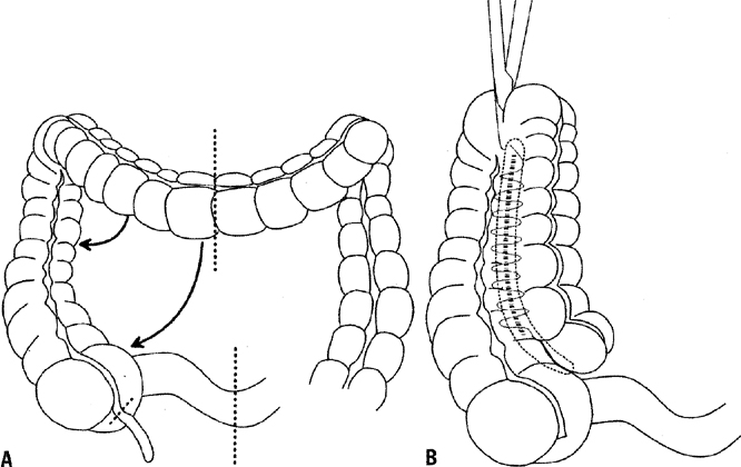
Reanastomosis of the Colon and Rectum

In patients who have undergone total pelvic exenteration, consideration should be given to reanastomosis of the colon and rectum. The development of the end-to-end circular stapling device has allowed the anastomosis of the colon to as little as 3 to 4 cm of rectal stump. Berek and associates55 reported good results with this technique, but recommended a diverting colostomy in previously irradiated patients. Barakat and coworkers60 noted a greater than 50% rate of anastomotic leaks in patients undergoing concomitant low rectal anastomosis.60 Hatch and associates61 reported success with this technique, but recommended bowel rest with 14 days of total parenteral nutrition instead of a diverting colostomy. An omental wrap of the anastomosis site was also recommended. They noted that most leaks were posterior and believed that it was important to cover this area with the omentum. The reservoir capacity of the rectum is reduced, and frequent defecation can be a problem after surgery. Hatch and associates61 recommended diphenoxylate and atropine sulfate (Lomotil) or loperamide (Imodium) every 2 hours until stool frequency is satisfactory. This problem usually diminishes with time, making consideration of reanastomosis reasonable. Frequent defecation as well as rectal tenesmus, which is another side effect of a low end-to-end anastomosis of the colon to rectum, can be reduced through the construction of a rectal J-pouch low-pressure reservoir, possibly obviating the need for antidiarrheal medication.62

Back to Top
POSTOPERATIVE CARE

Patients who have undergone exenterative surgery should be placed in an intensive care unit and monitored closely. The advantages of a pulmonary artery catheter over a central venous catheter are well documented.39 Many of these patients will be unable to tolerate significant oral intake for a number of days after surgery. Orr and colleagues30 demonstrated a reduction in postoperative weight loss from 10.6% of total body weight to 1.8% in patients who received parenteral and enteral hyperalimentation after exenterative surgery.

As expected with surgery of this magnitude, postoperative complications after exenterations are common. Febrile morbidity is the most frequently encountered complication, occurring in 75% to 85% of patients. Potential causes include pulmonary sources, pelvic cellulitis and abscess, wound infection, and occasionally, pelvic septic thrombophlebitis. Symptomatic pulmonary emboli occur in 1% to 5% of patients,63 and therefore, efforts should be made in the perioperative period to reduce the possibility of a pulmonary embolus. This may be accomplished by prophylactic low-dose heparin therapy or by a pneumatic antiembolic compression hose.64 Pyelonephritis after exenteration occurs between 5% and 20% of the time.31 Almost all conduits are colonized with bacteria within 1 week after surgery, making an accurate diagnosis difficult. The routine use of both prophylactic antibiotics and subcutaneous heparin has been shown to reduce the morbidity from pelvic exenteration.65

The most serious complication, because of its associated mortality and morbidity, is gastrointestinal obstruction or fistula. Reoperation for small bowel fistula is reported to have a perioperative mortality rate of 30% to 40%.31,66 For those patients surviving a fistula, long-term side effects include short bowel syndrome, persistent pelvic drainage, and chronic diarrhea.67 Rectovaginal fistula may occur after anterior exenteration and also carries a significant mortality rate.22,31 Every effort should be made in the preoperative evaluation and during the operation to avoid these potential complications.

Back to Top
NEW SURGICAL TECHNIQUES

Laterally extended endopelvic resection (LEER), developed by Hockel, has received attention as a new operation to treat infrailiac pelvic wall recurrences. This particular procedure extends the lateral resection plane of pelvic exenteration to the medial aspects of the acetabulum, obturator membrane, sacrospinous ligament, and sacral plexus/piriformis muscle. The preliminary results are interesting; however, this procedure does carry a high complication rate of approximately 22%.68

The addition of intraoperative interstitial brachytherapy has been examined to potentially reduce local recurrence, especially in those patients with microscopic or close margins at the time of pelvic exenteration.69,70,71 The preliminary findings are still variable, and further study is warranted at this time.

Back to Top
SUMMARY

Pelvic exenteration continues to be a challenge to surgeons who undertake the care of women with pelvic cancer. It provides the only hope for cure in women with pelvic malignancies that recur after radiation therapy. The potential for significant morbidity and even death secondary to this procedure should always be kept in mind and should guide in the selection of candidates for this operation.

Back to Top
REFERENCES

1. Brunschwig A: Complete excision of pelvic viscera for advanced carcinoma. Cancer 1:177, 1948

2. Ingersoll FM, Ulfelder H: Pelvic exenteration for carcinoma of the cervix. N Engl J Med 274:648, 1966

3. Brunschwig AD: Some reflections on pelvic exenteration after 20 years experience. In Sturgis SH, Taymos ML (eds): Progress in Gynecology. p 416, Vol 5:New York, Grune & Stratton, 1970

4. Miller B, Morris M, Levenback C, et al: Pelvic exenteration for primary and recurrent vulvar cancer. Gynecol Oncol 58:202, 1995

5. Geisler JP, Look KY, Moore DA, et al: Pelvic exenteration for malignant melanomas of the vagina or urethra with over 3 mm of invasion. Gynecol Oncol 59:338, 1995

6. Morris M, Alvarez RD, Kinney WK, et al: Treatment of recurrent adenocarcinoma of the endometrium with pelvic exenteration. Gynecol Oncol 60:288, 1996

7. Barakat RR, Goldman NA, Patel DA, et al: Pelvic exenteration for recurrent endometrial cancer. Gynecol Oncol 75:99, 1999

8. Rubin SC, Hoskins WJ, Lewis JLJ: Radical hysterectomy for recurrent cervical cancer following radiation therapy. Gynecol Oncol 27:316, 1987

9. Terada K, Morley GW: Radical hysterectomy as surgical salvage therapy for gynecologic malignancy. Obstet Gynecol 70:913, 1987

10. Adcock LL: Radical hysterectomy preceded by pelvic irradiation. Gynecol Oncol 8:152, 1979

11. Mattingly RF: Indications, contraindications, and method of total pelvic exenteration. Oncology 21:241, 1967

12. Shingleton HM, Soong S-J, Gelder MS, et al: Maxwell clinical and histopathologic factors predicting recurrence and survival after pelvic exenteration for cancer of the cervix. Obstet Gynecol 73:1027, 1989

13. Averette HE, Lichtinger M, Seven BU, et al: Pelvic exenteration: A 15-year experience in a general metropolitan hospital. Am J Obstet Gynecol 150:179, 1984

14. Hricak H, Hamm B, Semelka RC, et al: Carcinoma of the uterus: Use of gadopentetate dimeglumine in MR imaging. Radiology 181:95, 1991

15. Lawhead RAJ, Clark DGC, Smith DH, et al: Pelvic exenteration for recurrent or persistent gynecologic malignancies: A 10-year review of the Memorial Sloan-Kettering Cancer Center experience (1972–1981). Gynecol Oncol 33:279, 1989

16. Ketcham AS, Deckers PJ, Sugarbaker EV, et al: Pelvic exenteration for carcinoma of the uterine cervix: A 15-year experience. Cancer 26:513, 1970

17. Barber HRK, Roberts S, Brunschwig A: Prognostic significance of preoperative nonvisualizing kidney in patients receiving pelvic exenteration. Cancer 16:1614, 1963

18. Symmonds RE, Pratt JH, Webb MJ: Exenterative operations: Experience with 198 patients. Am J Obstet Gynecol 121:907, 1975

19. Karlen JR, Piver MS: Reduction of mortality and morbidity associated with pelvic exenteration. Gynecol Oncol 3:154, 1975

20. Shingleton HM, Orr JWJ: Recurrent cancer: Exenterative surgery. In Shingleton HM, Orr JWJ (eds): Cancer of the Cervix: Diagnosis and Treatment. p 223, New York, Churchill Livingstone, 1987

21. Matthews CM, Morris M, Burke TW, et al: Pelvic exenteration in the elderly patient. Obstet Gynecol 79:773, 1992

22. Morley GW, Hopkins MP, Lindenauer SM, et al: Pelvic exenteration, University of Michigan: 100 patients at 5 years. Obstet Gynecol 74:934, 1989

23. Miller B, Morris M, Rutledge F, et al: Aborted exenterative procedures in recurrent cervical cancer. Gynecol Oncol 50:94, 1993

24. Curry SL, Nahhas WA, Jahshan AE, et al: Pelvic exenteration: A 7-year experience. Gynecol Oncol 11:119, 1981

25. Dempsey GM, Buchsbaum HJ, Morrison J: Psychosocial adjustment to pelvic exenteration. Gynecol Oncol 3:325, 1975

26. Brown RS, Haddox V, Posada A, et al: Social and psychological adjustment following pelvic exenteration. Am J Obstet Gynecol 114:162, 1972

27. Turns D: Psychosocial issues: Pelvic exenterative surgery. J Surg Oncol 76:224, 2001

28. Andersen BL, Hacker NF: Psychosexual adjustment following pelvic exenteration. Obstet Gynecol 61:331, 1983

29. Hawighorst-Knapstein S, Schonefussrs G, Hoffmann SO, et al: Pelvic exenteration: Effects of surgery on quality of life and body image—a prospective longitudinal study. Gynecol Oncol 66:495, 1997

30. Orr J, James W, Shingleton HM: Importance of nutritional assessment and support in surgical and cancer patients. J Reprod Med 29:635, 1984

31. Orr J, James W, Hatch KD, et al: Gastrointestinal complications associated with pelvic exenteration. Am J Obstet Gynecol 145:325, 1983

32. Plante M, Roy M: The use of operative laparoscopy in determining eligibility for pelvic exenteration in patients with recurrent cervical cancer. Gynecol Oncol 59:401, 1995

33. Plante M, Roy M: Operative laparoscopy prior to a pelvic exenteration in patients with recurrent cervical cancer. Gynecol Oncol 69:94, 1998

34. Stanhope CR, Symmonds RE: Palliative exenteration: What, when, and why? Am J Obstet Gynecol 152:12, 1985

35. Rutledge FN, Smith JP, Wharton JT, et al: Pelvic exenteration: Analysis of 296 patients. Am J Obstet Gynecol 129:881, 1977

36. Jones WB: Surgical approaches for advanced or recurrent cancer of the cervix. Cancer 60:2094, 1987

37. Creasman WT, Rutledge F: Preoperative evaluation of patients with recurrent carcinoma of the cervix. Gynecol Oncol 1:111, 1972

38. Hatch KD, Shingleton HM, Soong SJ, et al: Anterior pelvic exenteration. Gynecol Oncol 31:205, 1988

39. Orr JWJ, Shingleton HM, Soong SJ, et al: Hemodynamic parameters following pelvic exenteration. Am J Obstet Gynecol 146:882, 1993

40. Butcher HRJ, Sugg WL, McAfee CA, et al: Ileal conduit method of ureteral urinary diversion. Ann Surg 156:682, 1962

41. Orr JWJ, Shingleton HM, Hatch KD, et al: Urinary diversion in patients undergoing pelvic exenteration. Am J Obstet Gynecol 142:883, 1982

42. Schlesinger RE, Berman ML, Ballon SC, et al: The choice of an intestinal segment for a urinary conduit. Surg Gynecol Obstet 148:45, 1979

43. Gilchrist RK, Merricks JW: Construction of a substitute bladder and urethra. Surg Clin North Am 36:1131, 1956

44. Kock NG, Nilson AE, Nilsson LO, et al: Urinary diversion via a continent ileal reservoir: Clinical results in 12 patients. J Urol 128:469, 1982

45. Rowland RG, Mitchell ME, Bihrle R: Alternative techniques for a continent urinary reservoir. Urol Clin North Am 14:797, 1987

46. Thuroff JW, Alken P, Riedmiller H, et al: The Mainz pouch (mixed augmentation ileum and cecum) for bladder augmentation and continent diversion. J Urol 136:17, 1986

47. Penalver MA, Bejany DE, Averette HE, et al: Continent urinary diversion in gynecologic oncology. Gynecol Oncol 34:274, 1989

48. Duckett JW, Snyder HMI: Use of the Mitrofanoff principle in urinary reconstruction. Urol Clin North Am 13:271, 1986

49. Rowland RG, Mitchell ME, Bihrle R, et al: Indiana continent urinary reservoir. J Urol 137:1136, 1987

50. Silver DF, Ashwell TR: Choices in creating continent urostomies following pelvic exenteration for gynecologic malignancies. Gynecol Oncol 82:510, 2001

51. El-Lamie IK: Preliminary experience with Mainz type II pouch in gynecologic oncology patients. Eur J Gynaecol Oncol 22:77, 2001

52. Benson MC, Olsson CA: Urinary diversion. Urol Clin North Am 19:779, 1992

53. Penalver MA, Angioli R, Mirhashemi R, et al: Management of early and late complications of ileocolonic continent urinary reservoir (Miami pouch). Gynecol Oncol 69:185, 1998

54. Powers JC, Fitzgerald JF, McAlvanah MJ: The anatomic basis for the surgical detachment of the greater omentum from the transverse colon. Surg Gynecol Obstet 143:105, 1976

55. Berek JS, Hacker NF, Lagasse LD: Rectosigmoid colectomy and reanastomosis to facilitate resection of primary and recurrent gynecologic cancer. Obstet Gynecol 64:715, 1984

56. Hatch KD: Construction of a neovagina after exenteration using the vulvobulbocavernosus myocutaneous graft. Obstet Gynecol 63:110, 1984

57. Loree TR, Hempling RE, Eltabbakh GH, et al: The inferior gluteal flap in the difficult vulvar and perineal reconstruction. Gynecol Oncol 66:429, 1997

58. Smith HO, Genesen MC, Runowicz CD, et al: The rectus abdominis myocutaneous flap: Modifications, complications, and sexual function. Cancer 83:510, 1998

59. Jurado M, Bazan A, Elejabeitia J, et al: Primary vaginal and pelvic floor reconstruction at the time of pelvic exenteration: A study of morbidity. Gynecol Oncol 77:293, 2000

60. Husain A, Curtin J, Brown C, et al: Continent urinary diversion and low-rectal anastomosis in patients undergoing exenterative procedures for recurrent gynecologic malignancies. Gynecol Oncol 78:208, 2000

61. Hatch KD, Shingleton HM, Potter ME, et al: Low rectal resection and anastomosis at the time of pelvic exenteration. Gynecol Oncol 31:262, 1988

62. Wheeless CRJ, Hempling RE: Rectal J pouch reservoir to decrease the frequency of tenesmus and defecation in low coloproctostomy. Gynecol Oncol 35:136, 1989

63. Girtanner RE, De Campo T, Alleyn JN, et al: Routine intensive care for pelvic exenterative operations. Surg Gynecol Obstet 153:657, 1981

64. Farquharson DIM, Orr JWJ: Thromboembolic complications in gynecology. J Reprod Med 29:845, 1984

65. Goldberg JM, Piver MS, Hempling RE, et al: Improvements in pelvic exenteration: Factors responsible for reducing morbidity and mortality. Ann Surg Oncol 5:399, 1998

66. Devereux DF, Sears HF, Ketcham AS: Intestinal fistula following pelvic exenterative surgery: Predisposing causes and treatment. J Surg Oncol 14:227, 1980

67. Miller B, Morris M, Gershenson DM, et al: Intestinal fistulae formation following pelvic exenteration: A review of the University of Texas M.D. Anderson Cancer Center experience, 1957–1990 Gynecol Oncol 56:207, 1995

68. Hockel M: Laterally extended endopelvic resection: Surgical treatment of infrailiac pelvic wall recurrences of gynecologic malignancies. Am J Obstet Gynecol 180:306, 1999

69. Beitler JJ, Anderson PS, Wadler S, et al: Pelvic exenteration for cervix cancer: Would additional intraoperative interstitial brachytherapy improve survival? Int J Radiat Oncol Biol Phys 38:143, 1997

70. Crowe PJ, Temple WJ, Lopez MJ, et al: Pelvic exenteration for advanced pelvic malignancy. Semin Surg Oncol 3:152, 1999

71. Chi DS, Gemignani ML, Curtin JP, et al: Long-term experience in the surgical management of cancer of the uterine cervix. Semin Surg Oncol 17:161, 1999

Back to Top