Chapter 33
Steroid Receptors and Selective Estrogen Receptor Modulation in Mammary and Gynecologic Malignancy
David J. Bentrem and V. Craig Jordan
Main Menu   Table Of Contents

Search

David J. Bentrem, MD
Robert H. Lurie Comprehensive Cancer Center, Northwestern University Medical School, Chicago, Illinois (Vol 4, Chap 33 )

V. Craig Jordan, PhD, DSc
Diana, Princess of Wales Professor of Cancer Research, Robert H. Lurie Comprehensive Cancer Center, Northwestern University Medical School, Chicago, Illinois (Vol 4, Chap 33)

LINK BETWEEN STEROID HORMONES AND CANCER
DISCOVERY OF THE ESTROGEN RECEPTOR
CLINICAL VALUE OF RECEPTOR STATUS
MOLECULAR MECHANISMS OF THE ESTROGEN RECEPTOR
TAMOXIFEN
THE ASSOCIATION OF TAMOXIFEN AND ENDOMETRIAL CANCER
RALOXIFENE
PREVENTION
STUDY OF TAMOXIFEN AND RALOXIFENE (STAR)
FUTURE DIRECTIONS
REFERENCES

LINK BETWEEN STEROID HORMONES AND CANCER

Malignancy of the breast, ovary, and endometrium is linked to endogenous hormones. Observations over the past 100 years have established this causal relationship. In 1900, Beatson and Boyd independently established that the removal of the ovaries from premenopausal women with metastatic breast cancer would, roughly one third of the time, cause regression of disease with improved prognosis.1,2 Therefore, an ovarian factor was implied in the promotion of carcinogenesis.

Allen and Doisey identified the “estrus stimulating principle,” endogenous estrogens that could activate tumors of the breast, and subsequent research in synthetic chemistry identified nonsteroidal estrogens. Dodds and associates described an extremely potent estrogen in animals, diethylstilbestrol (DES),4 but Haddow and colleagues5 first tested high-dose DES as a treatment for advanced breast cancer in postmenopausal women. Use of high dose estrogen treatment is somewhat paradoxical, but effective in one third of patients. The mechanism of action is unknown. Alternatively, reducing endogenous steroids by adrenalectomy improves prognosis of some postmenopausal women in similar proportions to oophorectomy.6 It is known that adrenal androstenedione and testosterone are converted to estrone and estradiol by peripheral aromatase enzyme systems.

Numerous studies have linked breast cancer risk to prolonged, unopposed exposure to estrogen as in early menarche,7 late menopause,8,9 and increased age at first pregnancy.10 Other hormonal risk factors are less well established. Reports on abortion and the risk of breast cancer have been equivocal.11–14 Studies of the effect of lactation on breast cancer risk have suggested that a longer duration of lactation reduces risk in premenopausal women.15 Postmenopausal obesity has been shown to increase risk.16,17

Similarly, obesity18 is a well-defined risk factor for endometrial cancer stemming from increased availability of peripheral estrogens especially in postmenopausal women with a compounding lack of progesterone.19,20 Estrogen is a proliferative hormone, whereas progesterone is a differentiating hormone in the uterus. Perhaps most important, periodic withdrawal bleeding sheds any malignant epithelial cells. Analogous factors related to high or unopposed estrogen levels also elevate the risk of endometrial cancer; these include early menarche, late menopause,21,22 nulliparity,23 and unopposed estrogen replacement therapy.23,24

The effects of exogenous hormones in the form of oral contraceptives and hormone replacement therapy (HRT) on breast cancer have been studied extensively. Some studies suggest that the long-term use of oral contraceptives in young women before their first pregnancy may increase breast cancer risk.25,26 Two meta-analyses of this effect demonstrate small but statistically significant increases in risk for HRT users.27,28 Steinberg and coworkers27 noted that after 5 years of estrogen use there is a proportional yearly increase in risk, whereas Sillero-Arenas and associates28 did not observe a significant association between duration of HRT and breast cancer risk. The Iowa Women's Health Study found a longer duration of HRT was associated with increased risk of invasive carcinoma with a favorable histology, with a relative risk (RR) of 1.81 for HRT use of less than 5 years duration versus a RR of 2.65 for use longer than 5 years.29 Even so, women who develop breast cancer while using HRT at the time of diagnosis have a similar prognosis in terms of type, size, or grade of tumor as that of nonusers.30 In breast epithelia, progesterone, like estradiol, also has a strong proliferative effect. Progesterone accelerates the appearance, growth rate, and number of tumors in carcinogen-induced rat mammary cancers.31 Furthermore, use of progestins in combination with estradiol for HRT has been linked with a higher risk of developing breast cancer than use of estradiol alone.32

Back to Top
DISCOVERY OF THE ESTROGEN RECEPTOR

In 1962, Jensen and Jacobson33 demonstrated that [3H] estradiol bound to and was retained by estrogen target tissues including the uterus, vagina, and pituitary gland in the female rat. In contrast, tissues that did not respond to estrogen did not retain [3H] estradiol. Jensen33 proposed that an estrogen receptor (ER) in estrogen target tissues must capture circulating steroids and initiate the cascade of biochemical events associated with estrogen action in that particular tissue. Toft and respective coworkers in two studies34,35 first identified the ER as an extractable protein from the rat uterus. Subsequently, Gorski and associates36 and Jensen and coworkers37 independently proposed subcellular models of estrogen action in target tissues. Jensen and associates38 then proposed the clinical ER assay to predict hormone responsive breast cancer. This established a mechanistic link between estrogen action and the growth of breast cancer.

Monoclonal antibodies to the ER subsequently established that the ER is a nuclear protein,39 and the technology of immunocytochemistry is now standard for the determination of receptor status in breast biopsies.40,41 The ER status of the patient is highly predictive of a treatment response to endocrine ablative surgery42 or antiestrogen therapy.43

Similar experimental techniques have been used with human endometrial tissue. The uptake of radiolabeled estrogen is highest during the first 2 weeks of the menstrual cycle,44 that is, during the time of unopposed estrogen action. In addition, quantitation of the human ER protein revealed that concentrations are markedly higher during the proliferative phase than in the secretory phase.45–48 In contrast, uptake of radiolabeled progesterone is higher in the secretory phase than in the proliferative phase of the menstrual cycle.49 The progesterone receptor (PgR) level is low during the proliferative phase, peaks at midcycle, and becomes lower again during the secretory phase.46,48

Endometrial cancers contain ERs and an inverse relationship exists between ER levels and tumor grade.50 However, cytosol receptor assays are inherently inaccurate in endometrial carcinoma because of the contribution from normal stromal and myometrial cells, which also contain receptors.51–53 Development of monoclonal antibodies to the PgRs allowed the quantitation of hormone receptor levels in endometrial cancer.

Molecular cloning of the cDNAs for the estrogen55 and progesterone receptors56,57 led to the realization that these proteins are members of the steroid and thyroid hormone receptor superfamily of ligand-responsive transcription factors.58 Both receptors are encoded by eight exons, which correspond to domains of functional significance.59–61

Biologic effects of estrogen are mediated by two receptors, ERα and ERβ.62,63 These two ERs share a conserved structure with six functional domains, A to F. ERβ is homologous to ERα at the ligand-binding domain (58%) and DNA-binding domain (95%). The remaining domains are not well conserved.64 Discovery of ERβ has advanced our understanding of estrogen signaling and may explain tissue responses to estrogen in which ERα is undetectable.65 Furthermore, the existence of ERα and β subtypes provides a possible explanation for the tissue selectivity of selective ER modulators (SERMs).

Back to Top
CLINICAL VALUE OF RECEPTOR STATUS

The responsiveness of neoplastic tissues to steroid hormones and antiestrogen therapy correlates with hormone receptor concentration in target cells. Neoplastic cells that retain a full or partial amount of steroid receptors are expected to respond to endocrine therapy whereas cells lacking receptors do not. Response to endocrine therapy in breast cancer correlates with levels of ER and PgR in the tumor,43 which makes receptor status useful in the management of breast cancer. In general, women who are ER- and PgR-positive have a longer disease-free interval and longer survival time than women with receptor-negative tumors.66,67

More than three fourths of endometrial carcinomas contain ER and PgR.50,68–73 ER levels in hyperplastic and neoplastic tissues are comparable with those found in early proliferative endometrium.74 Ehrlich and coworkers71,75 found a correlation between the response of patients with advanced endometrial cancer to progestin therapy and the presence of PgR. Early stage endometrial cancers are more likely to be receptor-positive and have higher receptor levels than more advanced tumors.68 Receptor concentrations are greater in well or moderately differentiated tumors than in anaplastic carcinomas. Receptor-poor tumors are generally more aggressive with decreased 5-year survival rates.68 Positive hormone receptor status is associated with improved disease-free and overall survival rates.13,76,77 When considered separately, positive PgR status in endometrial cancer appears to represent a more reliable predictor of prognosis than positive ER status.71,72,78,79 These studies suggest that there is a tendency for PgR-positive status to correlate with early stage tumors, that is, type I endometrial carcinoma. Receptor status does not correlate with invasion and metastasis found in advanced stages of disease.

Back to Top
MOLECULAR MECHANISMS OF THE ESTROGEN RECEPTOR

Both ERα and ERβ contain activation functions (AFs) that contribute to the ER's transcriptional activity. AF-1 is located in the amino-terminal region within the A/B region and is believed to be constitutively active and ligand-independent in ERα80,81 (Fig. 1). AF-2 is present at the carboxy-terminus, and is believed to be ligand dependent. Mutational analyses demonstrate the importance of this region for ER transcription because AF-2 can interact with a number of transcriptional coactivators in a ligand-dependent manner.82,83 AF-1 and AF-2 can each activate transcription independently but in most cases they synergize with one another as promoters within the specific cell context. The ER activates gene expression through binding to estrogen response elements (EREs) in responsive genes through the action of AF-1 and AF-2.84 ERβ also activates transcription of target genes through EREs.85 Additionally, estrogen can induce an AP-1 site in a reporter construct through ERα, but it is inactive through ERβ. Interestingly, ER antagonists activate ERβ to induce activity through an AP-1 site.86 These two receptors, ERα and ERβ, form functional heterodimers on DNA and stimulate transcription of a target gene. The existence of two rather than one ER portends a more complex mechanism of action than previously thought and remains a primary therapeutic target for new and existing SERMs.

Fig. 1. A subcellular model of estradiol (E2) action in target tissue. The steroid receptor complex undergoes a conformational change and dimerizes before binding to an estrogen response element (ERE) in the promoter region of an estrogen-responsive gene. A transcription unit is formed by interaction with coactivator (CoA) molecules to initiate RNA synthesis whereas corepressor (CoR) molecules are believed to prevent transcription. The ER is divided into six regions ( A-F ). The DNA-binding domain ( C) is essential for the interaction of the ER with the ERE. The ligand- binding domain ( E) is the site of E2 binding and the site of competitive binding by antiestrogens.

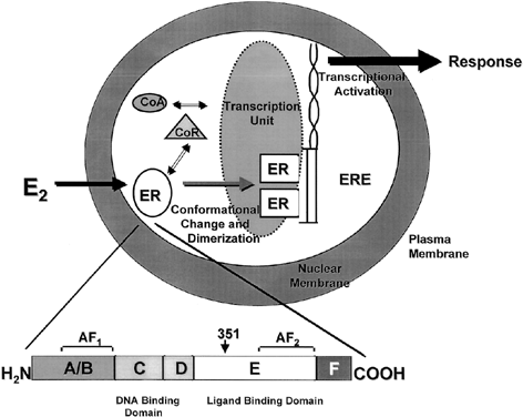
One of the functional areas of the ER, the E region, is the steroid-binding domain that undergoes a conformational change on binding with estrogen, locking the ligand into the hydrophobic pocket of the receptor. This allows the ER to dissociate from heat shock proteins, dimerize, and bind to specific DNA sequences of estrogen responsive genes. Changes in conformation of the ER allow for interaction with coregulatory proteins (coactivators or corepressors) that act as signaling intermediates between the ER and the transcriptional machinery.87 The crystal structure of the ligand binding domain of ERα was determined with estradiol88 and DES.89 A key feature of the agonist-receptor complex is the availability of a portion of the ligand binding site of ERα, helix 12, to lock the steroid in the hydrophobic pocket and allow for recruitment of coactivators to the AF2 site. The repositioning of helix 12 after ligand binding is an important mechanism for full estrogen action at ERα.90,91

The nuclear steroid receptors must associate with other nuclear proteins to form a transcription complex. Several coactivators, such as ERAP16092 and RIP140,93 have been identified based on their ability to interact with the agonist-bound receptor and not to the antagonist-bound receptor at AF-2. However, the SRC-1 protein has been identified and it also interacts with AF-194 and with ERb through phosphorylation of AF-1 by the MAP-kinase signaling cascade.95 It has been demonstrated that SRC-1 and another protein, p300/CBP, contain intrinsic acetyltransferase activity and can interact with other histone acetyl transferases (HATs). Acetylation by SRC-1 of histones bound at specific promoters could be a mechanism by which the AFs of ER and associated coactivators activate transcription of specific genes by enhancing formation of a stable preinitiation complex.96

Antiestrogens are competitive inhibitors of estrogen action; the shape of the ligand is essential to reducing the estrogenic efficacy. The side chain of the antiestrogen prevents helix 12 from closing so that activation cannot occur.88,97,98 It is known that tamoxifen silences AF-2 whereas AF-1 remains constituitively activated.81,99 Other antiestrogens, such as raloxifene, silence both AF-1 and AF-2.100,101 This is a possible explanation for the promiscuous estrogen like actions of tamoxifen compared with raloxifene.

Back to Top
TAMOXIFEN

Lerner and coworkers102 described the pharmacologic properties of a low potency nonsteroidal antiestrogen (ethamoxytriphetol (MER 25). In laboratory animals, MER 25 and the related compound MRL 37 demonstrated antifertility actions,103–105 stimulating a search for more potent agents for clinical applications. Clomiphene nafoxidine,106,107 nitromifene,109 and tamoxifen110,111 were all the result of that search, but clinical application as postcoital contraceptives was found to be inappropriate. The drugs stimulate ovulation in subfertile women.112 Clomiphene remains available as a profertility agent, inducing ovulation in subfertile women.

In the early 1970s, tamoxifen was tested successfully as an antiestrogen for the treatment of advanced breast cancer in postmenopausal women.113,114 Tamoxifen is now the endocrine therapy of choice for the treatment of all stages of ER-positive breast cancer. A 5-year course is currently considered to be optimal adjuvant therapy and produces a profound increase in disease free and overall survival.115 The 1998 Oxford Overview Analysis115 combined 55 randomized trials that began before 1990, comparing adjuvant tamoxifen therapy to no tamoxifen before recurrence. The study population of 37,000 women with node-positive and node-negative breast cancer, comprised 87% of the known, randomized clinical trials with tamoxifen. Of these women, fewer than 8,000 had a very low or zero level of ER, and 18,000 were classified as ER-positive. The ER status of the remaining nearly 12,000 women was unknown. Based on the normal distribution of ER in random populations, the authors estimated that two thirds would be ER-positive.

The recurrence reductions with tamoxifen in ER-positive patients are all significant whereas the therapeutic effect on ER-negative patients is minimal. In 5-year trials of tamoxifen treatment, the reductions of recurrence were 43% and 60% for patients with less than and greater than 100 fmol/mg cytosol of ER protein, respectively, translating to reductions in mortality rates of 23% and 36%.

The Overview Analysis also provides unequivocal proof of the laboratory principle that longer tamoxifen adjuvant therapy is more beneficial.116–118 Duration of therapy is extremely important for the ER-positive premenopausal woman with large amounts of circulating estrogen that can rapidly reverse the effect of short-term tamoxifen treatment (Fig. 2). The benefit of 1 year of tamoxifen in the premenopausal woman is virtually nonexistent, with a 20-fold increase in effectiveness with 5 years of therapy. In contrast, the effect of tamoxifen duration on women over the age of 60 is less dramatic because 1 year of tamoxifen is much more effective in postmenopausal women. The principle that longer therapy is more beneficial than shorter therapy is also demonstrated in the control of contralateral breast cancer (Fig. 3). Tamoxifen has no significant effect if 1 year of treatment is used but extending therapy to 5 years produces a 47% reduction in breast cancer incidence. This is a powerful demonstration of the effect of tamoxifen as a chemopreventive.

Fig. 2. The relationship between the duration of adjuvant tamoxifen therapy in estrogen receptor-positive premenopausal patients and the reduction in recurrence demonstrating the value of a longer duration of treatment.(Adapted from Fisher B, Costantino JP, Wickersham DL et al. Tamoxifen for prevention of breast cancer. J Natl Cancer Inst 90:137, 1998.)

Fig. 3. The relationship between the duration of adjuvant tamoxifen and the reduction in contralateral breast cancer.(Derived from Early Breast Cancer Trialists' Collaborative Group: Tamoxifen for early breast cancer: An overview of the randomised trials. Lancet 351:1451–1467, 1998.)

Back to Top
THE ASSOCIATION OF TAMOXIFEN AND ENDOMETRIAL CANCER

It is well known that tamoxifen produces a partial agonist action in the rat uterus,110 but until the late, little information was available about the actions of tamoxifen in the normal human uterus. Although tamoxifen has been used to treat endometrial cancer,119 the drug causes stromal thickening in the uterus120,121 and would be expected to encourage growth of preexisting endometrial cancer.122 Tamoxifen treatment is associated with a fourfold increase in the incidence of endometrial carcinoma in postmenopausal women120,121,123–125 (Fig. 4). Most important, the stage and grade of endometrial cancers observed in women taking tamoxifen are the same as those in the general population.123 It is known that the uterus harbors five times the amount of occult disease as is reported clinically.126 Any “estrogen-like” molecule enhances growth and promotes selection of hormone responsive disease.A recent report by Bernstein and colleagues127 has analyzed the incidence of endometrial cancer associated with tamoxifen therapy with known risk factors for the disease. This population of women warrant careful evaluation before the use of tamoxifen. The women most likely to develop endometrial cancer with tamoxifen therapy have previously used HRT and have known risk factors, such as obesity. There is no reported increase in risk of endometrial cancer in premenopausal women127 inasmuch as menstrual cycles continue during tamoxifen treatment for most women.

Fig. 4. The incidence of endometrial cancers screened for in women during the National Surgical Adjuvant Breast and Bowel Project tamoxifen prevention trial P-1. The increased incidence of endometrial cancer was seen primarily in postmenopausal patients.(Adapted from Fisher B, Costantino JP, Wickersham DL et al. Tamoxifen for prevention of breast cancer. J Natl Cancer Inst 90:137, 1998.)

Back to Top
RALOXIFENE

Raloxifene is a SERM currently approved for the treatment of osteoporosis. Evaluation of 60-mg daily raloxifene on bone homeostasis in postmenopausal women found that although the suppression of remodeling was greater for estrogen, the remodeling balance was the same for the two agents.128 Raloxifen increases bone by 2.4% in the lumbar spine and hip and has been shown to decrease spine fractures by 40%.129

Raloxifene inhibits the growth of 7,12-dimethylbenz[a]-anthracene (DMBA)-induced rat mammary carcinoma,130 and more important, for the proposed evaluation as a preventive therapy, raloxifene reduces the incidence of N-nitrosomethylurea-induced tumors if given after the carcinogen, but before the appearance of palpable tumors.131 As would be anticipated with a drug that has a short biologic half-life, raloxifene is not superior to tamoxifen at equivalent doses. Raloxifene and its analogues are clearly effective and potent inhibitors of the growth of breast cancer cells in culture,132,133 but the complication of first pass metabolism in vivo reduces potency. For this reason, doses above 60 mg daily have been tested in clinical trials to prevent osteoporosis.

Based on the hypothesis that raloxifene reduces incidence of breast cancer as a beneficial side effect of the prevention of osteoporosis,134 placebo-controlled trials with raloxifene are ongoing. The Multiple Outcomes of Raloxifene Evaluation (MORE) has randomized 7,704 postmenopausal women, mean age 66.5 years, with osteoporosis defined by hip or spine bone density at least 2.5 standard deviations (SDs) below normal or vertebral fractures, and no history of breast or endometrial cancer, to placebo, 60 mg, or 120 mg of raloxifene daily. Results at 3 years, with a total of 40 cases of breast cancer confirmed, indicate a 76% reduction in the risk of breast cancer.135 Another database pools all placebo-controlled trials and includes 10,553 women monitored for, on average, 3 years. In this group there is a 54% reduction in the incidence of breast cancer in the raloxifene treated patients.136 Similar to tamoxifen, raloxifene reduces the incidence of ER-positive breast cancer only.

Raloxifene and its analogues have limited estrogenic effects in the rat uterus.140 The raloxifene analogue LY117018 is able to block the estrogenic actions of tamoxifen in the rat uterus,140 however, raloxifene and its analogues cannot be classified as pure antiestrogens. There is not a complete lack of uterotropic properties,141,142 and estrogen-regulated genes, such as the PgR, are partially activated.

Raloxifene's effect in the human uterus is currently being evaluated. A study in women without preexisting endometrial abnormalities, shows that raloxifene, unlike estrogen, does not increase endometrial thickness.144 Raloxifene is less estrogenic than tamoxifen and only increases the growth of human endometrial carcinomas under laboratory conditions by approximately 50% of tamoxifen.122 Raloxifene has not been found to increase incidence of endometrial cancer during trials of osteoporosis treatment.135

Back to Top
PREVENTION

The decrease in contralateral breast cancers following adjuvant therapy and favorable toxicity profile in clinical practice suggested tamoxifen could play a role in breast cancer prevention. Furthermore, estrogenic hormones act over many years to promote a population of initiated cells. Given that the promotional stage is characterized by reversibility, much interest has been shown in preventing the consequences of prolonged, unopposed estrogenic stimulation of the breast.

The National Surgical Adjuvant Breast and Bowel Project (NSABP) P-1 opened in the United States and Canada in May 1992 with an accrual goal of 16,000 women from 100 North American sites. The specific aim was to test the utility of tamoxifen as a preventive for breast cancer. It closed after accruing 13,338 women in 1997. Women over the age of 60, or women between the ages of 35 and 59 whose 5-year risk of developing breast cancer, as predicted by the Gail model,145 was equal to that of a 60 year-old woman, were eligible for inclusion. Additionally, women over age 35 with a diagnosis of lobular carcinoma in situ (LCIS) treated by biopsy alone were eligible for entry. In the absence of LCIS, the risk factors necessary to enter the study varied with age, such that a 35-year-old woman needed an RR of 5.07, whereas the required RR for a 45-year-old woman was 1.79. Routine endometrial biopsies were performed in both arms of the study.

The breast cancer risk of women enrolled in the study was generally extremely high. Recruitment was also age balanced, with about one third younger than 50 years, one third between 50 and 60 years, and one third older than 60 years. Secondary endpoints of the study include the incidence of fractures and cardiovascular deaths. Most important, the study will provide information regarding tamoxifen's role in the treatment of women who carry somatic mutations in the BRCA-1 gene. Laboratory results are not yet available.

The first results of the NSABP study were reported in September 1998, after a mean followup of 47.7 months. In total, 363 invasive and noninvasive breast cancers occurred in the participants; 124 in the tamoxifen group and 239 in the placebo group. A 49% reduction in the risk of invasive breast cancer and a 50% reduction in the risk of noninvasive breast cancer were seen in the tamoxifen arm. A subset analysis of women at risk because of earlier LCIS demonstrated a 56% reduction of risk. The most dramatic risk reduction, 86%, was seen in women at risk due to atypical hyperplasia.

The benefits of tamoxifen were observed in all age groups, with an RR of breast cancer ranging from 0.45 in women aged 60 and older to 0.49 for those in the 50- to 59-year-old group, and 0.56 for women aged 49 and younger (Fig. 5). The benefit of tamoxifen was observed in all levels of breast cancer risk within the study and were not confined to a particular lower- or higher-risk subset.

Fig. 5. The overall reduction in invasive breast cancer observed in the National Surgical Adjuvant Breast and Bowel Project tamoxifen prevention trial P-1 in women at high risk for the disease, recruited to receive either tamoxifen (20 mg daily) or placebo.(Adapted from Fisher B, Costantino JP, Wickersham DL et al. Tamoxifen for prevention of breast cancer. J Natl Cancer Inst 90:137, 1998.)

As expected, tamoxifen affected the incidence of ER-positive tumors, which were reduced by 69% per year. The rate of ER-negative tumors in the tamoxifen group (1.46 per 1,000 women) did not significantly differ from that of the placebo group (1.20 per 1,000 women) (Fig. 6). Tamoxifen reduced the rate of invasive cancers of all sizes, but the greatest reduction was in the incidence of tumors 2.0 cm or smaller. Tamoxifen also reduced the incidence of both node-positive and node-negative breast cancer. The beneficial effects of tamoxifen were observed for each year of follow-up in the study. After year 1, the risk was reduced by 33%, and in year 5, by 69%.

Fig. 6. The incidence of estrogen receptor (ER)-positive and ER-negative breast cancer in the placebo and tamoxifen-treated arms of the National Surgical Adjuvant Breast and Bowel Project tamoxifen prevention trial P-1. The antiestrogen reduces the risk of developing ER-positive disease breast cancer, but there is no change in the incidence of ER-negative breast cancer.(Adapted from Fisher B, Costantino JP, Wickersham DL et al. Tamoxifen for prevention of breast cancer. J Natl Cancer Inst 90:137, 1998.)

Tamoxifen also reduced the incidence of osteoporotic fractures of the hip, spine, and radius by 19% (Fig. 7),123 approaching, but not reaching statistical significance. This reduction was greatest in women who were 50 years and older at study entry. No differences in the risks of myocardial infarction, angina, coronary artery bypass grafting, or angioplasty were noted between groups.123 The prevention study also confirmed the association between tamoxifen and endometrial carcinoma.125 The relative risk of endometrial cancer in the tamoxifen group was 2.5, with women over age 50 having an RR of 4.01. All endometrial cancers in the tamoxifen group were grade 1 and none of the women receiving tamoxifen died as a result of endometrial cancer. One endometrial cancer death occurred in the placebo group. Although no doubt remains that tamoxifen increases the risk of endometrial cancer, it is important to recognize that this increase translates to an incidence of 2.3 women per 1,000 per year.123

Fig. 7. The incidence of osteoporotic fractures of the hip, wrist, and spine observed in the placebo and tamoxifen-treated arms of the National Surgical Adjuvant Breast and Bowel Project tamoxifen prevention trial P-1.(Adapted from Fisher B, Costantino JP, Wickersham DL et al. Tamoxifen for prevention of breast cancer. J Natl Cancer Inst 90:137, 1998.)

More women in the tamoxifen group developed deep vein thrombosis (DVT),123 with the excess risk confined to women over 50 years. The RR of DVT in the older age group was 1.71 (95% confidence interval [CI], 0.85 to 3.58). An increase in pulmonary emboli was also seen in the older women taking tamoxifen, with an RR of approximately 3. Three deaths from pulmonary emboli occurred in the tamoxifen study arm, all in women with significant comorbidities. An increased incidence of stroke, RR 1.75, was also seen in the tamoxifen group, but this did not reach statistical significance.

An assessment of quality of life showed similar depression scores between groups. Hot flashes were reported in 81% of the women taking tamoxifen compared with 69% of the placebo group, and the tamoxifen-associated hot flashes appeared to be of comparable severity. Twenty-nine percent of the women in the tamoxifen group and 13% in the placebo group reported moderate or severe vaginal discharge. No differences in the occurrence of irregular menses, nausea, fluid retention, skin changes, or weight gain or loss were reported.

Back to Top
STUDY OF TAMOXIFEN AND RALOXIFENE (STAR)

Tamoxifen is approved for the reduction of risk for women at high risk for breast cancer using the Gail model. New agents are being developed to prevent and treat breast cancer, but preliminary data on the action of raloxifene to decrease the risk of breast cancer135 and the possibility that raloxifene might decrease the risk of endometrial cancer have resulted in the STAR trial, which is a phase III, double-blind trial that is assigning 22,000 eligible postmenopausal women to either 20 mg daily tamoxifen or 60 mg daily raloxifene for 5 years. Trial participants will complete a minimum of two additional years of follow-up after therapy is stopped.

The STAR trial's primary aim is to determine whether long-term raloxifene therapy is effective in preventing the occurrence of invasive breast cancer in high-risk postmenopausal women. It will additionally compare cardiovascular data, fracture data, and general toxicities for raloxifene and tamoxifen. It is clear that the activation or suppression of various target sites is similar for tamoxifen and raloxifene, but evaluation of the comparative benefits of the agents will provide an important new clinical database for raloxifene in postmenopausal women.

Premenopausal women at risk for breast cancer are currently ineligible for the STAR trial. Although extensive information is available about the efficacy of tamoxifen in premenopausal treatment and prevention of breast cancer,43,123 clinical experience with raloxifene is confined to postmenopausal women. The National Cancer Institute is currently conducting a randomized study of 60 mg and 300 mg daily raloxifene in high-risk women to address its effect on bone density. Additionally, short-term raloxifene treatment up to 28 days causes elevation in circulating estradiol but does not prevent ovulation,146 which is consistent with the known elevation of steroid hormones produced by tamoxifen in premenopausal breast cancer patients.147 The changes in endocrine function produced by raloxifene will also be assessed as a prelude to the recruitment of premenopausal high-risk women to the STAR trial.

Results of the STAR trial are expected by 2006. Clearly, these results will be invaluable to establish the overall benefits of the drugs with regard to breast cancer incidence, coronary heart disease, and osteoporosis. The comparisons of endometrial cancer will be especially interesting because the standard of care, that is, self-reporting, will be employed in the STAR trial rather than routine screening with annual biopsies.

Back to Top
FUTURE DIRECTIONS

The central issue for research on hormone receptor pharmacology is the discovery of mechanisms of target site specificity for the modulation of estrogenic and antiestrogenic response. The model must encompass the sum of pharmacologic consequences of signal transduction through ERα and ERβ with the simultaneous competition from endogenous estrogens at both sites. Dissection of the underlying pathways responsible for this activity may permit the development of tissue selective ER modulators.100

The development of ERβ monoclonal antibodies, the classification of target sites for the protein, and the continuing evaluation of ERβ and ERα,β knock-out mice will identify new therapeutic targets to modulate physiologic functions.148 Clearly, the successful crystallization of ERα with raloxifene88 has acted as a stimulus for the crystallization of ERβ with SERMs.149 However, the description of an agent that produces agonist or antagonist effects exclusively at ERα or ERβ will determine the therapeutic usefulness of ERβ as a target for disease prevention.

Major clinical questions remain in the current application of tamoxifen as a chemopreventive: when should the 5-year course be taken and how long will the effects last to protect a woman at elevated risk for breast cancer? It is known from the Overview analysis115 that the actions of tamoxifen to prevent contralateral breast cancer extend long after the treatment stops, but will a 5-year course of tamoxifen be sufficient for prolonged protection from breast cancer? Unfortunately, no rules can define when a high-risk woman will develop breast cancer, so it may be prudent to act immediately, rather than employ a watch-and-wait policy. Clearly, it will be important to discover the mechanism for the long-term beneficial effects of tamoxifen as a chemopreventive.

Similar questions about the optimal duration of raloxifene therapy need to be addressed in the future. In the case of raloxifene, this is not so important because long-term therapy for the prevention of osteoporosis is essential. Additionally, a key issue, which has yet to be addressed, is the cross-resistance with tamoxifen. Use of raloxifene for the prevention of osteoporois after 5 years of adjuvant tamoxifen cannot be presumed safe for the patient with breast cancer until this clinical issue is resolved.

Defining and stratifying “risk” is essential in selection of a target population. A major problem in the clinical identification of a woman at high risk is a lack of knowledge of the interactions among the various factors known to alter breast cancer risk. Most women have a combination of factors that both increase and decrease risk. Most data on risk come from studies in white women, and little is known about the impact of ethnicity on risk. In addition, it remains unclear whether the risk conferred by multiple risk factors is additive, multiplicative, or varies with the particular risk factor.

In the past decade, the incidence of breast cancer has increased,150 whereas the mortality has declined slightly for the first time in 30 years.151,152 Of particular importance is the full use of appropriate adjuvant radiation, chemotherapy, and antiestrogens in applying the best practices systematically and widely to patients after diagnosis. Laboratory concepts and clinical testing with tamoxifen and raloxifene have laid the foundation for a new generation of medicines that hold the promise of preventing breast and endometrial cancers, as well as targeting the prevention of osteoporosis and coronary heart disease.

Back to Top
REFERENCES

1. Beatson GT: On the treatment of inoperable cases of carcinoma of the mamma: Suggestions for a new method of treatment with illustrative cases. Lancet 2: 104– 107, 1896

2. Boyd S: On oophorectomy in cancer of the breast. BMJ 2: 1161– 1167, 1900

3. Allen E, Doisey DE: An ovarian hormone: Preliminary report on its localization, extraction and partial purification and action in test animals. JAMA 81: 819– 821, 1923

4. Dodds EC, Lawson W, Noble RL: Biological effects of the synthetic oestrogenic substance 4:4'-dihydroxy-alpha:beta-diethylstilbene. Lancet 1: 1389– 1391, 1938

5. Haddow A, Watkinson JM, Paterson E: Influence of synthetic oestrogens upon advanced malignant disease. BMJ 2: 393– 398, 1944

6. Kennedy BJ: Hormone therapy for advanced breast cancer. Cancer 18: 1551– 1557, 1965

7. MacMahon B, Trichopoulos D, Brown J et al: Age at menarche, probability of ovulation and breast cancer risk. Int J Cancer 29: 13– 16, 1982

8. Trichopoulos D, MacMahon B, Cole P: Menopause and breast-cancer risk. J Natl Cancer Inst 48: 605– 613, 1972

9. Kvale G, Heuch I: Menstrual factors and breast cancer risk. Cancer 62: 1625– 1631, 1988

10. MacMahon B, Purde M, Cramer D et al: Association of breast cancer risk with age at first and subsequent births: A study in the population of the Estonian Republic. J Natl Cancer Inst 69: 1035– 1038, 1982

11. Harris BM, Eklund G, Meirik O et al: Risk of cancer of the breast after legal abortion during first trimester: A Swedish register study. BMJ 299: 1430– 1432, 1989

12. Daling JR, Malone KE, Voigt LF et al: Risk of breast cancer among young women: Relationship to induced abortion. J Natl Cancer Inst 86: 1584– 1592, 1994

13. Newcomb PA, Storer BE, Longnecker MP et al: Pregnancy termination in relation to risk of breast cancer. JAMA 275: 283– 287, 1996

14. Melbye M, Wohlfahrt J, Olsen JH et al: Induced abortion and the risk of breast cancer. N Engl J Med 336: 81– 85, 1997

15. Kvale G, Heuch I: Lactation and cancer risk: Is there a relation specific to breast cancer? J Epidemiol Community Health 42: 30– 37, 1988

16. de Waard F, Baanders-van Halewijn EA: A prospective study in general practice on breast-cancer risk in postmenopausal women. Int J Cancer 14: 153– 160, 1974

17. De Waard F, Cornelis JP, Aoki K et al: Breast cancer incidence according to weight and height in two cities of the Netherlands and in Aichi prefecture, Japan. Cancer 40: 1269– 1275, 1977

18. Lawrence C, Tessaro I, Durgerian S et al: Smoking, body weight, and early-stage endometrial cancer. Cancer 59: 1665– 1669, 1987

19. Siiteri PK: Steroid hormones and endometrial cancer. Cancer Res 38: 4360– 4366, 1978

20. Enriori CL, Reforzo-Membrives J: Peripheral aromatization as a risk factor for breast and endometrial cancer in postmenopausal women: A review. Gynecol Oncol 17: 1– 21, 1984

21. Elwood JM, Cole P, Rothman KJ et al: Epidemiology of endometrial cancer. J Natl Cancer Inst 59: 1055– 1060, 1977

22. La Vecchia C, Franceschi S, Decarli A et al: Risk factors for endometrial cancer at different ages. J Natl Cancer Inst 73: 667– 671, 1984

23. Horwitz RI, Feinstein AR: Case-control study of oral contraceptive pills and endometrial cancer. Ann Intern Med 91: 226– 227, 1979

24. Parazzini F, Franceschi S, La Vecchia C et al: The epidemiology of ovarian cancer. Gynecol Oncol 43: 9– 23, 1991

25. Meirik O, Lund E, Adami HO et al: Oral contraceptive use and breast cancer in young women. A joint national case-control study in Sweden and Norway. Lancet 2: 650– 654, 1986

26. Oral contraceptive use and breast cancer risk in young women. UK National Case-Control Study Group. Lancet 1:973–982, 1989

27. Steinberg KK, Thacker SB, Smith SJ et al: A meta-analysis of the effect of estrogen replacement therapy on the risk of breast cancer. JAMA 265:1985-1990, 1991; published erratum appears in JAMA 266: 1362, 1991

28. Sillero-Arenas M, Delgado-Rodriguez M, Rodigues-Canteras R et al: Menopausal hormone replacement therapy and breast cancer: A meta- analysis. Obstet Gynecol 79: 286– 294, 1992

29. Gapstur SM, Morrow M, Sellers TA: Hormone replacement therapy and risk of breast cancer with a favorable histology: Results of the Iowa Women's Health Study. JAMA 281: 2091– 2097, 1999

30. Stallard S, Litherland JC, Cordiner CM et al: Effect of hormone replacement therapy on the pathological stage of breast cancer: Population based, cross sectional study. BMJ 320: 348– 349, 2000

31. Huggins C: Two principles in endocrine therapy of cancers: Hormone deprival and hormone interference. Cancer Res 25: 1163– 1167, 1965

32. Ross RK, Paganini-Hill A, Wan PC et al: Effect of hormone replacement therapy on breast cancer risk: Estrogen versus estrogen plus progestin. J Natl Cancer Inst 92: 328– 332, 2000

33. Jensen EV, Jacobson HI: Basic guides to the mechanism of estrogen action. Rec Prog Horm Res 18: 387– 414, 1962

34. Toft D, Gorski J: A receptor molecule for estrogens: Isolation from the rat uterus and preliminary characterization. Proc Natl Acad Sci USA 55: 1574– 1581, 1966

35. Toft D, Shyamala G, Gorski J: A receptor molecule for estrogens: Studies using a cell-free system. Proc Natl Acad Sci USA 57: 1740– 1743, 1967

36. Gorski J, Toft D, Shyamala G et al: Hormone receptors: Studies on the interaction of estrogen with the uterus. Recent Prog Horm Res 24: 45– 80, 1968

37. Jensen EV, Suzuki T, Kawashima T et al: A two-step mechanism for the interaction of estradiol with rat uterus. Proc Natl Acad Sci USA 59: 632– 638, 1968

38. Jensen EV, Block GE, Smith S et al: Estrogen receptors and breast cancer response to adrenalectomy. Natl Cancer Inst Monogr 34: 55– 70, 1971

39. King WJ, Greene GL: Monoclonal antibodies localize oestrogen receptor in the nuclei of target cells. Nature 307: 745– 747, 1984

40. King WJ, DeSombre ER, Jensen EV et al: Comparison of immunocytochemical and steroid-binding assays for estrogen receptor in human breast tumors. Cancer Res 45: 293– 304, 1985

41. DeSombre ER, Thorpe SM, Rose C et al: Prognostic usefulness of estrogen receptor immunocytochemical assays for human breast cancer. Cancer Res 46: 4256S- 4264S, 1986

42. McGuire WL, Chamness GC, Costlow ME et al: Steroids and human breast cancer. J Steroid Biochem 6: 723– 727, 1975

43. Early Breast Cancer Trialists' Collaborative Group: Tamoxifen for early breast cancer: An overview of the randomised trials. Lancet 351:1451–1467, 1998

44. Brush MG, Taylor RW, King RJ: The uptake of [6,7–3H]oestradiol by the normal human female reproductive tract. J Endocrinol 39: 599– 607, 1967

45. Hunter RE, Jordan VC: Detection of the 8 S oestrogen-binding component in human uterine endometrium during the menstrual cycle. J Endocrinol 65: 457– 458, 1975

46. Bayard F, Damilano S, Robel P et al: Cytoplasmic and nuclear estradiol and progesterone receptors in human endometrium. J Clin Endocrinol Metab 46: 635– 648, 1978

47. Janne O, Kauppila A, Kontula K et al: Female sex steroid receptors in normal, hyperplastic and carcinomatous endometrium. The relationship to serum steroid hormones and gonadotropins and changes during medroxyprogesterone acetate administration. Int J Cancer 24: 545– 554, 1979

48. Levy C, Robel P, Gautray JP et al: Estradiol and progesterone receptors in human endometrium: Normal and abnormal menstrual cycles and early pregnancy. Am J Obstet Gynecol 136: 646– 651, 1980

49. Edwards R, Brush MG, Taylor RW: The uptake and intracellular distribution of (1,2–3H)progesterone by human endometrium. J Endocrinol 45 (Suppl): 3– 4, 1969

50. Hunter RE, Longcope C, Jordan VC: Steroid hormone receptors in adenocarcinoma of the endometrium. Gynecol Oncol 10: 152– 161, 1980

51. Bergeron C, Ferenczy A, Shyamala G: Distribution of estrogen receptors in various cell types of normal, hyperplastic, and neoplastic human endometrial tissues. Lab Invest 58: 338– 345, 1988

52. Press MF, Udove JA, Greene GL: Progesterone receptor distribution in the human endometrium: Analysis using monoclonal antibodies to the human progesterone receptor. Am J Pathol 131: 112– 124, 1988

53. Segreti EM, Novotny DB, Soper JT et al: Endometrial cancer: Histologic correlates of immunohistochemical localization of progesterone receptor and estrogen receptor. Obstet Gynecol 989;73:780–785, 1989

54. Clarke CL, Zaino RJ, Feil PD et al: Monoclonal antibodies to human progesterone receptor: Characterization by biochemical and immunohistochemical techniques. Endocrinology 121: 1123– 1132, 1987

55. Green S, Walter P, Kumar V et al: Human oestrogen receptor cDNA: sequence, expression and homology to v-erb-A. Nature 320: 134– 139, 1986

56. Misrahi M, Atger M, d'Auriol L et al: Complete amino acid sequence of the human progesterone receptor deduced from cloned cDNA. Biochem Biophys Res Commun 143: 740– 748, 1987

57. Kastner P, Krust A, Turcotte B et al: Two distinct estrogen-regulated promoters generate transcripts encoding the two functionally different human progesterone receptor forms A and B. EMBO J 9: 1603– 1614, 1990

58. Evans RM: The steroid and thyroid hormone receptor superfamily. Science 240: 889– 895, 1989

59. Ponglikitmongkol M, Green S, Chambon P: Genomic organization of the human oestrogen receptor gene. EMBO J 7: 3385– 3388, 1988

60. Jeltsch JM, Turcotte B, Garnier JM et al: Characterization of multiple mRNAs originating from the chicken progesterone receptor gene. Evidence for a specific transcript encoding form A. J Biol Chem 265: 3967– 3974, 1990

61. Misrahi M, Venencie PY, Saugier-Veber P et al: Structure of the human progesterone receptor gene. Biochim Biophys Acta 1216: 289– 292, 1993

62. Mosselman S, Polman J, Dijkema R: ER beta: Identification and characterization of a novel human estrogen receptor. FEBS Lett 392: 49– 53, 1996

63. Kuiper GG, Enmark E, Pelto-Huikko M: Cloning of a novel receptor expressed in rat prostate and ovary. Proc Natl Acad Sci USA 93: 5925– 5930, 1996

64. Enmark E, Pelto-Huikko M, Grandien K et al: Human estrogen receptor beta-gene structure, chromosomal localization, and expression pattern. J Clin Endocrinol Metab 82: 4258– 4265, 1997

65. Couse JF, Curtis SW, Washburn TF et al: Analysis of transcription and estrogen insensitivity in the female mouse after targeted disruption of the estrogen receptor gene. Mol Endocrinol 9: 1441– 1454, 1995

66. McGuire WL, Horwitz KB, Zava DT et al: Hormones in breast cancer: Update 1978. Metabolism 27: 487– 501, 1978

67. Thorpe SM, Rose C, Rasmussen BB et al: Prognostic value of steroid hormone receptors: Multivariate analysis of systemically untreated patients with node-negative primary breast cancer. Cancer Res 47: 6126– 6133, 1987

68. Kauppila AJ, Isotalo HE, Kivinen ST et al: Prediction of clinical outcome with estrogen and progestin receptor concentrations and their relationships to clinical and histopathological variables in endometrial cancer. Cancer Res 46: 5380– 5384, 1986

69. Lindahl B, Alm P, Ferno M et al: Relapse of endometrial carcinoma related to steroid receptor concentration, staging, histologic grading and myometrial invasion. Anticancer Res 6: 1317– 1320, 1986

70. Utaaker E, Iversen OE, Skaarland E: The distribution and prognostic implications of steroid receptors in endometrial carcinomas. Gynecol Oncol 28: 89– 100, 1987

71. Ehrlich CE, Young PC, Stehman FB et al: Steroid receptors and clinical outcome in patients with adenocarcinoma of the endometrium. Am J Obstet Gynecol 158: 796– 807, 1988

72. Palmer DC, Muir IM, Alexander AI et al: The prognostic importance of steroid receptors in endometrial carcinoma. Obstet Gynecol 72: 388– 393, 1988

73. van der Putten HW, Baak JP, Koenders TJ et al: Prognostic value of quantitative pathologic features and DNA content in individual patients with stage I endometrial adenocarcinoma. Cancer 63: 1378– 1387, 1989

74. Tseng L, Gusberg SB, Gurpide E: Estradiol receptor and 17 beta-dehydrogenase in normal and abnormal human endometrium. Ann NY Acad Sci 286: 190– 198, 1977

75. Ehrlich CE, Young PC, Cleary RE: Cytoplasmic progesterone and estradiol receptors in normal, hyperplastic, and carcinomatous endometria: Therapeutic implications. Am J Obstet Gynecol 141: 539– 546, 1981

76. Kauppila A: Oestrogen and progestin receptors as prognostic indicators in endometrial cancer: A review of the literature. Acta Oncol 28: 561– 566, 1989

77. Soper JT, Christensen CW: Steroid receptors and endometrial cancer. Clin Obstet Gynaecol 13: 825– 842, 1986

78. Creasman WT, Soper JT, McCarty KS Jr et al: Influence of cytoplasmic steroid receptor content on prognosis of early stage endometrial carcinoma. Am J Obstet Gynecol 151: 922– 932, 1985

79. Kleine W, Maier T, Geyer H, Pfleiderer A: Estrogen and progesterone receptors in endometrial cancer and their prognostic relevance. Gynecol Oncol 38: 59– 65, 1990

80. Kumar V, Green S, Stack G et al: Functional domains of the human estrogen receptor. Cell 51: 941– 951, 1987

81. Metzger D, Ali S, Bornert JM et al: Characterization of the amino-terminal transcriptional activation function of the human estrogen receptor in animal and yeast cells. J Biol Chem 270: 9535– 9542, 1995

82. Beato M, Sanchez-Pacheco A: Interaction of steroid hormone receptors with the transcription initiation complex. Endocr Rev 17: 587– 609, 1996

83. L'Horset F, Dauvois S, Heery DM et al: RIP-140 interacts with multiple nuclear receptors by means of two distinct sites. Mol Cell Biol 16: 6029– 6036, 1996

84. Kraus WL, McInerney EM, Katzenellenbogen BS: Ligand-dependent, transcriptionally productive association of the amino- and carboxyl-terminal regions of a steroid hormone nuclear receptor. Proc Natl Acad Sci USA 92: 12314– 12318, 1995

85. Pace P, Taylor J, Suntharalingam S et al: Human estrogen receptor beta binds DNA in a manner similar to and dimerizes with estrogen receptor alpha. J Biol Chem 272: 25832– 25838, 1997

86. Paech K, Webb P, Kuiper GG et al: Differential ligand activation of estrogen receptors ERalpha and ERbeta at AP1 sites. Science 277: 1508– 1510, 1997

87. Horwitz KB, Jackson TA, Bain DL et al: Nuclear receptor coactivators and corepressors. Mol Endocrinol 10: 1167– 1177, 1996

88. Brzozowski AM, Pike AC, Dauter Z et al: Molecular basis of agonism and antagonism in the oestrogen receptor. Nature 389: 753– 758, 1997

89. Shiau AK, Barstad D, Loria PM et al: The structural basis of estrogen receptor/coactivator recognition and the antagonism of this interaction by tamoxifen. Cell 95: 927– 937, 1998

90. Feng W, Ribeiro RC, Wagner RL et al: Hormone-dependent coactivator binding to a hydrophobic cleft on nuclear receptors. Science 280: 1747– 1749, 1998

91. Moras D, Gronemeyer H: The nuclear receptor ligand-binding domain: structure and function. Curr Opin Cell Biol 10: 384– 391, 1998

92. Halachmi S, Marden E, Martin G et al: Estrogen receptor-associated proteins: Possible mediators of hormone-induced transcription. Science 264: 1455– 1458, 1994

93. Cavailles V, Dauvois S, L'Horset F et al: Nuclear factor RIP140 modulates transcriptional activation by the estrogen receptor. EMBO J 14: 3741– 3751, 1995

94. Webb P, Nguyen P, Valentine C et al: The estrogen receptor enhances AP-1 activity by two distinct mechanisms with different requirements for receptor transactivation functions. Mol Endocrinol 13: 1672– 1685, 1999

95. Tremblay A, Tremblay GB, Labrie F et al: Ligand-independent recruitment of SRC-1 to estrogen receptor beta through phosphorylation of activation function AF-1. Mol Cell 3: 513– 519, 1999

96. Spencer TE, Jenster G, Burcin MM et al: Steroid receptor coactivator-1 is a histone acetyltransferase. Nature 389: 194– 198, 1997

97. Lieberman ME, Jordan VC, Fritsch M et al: Direct and reversible inhibition of estradiol-stimulated prolactin synthesis by antiestrogens in vitro. J Biol Chem 258: 4734– 4740, 1983

98. Tate AC, Greene GL, DeSombre ER et al: Differences between estrogen- and antiestrogen-estrogen receptor complexes from human breast tumors identified with an antibody raised against the estrogen receptor. Cancer Res 44: 1012– 1018, 1984

99. McInerney EM, Weis KE, Sun J et al: Transcription activation by the human estrogen receptor subtype beta (ER beta) studied with ER beta and ER alpha receptor chimeras. Endocrinology 139: 4513– 4522, 1998

100. Levenson AS, Jordan VC: Selective oestrogen receptor modulation: Molecular pharmacology for the millennium. Eur J Cancer 35: 1628– 1639, 1999

101. Schafer JI, Liu H, Tonetti DA et al: The interaction of raloxifene and the active metabolite of the antiestrogen EM-800 (SC 5705) with the human estrogen receptor. Cancer Res 59: 4308– 4313, 1999

102. Lerner LJ, Holthaus JF, Thompson CR: A non-steroidal estrogen antagonist 1-(p-2-diethylaminoethoxyphenyl)1-phenyl-2-p-methoxyphenylethanol. Endocrinology 63: 295– 318, 1958

103. Segal JS, Nelson WO: An orally active compound with antifertility effects in rats. Proc Soc Exp Biol Med 98: 431– 436, 1958

104. Chang MC: Degeneration of ova in the rat and rabbit following oral administration of 1-(p-diethylaminoethoxyphenyl)-1-phenyl-2-p-anisylethanol. Endocrinology 65: 339– 342, 1959

105. Barnes JE, Meyer RK: Effects of ethamoxytriphetol, MRL37 and clomiphene or reproduction in rats. Fertil Steril 13: 472– 480, 1962

106. Segal JS, Nelson WO: Antifertility action of ahloramiphene. Anat Rec 139: 273, 1961

107. Holtkamp DE, Greslin SC, Root CA et al: Gonadotropin inhibiting and antifecundity effects of chloramiphene. Proc Soc Exp Biol Med 105: 197– 201, 1960

108. Duncan GW, Lyster SC, Clark JJ et al: Antifertility activities of two diphenyl-dihydromaphthalene derivatives. Proc Soc Exp Biol Med 112: 439– 442, 1963

109. Callantine MR, Humphrey RR, Lee SL et al: Action of an estrogen antagonist on reproductive mechanisms in the rat. Endocrinology 79: 153– 169, 1966

110. Harper MJ, Walpole AL: A new derivative of triphenylethylene: Effect on implantation and mode of action in rats. J Reprod Fertil 13: 101– 119, 1967

111. Harper MJ, Walpole AL: Mode of action of I.C.I. 46,474 in preventing implantation in rats. J Endocrinol 37: 83– 92, 1967

112. Greenblatt RB, Barfield WE, Jungck EC et al: Induction of ovulation with MRL-41: Preliminary report. JAMA 178: 101– 104, 1961

113. Cole MP, Jones CT, Todd ID: A new anti-oestrogenic agent in late breast cancer: An early clinical appraisal of ICI46474. Br J Cancer 25: 270– 275, 1971

114. Ward HW: Anti-oestrogen therapy for breast cancer: A trial of tamoxifen at two dose levels. BMJ 1: 13– 14, 1973

115. Early Breast Cancer Trials Collaborative Group: Tamoxifen for early breast cancer: An overview of the randomised trials. Lancet 351:1451–1467, 1998

116. Jordan VC, Dix CJ, Allen KE: The effectiveness of long term tamoxifen treatment in a laboratory model for adjuvant hormone therapy of breast cancer. Vol 2. In: Salmon SE, Jones SE (eds): Adjuvant Therapy of Cancer. New York, Grune & Stratton, 1979

117. Jordan VC, Allen KE: Evaluation of the antitumour activity of the non-steroidal antioestrogen monohydroxytamoxifen in the DMBA-induced rat mammary carcinoma model. Eur J Cancer 16: 239– 251, 1980

118. Jordan VC: Laboratory studies to develop general principles for the adjuvant treatment of breast cancer with antiestrogens: Problems and potential for future clinical applications. Breast Cancer Res Treat 3: S73- S86, 1983

119. Bonte J, Ide P, Billiet G et al: Tamoxifen as a possible chemotherapeutic agent in endometrial adenocarcinoma. Gynecol Oncol 11: 140– 161, 1981

120. Goldstein SR: Unusual ultrasonographic appearance of the uterus in patients receiving tamoxifen. Am J Obstet Gynecol 170: 447– 451, 1994

121. DeCensi A, Fontana V, Bruno S et al: Effect of tamoxifen on endometrial proliferation. J Clin Oncol 14: 434– 440, 1996

122. Gottardis MM, Ricchio ME, Satyaswaroop PG et al: Effect of steroidal and nonsteroidal antiestrogens on the growth of a tamoxifen-stimulated human endometrial carcinoma (EnCa101) in athymic mice. Cancer Res 50: 3189– 3192, 1990

123. Fisher B, Costantino JP, Wickerham DL et al: Tamoxifen for prevention of breast cancer: Report of the National Surgical Adjuvant Breast and Bowel Project P-1 Study. J Natl Cancer Inst 90: 1371– 1388, 1998

124. Fornander T, Rutqvist LE, Cedermark B et al: Adjuvant tamoxifen in early breast cancer: Occurrence of new primary cancers. Lancet 1: 117– 120, 1989

125. Fisher B, Costantino JP, Redmond CK et al: Endometrial cancer in tamoxifen-treated breast cancer patients: Findings from the National Surgical Adjuvant Breast and Bowel Project (NSABP) B-14. J Natl Cancer Inst 86: 527– 537, 1994

126. Horwitz RI, Feinstein AR, Horwitz SM et al: Necropsy diagnosis of endometrial cancer and detection-bias in case/control studies. Lancet 2: 66– 68, 1981

127. Bernstein L, Deapen D, Cerhan JR et al: Tamoxifen therapy for breast cancer and endometrial cancer risk. J Natl Cancer Inst 91: 1654– 1662, 1999

128. Heaney RP, Draper MW: Raloxifene and estrogen: Comparative bone-remodeling kinetics. J Clin Endocrinol Metab 82: 3425– 3429, 1997

129. Delmas PD, Bjarnason NH, Mitlak BH et al: Effects of raloxifene on bone mineral density, serum cholesterol concentrations, and uterine endometrium in postmenopausal women. N Engl J Med 337: 1641– 1647, 1997

130. Clemens JA, Bennett DR, Black LJ et al: Effects of a new antiestrogen, keoxifene (LY156758), on growth of carcinogen-induced mammary tumors and on LH and prolactin levels. Life Sci 32: 2869– 2875, 1983

131. Gottardis MM, Jordan VC: Antitumor actions of keoxifene and tamoxifen in the N-nitrosomethylurea-induced rat mammary carcinoma model. Cancer Res 47: 4020– 4024, 1987

132. Jiang SY, Parker CJ, Jordan VC: A model to describe how a point mutation of the estrogen receptor alters the structure-function relationship of antiestrogens. Breast Cancer Res Treat 26: 139– 147, 1993

133. Poulin R, Merand Y, Poirier D et al: Antiestrogenic properties of keoxifene, trans-4-hydroxytamoxifen, and ICI 164384, a new steroidal antiestrogen, in ZR-75-1 human breast cancer cells. Breast Cancer Res Treat 14: 65– 76, 1989

134. Lerner LJ, Jordan VC: Development of antiestrogens and their use in breast cancer: Eighth Cain Memorial Award lecture. Cancer Res 50: 4177– 4189, 1990

135. Cummings SR, Eckert S, Krueger KA et al: The effect of raloxifene on risk of breast cancer in postmenopausal women: Results from the MORE randomized trial. Multiple Outcomes of Raloxifene Evaluation. JAMA 281: 2189– 2197, 1999

136. Jordan VC, Glusman JE, Eckert S, et al: Incident primary breast cancer are reduced by raloxifene: Integrated data from multicenter, double blind, randomized trials in 12,000 postmenopausal women. Breast Cancer Res Treat 50: 227, 1998

137. Jones CD, Jevnikar MG, Pike AJ et al: Antiestrogens. 2. Structure-activity studies in a series of 3-aroyl-2- arylbenzo[b]thiophene derivatives leading to [6-hydroxy-2-(4hydroxyphenyl)benzo[b]thien-3-yl] [4-[2-(1-piperidinyl)ethoxy]- phenyl]methanone hydrochloride (LY156758), a remarkably effective estrogen antagonist with only minimal intrinsic estrogenicity. J Med Chem 27: 1057– 1066, 1984

138. Black LJ, Goode RL: Uterine bioassay of tamoxifen, trioxifene and a new estrogen antagonist (LY117018) in rats and mice. Life Sci 26: 1453– 1458, 1980

139. Black LJ, Jones CD, Falcone JF: Antagonism of estrogen action with a new benzothiophene derived antiestrogen. Life Sci 32: 1031– 1036, 1983

140. Jordan VC, Gosden B: Inhibition of the uterotropic activity of estrogens and antiestrogens by the short acting antiestrogen LY117018. Endocrinology 113: 463– 468, 1983

141. Grese TA, Cho S, Finley DR et al: Structure-activity relationships of selective estrogen receptor modulators: Modifications to the 2-arylbenzothiophene core of raloxifene. J Med Chem 40: 146– 167, 1997

142. Grese TA, Sluka JP, Bryant HU et al: Molecular determinants of tissue selectivity in estrogen receptor modulators. Proc Natl Acad Sci USA 94: 14105– 14110, 1997

143. Jordan VC, Gosden B: Differential antiestrogen action in the immature rat uterus: A comparison of hydroxylated antiestrogens with high affinity for the estrogen receptor. J Steroid Biochem 19: 1249– 1258, 1983

144. Boss SM, Huster WJ, Neild JA et al: Effects of raloxifene hydrochloride on the endometrium of postmenopausal women. Am J Obstet Gynecol 177: 1458– 1464, 1997

145. Gail MH, Brinton LA, Byar DP et al: Projecting individualized probabilities of developing breast cancer for white females who are being examined annually. J Natl Cancer Inst 81: 1879– 1886, 1989

146. Baker VL, Draper M, Paul S et al: Reproductive endocrine and endometrial effects of raloxifene hydrochloride, a selective estrogen receptor modulator, in women with regular menstrual cycles. J Clin Endocrinol Metab 83: 6– 13, 1998

147. Jordan VC, Fritz NF, Langan-Fahey S et al: Alteration of endocrine parameters in premenopausal women with breast cancer during long-term adjuvant therapy with tamoxifen as the single agent. J Natl Cancer Inst 83: 1488– 1491, 1991

148. Couse JF, Korach KS: Estrogen receptor null mice: what have we learned and where will they lead us? Endocr Rev 20:358–417, 1999; published erratum appears in Endocr Rev 20: 459, 1999

149. Pike AC, Brzozowski AM, Hubbard RE et al: Structure of the ligand-binding domain of oestrogen receptor beta in the presence of a partial agonist and a full antagonist. EMBO J 18: 4608– 4618, 1999

150. Ries LA, Wingo PA, Miller DS et al: The annual report to the nation on the status of cancer, 1973-1997, with a special section on colorectal cancer. Cancer 88: 2398– 2424, 2000

151. Byers T, Mouchawar J, Marks J et al: The American Cancer Society challenge goals. How far can cancer rates -decline in the U.S. by the year 2015? Cancer 86: 715– 727, 1999

152. Cole P, Rodu B: Declining cancer mortality in the United States. Cancer 78: 2045– 2048, 1996

Back to Top