Chapter 8
Regulation of the Pituitary Response to Gonadotropin-Releasing Hormone
Seth G. Derman, Howard D. McClamrock and Eli Y. Adashi
Main Menu   Table Of Contents

Search

Seth G. Derman, MD
Instructor, Department of Obstetrics and Gynecology, Division of Reproductive Endocrinology, University of Maryland School of Medicine, Baltimore, Maryland (Vol 5, Chap 8)

Howard D. McClamrock, MD
Associate Professor, Department of Obstetrics and Gynecology, Division of Reproductive Endocrinology, The University of Maryland School of Medicine, Baltimore, Maryland (Vol 5, Chap 8)

Eli Y. Adashi, MD
John A. Dixon Presidential Endowed Chair; Professor and Chair, Department of Obstetrics and Gynecology; Professor, Department of Pediatrics, University of Utah Health Sciences Center, Salt Lake City, Utah (Vol 5, Chaps 8, 11, 36, 37)

INTRODUCTION
PITUITARY RESPONSE TO GnRH
PITUITARY RESPONSE TO GnRH IN SPECIAL CIRCUMSTANCES
PITUITARY RESPONSE TO GnRH ANALOGS
REFERENCES

INTRODUCTION

The synthesis and release of both luteinizing hormone (LH) and follicle-stimulating hormone (FSH) from the anterior pituitary gland is primarily under the control of gonadotropin-releasing hormone (GnRH). There is some empirical evidence in the rat for the existence of a separate follicle-stimulating releasing hormone principle yet to be isolated.1, 2 In the primate, however, GnRH induces the synthesis and release of both LH and FSH from pituitary gonadotrophs. A decapeptide (Glu 1-His 2-Trp 3-Ser 4-Tyr 5-Gly 6-Leu 7-Arg 8-Pro 9-Gly 10-amide) GnRH of arcuate nucleus origin reaches the pituitary gonadotrophs through the portal vessels. This chapter reviews the pituitary response to GnRH and its modulation by sex steroids throughout the menstrual cycle and in pathologic states.

The GnRH Gene

The structure of the peptide encoded by the GnRH gene is shown in Figure 1. The gene encodes for a signal peptide sequence, the GnRH sequence itself, and for a posttranscriptional processing signal (three amino acids long). In addition, it encodes for a 56-amino acid peptide that has been termed GnRH-associated peptide (GAP).3 The structure of GAP is similar in the human, rat, and mouse, and because it is present in the same neurons as GnRH in the primate brain, it is believed to be cosecreted with GnRH.4 Although less potent, GAP, like GnRH stimulates the in vitro release of gonadotropins. Perhaps more important than its effect on gonadotropin release, GAP inhibits prolactin release from rat anterior pituitary cell cultures. However, when administered systemically to rats, GAP exhibits both an inhibitory activity on the pituitary release of prolactin and a secretagogue action on FSH.5 The physiologic role of GAP has not been defined. For instance, unlike dopamine, the putative prolactin inhibitory factor, GAP is unable to inhibit prolactin secretion by human pituitary adenoma cells in culture.6

Fig. 1. Structure of the peptide encoded by the GnRH gene.

Another compound under study is a 13-amino acid long peptide within GAP that has been termed precursor human GnRH(14–26) (see Fig. 1). This smaller peptide stimulates the secretion of gonadotropins when applied to baboon and human pituitary cell cultures.7, 8 Nevertheless, unlike GAP, precursor human GnRH(14–26) has no effect on prolactin secretion. Interestingly, its action on the release of gonadotropins is not inhibited by GnRH antagonists and, therefore, does not appear to be mediated via the pituitary GnRH receptor. It is tempting to speculate that sustained precursor human GnRH(14–26) release may be responsible for the residual secretion of immunoreactive, but not bioactive, gonadotropins in GnRH antagonist-treated men or women.9, 10

GnRH Secretion

GnRH is secreted in a pulsatile fashion that has been described in nearly every mammal.11 This phenomenon depends on a pulse generator with intrinsic rhythmicity12 located in the area of the arcuate nucleus of the mediobasal hypothalamus. Each pulsatile secretory episode of LH results from a corresponding hypothalamic discharge of GnRH.13 In contrast, although GnRH is necessary for FSH secretion, GnRH and FSH pulses are not always temporally related.14 LH does not interfere with the “pulse generator” (short feedback).12

The GnRH Receptor

GnRH binds to specific membrane receptors at the level of the pituitary gonadotroph from which it disassociates rapidly.15 Once unbound, GnRH is highly susceptible to proteases in the hypothalamus, pituitary, and blood.16 Upon the binding of GnRH, microaggregation of receptors and internalization occurs, a phenomenon that is markedly reduced in vitro in the absence of calcium.17 The binding of GnRH to its receptor results in two cellular responses (Fig. 2). On the one hand, a rapid influx of Ca++ from the extracellular pool into the cells takes place. This ion, in turn, activates a calcium-binding protein, calmodulin. On the other hand, the binding of GnRH to its receptors stimulates the cellular production of specific membrane-associated lipid-like diacylglycerols, which, acting as a second messenger, activate the enzyme protein kinase C (PKC). Independently, activated calmodulin and activated PKC can promote the release of gonadotropins. However, the formation of diacylglycerols amplifies the action of Ca++-calmodulin, thereby synergistically enhancing the release of gonadotropins.18

Fig. 2. Mechanism of action of GnRH.

In recent years, the human GnRH receptor (GnRHr) has been cloned,19 as has been accomplished in the rat,20, 21 mouse,22 and sheep.23 The human GnRHr cDNA is 1330 nucleotides long, and contains an apparent poly (A+)- tail at the 3' end.19 The open reading frame of this gene encodes for a 328 amino acid protein receptor, the structure of which is illustrated in Figure 3. Analysis of the peptide sequence suggests the presence of seven hydrophobic transmembrane domains, as well as several potential phosphorylation and glycosylation sites. Particularly noteworthy is the fact that the GnRHr is unique among the 7-transmembrane domain, G-protein coupled receptors in that it lacks a C-terminal cytoplasmic domain. It has been suggested that the absence of this domain may be related to the propensity of the GnRHr to desensitize.19

Fig. 3. The proposed structure of the human GnRH receptor. Sites for potential glycosylation are indicated by (Y); sites for phosphorylation by protein kinase C are shown by *; and extracellular cysteine residues by ().(Kakar SS, Musgrove LC, Devor DC et al: Cloning, sequencing, and expression of human gonadotropin releasing hormone [GnRH] receptor. Biochem Biophys Res Commun 189:289, 1992)

Cloning of the GnRH receptor has enabled investigators to probe various tissues for the presence of mRNA corresponding to the GnRH receptor. GnRHr transcripts are expressed in the pituitary,19 pituitary adenomas,24 ovary, breast, testis, prostate,19 and granulosa-luteal cells from patients undergoing in vitro fertilization procedures.25 The human GnRHr gene produces an 800 bp polymerase chain reaction product found in multiple tissues, and a 5.0 kb mRNA on northern analysis. Interestingly, GnRHr mRNA expression in granulosa-theca cells is upregulated by GnRH, suggesting a potential paracrine role for GnRH in the ovary. In summary, the recent cloning of the human GnRHr gene has provided new avenues to better understand the mechanisms underlying GnRHr function.

Modulation of the GnRH Pulse Generator by Sex Steroids

The relative importance of the modulation of the pulse generator by sex hormones has not been well-defined. Sex steroids modulate the pulse generator directly and indirectly. For example, both testosterone and progesterone can each decelerate GnRH pulse frequency, a phenomenon mediated, in part, by the opioidergic system.26, 27 A role for estradiol is suggested by the observation that in those treated with estradiol, the female rhesus monkeys, as well as preovulatory women, display increased GnRH amplitude and frequency.28, 29 Nonetheless, estradiol does not seem to play a major physiologic role in the modulation of the “pulse generator.” In fact, ovulation can be induced in women with hypothalamic failure using GnRH delivered in a fixed pulsatile pattern. Interestingly, estradiol decreases pulse generator activity in castrated female monkeys.30 This observation points to a putative, yet indeterminate, ovarian factor that at least partially protects the neural system from the influence of estrogen. During the luteal phase, the influence of estrogens and progesterone on the pulse generator, rather than on the pituitary responsiveness to GnRH, results in a slower LH pulse frequency.31 Taken together, the data presented suggest that the most important modulating effect of sex steroids is exerted at the pituitary level. Estrogens and progesterone, however, facilitate the actual release of GnRH from the hypothalamus.9

Back to Top
PITUITARY RESPONSE TO GnRH

The sustained release of LH and FSH depends on the pulsatile stimulation of the gonadotroph by GnRH. For instance, within a narrow pulse range, increases in GnRH pulse frequency result in a reduction in gonadotropin secretion, regardless of the magnitude of the pulse (Fig. 4).32 This paradoxic effect is believed to result from a reduction (down-regulation) in the number of pituitary GnRH receptors.17, 33 At lower frequencies, however, a self-priming effect operates as well.

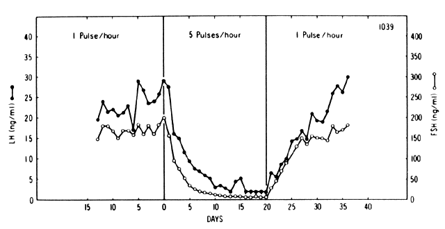
Fig. 4. Pituitary response to pulsatile and continuous administration of GnRH.(Belchetz PE, Plant TM, Nakai Y et al: Hypophysial responses to continuous and intermittent delivery of hypothalamic gonadotropin-releasing hormone. Science 202:631, 1978. Copyright 1978 by the AAAS)

Accordingly, the provision of GnRH heightens the pituitary response to a subsequent similar stimulus.

In the ovariectomized and hypothalamus-pituitary disconnected ewe, a decrease in GnRH pulse frequency results in decreased LH pulse amplitude, secondary to a reduction in the amount of releasable LH.34 The frequency of GnRH pulses has a qualitative effect as well. For instance, in monkeys, changes in the frequency of GnRH pulses alters the FSH/LH ratio, whereas changes in the amplitude of the pulses have a minor effect.35 For example, the pulsatile administration (every 2 hours) of GnRH to hypogonadotropic men coincides with the release of LH and the α-subunit common to both gonadotropins, but not with the release of FSH. When GnRH is administered every 30 minutes, the release of α-subunits becomes erratic and baseline LH increases, although the coincidence with α-subunit pulses is lost. In addition, FSH decreases to barely detectable levels, resulting in an increased LH/FSH ratio.36 In contrast to the critical importance of the frequency of GnRH pulses, changes in the amplitude seem to have a lesser role.11 Nevertheless, in ovariectomized sheep, the pulse amplitudes of GnRH and LH are highly correlated.37

In women of reproductive age, a single bolus of exogenous GnRH elicits an acute release of LH and, to a lesser extent, of FSH. When GnRH is given at 2-hour intervals, a dose-response relationship exists between GnRH (from at least 10 mg and up to 300 mg doses) and LH.38 The response is detected 5 minutes after intravenous injection of GnRH and reaches its peak between 30 to 45 minutes later. In response to GnRH, the pituitary may release prolactin as well. Increased circulating prolactin following a 4-hour infusion of GnRH was first reported in a group of patients suffering from anorexia nervosa, who had gained weight during the week preceding the test.39 A similar effect was described in normal women, in whom the finding was more pronounced during the peri-ovulatory period.40 Because the prolactin increment following infusion of GnRH correlates with both the FSH response41 and with the LH response,42 the observed increase is believed to be the consequence of a paracrine interaction between gonadotropin and prolactin. However, elevations of prolactin in response to GnRH administration have been documented in 7 of 10 women during the midtrimester of gestation, despite the lack of gonadotropin response to GnRH. This nonspecific prolactin response could also have been explained by the positive effect of high circulating levels of estrogens during gestation on prolactin production and release, thus sensitizing the lactotroph to nonspecific stimuli.43 Moreover, GnRH sensitizes the lactotrophs to thyrotropin-releasing hormone action in normal women.44 Finally, GnRH may bind to somatotrophic and thyrotrophic cells as well, resulting 
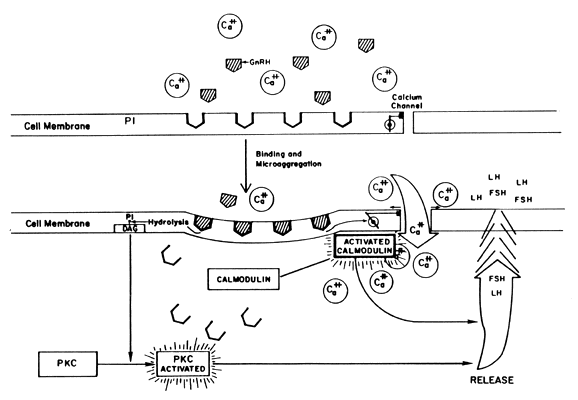
in actions not yet clarified.15

Self-Priming Action of GnRH

Gonadotropin-releasing hormone induces two separate phenomena in the pituitary. On 
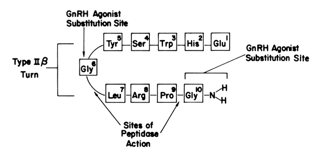
the one hand, GnRH primes (sensitizes) the gonadotroph to subsequent stimulation by increasing the number of its own receptors (Fig. 5). A similar phenomenon of increased pituitary responsiveness to GnRH is involved in the maturation of the hypothalamic-hypophyseal axis during puberty,45 a phenomenon that is used to diagnose true precocious puberty. On the other hand, GnRH triggers the release of LH and FSH. Both the “sensitization” and the “release” effects are dose-dependent, but functionally separate with the lower doses promoting priming, the higher doses favoring release. The responses of FSH parallel those of LH but on a much smaller scale.46

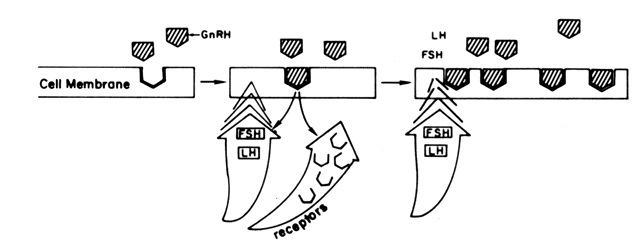
Fig. 5. Results of the binding of GnRH to its pituitary receptors.

One way by which the endocrine milieu modulates the pituitary response to GnRH is by regulating the number of GnRH receptors rather than by altering their affinity to GnRH. Indeed, in the rat, pituitary responsiveness correlates well with the number of GnRH receptors.18 In this connection, estradiol seems necessary to unmask the self-priming effect of GnRH.47 In the rat, low levels of prolactin are also necessary for GnRH to increase its own receptor number. Nevertheless, a different mechanism may be at play as well because both bromocriptine, a dopaminergic agent that decreases prolactin release, or estradiol alone can sensitize the pituitary to GnRH without necessarily increasing GnRH receptors.48

More recently, it has been demonstrated that inhibitors of PKC may inhibit the self-priming actions of GnRH.49 Furthermore, it has been postulated the effects of the sex steroids on the pituitary response may be directly related to an effect on PKC, independent of LH and Ca++ influx.50

Effect of Estrogens on the Pituitary Response to GnRH

In women, estrogens, in addition to unmasking GnRH self-priming, appear to protect from pituitary desensitization. In this connection, estrogen is antagonized by progesterone.51 Moreover, estradiol, at physiologic doses, increases the pituitary response to GnRH.52 In contrast, the provision of pharmacologic doses of estradiol to normally cycling women, resulting in serum levels of 700 to 1200 pg/mL, induces a marked diminution in pituitary responsiveness to even high doses (300 mg) of GnRH.53 The modulation of the pituitary response to GnRH by estradiol is duration-dependent. For instance, the administration of estradiol at doses resulting in serum levels of 100 to 200 pg/mL enhances the LH response to 100 mg of GnRH, provided the preceding exposure to estrogen was at least 84 hours in duration.54 The pituitary responsiveness is concentration-dependent and duration-dependent. Thus, LH and FSH response to GnRH is proportional to the dose of estradiol administered for 6 days. Minimal estradiol concentrations, resulting in increased gonadotropin responsiveness to 100 mg of GnRH, are 90 pg/mL for LH and 145 pg/mL for FSH.55 Interestingly, the administration of estradiol benzoate to normal women at a dose resulting in serum estradiol concentrations similar to those seen at midcycle can trigger an LH surge similar to that observed prior to ovulation. The above studies, however, do not accurately reflect the in vivo situation because the GnRH doses evaluated are not physiologic and the peptide is not given in a pulsatile fashion. The ovariectomized monkey with hypothalamic lesions in which gonadotropin secretion is re-established by chronic pulsatile GnRH administration serves as a better model to study the role of estrogen in modulating pituitary sensitivity to GnRH. The administration of estradiol to such animals results in an initial decline in gonadotropin levels, followed by a discharge of LH and FSH. This suggests that estradiol exerts both a negative and a positive feedback action at the level of the pituitary gland.56

To explain the observed effects of GnRH on the pituitary release of gonadotropins, the existence of two pools of gonadotropins has been postulated,38 namely, a readily “releasable” pool and a “reserve” pool. The former reflects pituitary “sensitivity” to a discrete GnRH stimulus. The latter represents intracellular gonadotropin not immediately available for release. The so-called capacity of the pituitary is defined as the amount of gonadotropins released in response to a larger and, therefore, more prolonged stimulation with GnRH. In this context, pituitary capacity consists of the releasable and reserve pools, as well as the amount of newly synthesized LH as the pituitary is continuously challenged.57

To assess the sensitivity of the gonadotroph or the status of the “releasable pool,” submaximal doses of GnRH must be used. Because the plasma clearance of exogenous GnRH is reduced by at least 50% when the dose is increased from 10 to 100 mg,58 the stimulus provided by relatively high doses of GnRH can be long enough to evoke a release from the reserve pool as well. When 10 mg of GnRH is administered every 2 hours for 10 hours, the LH response to the first dose reflects the releasable pool. The integrated concentrations of gonadotropins over time or the area under the LH and FSH curve during the 10-hour period reflects the reserve pool because the initial releasable pool is considered exhausted. Interpreted in this manner, the administration of estradiol benzoate to mimic follicular phase levels of estradiol results in increased releasable and reserve pools.59 Estrogens, however, seem preferentially to increase the reserve pool. For instance, as the follicular phase progresses and estradiol levels increase, the disparity between the response to 10 mg (releasable pool response) and the response to 150 mg (releasable plus reserve pools) increases in favor of the latter.60 In contrast, hypogonadal women have increased sensitivity to GnRH,59, 60 particularly if the baseline estradiol levels are less than 15 pg/mL.61 Such patients respond similarly to low or high doses of GnRH, indicating the absence of an adequate reserve pool.60 This condition is reversed by the administration of estradiol,61 again confirming that estradiol preferentially increases the reserve pool. Similarly, increased sensitivity is seen in postmenopausal patients. When estradiol is replaced in these women, the acute releasable pool is decreased at the second day of replacement but recovers on the fifth day, describing a U-shaped curve. The reserve pool (measured as the pituitary response to the second and third 10 mg dose of GnRH given every 2 hours) increases on the fifth day of estrogen replacement.62 The addition of progesterone at the end of the estradiol treatment further amplifies the response to a GnRH stimulus. As mentioned before, in addition to the effects described, estrogen may influence the pituitary response in more ways than one. For instance, estrogen inhibits pituitary desensitization due to the down-regulation of receptors that follows the continuous administration of GnRH, an effect that is antagonized by progesterone.51 Finally, estrogen administration to oophorectomized ewes has produced a LH pulse profile, suggesting a more prolonged LH response to each GnRH pulse.63 A similar observation was made in women during the periovulatory period.29

Effect of Progesterone on the Pituitary Response to GnRH

Progesterone given after the administration of estrogens further amplifies the estrogen-enhanced responsiveness of the pituitary to GnRH.59, 64 In fact, progesterone, but not 17α-hydroxyprogesterone, is able to trigger an LH and FSH surge in most normal estrogen-primed women.64, 65, 66 Indeed, the small preovulatory rise of progesterone may play a role in the generation of the midcycle LH surge. The above notwithstanding, progesterone antagonizes estrogen as it reduces the extent of pituitary desensitization in response to a continuous GnRH infusion.51 Interestingly, combination oral contraceptives (containing both estrogens and progestins) do not appear to affect the pituitary sensitivity to GnRH.67

Other Modulating Factors of the Pituitary Response to GnRH

The FSH and LH response to GnRH may be modulated by dopamine as well. Hyperprolactinemic women, presumably dopamine deficient, display an increased LH and FSH response to GnRH. Exogenous dopamine infusion decreases this response in these patients as it does in normal women.68

Neuropeptide Y, a putative modulator of GnRH release,69 has been shown to enhance GnRH-stimulated LH release.70, 71

Gonadotropin surge attenuating factor (GnSAF) is a proposed modulator of the midcycle gonadotropin surge, which is found in follicular fluid.72 It has been suggested that GnSAF attenuates both the primed and unprimed response of the pituitary to GnRH.73, 74 One investigator has gone so far as to speculate that the self-priming phenomenon is, in fact, a result of the interplay between GnRH and GnSAF.75 Nonetheless, the very existence of GnSAF and the physiologic role for this agent are unclear.

Inhibin has been shown to inhibit the GnRH-stimulated secretion of FSH, as well as to diminish the LH pulse amplitude,76 independent from the GnRH stimulation protocol.77

Acute intake of alcohol appears to diminish the response of LH and free α-subunit, but not FSH to the GnRH stimulus in men.78

Age brings about an impaired LH secretory capacity in men. While baseline parameters remain unchanged, the bioactive/immunoactive ratio of LH is decreased in response to an exogenous GnRH stimulus in older men.79 Additionally, older postmenopausal women have a decreased sensitivity to estrogen feedback as evidenced by the response to a clomiphene citrate challenge than do recently postmenopausal women.80 The significance of these findings remain to be determined.

Finally, short-term fasting increases the sensitivity of the gonadotroph to GnRH in men and women, an action independent of the level of plasma glucose.81

Changes in the Pituitary Response to GnRH During the Menstrual Cycle

Two mechanisms may play a role in the control of the hypothalamic-pituitary axis, which culminates in the midcycle LH surge and subsequent ovulation. The first, as previously described, is the modulation of the pituitary response to GnRH by sex steroids. The second may be the influence of these steroids on the GnRH pulse generator. Although the GnRH pulse generator appears to play only a permissive role in gonadotropin release, the gonadotropin pulse pattern varies during the cycle according to the hormonal status (i.e., high frequency, low amplitude LH pulses during the follicular phase and low frequency, high amplitude LH pulses during the luteal phase). The female rhesus monkey exposed to estradiol benzoate displays increased GnRH amplitude before and during an estradiol-triggered LH surge compared with nonestrogen exposed controls. It is, therefore, conceivable that, although not essential, an estradiol-triggered midcycle GnRH surge may play a role in the generation of the midcycle LH surge.28 Similarly, the high levels of preovulatory estradiol may be responsible for the preovulatory increase in pulse frequency and amplitude of LH and thus, by inference, of GnRH, as observed in normal women.29 By the same token, during the luteal phase, the luteal reaction in LH pulsatility appears to result from the action of estradiol and progesterone at the level of the GnRH pulse generator, rather than the pituitary gonadotrophs.31

Aside from steroidal interactions at the level of the hypothalamus, gonadotropin release is modulated by altering the pituitary response. Before demonstrating the influence of sex steroids on the pituitary response, the 
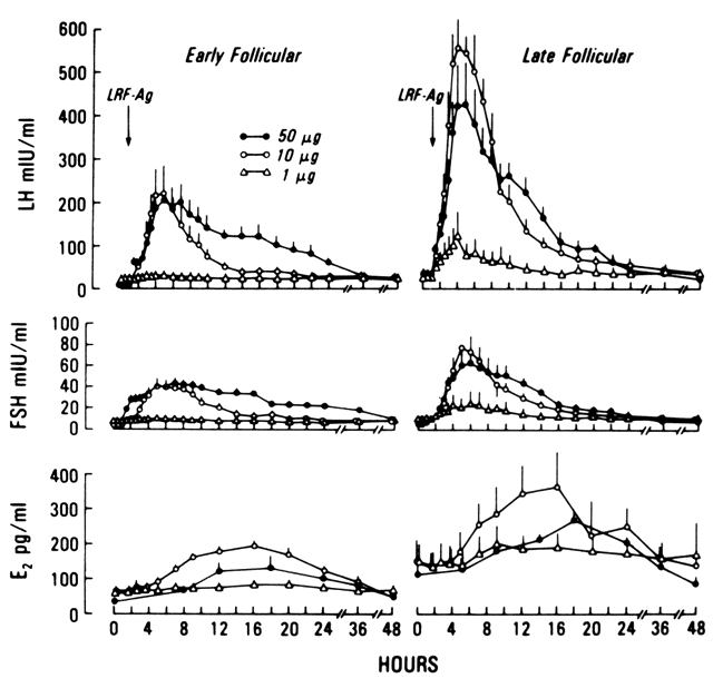
effect of 100 to 150 mg of GnRH was shown to vary during the menstrual cycle. While a more pronounced response is described during the late follicular phase, various reports disagree over whether the response is greater in the luteal phase82, 83 or in the late follicular phase.84

It is evident that the maximal sensitivity to GnRH occurs at midcycle, a phenomenon modulated mainly by the circulating levels of estradiol. As the follicular phase progresses, there is a change in the pituitary response to large (150 mg), but not to smaller (10 mg), doses of GnRH,38 indicating that estradiol exerts a preferential effect on the reserve pool. Additionally, the self-priming effect of GnRH may be an important determinant of pituitary sensitivity. Thus, the administration of 10 mg of GnRH at midcycle results in a tenfold increase in pituitary sensitivity.38 A U-shaped curve best describes the pituitary response to GnRH during the follicular phase (Fig. 6). During the early follicular stage, when circulating estradiol levels are at their lowest, both the releasable (sensitivity) and reserve pools appear depleted. However, at even lower estradiol concentrations (i.e., hypogonadism), pituitary sensitivity and capacity appear to increase,61, 62 perhaps due to an increased GnRH output, resulting in a self-priming effect. On the other hand, levels of estradiol consistent with the midfollicular and late-follicular phase increase pituitary sensitivity. More important, however, is the stimulatory effect of estrogens on the gonadotrophic capacity. Consequently, the observed negative feedback action of estrogens on the pituitary is exerted more at the level of gonadotropin release rather than at the level of gonadotropin synthesis.56 During the follicular phase, transfer of gonadotropins from the reserve to the releasable pool may occur (Fig. 7). The LH surge thus may be the consequence of multiple factors including estrogens acting in concert with perhaps the amplifying action of progesterone59, 64, 65, 66, 85; the estrogen-induced self-priming effect of GnRH86; the prolongation of GnRH-dependent LH pulses in the presence of estradiol34; and the possible, but not crucial, estrogen effect on GnRH release.28

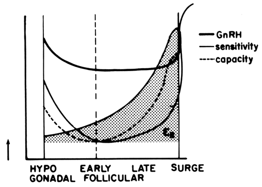
Fig. 6. Pituitary sensitivity and responsive capacity to GnRH according to the estrogenic milieu and throughout the follicular phase of the menstrual cycle.(Modified from Yen SSC, Lein A: The apparent paradox of the negative and positive feedback cortisol system on gonadotrophin secretion. Am J Obstet Gynecol 126:942, 1976)

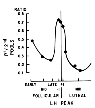
Fig. 7. Ratio between the first (releasable) pituitary gonadotropin pool and the second (reserve) pool in response to GnRHthroughout the menstrual cycle. (Hoff JD, Lasley BL, Wang CF,Yen SSC: The two pools of pituitary gonadotropins: Regulation during the menstrual cycle. J Clin Endocrinol Metab 44:302. Copyright 1977 by the Endocrine Society.)

In recent years, a great debate has raged over whether or not GnRH plays a pivotal role in the midcycle gonadotropin surge. In support of this 
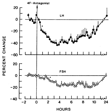
notion is the demonstration that GnRH antagonists can effectively prevent the midcycle LH surge.87, 88, 89, 90 Additionally, there exists the finding that the frequency of multiactivity unit volleys emanating from the region of the hypothalamus decreases just before the LH surge, a phenomenon most likely related to the preovulatory E2 surge.91, 92 In this regard, however, evidence also exits refuting a role for GnRH in the midcycle surge. For instance, it has been known for some time that women with hypogonadotropic hypogonadism treated with pulsatile GnRH do not require additional GnRH for the LH surge to occur.93 More importantly, GnRH pulse frequency (as evidenced by LH and free α-subunit pulse frequency) does not change at midcycle.94 Clearly, the role of GnRH in the midcycle gonadotropin surge is not well understood at this time.

In summary, during the follicular phase, estradiol amplifies GnRH action at the pituitary level by increasing the reserve pool while at the same time inhibiting the release of gonadotropins. In the late follicular phase, however, increasing amounts of estradiol may directly increase both GnRH pulse frequency and amplitude and the transfer of LH from the reserve into the releasable pool. This results in the paroxysmal LH surge despite the inhibitory action of estradiol on LH release.

Back to Top
PITUITARY RESPONSE TO GnRH IN SPECIAL CIRCUMSTANCES

Pregnancy and Puerperium

The total content of pituitary LH may decrease during pregnancy, the lowest values being noted at term.95 This finding is consistent with the observation of very low levels of circulating pituitary gonadotropins and a marked suppression of the gonadotropin response to GnRH by as early as 8 weeks of gestation.43, 96 Since pituitary insensitivity to GnRH persists for the first 2 to 3 weeks after delivery, despite the abrupt decline of estrogens and progesterone during the puerperium, the lack of response after birth may reflect the time needed for the recovery in the synthesis of gonadotropins. The puerperium is characterized by a period of relative pituitary refractoriness, followed by increased responsiveness to GnRH after the third week for FSH and the fifth to eighth weeks for LH.97 The lack of gonadotropic response may play a major role in postpartum amenorrhea. Others have shown that lactating women, when compared with their midfollicular phase counterparts, display no LH pulses; lower basal circulating levels of LH; a similar LH response to GnRH, and a significantly higher response of FSH (relative to LH) in the face of a comparable stimulus.98 Interpreted in light of studies in the subhuman primate wherein decreased GnRH pulsatility results in lower LH and higher FSH levels,28 the described response in puerperal women can perhaps be attributed to the slower frequency of GnRH release.

Hypothalamic Amenorrhea

Women with congenital absence of GnRH present a blunted pituitary response to GnRH, indicating low reserve and releasable pools.99 However, the low response to a single GnRH bolus could be due to the absence of GnRH receptors. Therefore, repeated doses of GnRH or steroid priming is required to bring about a normal response.100

Patients with secondary amenorrhea of suprapituitary origin display low to normal gonadotropin levels in the absence of organic pituitary lesions. Adult patients with very low circulating levels of estradiol (less than 15 pg/mL) show an exaggerated response to low doses of GnRH (10 mg) as compared with those with higher estradiol levels consistent with those observed in normal women in the early follicular phase.61 Although it is clear that these women have an altered response to estradiol, the pituitary gonadotrophs are fully capable of responding to ovulation induction with pulsatile GnRH.

Hyperprolactinemic Amenorrhea

The pituitary response to GnRH in hyperprolactinemia will, as might be expected, obviously depend on the cause of the abnormal condition. Thus, an expansive macroadenoma may compromise the pituitary severely enough to cause pituitary failure and, therefore, a lack of response to GnRH or other releasing factors. In cases of hyperprolactinemia in which the pituitary is otherwise largely functional (i.e., microadenoma, idiopathic hyperprolactinemia), an increase in both the releasable and reserve pools of FSH, with a decrease in the circulating levels of both LH and FSH has been demonstrated.64, 101 These findings were attributed to the hypoestrogenism, secondary to the hyperprolactinemia, and the consequent decrease in the negative feedback effect of estradiol on FSH release. The administration of bromocriptine, a dopaminergic agent capable of correcting the hyperprolactinemia, dopamine itself, or the surgical excision of a microadenoma reversed these findings. Given hyperprolactinemic women with normal sellar tomograms, no correlation exists between the degree of hyperprolactinemia and the response to GnRH, which is similar to that of normal controls. This suggests that prolactin does not interfere with the pituitary response to GnRH,102 but that the hypogonadotropic state is caused by a dopamine deficiency.64, 103

Anovulation and Dysfunctional Uterine Bleeding

Normoestrogenic women with dysfunctional uterine bleeding and adolescents with anovulation have normal responses to GnRH.104, 105

Polycystic Ovarian Disease

Patients with polycystic ovarian disease have an exaggerated LH and FSH response to GnRH,38, 106 a response correlated with the basal level of circulating LH.107, 108 While some investigators have found a decreased response to repetitive (every 2 hours) GnRH administration (10 mg),108 others have found increasing LH responses to repeat doses of 25 mg of GnRH.107 The increased pituitary sensitivity and the increase in the basal circulating levels of LH may be related to the increased circulating estrogen levels.107 Compared with normal women in the follicular phase, women with polycystic ovarian disease respond to 150 mg of GnRH with increased LH and FSH release.108 However, the FSH response does not change when GnRH is increased from a low (10 mg) to a high (150 mg) dose, perhaps due to the preferential inhibitory action of estrogen on FSH release.109, 110 More recently, it has been demonstrated that the frequency of the GnRH pulse generator is elevated in polycystic ovarian disease patients,111 as evidenced by elevations in pulsatile LH and free α-subunit release.112 In addition, through the use of a GnRH antagonist (Nal-Glu), it has been shown that these patients do not experience an increase in the amplitude of GnRH pulses.112 These data lend support to the hypothesis that an increase in the GnRH pulse frequency leads to a partial desensitization of the gonadotrope, which, in turn, is responsible for the abnormal gonadotropin secretory dynamics seen in polycystic ovarian disease.111

Isolated Gonadotropin Deficiency

By definition, patients with isolated gonadotropin deficiency may lack any response to GnRH. However, the diagnosis requires prolonged intermittent GnRH stimulation because some patients with hypothalamic hypogonadotropic hypogonadism and others with delayed puberty will not respond to GnRH, due to lack of a prior self-priming effect of GnRH.100, 113

Isosexual Precocious Puberty

Physiologic puberty is associated with increasing pituitary responsiveness to GnRH.46 This is because of the self-priming action of the decapeptide and increased estrogenic exposure, resulting in an adult type response. Conversely, precocious puberty secondary to exogenous or autonomous estrogen exposure is generally characterized by suppressed responses to GnRH.

Hypergonadotropic Hypogonadism

Women with this condition (i.e., Turner's syndrome) manifest increased pituitary sensitivity when circulating levels of estradiol are very low.114 This response is similar to the one seen in patients with hypothalamic amenorrhea.61

The GnRH Challenge Test

The administration of GnRH to women with hypogonadotropic amenorrhea can differentiate patients with hypothalamic hypogonadism from those with isolated pituitary gonadotropin failure. The latter will not respond, whereas those with hypothalamic hypogonadism will respond, provided the stimulus is repetitive enough to “awaken” (possibly through up-regulation of pituitary receptors to GnRH) the biosynthesis and release capabilities of the gonadotroph. In addition, the gonadotropin response to intravenous administration of GnRH (100 to 150 mg) over 30 seconds may segregate patients with central (true) from secondary (pseudo) precocious puberty.115 Thus, as a result of prior exposure to pulsatile GnRH, girls with true precocious puberty will have an adult-like response. Conversely, those whose development is due to nonhypothalamic-pituitary-induced steroidogenesis will not respond. Incidentally, the administration of GnRH will not affect the action of other releasing factors on the pituitary. In fact, a combined sequential anterior pituitary function test using corticotropin-releasing hormone, GnRH, growth hormone-releasing hormone, and thyrotropin-releasing hormone has been described.116

In the delicate balance characteristic of the hypothalamic-pituitary-ovarian axis, sex steroids seem to exert the most important modulatory action because their influence changes during each cycle, while the pituitary responds to what appears to be a permissive role for the hypothalamic GnRH pulse generator.

Back to Top
PITUITARY RESPONSE TO GnRH ANALOGS

The elucidation of the structure and sequence of GnRH quickly led to efforts to synthesize more potent and long-acting analogs of the hormone.

Many analogs have now been synthesized, some acting as agonists, others having predominantly antagonistic properties.117 The potential for further development and the indications for clinical use continue to increase, making an understanding of the physiology and pharmacology of the compounds important for the clinician.

GnRH Agonists

Gonadotropin-releasing hormone agonists were developed in the hope of yielding compounds with potential ovulation-promoting properties. However, it was soon learned that treatment with GnRH agonists, as with continuous high-dose infusions of native GnRH, cause a biphasic response in gonadotropin release from the pituitary and a paradoxic gonadal suppression.32, 118 Thus, administration of GnRH agonists of prolonged action results in paradoxic antireproductive effects, which ultimately lead to a hypogonadal state. Gonadal steroids approach undetectable levels after 1 to 3 weeks of treatment; a similar time is required for recovery.

As with native GnRH, the pituitary response to acute administration of GnRH agonists is variable, depending on the dose of agonist used and the phase in the menstrual cycle during which the compound is administered. It has been shown that the pituitary responds more briskly and to lower doses of GnRH agonists in the late follicular phase compared with the early follicular phase (Fig. 8).119 This can be explained by the priming of the pituitary with GnRH in the early follicular phase, resulting in an increased releasable pool of gonadotropin in the high-estrogen phase of the cycle. Higher dosages of GnRH agonists not only cause an acute rise in gonadotropins but also a sustained elevation over several hours to days. Similarly, the duration of the gonadotropin rise appears to be longer in the early follicular phase than in the late follicular phase (13 to 36 hours and 36 to 48 hours, respectively) (see Fig. 8).119 ln the midluteal phase, the response to an identical dose of the same GnRH agonist is almost identical to that observed in the early follicular phase.120 It is apparent that the administration of GnRH agonists mimics the infusion of high doses of naturally occurring GnRH, which stimulate an increase in LH and FSH levels for the first 4 to 6 hours, followed by a sustained decline to normal levels if GnRH is discontinued or to low to nondetectable levels if administration continues.121 The GnRH-induced shutdown of pituitary function is felt to be due to “desensitization” and “down-regulation” of the pituitary gonadotroph. Desensitization refers to an uncoupling of the GnRH receptor from the secretory signal (i.e., gonadotropin release), whereas down-regulation is thought to result from a decreased number of available membrane receptors for GnRH following internalization of the GnRH-receptor complexes.122

Fig. 8. Pituitary response to GnRH-agonists during the early and late follicular phase.(Casper RF, Sheehan KL, Yen SSC:Gonadotropin-estradiol responses to a superactive luteinizing hormone releasing hormone agonist in women. J Clin Endocrinol Metab 50:179. Copyright 1980 by the Endocrine Society.)

Structure of GnRH-Analogs

Peptidases in the pituitary gland and hypothalamus rapidly degrade naturally occurring GnRH by cleaving the decapeptide molecule between amino acids Gly6 to Leu7 and at the Pro9 to Gly10 bond (Fig. 9).123, 124 Thus, the circulating half-life of GnRH is relatively short (2 to 8 minutes). It is believed that the bond between amino acids 6 and 7 is the most vulnerable to degradation by pituitary endopeptidases because of its location at the type IIb turn (hairpin turn) of the GnRH molecule.123, 124 Adequate hormone-to-receptor binding is thought to be due to the configuration of the molecule that is maintained by amino acids (Glul, Gly6, Gly10). GnRH agonists were developed to increase potency over naturally occurring GnRH through enhanced binding affinity to the GnRH receptor and decreased enzymatic proteolysis.123,125,126 Substitutions by metabolically stable residues in the above-mentioned positions (amino acids 6 and 10) result in increased potency and prolonged half-life.122 It also has been suggested that protection of the agonists from renal elimination by albumin binding may also account for their prolonged action.127

Fig. 9. Schematic structure of the GnRH decapeptide.

GnRH Antagonists

Unlike GnRH agonists, which act by decreasing receptor availability and uncoupling, GnRH antagonists act by competing with GnRH for receptor sites, never causing a stimulatory signal. Therefore, antagonists have the advantage of a rapid onset of action without the initial stimulatory burst of gonadotropins. The pituitary response to GnRH antagonists differs in several ways when compared with their agonistic counterparts. The circulating levels of LH and FSH decline rapidly after the administration of a GnRH antagonist (maximal reduction occurring at 4 and 9 hours after an intravenous dose, respectively)128 and their pulse amplitudes are lowered (Fig. 10). Circulating gonadotropins return to basal levels and resume pulsatile release between 10 and 24 hours following GnRH antagonist administration. Contrary to the response to the agonists, the response to GnRH antagonists does not seem to vary during the menstrual cycle. The maximal decline of FSH, however, is slightly more profound in the midluteal phase.129 There appears to be a dose-dependent relationship between the GnRH antagonist administered and the LH response, a phenomenon also seen with GnRH agonists. Higher doses of GnRH antagonists act faster to abolish LH pulsation.130 The duration of LH suppression is dose-dependent as well.130 Nevertheless, suppression of plasma immunoreactive FSH was found to be insignificant at all doses and not dose-dependent. Thus, LH release is more profoundly inhibited than FSH, suggesting that a GnRH-independent releasing factor for FSH may operate as well.130

Fig. 10. Gonadotropin response to GnRH antagonists.(Cetel NS, Rivier J, Vale W, Yen SSC: The dynamics of gonadotropin inhibition in women induced by an antagonistic analog of gonadotropin releasing hormone. J Clin Endocrinol Metab 57:62–65. Copyright 1983 by the Endocrine Society.)

Interestingly, the antagonists were actually synthesized before the agonists.131 The idea of preventing ovulation by blocking the release of gonadotropins was initially exciting but later proved disappointing due to difficulties in developing a clinically useful GnRH antagonist.132 Indeed, the earlier generations of GnRH antagonists stimulated histamine release from mast cells, and therefore had anaphylactoid potential.133,134 Furthermore, some have agonistic properties.128 Particularly strenuous demands are put on GnRH antagonists for clinical use because as little as 10% receptor occupancy by GnRH can cause LH release,122,135 requiring the antagonist to be present continually at the receptor to block native GnRH binding. To accomplish their inhibitory action, antagonists are endowed with a long half-life and a high receptor affinity.132

In recent years, gonadotropin-releasing hormone antagonists that elicit minimal histamine release have been evaluated for clinical use. The potential benefits of an agent that lacks the initial stimulatory phase of the response to the agonist are self-evident. In clinical studies, GnRH antagonists have been shown capable of preventing premature LH surges in women undergoing ovulation induction.87,136,137 These agents have also been studied in men for the treatment of sex steroid-dependent disorders, such as benign prostatic hypertrophy.138 The promising data suggest the enormous potential for the application of GnRH antagonists in disorders in which GnRH agonists are currently indicated, including endometriosis and uterine leiomyomata,139 as well as the possibility of their use in the ovarian hyperstimulation syndrome, and breast and ovarian cancer.140

Back to Top
REFERENCES

1. Mizinuma H, Sampson WK, Lumpkin MD, McCann SM: Evidence for an FSH releasing factor in the posterior portion of the rat median eminence. Life Sci 33: 2003, 1983

2. Mizinuma H, Sampson WK, Lumpkin MD et al: Purification of a bio-active FSH-releasing factor (FSH-LH). Brain Res Bull 10: 623, 1983

3. Nikolics K, Mason AJ, Szonyi E et al: A prolactin-inhibiting factor within the precursor for human gonadotropin releasing hormone. Nature 316: 511, 1985

4. Song T, Nikolics K, Seeburg PH, Goldsmith PC: GnRH prohormone-containing neurons in the primate brain: Immunostaining for the GnRH associated peptide. Peptides 8: 335, 1987

5. Seeburg PH, Mason AJ, Stewart TA, Nikolics K: The mammalian GnRH gene and its pivotal role in reproduction. Recent Prog Horm Res 43: 96, 1987

6. Ishibashi M, Yamaji T, Takaku F et al: Effect of GnRH-associated peptide on prolactin secretion from human lactotrope adenoma cells in culture. Acta Endocrinol 116: 81, 1987

7. Millar RP, Wormald PJ, Milton RC de L: Stimulation of gonadotropin release by a non-precursor peptide sequence of the GnRH precursor. Science 232: 68, 1986

8. Milton RC de L, Wormald PJ, Brandt W, Millar RP: The delineation of a decapeptide gonadotropin-releasing sequence in the carboxyl-terminal extension of the human gonadotropin releasing hormone precursor. J Biol Chem 261: 16990, 1986

9. Dahl KD, Pavlou SN, Kovacs WJ, Hsueh AJW: The changing ratio of bioactive to immunoreactive follicle-stimulating hormone in normal men following treatment with a potent gonadotropin releasing hormone antagonist. J Clin Endocrinol Metab 63: 792, 1986

10. Kessel B, Dahl KD, Kazer RR et al: The dependency of bioactive follicle-stimulating hormone secretion on gonadotropin-releasing hormone in hypogonadal and cycling women. J Clin Endocrinol Metab 66: 361, 1988

11. Phol CR, Knobil E: The role of the central nervous system in the control of ovarian function in higher primates. Annu Rev Physiol 44: 583, 1982

12. Kessner JS, Kaufman JM, Wilson RC, Knobil E: On the short-loop feedback regulation of the hypothalamic luteinizing hormone releasing hormone “pulse-generator” in the rhesus monkey. Neuroendocrinology 42: 109, 1986

13. Clark IJ, Cummins JT: The temporal relationship between gonadotropin releasing hormone (GnRH) and the luteinizing hormone (LH) secretion in ovariectomized ewes. Endocrinology 111: 1737, 1982

14. Culler MD, Negro-Villar A: Evidence that pulsatile follicle-stimulating hormone secretion is dependent on endogenous luteinizing hormone-releasing hormone. Endocrinology 118: 606, 1986

15. Hopkins CR, Gregory H: Topographical localization of the receptor for luteinizing hormone releasing hormone on the surface of dissociated pituitary cells. J Cell Biol 75: 528, 1977

16. Conn MP, Marian J, McMillan M et al: Gonadotropin-releasing hormone action in the pituitary: A three step mechanism. Endocr Rev 2: 174, 1981

17. Hazum E, Cuatrecasas P, Marian J, Conn PM: Receptor mediated internalization of fluorescent gonadotropin releasing hormone by pituitary gonadotropes. Proc Natl Acad Sci USA 77: 6692, 1980

18. Conn MP: The molecular basis of gonadotropin-releasing hormone action. Endocr Rev 7: 3, 1986

19. Kakar SS, Musgrove LC, Devor DC et al: Cloning, sequencing and expression of human gonadotropin releasing hormone (GnRH) receptor. Biochem Biophys Res Commun 189: 289, 1992

20. Kaiser UB, Zhao D, Cardona R et al: Isolation and characterization of cDNAs encoding rat pituitary gonadotropin-releasing hormone receptor. Biochem Biophys Res Commun 189: 1645, 1992

21. Perrin MH, Bilezikjian LM, Hoeger C et al: Molecular and functional characterization of GnRH receptors cloned from rat pituitary and a mouse pituitary tumor cell line. Biochem Biophys Res Commun 191: 1139, 1993

22. Reinhart J, Mertz LM, Catt KJ et al: Molecular cloning and expression of cDNA encoding for the murine gonadotropin-releasing hormone receptor. J Biol Chem 267: 21281, 1992

23. Brooks J, McNeilly AS: Regulation of gonadotrophin-releasing hormone receptor mRNA expression in the sheep. Endocrinology 143: 175, 1994

24. Alexander JM, Klibanski A: Gonadotropin-releasing hormone receptor mRNA expression by human pituitary tumors in vitro. J Clin Invest 93: 2332, 1994

25. Peng C, Fan NC, Ligier M et al: Expression and regulation of gonadotropin-releasing hormone (GnRH) and GnRH receptor messenger ribonucleic acids in human granulosa-luteal cells. Endocrinology 135: 1740, 1994

26. Plant TM: Gonadal regulation of hypothalamic gonadotropin-releasing hormone release in primates. Endocr Rev 7: 75, 1986

27. Soules MR, Steiner RA, Clifton DK et al: Progesterone modulation of pulsatile luteinizing hormone secretion in normal women. J Clin Endocrinol Metab 58: 378, 1984

28. Levine JE, Norman Rl, Gliessman FR et al: In vivo gonadotropin-releasing hormone release and serum luteinizing hormone measurements in ovariectomized, estrogen-treated rhesus macaques. Endocrinology 117: 711, 1985

29. Djahanbakhch O, Warner P, McNeilly AS, Baird DT: Pulsatile release of LH and estradiol during the preovulatory period in women. Clin Endocrinol 20: 579, 1984

30. Kesner JS, Wilson RC, Kaufman JM et al: Unexpected responses of the hypothalamic gonadotropin-releasing hormone “pulse generator” to physiological estradiol inputs in the absence of the ovary. Proc Natl Acad Sci USA 84: 8745, 1987

31. Nippold TB, Khoury S, Barkan A et al: Gonadotrophin responses to GnRH pulses in hypogonadotrophic hypogonadism: LH responsiveness is maintained in the presence of luteal phase concentration of estrogen and progesterone. Clin Endocrinol 26: 293, 1987

32. Belchetz PE, Plant TM, Nakai Y et al: Hypophysial responses to continuous and intermittent delivery of hypothalamic gonadotropin-releasing hormone. Science 202: 631, 1978

33. Katt JA, Duncan JA, Herbon L et al: The frequency of gonadotropin-releasing hormone stimulation determines the number of pituitary gonadotropin-releasing hormone receptors. Endocrinology 116: 2113, 1985

34. Clarke IJ, Cummins JT: GnRH pulse frequency determines LH amplitude by altering the amount of releasable LH in the pituitary gland of ewes. J Reprod Fertil 73: 425, 1985

35. Wild L, Hausler A, Marshall G et al: Frequency and amplitude of gonadotropin releasing hormone stimulation and gonadotropin secretion in the rhesus monkey. Endocrinology 109: 376, 1981

36. Spratt Dl, Chin WW, Ridgways EC, Crowley WF Jr: Administration of low-dose pulsatile gonadotropin-releasing hormone (GnRH) to GnRH-deficient men regulates free α-subunit secretion. J Clin Endocrinol Metab 62: 102, 1986

37. Levine JE, Pau K-Y P, Ramirez VD, Jackson GL: Simultaneous measurements of luteinizing hormone-releasing hormone and luteinizing hormone release in unanesthetized, ovariectomized sheep. Endocrinology 111: 1449, 1982

38. Yen SSC, Lasley BL, Wang CF et al: The operating characteristics of the hypothalamic-pituitary system during the menstrual cycle and observations of biological action of somatostatin. Recent Prog Horm Res 31: 312, 1975

39. Beumon PJV, Abraham SF, Turtle J: Paradoxical prolactin response to gonadotropin releasing hormone during weight gain in patients with anorexia nervosa. J Clin Endocrinol Metab 51: 1283, 1980

40. Mais V, Melis GB, Paoletti AM et al: Prolactin-releasing action of a low dose of exogenous gonadotropin-releasing hormone through the human menstrual cycle. Neuroendocrinol 44: 326, 1986

41. Mais V, Yen SSC: Prolactin-releasing action of gonadotropin-releasing hormone in hypogonadal women. J Clin Endocrinol Metab 62: 1089, 1986

42. Tan YM, Steele PA, Judd SJ: The effect of physiological changes in ovarian steroids on the prolactin secretion response to gonadotropin releasing factor. Clin Endocrinol 24: 71, 1986

43. Sowers JR, Colantino M, Fayez J, Jonas H: Pituitary response to LHRH in midtrimester pregnancy. Obstet Gynecol 52: 685, 1978

44. Minakami H, Kimura K, Ljima K et al: Evaluation of GnRH administration on the prolactin response to thyrotropin-releasing hormone in normal women. Endocrinol Jpn 33: 511, 1986

45. Spratt Dl, Crowley WF Jr: Pituitary and gonadal responsiveness is enhanced during GnRH-induced puberty. Am J Physiol 254: E625, 1988

46. Hoff JD, Lasley BL, Yen SSC: The functional relationship between priming and releasing actions of luteinizing hormone-releasing hormone. J Clin Endocrinol Metab 49: 8, 1979

47. Veldhuis JD, Evans WS, Rogol AD et al: Pituitary self priming action of gonadotropin-releasing hormone. J Clin Invest 77: 1849, 1986

48. Duncan JA, Barkan A, Herbon L, Marshall JC: Regulation of pituitary gonadotropin-releasing hormone (GnRH) receptors by pulsatile GnRH in female rats: Effects of estradiol and prolactin. Endocrinology 118: 320, 1986

49. Johnson MS, Mitchell R, Thompson FJ: The priming effect of luteinizing hormone-releasing hormone (LHRH) but not LHRH-induced gonadotropin release can be prevented by certain protein kinase C inhibitors. Mol Cell Endocrinol 85: 183, 1992

50. Ortmann O, Tilse B, Emons G: Modulatory actions of estradiol and progesterone on phorbol ester-stimulated LH secretion from cultured rat pituitary cells. J Steroid Biochem Mol Biol 43: 619, 1992

51. Araki S, Chikazawa K, Motoyama M et al: Reduction in pituitary desensitization and prolongation of gonadotropin release by estrogen during continuous administration of gonadotropin-releasing hormone in women: Its antagonism by progesterone. J Clin Endocrinol Metab 60: 590, 1985

52. Jaffe RB, Keye WR: Estradiol augmentation of pituitary responsiveness to gonadotropin releasing hormone in women. J Clin Endocrinol Metab 39: 850, 1974

53. Yen SSC, Vandenberg G, Siler TM: Modulation of pituitary responsiveness to LRF by estrogen. J Clin Endocrinol Metab 39: 170, 1974

54. Keye WR, Jaffe RB: Strength duration characteristics of estrogen effects on gonadotropin response to gonadotropin-releasing hormone in women. I. Effects of varying duration of estradiol administration. J Clin Endocrinol Metab 1: 1003, 1975

55. Young JR, Jaffe RB: Strength-duration characteristics of estrogen effect on gonadotropin response to gonadotropin-releasing hormone in women. II. Effect of varying concentrations of estradiol. J Clin Endocrinol Metab 42: 432, 1976

56. Nikai Y, Plant TM, Hess DL et al: On the site of negative and positive feedback actions of estradiol in the control of gonadotropin secretion in the rhesus monkey. Endocrinology 102: 1008, 1978

57. Yen SSC, Lein A: The apparent paradox of the negative and positive feedback control system on gonadotropin secretion. Am J Obstet Gynecol 126: 942, 1976

58. Kelch RP, Clemens LE, Markovs M et al: Metabolism and effects of synthetic gonadotropin-releasing hormone (GnRH) in children and adults. J Clin Endocrinol Metab 40: 53, 1975

59. Lesley BL, Wang CF, Yen SSC: The effects of estrogen and progesterone on the functional capacity of the gonadotrophs. J Clin Endocrinol Metab 41: 820, 1975

60. Wang CF, Lasley BL, Yen SSC: Gonadotropin secretion in response to low and high doses of LRF in normal and hypogonadal women (functional capacity of the gonadotrophs). J Clin Endocrinol Metab 42: 427, 1976

61. Rebar RW, Harman SM, Vaitukaitis JL: Differential responsiveness to LRF after estrogen therapy in women with hypothalamic amenorrhea. J Clin Endocrinol Metab 46: 48, 1978

62. Lachelin GCL, Yen SSC: Biphasic change in pituitary capacity induced by estrogen in hypogonadal women. J Clin Endocrinol Metab 46: 369, 1978

63. Clarke IJ, Cummins JT: Increased GnRH pulse frequency associated with estrogen induced LH surges in ovariectomized ewes. Endocrinology 116: 2376, 1986

64. Chang RJ, Jaffe RB: Progesterone effects on gonadotropin release in women pretreated with estradiol. J Clin Endocrinol Metab 47: 119, 1978

65. Leyendeker G, Wildt L, Gips H et al: Experimental studies on the positive feedback of progesterone, 17-hydroxy-progesterone on the pituitary release of LH and FSH in the human female. Arch Gynaekol 221: 29, 1976

66. Rakoff JS, Rigg LA, Yen SSC: Assessment of progesterone(O)-induced LH release as a test for the hypothalam-ic-gonadotropin function. Gynecol Invest 8:31(A), 1977

67. Hemrika DJ, Slaats EH, Schoemaker J: The response of the pituitary-ovarian axis to pulsatile administration of gonadotropin-releasing hormone in long-term oral contraceptive users. Am J Obstet Gynecol 170: 462, 1994

68. Nicoletti I, Ambrosi F, Giammartino C et al: Catecholamines and pituitary function. V. Effect of low dose dopamine infusion on basal gonadotropin-releasing hormone stimulated gonadotropin release in normal cycling women and patients with hyperprolactinemic amenorrhea. Horm Metab Res 18: 479, 1986

69. Minamini S, Frautschy SA, Plotsky PM et al: Facilitory role of neuropeptide on the onset of puberty: Effect of immunoneutralization of neuropeptide Y on the release of luteinizing hormone-releasing hormone. Neuroendocrinol 52: 112, 1990

70. Bauer-Dantoin AC, McDonald JK, Levine JE: Neuropeptide Y potentiates luteinizing hormone (LH)-releasing hormone-induced LH secretion only under conditions leading to preovulatory LH surges. Endocrinology 131: 2946, 1992

71. Kalra SP, Crowley WR: Neuropeptide Y: a novel neuroendocrine peptide in the control of pituitary secretion, and its relationship to luteinizing hormone. Front Neuroendocrinol 13: 1, 1992

72. Messinis IE, Templeton A: Attenuation of gonadotrophin release and reserve in superovulated women by gonadotrophin surge attenuating factor (GnSAF). Clin Endocrinol 34: 259, 1991

73. Fowler PA, Cunningham P, Fraser M et al: Circulating gonadotrophin surge-attenuating factor from superovulated women suppresses in vitro gonadotrophin-releasing hormone self-priming. J Endocrinol 143: 45, 1994

74. deKoning J, Westhoff WE, Koppenaal DW et al: On the dynamics of gonadotrophin releasing hormone (GnRH): role of self-priming and desensitization in the luteinizing hormone response to GnRH after follicle stimulating hormone treatment. Hum Reprod 9: 1600, 1994

75. Koppenaal DW, Tijssen AM, deKoning J: The effect of gonadotrophin-surge inhibiting factor on the self-priming action of gonadotrophin-releasing hormone in female rats in vitro. J Endocrinol 134: 427, 1992

76. Clarke IJ, Findlay JK, Cummins JT, Evans WJ: Effects of ovine follicular fluid on plasma LH and FSH secretion in ovariectomized ewes to indicate the action of inhibin. J Reprod Fertil 77: 575, 1986

77. Charlesworth MC, Grady RR, Shin L et al: Differential suppression of FSH and LH secretion by follicular fluid in the presence or absence of GnRH. Neuroendocrinol 38: 199, 1984

78. Zgliczynski W, Nowakowski J, Roslonowska E et al: Alcohol decreases the alpha subunit, LH and testosterone secretion in response to LH-RH. Endokrynol Pol 43: 257, 1992

79. Urban RJ, Veldhuis JD, Blizzard RM, Dufau ML: Attenuated release of biologically active luteinizing hormone in healthy aging men. J Clin Invest 81: 1020, 1988

80. Rossmanith WG, Reichelt C, Scherbaum WA: Neuroendocrinology of aging in humans: attenuated sensitivity to sex steroid feedback in elderly postmenopausal women. Neuroendocrinol 59: 355, 1994

81. Rodjmark S: Increased gonadotropin responsiveness to gonadotropin-releasing hormone during fasting in normal subjects. Metabolism 36: 21, 1987

82. Yen SSC, Vandenberg R, Rebar R, Ehara Y: Variation of pituitary responsiveness to synthetic LRH during different phases of the menstrual cycle. J Clin Endocrinol Metab 35: 931, 1972

83. Faure N, Olivier GC: Hypersensitivity of the human gonadotrophs to the repeated administration of small doses of LH-RH. Horm Res 9: 12, 1978

84. Nillius SJ, Wide L: Variation in LH and FSH response to LH-releasing hormone during the menstrual cycle. J Obstet Gynaecol Br Commonw 79: 865, 1972

85. Clark IJ, Cummins JT: Direct pituitary effects of estro- gen and progesterone on gonadotropin secretion in the ovariectomized ewes. Endocrinology 39: 267, 1984

86. Veldhuis JD, Evans WS, Rogol AD et al: Pituitary self priming action of gonadotropin-releasing hormone. J Clin Invest 77: 1849, 1986

87. Frydman R, Cornel C, deZiegler D et al: Spontaneous luteinizing hormone surges can be reliably prevented by the timely administration of a gonadotrophin releasing hormone antagonist (Nal-Glu) during the late follicular phase. Hum Reprod 7: 930, 1992

88. Dubourdieu S, Charbonnel B, d'Acremont MF et al: Effect of administration of a gonadotropin-releasing hormone (GnRH) antagonist during the periovulatory period: the luteinizing hormone surge requires the secretion of GnRH. J Clin Endocrinol Metab 78: 343, 1994

89. Hall JE, Taylor AE, Martin KA et al: Decreased release of a gonadotropin-releasing hormone during the preovulatory midcycle luteinizing hormone surge in normal women. Proc Natl Acad Sci USA 91: 6894, 1994

90. Leroy I, d'Acremont M, Brailly-Tabard S et al: A single injection of a gonadotropin-releasing hormone (GnRH) antagonist (Cetrorelix) postpones the luteinizing hormone surge: further evidence for the role of GnRH during the LH surge. Fertil Steril 62: 461, 1994

91. O'Byrne KT, Thalabard JC, Grosser PM et al: Radiotelemetric monitoring of hypothalamic gonadotropin-releasing hormone pulse generator activity throughout the menstrual cycle of the rhesus monkey. Endocrinology 129: 1207, 1991

92. O'Byrne KT, Knobil E: Electrophysiologic approaches to gonadotrophin releasing hormone pulse generator activity throughout the menstrual cycle of the rhesus monkey. Hum Reprod 8: 37, 1993

93. Santoro N, Filicori M, Crowley WF: Hypogonadotropic disorders in men and women: diagnosis and therapy with pulsatile GnRH. Endocr Rev 7: 11, 1986

94. Adams JM, Taylor AE, Schoenfeld DA et al: The midcycle gonadotropin surge in normal women occurs in the face of an unchanging gonadotropin-releasing hormone pulse frequency. J Clin Endocrinol Metab 79: 858, 1994

95. De la Lastra M, Llados C: Luteinizing hormone content of the pituitary gland in pregnant and non-pregnant women. J Clin Endocrinol Metab 44: 921, 1977

96. Rubinstein LM, Parlow AF, Derzko C, Hershman J: Pituitary gonadotropin response to LHRH in human pregnancy. Obstet Gynecol 52: 172, 1978

97. Keye WR, Jaffe RB: Changing patterns of FSH and LH response to gonadotropin releasing hormone in the puerperium. J Clin Endocrinol Metab 42: 1133, 1976

98. Morishita H, Higuchi K, Hashimoto T et al: Study on the responsiveness of pituitary FSH to GnRH 6 weeks after delivery. Gynecol Obstet Invest 23: 110, 1987

99. Kletzky OA, Nicoloff J, Davajan V et al: Idiopathic hypogonadotrophic hypogonadal primary amenorrhea. J Clin Endocrinol Metab 46: 808, 1978

100. Snyder PJ, Rudenstein RS, Gardner DF et al: Repetitive infusion of gonadotropin-releasing hormone distinguishes hypothalamic from pituitary hypogonadism. J Clin Endocrinol Metab 48: 864, 1979

101. Lachelin GCL, Abu-Fadil S, Yen SSC: Functional delineation of hyperpro-lactinemic-amenorrhea. J Clin Endocrinol Metab 44: 1163, 1987

102. Spellacy WN, Cantor B, Kalra PS et al: The effect of varying prolactin levels on pituitary luteinizing hormone and follicle-stimulating hormone response to gonadotropin-releasing hormone. Am J Obstet Gynecol 132: 157, 1978

103. Quigley ME, Gilliland GB, Yen SSC: Effects of gonadotropin antagonist on the release of gonadotropin and prolactin in normal women and women with hyperprolactinemic anovulation. J Clin Endocrinol Metab 48: 718, 1979

104. Van Look PFA, Hunter WM, Fraser IS, Baird DT: Impaired estrogen-induced luteinizing hormone release in young women with anovulatory dysfunctional uterine bleeding. J Clin Endocrinol Metab 46: 816, 1978

105. Fraser IS, Michie EA, Wide L, Baird DT: Pituitary gonadotropins and ovarian function in adolescent dysfunctional uterine bleeding. J Clin Endocrinol Metab 37: 407, 1973

106. Baird DT, Corker CS, Davidson DW et al: Pituitary ovarian relationship in polycystic ovarian syndrome. J Clin Endocrinol Metab 45: 798, 1977

107. Moltz L, Rommler A, Schwartz V: Peripheral steroid-gonadotropin interactions and diagnostic significance of double-stimulation tests with luteinizing hormone-releasing hormone in polycystic ovary syndrome. Am J Obstet Gynecol 134: 813, 1979

108. Rebar R, Judd HL, Yen SSC et al: Characterization of the inappropriate gonadotropin secretion in polycystic ovary syndrome. J Clin Invest 57: 1320, 1976

109. Futterweit W: Polycystic Ovarian Disease, pp 50–53. New York, Springer-Verlag, 1984

110. Adashi EY: Hypothalamic-pituitary dysfunction in polycystic ovarian disease. Endocrinol Metab Clin 17: 649, 1988

111. Hall JE, Taylor AE, Martin KA, Crowley WF: New approaches to the study of the neuroendocrine abnormalities of women with the polycystic ovarian syndrome. Ann N Y Acad Sci USA 22: 182, 1994

112. Hall JE, Whitcomb RW, Rivier JE et al: Differential regulation of luteinizing hormone, follicle-stimulating hormone and free α-subunit secretion from the gonadotrope by gonadotropin-releasing hormone (GnRH): evidence from the use of two GnRH antagonists. J Clin Endocrinol Metab 70: 328, 1990

113. Yoshimoto Y, Moridera K, Imura H: Restoration of normal pituitary gonadotropin reserve by administration of luteinizing-hormone-releasing hormone in patients with hypogonadotropic hypogonadism. N Engl J Med 292: 242, 1975

114. Boyar RM, Ramsey J, Chapman J et al: Luteinizing hormone and follicle-stimulating hormone secretory dynamics in Turner's syndrome. J Clin Endocrinol Metab 47: 1078, 1978

115. Zipf WB, Kelch RP, Hopwood NJ et al: Suppressed responsiveness to gonadotropin-releasing hormone in girls with unsustained isosexual precocity. J Pediatr 94: 38, 1979

116. Sheldon WR Jr, DeBold CR, Evans WS et al: Rapid sequential intravenous administration of four hypothalamic releasing hormones as a combined anterior pituitary function in normal subjects. J Clin Endocrinol Metab 60: 623, 1985

117. Karten MJ, Rivier JE: Gonadotropin-releasing hormone analog design. Structure-function studies toward the development of agonists and antagonists: rationale and perspective. Endocr Rev 7: 44, 1986

118. Rabin D, McNeil LW: Pituitary and gonadal desensitization after continuous luteinizing hormone-releasing hormone infusion in normal females. J Clin Endocrinol Metab 51: 873, 1980

119. Casper RF, Sheehan KL, Yen SSC: Gonadotropin-estradiol responses to a superactive luteinizing hormone releasing hormone agonist in women. J Clin Endocrinol Metab 50: 179, 1980

120. Sheehan KL, Casper RF, Yen SSC: Effects of a superactive luteinizing hormone-releasing factor agonist on gonadotropin and ovarian function during the menstrual cycle. Am J Obstet Gynecol 135: 759, 1979

121. Davies TF, Gomez-Pan A, Watson MJ et al: Reduced gonadotropin response to releasing hormone after chronic administration in impotent men. Clin Endocrinol 6: 213, 1977

122. Vickery BH, Nestor JJ: Luteinizing hormone-releasing hormone analog: Development and mechanism of action. Semin Reprod Endocrinol 5: 353, 1987

123. Horsthenke B, Knisatschek H, Rivier J et al: Degradation of luteinizing hormone-releasing hormone and analogs by adeno-hypophyseal peptidases. Biochem Biophys Res Commun 100: 753, 1981

124. Marks N, Stern F: Enzymatic mechanisms for the inactivation of luteinizing hormone-releasing hormone (LHRH). Biochem Biophys Res Commun 61: 1458, 1974

125. Clayton RN, Catt KJ: Receptor-binding affinity of gonadotropin-releasing hormone analogs: analysis of radio-ligand-receptor-assay. Endocrinology 106: 1154, 1980

126. Loumaye E, Naor Z, Catt KJ: Binding affinity and biological activity of gonadotropin-releasing hormone agonists in isolated pituitary cells. Endocrinology 111: 730, 1982

127. Nestor JJ Jr, Ho TL, Simpson RA et al: Synthesis and biological activity of some very hydrophobic superagonist analogues of luteinizing hormone releasing hormone. J Med Chem 25: 795, 1982

128. Cetel NS, Rivier J, Vale W, Yen SSC: The dynamics of gonadotropin inhibition in women induced by an antagonistic analog of gonadotropin releasing hormone. J Clin Endocrinol Metab 57: 62, 1983

129. Mais V, Kazer RR, Cetel NS et al: The dependency of folliculogenesis and corpus luteum function on pulsatile gonadotropin secretion in cycling women using a gonadotropin-releasing hormone antagonist as a probe. J Clin Endocrinol Metab 62: 1250, 1986

130. Hall JE, Brodie TD, Badger TM et al: Evidence of differential control of FSH and LH secretion by gonadotropin-releasing hormone (GnRH) from the use of a GnRH antagonist. J Clin Endocrinol Metab 67: 524, 1988

131. Vale W, Grant G, Rivier J et al: Synthetic polypeptide antagonists of the hypothalamic luteinizing hormone releasing factor. Science 176: 933, 1972

132. Schally AV, Kastin AJ: Stimulation and inhibition of fertility through hypothalamic agents. Drug Therapy 1: 29, 1971

133. Hahn DW, McGune JL, Vale WW, Rivier J: Reproductive/endocrine and anaphylactoid properties of an LHRH antagonist ORL 18260 [Ac-D-Nal(2)1, 4FD-Phe2, D-Trp3, D-Arg3 ]-GnRH. Life Sci 37: 506, 1985

134. Schmidt F, Sundaram K, Thau RB, Bardin CW: [Ac-D-Nal(2)1, 4FD-Phe2, D-Trp3, D-Arg6 ]-LHRH a potent antagonist of LHRH, produces transient edema and behavioral change in rats. Contraception 29: 283, 1984

135. Clayton RN: Gonadotropin-releasing hormone modulation of its own pituitary receptors: evidence for biphasic regulation. Endocrinology 111: 152, 1982

136. Olivennes F, Fanchin R, Bouchard P et al: The single or dual administration of the gonadotropin-releasing hormone antagonist Cetrorelix in an in vitro fertilization-embryo transfer program. Fertil Steril 62: 468, 1994

137. Diedrich K, Diedrich C, Santos E: Suppression of the endogenous LH- surge by the GnRH-antagonist Cetrorelix during ovarian stimulation. Hum Reprod 9: 788, 1994

138. Behre HM, Klein B, Steinmeyer E: Effective suppression of luteinizing hormone and testosterone by single doses of the new gonadotropin-releasing hormone antagonist Cetrorelix (SB-75) in normal men. J Clin Endocrinol Metab 75: 393, 1992

139. Reissmann T, Diedrich K, Comaru-Schally AM, Schally AV: Introduction of LHRH-antagonists into the treatment of gynecological disorders. Hum Reprod 9: 767, 1994

140. Emons G, Schally AV: The use of luteinizing hormone releasing hormone agonists and antagonists in gynaecologic cancer. Hum Reprod 9: 1364, 1994

Back to Top