Chapter 16
Precocious Puberty
Andrea Haqq, Bruce Boston and Stephen LaFranchi
Main Menu   Table Of Contents

Search

Dndrea Haqq, MD
Department of Pediatrics, Duke University Medical Center, Division of Endocrinology and Diabetes, Durham, North Carolina (Vol 5, Chap 16)

Bruce Boston, MD
Assistant Professor, Division of Endocrinology, Department of Pediatrics, Doernbecher Children's Hospital, Oregon Health Sciences University, Portland, Oregon (Vol5, Chap 16)

Stephen LaFranchi, MD
Professor, Department of Pediatrics, and Head, Pediatric Endocrinology, Doernbecher Childern's Hospital, Oregon Health Sciences University, Portland, Oregon (Vol 5, Chap 16)

 
NORMAL PUBERTY
CLINICAL FEATURES OF PRECOCIOUS PUBERTY
CLASSIFICATION OF PRECOCIOUS PUBERTY
DISORDERS PRODUCING PRECOCIOUS PUBERTY AND THEIR SPECIFIC CLINICAL FEATURES
LABORATORY DIAGNOSIS
TREATMENT
PROGNOSIS
REFERENCES

At puberty, maturation of the hypothalamic-pituitary-gonadal axis results in the profound physical, sexual, and emotional changes that transform a girl into a woman and a boy into a man. Difficult under normal circumstances, puberty out of synchrony with friends and classmates has the potential for significant emotional distress. If one accepts the mean ±2.5 SDs as encompassing the normal range, puberty is precocious if changes occur in a girl before 8 years (or possibly 7 years if a more recent study is correct) and in a boy before 9 years. Studies show a younger age of onset in African-American girls. Precocious puberty requires a careful history, physical examination, laboratory evaluation, and imaging studies to search for an underlying cause; however, in most children, especially girls, no abnormal cause is identified. Treatment is available for most cases of gonadotropin-dependent and gonadotropin-independent forms of precocious puberty.

Although children with precocious puberty undergo early physical and sexual development, their emotional and intellectual development is commensurate with their chronologic age. They and their parents require special counseling to help them adjust to these changes.

Back to Top
NORMAL PUBERTY

Physical Changes of Puberty

The detailed longitudinal studies of British children by Tanner and Marshall1,2,3 provide information about the sequence and timing of the physical and sexual changes that occur throughout puberty. For convenience, breast and pubic hair development in girls and genital and pubic hair development in boys were arbitrarily divided into five stages (Tables 1 and 2), referred to as Tanner breast, pubic hair, and genital stages.


TABLE 1. Tanner Staging of Female Puberty


Stage

Breasts

Pubic Hair

1—Prepubertal

Elevation of papilla only

Any vellus over pubes no different from abdominal hair

2

Breast bud with elevation of breast and papilla and enlargement of areola

Slightly pigmented, downy hair along the labia

3

Further enlargement of breast and papilla with no separation of their contours

Darker, coarser, more curled hair over pubes

4

Projection of areola and papilla to form a secondary mound

Adult pubic hair that does not reach thighs (axillary hair)

5—Adult

Mature breast, projection of papilla only as areola conforms to breast contour

Adult hair now on thighs


TABLE 2. Tanner Staging of Male Puberty


Stage

Genitalia

Pubic Hair

1—Prepubertal

Testes, penis, and scrotum same size and proportion as early childhood

Any vellus over pubes no different from abdominal hair

2

Early enlargement of testes >2 cm3; scrotal skin reddens and changes in texture

Slightly pigmented, downy hair at base of penis

3

Penis lengthens; testes enlarge 3–6 cm3; growth of scrotum

Darker, coarser, more curled, over pubes

4

Further penile and scrotal growth; testes 8–12 cm3

Adult pubic hair that does not reach thighs (axillary hair)

5—Adult

Genitalia adult in size and shape, testes 15–25 cm3

Adult hair now on thighs


Several factors seem to influence the time of initiation of puberty, including genetic factors, geographic location, exposure to light, and general health and nutrition. In family studies, the onset of the mother’s puberty seems more important than the father’s puberty; the daughters and sons of early-maturing mothers are early to mature.4 Children closer to the equator, children at lower altitudes, children in urban areas, and obese children start earlier than children in northern latitudes, children at higher elevation above sea level, rural children, and normal-weight children. British girls begin menarche at a mean age of 13.5 years, whereas a 1970 study showed that American girls begin menarche at a mean age of 12.8 years.5

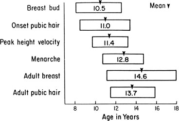
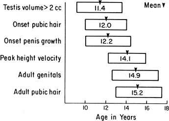
The sequence and timing of pubertal changes in girls and boys are shown in Figures 1 and 2. Figure 1 has been modified from the data of Marshall and Tanner2 to take into account the fact that menarche in American girls is 0.7 years earlier than in British girls. The first sign of puberty in a girl is breast budding, which occurs at a mean age of 10.5 years; the first sign of puberty in boys is testicular enlargement, which occurs a year later at a mean age of 11.5. The size of the testes can be measured accurately by comparison with a set of elliptical beads of increasing volume (the Prader orchidometer). Normal values for testicular volume are presented in Figure 3.6 The normal range (mean ± 2.5 SD) is 8 to 13 years for breast budding and 9 to 14 years for testicular enlargement. In about 20% of children, pubic hair growth is the first sign of puberty. A cross-sectional study based on pubertal staging of more than 17,000 girls from age 3 to 12 years was carried out in pediatric office practices across the United States to evaluate the age of onset of puberty in African-American and white girls. This study reported that white girls had onset of pubic hair growth at a mean of 10.51 years, breast development at 9.96 years, and menarche at 12.88 years. In comparison, African-American girls had the onset of pubic hair growth at a mean of 8.78 years (range, 4.8 to 12.8 years), breast development at 8.87 years (range, 5 to 12.7 years), and menarche at 12.16 years (range, 9.7 to 14.6 years).7 The maximal growth rate occurs 1 year after onset of breast budding in girls at a mean age of 11.5 years and 2.5 years after testicular enlargement in boys at a mean age of 14.1 years. A criticism of this cross-sectional study is the fact that pubertal staging was rated by inspection only and not palpation. It is possible that occasional erroneous classification of a Tanner 1 girl as a Tanner 2 girl may have occurred. This cross-sectional study is supported, however, by data from the National Health Examination Surveys (NHES) from 1963 to 1970 and the National Health Examination Survey (NHANES III) from 1988 to 1994. The NHANES data indicate that 12% to 14% of girls have Tanner 2 breast development by age 8 years. The median age of Tanner 2 breast development is 9.5 years in the aforementioned cross-sectional study and 9.7 years in NHANES. The onset of menses is 12 years in all three studies, supporting the idea that although the onset of puberty is occurring earlier, it is not being completed earlier.8 Based on these studies, it has been proposed that the lower limit of normal onset of puberty be redefined to 7 years for white girls and 6 years for African-American girls. An organic cause for precocious puberty should be sought in white girls less than 7 years old and African-American girls less than 6 years old. No change has been made in guidelines for the assessment of precocious puberty in boys, who still require investigation for organic causes if the onset of puberty occurs at less than 9 years of age.9 Girls and boys who experience rapid progression of pubertal development, even if it occurs after the above-mentioned age cutoffs, require diagnostic studies to determine the cause of their accelerated puberty.

Fig. 1. Mean age of onset with a range of ±2.5 SDs for several events of female puberty. These data are adapted from Marshall and Tanner2 to take into account the fact that menarche is 0.7 years earlier in American girls than in British girls.

Fig. 2. Mean age of onset with a range of ±2.5 SDs for several events of male puberty. These data are adapted from Marshall and Tanner3 to take into account the fact that puberty appears 0.7 years earlier in American boys than in British boys.

Fig. 3. Testicular volumes in relation to chronologic age. Mean values ±1 SD.(Zachmann M, Prader A, Kind HP, et al: Testicular volume during adolescence: Cross-sectional and longitudinal studies. Helv Paediatr Acta 29:61, 1974.)

The pattern of growth velocity from birth to adult life, including the timing of the maximal pubertal growth spurt, is illustrated in Figure 4. The maximal growth rate always precedes menarche. The mean age of menarche is 12.8 years, with a normal range of 10.3 to 15.3 years (mean ± 2.5 SD). Menarche occurs 2.3 ± 2.1 years after breast budding. Although there is not a corresponding landmark in boys, a Scandinavian study reported the average age of spermarche (as judged by appearance of spermatozoa in the urine) to be 13.4 years (range, 11.7 to 15.3 years).10 The age of menarche has been decreasing at a rate of about 1 year for every 25 years from 1830 to 1960. This change probably is related to improved general health and nutrition in particular. Over the last generation, this trend has leveled off.

Fig. 4. The pattern of growth velocity from birth through 18 years, showing the adolescent growth spurt in girls and boys.(Tanner JM: Growth at Adolescence, 2nd ed. Oxford, Blackwell Scientific Publishers, 1962.)

All these figures may vary depending on the individual’s genetic and environmental background, and there is a wide variation of normal for the onset and sequence of each stage. The pubertal growth spurt coincides with increasing serum insulin-like growth factor I (IGF-I) concentrations concomitant with the rise in sex steroid levels.11 Evidence supports the conclusion that estrogens and androgens augment human pituitary growth hormone (hGH) secretion. Studies using 24-hour sampling show higher mean hGH concentrations, with more hGH peaks and higher peak amplitude.12 Children with precocious puberty and hGH deficiency still undergo a growth spurt, however, although less than normal, and manifest serum IGF-I concentrations intermediate between prepubertal children with hGH deficiency and children with sexual precocity and intact hGH secretion.13 These studies support a smaller but direct effect of sex steroids on IGF-I production.

Other important somatic changes accompany the pubertal growth spurt. In girls, total body fat increases from 16% to 23.5% of total body weight; in boys, total body fat decreases in early to mid puberty. In boys, muscle mass, as measured by radiologic studies of muscle cross-sectional area, increases fourfold; in girls, muscle mass increases twofold.

It has been proposed that when girls reach a critical weight of 47.8 kg, menarche is triggered (the so-called Frische-Revelle hypothesis).14 Other studies show that there is a large variation in total body weight at menarche, and menarche seems better correlated with the increase in total body fat to 23.5%. There are exceptions to this hypothesis, however: Girls with idiopathic central precocious puberty may undergo menarche with a total body fat of 19%, children with sexual precocity secondary to hypothyroidism have a total body fat of 29%, and obese girls with no signs of puberty may have a total body fat of 27%.15 It seems more reasonable to hypothesize that central mechanisms activate the hypothalamic-pituitary axis (HPA), leading to gonadotropin stimulation of ovarian sex steroids that in turn stimulate growth to the critical weight and an increase in body fat composition to 23.5%.

The cross-sectional study cited earlier documented a trend toward initiation of puberty at earlier ages in white and African-American girls. This trend may be linked to an increase in the prevalence of obesity in young girls. Significantly higher body mass index z scores have been shown in white pubertal versus prepubertal 6- to 9-year-old girls. In African-American girls, a smaller difference in body mass index z score was detected, suggesting that other genetic and environmental influences may play a role in their earlier pubertal onset. The practical implications of these findings for clinicians are that white and African-American girls who are overweight may be more likely to display signs of early puberty. Physicians should consider the contribution of obesity to the incidence of early puberty when decisions regarding searching for pathologic causes or consideration of treatment of early puberty is made.16

Leptin produced by adipocytes may play a role in modulating reproduction. Leptin-deficient Ob/Ob mice are infertile, with fertility restored with leptin replacement. In rats, minimal leptin levels seem to be required to initiate puberty. Serum leptin concentrations are correlated positively with Tanner stages, and adolescent girls have higher levels than boys. Obese adolescent girls have higher serum leptin concentrations, suggesting that these girls overcome leptin resistance to achieve reproductive capacity.17

Endocrine Changes at Puberty

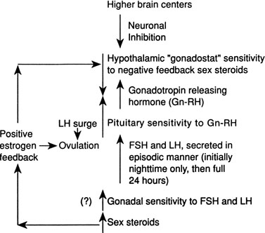
Although several pieces of the puzzle of the mechanism of onset of puberty have been put into place, the complete picture of the factors responsible for activation of the hypothalamic-pituitary-gonadal axis is not clear. During fetal life, pituitary follicle-stimulating hormone (FSH) and luteinizing hormone (LH) concentrations rise and peak near midgestation; gonadotropin levels then fall, perhaps under the inhibition of placental steroids. Early in infancy, again there is a rise in gonadotropins, perhaps resulting from loss of placental steroid inhibition, with FSH predominating in girls and LH in boys. Serum gonadotropin concentrations reach levels seen in adults (FSH, 1 to 31 mIU/mL in women; LH, 3 to 25 mIU/mL in men), peak around 2 to 4 weeks of life, then fall to prepubertal low levels over the next several months.18,19 These pituitary gonadotropins stimulate temporary rises in estradiol in girls (10 to 60 pg/mL) and testosterone in boys (60 to 400 ng/dL).20,21 Even during the quiescent prepubertal period, sensitive immunofluorometric assays show pulsatile secretion, albeit at low amplitude, of LH and FSH, with nocturnal levels higher than daytime levels.22 The prepubertal pituitary can respond to exogenous gonadotropin-releasing hormone (GnRH) stimulation, with an FSH peak predominating in girls and a slight LH peak predominating in boys.23 The prepubertal gonad also shows a rise in sex steroids in response to exogenous gonadotropin stimulation.24 Immaturity of the prepubertal pituitary-gonadal axis does not seem to prevent the onset of puberty. These studies indirectly support the hypothesis that an inhibitor from higher brain centers restrains the HPA. Although the nature of this inhibitor is unknown, opioid peptides are a potential candidate. In a study of pubertal boys, administration of the opioid receptor antagonist naltrexone resulted in significantly higher mean LH concentration, LH pulse frequency, and area under LH time curve.25 Alternatively, animal studies support a neuronal-to-glial signaling pathway, perhaps via increased production of growth factors, such as transforming growth factor α, directly stimulating GnRH.26

The onset of puberty may result from diminished brain inhibition or positive glial-neuronal stimulation of GnRH secretion (Fig. 5). The maturation process that triggers this pubertal alarm clock is unknown, but it seems to be tied to normal development of the HPA during fetal life and infancy, physical growth and body composition changes, and skeletal maturation.

Fig. 5. Proposed mechanism of the onset of puberty. Decreasing central nervous system neuronal inhibition of the hypothalamic “gonadostat” results in increasing gonadotropin-releasing hormone (Gn-RH) secretion, which stimulates pituitary gonadotropin secretion and gonadal sex steroid production.

GnRH is secreted in a pulsatile pattern about once every 90 minutes. The frequency and amplitude of this GnRH pulse generator determine the pattern of FSH and LH secretion. During infancy, GnRH secretion is elevated followed by a long period of relative quiescence that occurs until late childhood, when pubertal maturation begins. The transition period from childhood quiescence to an adolescent pattern of GnRH secretion occurs gradually rather than abruptly. Animal models and human studies indicate that hypothalamic neurons are capable of secreting GnRH throughout childhood, and small pulses of GnRH-induced LH and FSH secretion are found in 4-year-olds.27 In girls in early puberty, FSH and LH are secreted at higher frequency and amplitude at night, and there is an equal FSH and LH response to GnRH stimulation. In mid to late puberty, FSH and LH are secreted during the day and night, with higher LH peak amplitudes, both spontaneous and in response to exogenous GnRH stimulation.23 In boys in early puberty, nocturnal LH secretion predominates; in late puberty, daytime LH secretion predominates. LH secretion, spontaneous and in response to exogenous GnRH stimulation, is greater than FSH secretion throughout puberty in boys.23

Increased pituitary gonadotropin secretion results in growth and development of the gonads and a gradual increase in sex steroid production. The normal ranges of serum gonadotropins and sex steroids through the Tanner stages are presented for girls in Table 3 and for boys in Table 4. Before the pubertal rise in gonadotropins, an increase in adrenal sex steroids, such as dehydroepiandrosterone (DHEA) and androstenedione, has been documented.28 Evidence supports separate control mechanisms regulating adrenarche and gonadarche, although some investigators have speculated that the prepubertal rise in these adrenal steroids may play a role in the activation of the hypothalamic “gonadostat.”29 A disproportionate number of girls with premature adrenarche, resulting, for example, from late-onset congenital adrenal hyperplasia (CAH), experience an early onset of menarche.30


TABLE 3. Blood Hormone Concentrations During Female Puberty*


Tanner Stage

FSH (mIU/ml)

LH (mIU/ml)

Estradiol (pg/ml)

DHEA (ng/dL)

Androstenedione (ng/dL)

17-OH-Progesterone (ng/dL)

Progesterone (ng/dL)

1—Prepubertal

2.16 ± 1.14

0.03 ± 0.03

1–20

31–345

8–50

3–82

<10–33

2

3.44 ± 1.58

0.71 ± 1.04

10–24

150–570

42–100

11–98

<10–55

3

4.88 ± 2.11

2.10 ± 2.33

7–60

200–600

80–190

11–155

10–450

4

6.19 ± 2.55

3.67 ± 2.22

21–85

200–780

77–225

18–230

10–1300

5—Adult: Follicular

6.63 ± 2.19

5.76 ± 3.46

3–100

215–850

82–240

15–70

15–70

Luteal


70–300


35–290

200–2500


To convert to International SI units, multiply:
FSH × 1 = IU/L   LH × 1 = IU/L  
Estradiol × 3.67 = pmol/L   DHEA × 34.67 = pmol/L   Androstenedione × 34.92 = pmol/L   17-OH-progesterone × 30.26 = pmol/L   Progesterone × 31.80 = pmol/L
* Taken from normal ranges reported by Endocrine Sciences, Tarzana, CA, 1990, except as noted below.
Mean ± 1 SD taken from Neely EK, Hintz RL, Wilson DM et al: Normal ranges for immunochemiluminometric gonadotropin assays. J Pediatr 127:40, 1995


TABLE 4. Blood Hormone Concentrations During Male Puberty*


Tanner Stage

FSH (mIU/ml)

LH (mIU/ml)

Testosterone (ng/dL)

DHT (ng/dL)

DHEA (ng/dL)

Androstenedione (ng/dL)

17-OH-Progesterone (ng/dL)

1—Prepubertal

0.97 ± 0.59

0.05 ± 0.05

<3–10

<3

31–345

8–50

3–90

2

2.75 ± 1.84

1.80 ± 1.30

18–150

3–17

110–495

31–65

5–115

3

2.94 ± 1.55

1.86 ± 1.41

100–320

8–33

170–585

50–100

10–138

4

4.47 ± 1.88

2.65 ± 1.81

200–620

22–52

160–640

48–140

24–180

5—Adult

4.91 ± 2.02

4.51 ± 1.99

350–970

24–65

250–900

65–210

24–175


To convert to International SI units, multiply:
FSH × 1 = IU/L; LH × 1 = IU/L;
Testosterone × 34.67 = pmol/L; Dihydrotestosterone × 34.44 = pmol/L; DHEA × 34.67 = pmol/L; Androstenedione × 34.92 = pmol/L; 17-OH-progesterone × 30.26 = pmol/L
* Taken from normal ranges reported by Endocrine Sciences, Tarzana, CA, 1990, except as noted below.
Mean ± 1 SD taken from Neely EK, Hintz RL, Wilson DM et al: Normal ranges for immunochemiluminometric gonadotropin assays. J Pediatr 127:40, 1995


In mid to late puberty in girls, there is development of positive estrogen feedback on the hypothalamus.31 This feedback seems to be important in the maturation of the hypothalamic cyclic center in girls, and it is this positive estrogen feedback that stimulates the midcycle LH surge, which is important for ovulation.

Back to Top
CLINICAL FEATURES OF PRECOCIOUS PUBERTY

Signs of prepubertal development in a white girl before 7 years, an African-American girl before 6 years, or a boy before 9 years are beyond 2.5 SD from the mean and represent precocious puberty. Pubertal development of the same sex, seen predominantly as feminization in a girl, is termed isosexual precocious puberty; the occurrence of pubertal changes of the opposite sex, seen predominantly as virilization with hirsutism and clitoral enlargement in a girl or gynecomastia in a boy, is termed heterosexual precocious puberty.

In girls, sexual precocity usually begins with breast development, although pubic hair growth or vaginal bleeding may be the initial manifestation. Other signs of estrogen secretion include nipple enlargement and pigmentation, thickening of the labia minora, a change in the vaginal mucosa from shiny red to dull purple, and vaginal mucus production. Some emotional lability and cyclic mood swings may be present. Percentage of body fat composition increases, and a female body contour develops.

In boys, sexual precocity may begin with an increase in genital size, which may or may not include the testes, depending on the underlying cause. Pubic hair growth may be the first sign. Testes volume of greater than 2 mL indicates pubertal development. The testes progressively increase in volume through puberty (see Fig. 3). Other androgen effects include an increase in muscle bulk and strength, deepening of the voice, and eventually facial hair growth. Boys also may experience more frequent erections, penile emissions, and behavior changes, including increased libido and perhaps aggressive behavior.

Girls and boys develop apocrine gland activity, with perspiration and body odor, and sebaceous gland secretion, accompanied by oily skin and hair and sometimes acne. The secretion of estrogen in girls and androgen in boys results in increased growth velocity and accelerated skeletal maturation. These growth and skeletal changes are associated with increased serum IGF-I and IGF binding protein-3 concentrations and increased bone density; these changes are concordant with skeletal rather than chronologic age.32,33

Complete forms of sexual precocity eventually result in the development of all secondary sex characteristics and acceleration in growth and skeletal maturation seen with normal pubertal development. These children must be investigated thoroughly to determine the underlying cause so that appropriate treatment, if indicated, can be selected. Incomplete forms of sexual precocity are less urgent. Isolated breast development in a girl before 7 to 8 years is termed premature thelarche. Isolated pubic or axillary hair growth before 7 to 8 years in a girl or 9 years in a boy is termed premature pubarche. These latter changes are usually the result of adrenal androgen secretion, so the term premature adrenarche often is used interchangeably with premature pubarche. Isolated vaginal bleeding before 8 years is termed premature menarche. Each of these incomplete forms of precocious puberty requires observation over time. If no other pubertal changes develop and growth and skeletal maturation remain normal, they are usually benign conditions.

Back to Top
CLASSIFICATION OF PRECOCIOUS PUBERTY

Precocious puberty is divided most easily into gonadotropin-dependent and gonadotropin-independent sexual precocity. Gonadotropin-dependent precocity is almost always pituitary-mediated and so is also termed central precocious puberty. Gonadotropin-independent precocity, usually a result of a gonadal or adrenal disorder, is also termed peripheral precocious puberty. Precocious puberty is three to five times more common in girls than in boys. A classification and relative occurrence of causes of sexual precocity are presented in Table 5, which is based on a summary of seven series previously reported.34,35,36,37,38,39,40


TABLE 5. Classification and Relative Occurrence of Causes of Precocious Puberty*


Female (%)

Male (%)

Gonadotropin dependent (central)


 Idiopathic

65

21

 Hypothalamic hamartoma

15

20

 Other cerebral lesions

7

26

 Ectopic gonadotropins

0.5

0.5

 Primary hypothyroidism


Gonadotropin independent (peripheral)


 Ovarian (cyst or tumor)

5


 Testicular (including familial male precocious puberty)


10

 McCune-Albright syndrome

5

1

 Adrenal-feminizing

1

0

 Adrenal-virilizing

1

22

Combined gonadotropin-independent and gonadotropin-dependent    

* The relative occurrence of each disorder is based on a summary of seven series of precocious puberty.
These cases are included in the gonadotropin-independent cases.


Most cases of precocious puberty are gonadotropin-dependent (87.5% in girls and 67.5% in boys). In 65% of girls, no underlying abnormality is found; this percentage has decreased from 80% in the early 1990s, resulting from the discovery of hamartomas in 15% of cases by newer imaging techniques. In contrast, two thirds of boys with central precocious puberty have an underlying central nervous system abnormality identified; pineal tumors and hamartomas are the most common causes.

Of gonadotropin-independent causes in girls, most represent autonomous ovarian lesions—cysts, granulosa-theca cell tumors, or part of McCune-Albright syndrome. In boys, the most common gonadotropin-independent cause is virilizing CAH. Because simple virilizing CAH represents the non–salt-losing form, it generally does not present in infancy. Autonomous testicular causes are increasingly recognized, the most common being familial male precocious puberty (also known as testotoxicosis or familial Leydig and germ cell hyperplasia). Lastly, initially autonomous gonadal or adrenal disorders may accelerate physical and skeletal maturation to the point where activation of central or pituitary mediated precocity develops. Although the mechanism of this activation is unknown, it most likely occurs with intermittent sex steroid production so that there is incomplete inhibition of pituitary gonadotropin secretion or after the underlying gonadal or adrenal disorder is treated, leading to reduced sex steroid inhibition. These children may have a combination of gonadotropin-independent and gonadotropin-dependent sexual precocity.

Back to Top
DISORDERS PRODUCING PRECOCIOUS PUBERTY AND THEIR SPECIFIC CLINICAL FEATURES

Sexual Precocity in Girls

GONADOTROPIN-DEPENDENT.

Idiopathic.

If the normal population is defined to include 2.5 SD around the mean, or about 97% of girls, 1 or 2 normal girls per 100 will have the onset of puberty just before age 7 to 8 years. These girls with constitutional precocious puberty often have a family history of slightly early onset of puberty. Girls with idiopathic sexual precocity usually start puberty much earlier than 8 years, however, and there is usually no family history of early onset of puberty. Familial precocious puberty is rare in girls41; in boys, however, many family pedigrees of sexual precocity have been reported.

Although idiopathic precocious puberty is the most common situation with female precocity, it is a diagnosis of exclusion. Idiopathic sexual precocity seems to be the result of early activation of the HPA with loss of neuronal inhibition of the hypothalamic gonadostat, the same mechanism (unknown) that induces the onset of normal puberty. Electroencephalographic abnormalities are common in children with true precocious puberty, further supporting a central nervous system abnormality.

The progression of pubertal changes with precocity varies. Some children show a normal tempo of sexual maturation from the time that puberty is initiated, whereas others show a staccato pattern of maturation that progresses to a more gradual pubertal development. Finally, other children who begin early puberty show steady progression but often at a tempo that is much slower than with normal puberty. If sexual precocity begins in infancy (<1 year old), only 15% to 20% of girls menstruate by age 2.5 years. Although the average interval from normal breast development to menarche is 2 to 2.5 years, about 20% of girls do not menstruate for 4 or more years after the first signs of puberty. Ovulatory cycles occur in girls with sexual precocity, with the earliest reported pregnancy resulting in delivery when the mother was 5 years, 7 months.34 Sexual precocity is consistent with a normal reproductive life and does not seem to be associated with premature menopause.

The most severe effect of precocious puberty is the resultant short stature as an adult. The skeleton is a sensitive end organ for sex steroids, especially estrogen. Estrogen seems to be primarily responsible for final skeletal maturation and epiphyseal fusion.42,43 Although early on these children are tall for their age, early exposure to sex steroids causes accelerated skeletal maturation, which results in early epiphyseal fusion and short stature. About 50% of girls with precocious puberty are less than 5 feet tall as adults (range, 4 feet, 5 inches to 5 feet, 8 inches).44 Dental eruption corresponds more closely with chronologic age than skeletal age.

Girls with precocious puberty may have emotional problems resulting from teasing and feelings of being different from their friends and classmates. Hormone changes may lead to feelings and behavior that they are not yet mature enough to deal with. They require reassurance that these changes are normal, albeit early. It is important to remember that intellectual and psychosocial development are commensurate with chronologic age rather than the state of puberty. Parents, siblings, other relatives, peers, and teachers often respond as if the child were older, with expectations of greater intellectual and physical performance and more mature, even assertive behavior. These children inappropriately may appear developmentally delayed, uncoordinated, and immature. Parents and teachers should be counseled not to put excessive demands on these girls but rather to relate to them at the appropriate chronologic age level.

Hypothalamic Hamartoma.

Hypothalamic hamartomas, previously often unrecognized, are now discovered in about 15% of girls with precocious puberty; as such, they are the most common central nervous system tumor causing sexual precocity in girls.45,46 Central nervous system hamartomas represent heterotopic and hyperplastic neuronal tissue. They are isodense with brain on imaging techniques and do not enhance with the administration of contrast agents. They are well circumscribed and are usually 0.5 to 2 cm in diameter, occasionally larger. They are usually in the region of the tuber cinereum, and they may develop as pedunculated masses from the floor of the third ventricle. The exact mechanism leading to precocious puberty is unknown, but it most likely involves either production of GnRH by the hamartoma itself or interference with the neuronal inhibition restraining activation of the HPA. In at least one case, immunofluorescent studies showed the presence of GnRH in the hamartoma, along with thyroid-stimulating hormone (TSH) and somatostatin.47 This latter hamartoma resembled an autonomously functioning accessory hypothalamus that was found to be responsive to hormonal stimulation and negative feedback. The clinical features are similar to those of idiopathic precocious puberty.

The natural history of central nervous system hamartomas is to grow slowly and to stop growing when the brain finishes growing, around 8 to 12 years. They usually are not associated with visual impairment, signs of increased intracranial pressure, focal neurologic abnormalities, gelastic seizures, or pituitary hormone defects, although any of these features may occur. In the absence of any of these features, management involves careful observation with repeat imaging studies at periodic intervals. Some clinicians advocate neurosurgical removal of easily accessible, pedunculated hamartomas in young children to prevent further progression of precocious puberty and the need for long-term GnRH agonist therapy.48 Therapy with the newer agonists has been so successful, however, that even the minimal complications of neurosurgery in the aforementioned setting need not be risked.

Other Cerebral Lesions.

Abnormalities of the central nervous system resulting in precocious puberty include other tumors, congenital brain defects, and acquired lesions after insults such as central nervous system radiation, meningoencephalitis, or trauma. Together, these account for about 7% of cases of female sexual precocity.

Tumors (other than hypothalamic hamartoma [discussed earlier]) include optic gliomas, dysgerminomas, and teratomas. Craniopharyngiomas, the most common childhood pituitary tumor, are usually destructive, but uncommonly they may be associated with precocious puberty.49 Of the phakomatoses, neurofibromatosis and tuberous sclerosis may result in sexual precocity; in about half of neurofibromatosis cases, optic gliomas are present.50 Congenital brain defects associated with sexual precocity include the syndrome of septo-optic dysplasia (the triad of optic nerve dysplasia, absent septum pellucidum, and hypothalamic hypopituitarism)51 and congenital blindness, suprasellar cysts, hydrocephalus, and meningomyelocele.52 In the syndrome of septo-optic dysplasia, about one third of children manifest either precocious or rapidly progressing pubertal development.53 In some cases, precocity may be reversible after correction of the underlying process, as reported in one case after removal of a congenital arachnoid cyst.54 Sexual precocity may follow encephalitis, head trauma,55 or severe prematurity, and in one report it was seen in 20% of children who received radiation therapy (2500 to 4500 rad) as treatment for head and neck tumors.56 The exact mechanism of precocity associated with these latter disorders is unknown, but it is presumed that the underlying cerebral lesion interferes with the neuronal inhibitor restraining activation of the HPA.

The clinical manifestations are similar to those of idiopathic precocious puberty; in addition, these patients may have neurologic symptoms and signs secondary to the cerebral abnormality. A careful neurologic examination, including ophthalmologic examination with visual fields and imaging of hypothalamus and pituitary with magnetic resonance imaging (MRI) with contrast administration, is essential in separating the idiopathic from the cerebral type of sexual precocity. Girls with apparent idiopathic sexual precocity should be re-evaluated periodically because signs of an underlying cerebral lesion may surface years after the precocity has developed. The progression of precocious puberty may be more rapid in girls with underlying cerebral abnormalities than normal pubertal events.

Ectopic Gonadotropin Production.

Tumors that ectopically produce human chorionic gonadotropin (hCG) are rare causes of sexual precocity, accounting for less than 0.5% of cases. The most common tumors that produce hCG are chorioepitheliomas and dysgerminomas of the ovary and hepatoblastomas.57,58 These tumors produce not only hCG, but also other tumor markers, such as alpha-fetoprotein and pregnancy-specific β1-glycoprotein. Tumor spread may be present at the time of precocious puberty, and this may produce pelvic or abdominal masses and ascites.

Primary Hypothyroidism.

Primary thyroid disease that produces severe or long-standing hypothyroidism may paradoxically be associated with sexual pseudoprecocity, manifested by breast development and sometimes vaginal bleeding. Although these features suggest precocious puberty, short stature and delayed skeletal maturation (bone age) separate these children from children with true sexual precocity. Low serum thyroxine levels lead by negative feedback to elevated levels of hypothalamic thyrotropin-releasing hormone (TRH), which stimulates TSH and prolactin secretion and so may result in galactorrhea. The exact mechanism of the pseudoprecocity is unknown. Serum FSH concentration is often elevated. FSH and LH are glycoproteins consisting of α and β chains, the α chain being identical to that in TSH. An initial theory was that with increased α chain formation to produce more TSH, some might spill over to produce more FSH and LH.59 With the development of α chain assays, however, elevated serum α chain concentrations have not been found. There is some evidence that elevated TRH levels result in increased FSH secretion.60 It has been reported that TSH has some activity at the FSH receptor, and high levels may stimulate sex steroid production.61 The sella turcica may be enlarged as a result of pituitary thyrotropin hypertrophy. Thyroxine replacement lowers serum gonadotropin levels into the prepubertal range, and the clinical features of sexual precocity regress, galactorrhea stops, and thyrotropin hyperplasia returns to normal.

GONADOTROPIN-INDEPENDENT.

Ovarian Tumors and Cysts.

Ovarian tumors and cysts account for about 5% of cases of female sexual precocity. The granulosa-theca cell tumor is the most common ovarian tumor producing precocious puberty,62 although gonadoblastomas, teratomas, lipoid tumors, cystadenomas, and ovarian carcinoma also have been reported as rare causes of sexual precocity. Ovarian sex-cord tumors have been associated with the Peutz-Jeghers syndrome.63

These ovarian tumors represent an autonomous source of estrogen secretion that produces nipple and breast growth, genital development, and accelerated growth rate and skeletal maturation. Vaginal bleeding is irregular, often menorrhagic, and anovulatory. These signs of selective estrogen stimulation may be mimicked by an exogenous source of estrogen, and a careful history to exclude this possibility must be taken. Pubic hair growth may occur initially, or it may be delayed for many years. Ovarian tumors are large and palpable in about 80% of girls at the time they present with signs of sexual precocity.62 About 20% of granulosa-theca cell tumors seem to be malignant.

Another ovarian tumor, a luteoma, may produce estrogens and progesterone that can result in sexual precocity. Luteomas are usually small and not palpable. Follicular and luteal cysts also are reported to be capable of secreting sufficient estrogen to produce sexual precocity.64 These cysts may enlarge and involute and recur, so that the signs of sexual precocity and episodes of vaginal bleeding tend to come and go. Autonomous cysts are usually unilateral, large (>3 cm in diameter), and palpable on pelvic examination. Follicular cysts also may be seen in true precocious puberty as a result of gonadotropin secretion, but these are usually small (<3 cm) and bilateral. Serial ultrasound examinations may help separate these two categories. Bilateral follicular cysts tend to come and go over time; even unilateral autonomous cysts less than 5 cm tend to undergo spontaneous resolution.65 In most cases, GnRH stimulation separates gonadotropin-dependent precocious puberty from autonomous cysts. Torsion of large cysts with ovarian infarction remains a potential complication.66

McCune-Albright Syndrome (Polyostotic Fibrous Dysplasia).

This syndrome, which accounts for about 5% of cases of female sexual precocity, consists of the triad of irregular café-au-lait pigmentations (“coast of Maine”), bone cysts (primarily in the long bones and skull), and precocious puberty. Sexual precocity is now shown to be the result of autonomous early production of estrogens by the ovary.67 In addition, Cushing’s disease, gigantism, and hyperthyroidism have been reported in association with McCune-Albright syndrome. McCune-Albright syndrome is caused by a postzygotic somatic mutation in affected tissues resulting in constitutive activation of the α subunit of the stimulatory G protein, leading to increased levels of intracellular cyclic adenosine monophosphate (cAMP).68 Girls with McCune-Albright syndrome may have abrupt onset of breast development and nipple enlargement associated with autonomous ovarian estrogen secretion from a unilateral cyst. Asymmetry of ovarian development, with one large, cystic ovary as shown on pelvic ultrasound examination, seems to be characteristic of this syndrome.69 Cyst size and estrogen secretion may wax and wane, leading to intermittent breast enlargement and vaginal bleeding. Although café-au-lait skin pigmentations and fibrous dysplasia are part of the triad, they may not be present at the time sexual precocity occurs but may develop at a later age. With persistent estrogen secretion, growth velocity is increased, and skeletal maturation is accelerated. Early on, GnRH stimulation is compatible with gonadotropin-independent sexual precocity, although with time the clinical and endocrine picture progresses to a combination of gonadotropin-independent and central, gonadotropin-dependent precocious puberty.70

Feminizing Adrenal Tumors.

Adrenal tumors are uncommon in children, and solely feminizing adrenal tumors are rare, accounting for only 1% of cases of sexual precocity. Most adrenal tumors result in some virilization and hypercortisolism (see later), but adrenal carcinomas that are primarily estrogen secreting and result in isosexual precocity have been reported.71 These patients present with breast and genital development, marked acceleration of growth, and usually some pubic hair growth and acne. Feminizing adrenal tumors are usually not palpable.

Virilizing Ovarian and Adrenal Lesions.

Heterosexual precocious puberty is the result of excessive androgen production from any source. Ovarian virilizing tumors are rare; the most common is an arrhenoblastoma, of which 25% are malignant. Dysgerminomas (usually hormonally inactive), lipoid cell tumors, cystadenomas, gonadoblastomas, and ovarian carcinomas also can produce virilization. Although most girls with CAH present with ambiguous genitalia in infancy, occasionally this is overlooked, and these children present with virilizing changes in early childhood. The most common adrenal enzyme deficiency, 21-hydroxylase, produces virilization in all patients, whereas two thirds are salt-wasters; 11-hydroxylase deficiency produces virilization in all and hypertension in half of affected patients.

More recently described late-onset forms of adrenal hyperplasia may present with pubic hair growth, acne, oily skin, and mild acceleration of growth and bone age. These later forms usually are not severe enough to produce true virilization, such as clitoral enlargement and temporal balding. Late in adolescence, they resemble polycystic ovarian syndrome. The two best-described forms of late-onset adrenal hyperplasia are due to 21-hydroxylase or 3β-hydroxysteroid dehydrogenase deficiency.72,73

Other adrenal causes of heterosexual precocious puberty include adrenal adenomas, carcinoma, and bilateral adrenal hyperplasia resulting from an adrenocorticotropic hormone (ACTH)–secreting pituitary adenoma (Cushing’s disease).74 These girls usually present with pubic hair growth, which, as mentioned previously, also can be the first sign of isosexual precocious puberty. Other clinical signs of virilization include clitoral hypertrophy, facial hair growth, temporal hair recession, acne, deepening of the voice, muscular development, and acceleration of growth rate and skeletal maturation. Hypercortisolism may produce central obesity, moon facies, striae, and hypertension. Rarely an exogenous source of androgen results in heterosexual precocious puberty. With the use of androgen gels and patches, however, this cause of precocious puberty seems to be increasing. When enlargement of the clitoris alone is present, a tumor of the clitoris must be considered.

INCOMPLETE SEXUAL PRECOCITY.

Premature Thelarche.

Appearance of isolated breast development before age 8 years, unaccompanied by other pubertal changes or an increase in height velocity, is termed premature thelarche and is relatively common. The peak age of onset of premature thelarche is 12 months. Neonatal breast tissue may persist for 6 months; if it lasts longer, this also may be termed premature thelarche. The breast tissue may be unilateral or bilateral, and often it is asymmetric. There is no genital development, although some changes in the labia minora may be present; no pubic hair growth or acceleration of growth rate or skeletal maturation occurs. Studies of the natural history of thelarche show that the breast tissue tends to regress in about half of these girls (range, 6 months to 6 years). It persists without enlargement in about one third, and it progresses in about 20% until puberty starts.75,76 The rest of pubertal development occurs at the normal time. Early on, it may be difficult to separate premature thelarche from beginning true sexual precocity. Most girls with true sexual precocity show a predominant increase of LH with GnRH stimulation that is similar to pubertal girls or women; however, girls with incomplete sexual precocity show a predominant increase in FSH. This pattern may be seen in other girls with early forms of true central precocious puberty, however.77 The cause of premature thelarche is unknown, although it seems to represent a response to transient gonadotropin stimulation of ovarian estrogen secretion. Some epidemics of thelarche are thought to be caused by exogenous estrogen contamination in food ingested by children or their mothers who are breast-feeding them.78

Premature Pubarche.

Appearance of pubic or axillary hair before age 8 years without other signs of pubertal development or virilization is termed premature pubarche. This condition is more common in girls than boys; it also seems to be more common in African-American than in white girls. It usually is seen in girls between 6 and 8 years old. Other signs of androgen effect, such as sweat gland and sebaceous gland function, result in body odor, oily skin, and acne. A slight increase in height age and bone age is seen in premature pubarche, but other pubertal events occur at the normal time. Early on, it may be difficult to separate premature pubarche from beginning isosexual or heterosexual precocity. This condition seems to be due to secretion of adrenal androgens; the term premature adrenarche is used interchangeably. Levels of serum DHEA and 24-hour urinary 17-ketosteroids are elevated slightly into the Tanner stage 2 range. Some studies report that about 10% of children with premature pubarche have a late-onset form of adrenal hyperplasia.79,80

Girls with premature pubarche and a history of low birth weight have been shown to be at increased risk of development of hyperinsulinism; dyslipidemia; and ovarian hyperandrogenism characterized by hirsutism, menstrual irregularities, anovulation, and 17-hydroxyprogesterone hyperresponsiveness to GnRH agonist administration.81 A longitudinal study looking at data from 47 girls with premature pubarche at three time points—at birth, at diagnosis, and further on in adolescence—revealed that more than one third of these girls went on to develop functional adrenal hyperandrogenism characterized by ACTH-dependent 17-ketosteroid overproduction. The only predictor of development of functional adrenal hyperandrogenism in adolescence was low birth weight. Baseline DHEAS, androstenedione, and post-ACTH 17-hydroxyprogesterone values at diagnosis were not significant predictors.82

A follow-up study examined the peripubertal ontogeny of this low-birth-weight effect on the development of hyperinsulinism, dyslipidemia, and ovarian dysfunction after premature pubarche. No differences in girls of low birth weight and normal birth weight were seen before puberty. By early puberty, the dyslipidemia (hypertriglyceridemia and elevated low-density lipoprotein cholesterol levels) became apparent in the low-birth-weight subgroup. At the postmenarchal stage, evidence of reduced insulin sensitivity and ovarian dysfunction became overt in the low-birth-weight subgroup. The difference seen in these two subgroups was not attributable to a difference in body mass index. Further studies need to determine whether initiation of insulin-sensitizing therapy in the early postmenarche period may prevent the low-birth-weight precocious adrenarche subgroup of girls from progression to a polycystic ovary syndrome–like condition.83 Lastly, there seems to be a higher incidence of premature adrenarche in girls with scoliosis.

Premature Menses.

Isolated vaginal bleeding in prepubertal girls is relatively rare. Vaginal bleeding alone may be due to insertion of a foreign body into the vagina, local trauma, sexual abuse, vaginitis, prenatal exposure to diethylstilbestrol, or a genital tumor.84 The isolated finding of irregular vaginal bleeding requires a history investigating drug exposure during pregnancy and a careful vaginal examination. This examination may be accomplished using an otoscope in a cooperative girl, although consideration should be given to examination under sedation or anesthesia in all cases. A careful examination should be performed to find the source of bleeding; look for other signs of sexual abuse; and exclude vaginal foreign bodies, tumors, or infection. Studies suggest that, similar to premature thelarche, premature menses is the result of transient ovarian estrogen secretion resulting from hypothalamic-pituitary gonadotropin stimulation.85

Sexual Precocity in Boys

GONADOTROPIN-DEPENDENT.

Idiopathic.

The appearance of secondary sex characteristics before age 9 years in a boy is precocious, and an abnormal source of sex steroid should be sought. If the normal population is defined to include 2.5 SD around the mean, or about 97% of boys, however, 1 or 2 normal boys per 100 have the onset of puberty just before age 9 years. These boys with constitutional precocious puberty often have a family history of early pubertal development.

As with girls, idiopathic sexual precocity in boys is a diagnosis of exclusion; in contrast to girls, idiopathic precocity accounts for only about 20% of cases in boys. It seems to be the result of early activation of the HPA, the same mechanism that induces normal puberty. Pituitary gonadotropin secretion stimulates bilateral testicular enlargement, a finding that usually signifies true (central) sexual precocity, although testicular enlargement also is seen with familial male precocious puberty and to some extent hCG-secreting tumors. Serum testosterone levels are variably elevated, and GnRH stimulation testing shows a predominant LH pubertal response. Imaging studies such as computed tomography (CT) or MRI of the brain are normal. The progression of pubertal events is variable, and as with girls, the time course may be slower or faster than that of normal puberty. The most severe side effect is short stature, which is the result of skeletal maturation that progresses even faster than growth acceleration and results in early epiphyseal fusion. Boys with sexual precocity have normal fertility as adults. Boys suffer from the same psychological problems as do girls with precocious puberty.

Cerebral Lesions.

Hypothalamic hamartomas represent a significant emerging cause of gonadotropin-dependent precocity, accounting for about 20% of cases in boys. The same mechanisms of activation of the HPA and natural course as described for girls apply to boys. Other abnormalities of the central nervous system account for about 26% of cases of sexual precocity in boys. The central nervous system abnormalities include the same disorders listed under sexual precocity in girls with one important addition: pineal tumors. In some series, pineal tumors account for greater than 50% of cerebral causes of precocious puberty in boys. Pineal tumors may present with just sexual precocity, or they may produce symptoms and signs of increased intracranial pressure, visual disturbances, cerebellar signs, and other pituitary hormone deficiencies. Increased intracranial pressure may present with headaches, vomiting, and papilledema. Visual disturbances include diplopia, blurred vision, areflexic pupils, gaze palsies, nystagmus, and bitemporal field cuts. Cerebellar signs include incoordination and ataxia. Diabetes insipidus may be associated with anterior pituitary hormone deficiencies. The mechanism of sexual precocity is not known with certainty, but three theories have been proposed. First, the pineal tumor, through mass effect and pressure on the HPA, may result in release of GnRH and gonadotropins. Second, certain pineal tumors of germ cell origin may secrete ectopic GnRH or gonadotropins. Third, the tumor may destroy the neuronal inhibitor restraining GnRH secretion.86 Different mechanisms may act in different patients. These boys present with bilateral testicular enlargement and all the male secondary sex characteristics seen in true central precocious puberty. Pineal calcification seen on skull radiograph is often present. A careful history and neurologic and ophthalmologic examinations and head imaging with MRI are essential to look for other abnormal central nervous system causes of male sexual precocity. Sexual precocity in boys also has been associated with hyperprolactinemia resulting from prolactinomas.87

Ectopic Gonadotropin Production.

In boys, germ cell tumors, such as teratomas, choriocarcinomas, yolk sac tumors, and seminomas, may be sources of ectopic hCG production. Production of other proteins, such as alpha-fetoprotein and pregnancy-specific β1-glycoprotein, may serve as useful tumor markers. There seems to be a high occurrence of sex chromosome abnormalities, including Klinefelter’s syndrome, in boys with germ cell tumors.88

Primary Hypothyroidism.

Boys with prolonged primary hypothyroidism may develop pseudoprecocious puberty characterized by enlargement of the penis and testes.89 Growth and bone age are retarded, however. The mechanism of pituitary function and the natural course are the same as for girls.

GONADOTROPIN-INDEPENDENT.

Familial Male Precocious Puberty (Testotoxicosis).

A distinct form of sexual precocity inherited in an autosomal dominant, male-limited fashion has been reported in several families.90,91,92 Boys present in infancy or early childhood with rapid development of male secondary sex characteristics, including bilateral testicular enlargement. Serum testosterone levels are elevated into the pubertal or adult range. This condition has been proved to result from activating mutations of the LH receptor. Boys show prepubertal basal levels of LH, absence of a pubertal pulsatile pattern of LH secretion, and prepubertal LH response to GnRH, even after long-standing precocious sexual development.90 Testicular biopsy specimens show pubertal maturation of Leydig cells and spermatogenic element.91 Boys grow rapidly, their epiphyses fuse early, and they usually are short as adults (152 to 162 cm)92; fertility is preserved. An Asp 578 Gly mutation in the sixth transmembrane domain of the LH receptor is the most frequently observed activating mutation, although others can occur.93,94,95 Another report described a temperature-sensitive activating mutation in the stimulatory Gsα subunit in a boy with testotoxicosis and Albright's hereditary osteodystrophy. At the slightly lower temperature of the testes, Gsα is activated. At higher body temperatures, however, Gsα is inactive, resulting in the Albright's hereditary osteodystrophy phenotype.96

Testicular Tumors.

Unilateral testicular enlargement that may be accompanied by atrophy of the opposite testis is an important finding that strongly suggests a testicular tumor as a cause of the precocity. Testicular tumors account for approximately 5% of precocious puberty in boys. Leydig cell tumors are the most common testicular tumor producing sexual precocity. Seminomas may be a rare cause, although if they are hormonally active, they more commonly produce gynecomastia. These testicular tumors often occur before 5 years and are not associated with undescended testes as are other testicular tumors. Occasionally, tumors may be present in both testes and produce bilateral enlargement that mimics true isosexual precocity. Suppressed serum gonadotropins and lack of response to gonadotropin stimulation are usually, but not always, helpful in pointing to a testicular disorder. These testicular tumors represent autonomous sources of androgen secretion (or estrogen secretion in some instances), and they produce complete male secondary sex characteristics except for sperm production, which is not seen in pseudoprecocity.

Rarely, unilateral scrotal enlargement is the result of growth of adrenal rests associated with CAH. Nearly all males have adrenal rests in the scrotum, and these enlarge under ACTH stimulation.97 It is extremely difficult to differentiate Leydig cell tumors from adrenocortical rests on histologic examination.

McCune-Albright Syndrome (Polyostotic Fibrous Dysplasia).

McCune-Albright syndrome seems to be less common in boys compared with girls. Autonomous testicular function is analogous to the mechanism seen in girls. Information regarding the clinical features, endocrine findings, and natural course as presented for girls applies to boys.

Adrenal Lesions.

If both testes are prepubertal in size (<2 mL in volume) in a boy with sexual precocity, a virilizing adrenal lesion should be strongly suspected. Adrenal virilizing lesions account for approximately 22% of precocious puberty in boys, and CAH accounts for greater than 80% of these adrenal lesions. The most common adrenal enzyme defect, 21-hydroxylase deficiency, results in virilization in all children and salt-wasting in approximately two thirds; salt wasting is the initial presentation in most affected boys. The other virilizing adrenal enzyme, 11-hydroxylase deficiency, produces hypertension in approximately half of affected children.98 Other adrenal causes of sexual precocity include adrenal adenomas and carcinomas. These lesions result in complete male secondary sex characteristics except for sperm production. Adrenal carcinomas may produce symptoms and signs of glucocorticoid excess, such as obesity, moon facies, striae, and hypertension. When a boy presents with sexual precocity and prepubertal testes, an exogenous source of androgens producing virilization must also be considered and should be sought for in the history.

Combined Gonadotropin-Dependent and Gonadotropin-Independent

In any form of gonadotropin-independent precocious puberty, activation of the HPA may develop, resulting in a combination of central and peripheral sexual precocity. This process commonly occurs with lifelong disorders, such as CAH, McCune-Albright syndrome, and familial male precocious puberty. The exact mechanism underlying this process is unclear, but it seems to be associated with changes in physical growth, body composition, skeletal maturation, and sex steroid production, somehow triggering the onset of central puberty. This triggering typically occurs when bone age has advanced beyond 10 years in a girl and 11 years in a boy, although achieving this bone age is not always necessary. The intermittent sex hormone production may lead to these physical changes but may be not persistent enough to suppress pituitary gonadotropin secretion. Alternatively, therapeutic intervention may decrease peripheral hormones, removing negative feedback on the HPA. Investigation of gonadotropin secretion shows a gonadotropin-dependent pattern, and treatment, if needed, must include suppression of pituitary gonadotropins.

Back to Top
LABORATORY DIAGNOSIS

A careful history and physical examination are important in directing the laboratory evaluation. A search for exogenous estrogens (or androgens) should include not only questions regarding birth control pills, but also estrogen-containing cosmetics and “health foods.” Symptoms and signs of hypothyroidism, possibly associated with galactorrhea, suggest this disorder as the cause of sexual precocity. Irregular café-au-lait spots and orthopedic problems or bone cysts on radiologic examination point toward McCune-Albright syndrome, whereas round café-au-lait spots, axillary freckling, and skin neurofibromas point toward von Recklinghausen’s disease. Neurologic and ophthalmologic abnormalities should raise suspicion of a cerebral abnormality associated with precocious puberty. A summary of the results of laboratory tests in disorders producing sexual precocity is presented in Table 6.


TABLE 6. Laboratory Findings in Disorders Producing Precocious Puberty


Disorder

Gonadal Size (Examination or Ultrasound)

Basal FSH and LH

Estradiol or Testosterone

24-h Urinary 17-Ketosteroids

LH-RH Stimulation Test

Head CT Scan or MRI

Idiopathic

Increased

±, ↑

Pubertal

Negative

Cerebral

Increased

±, ↑

Pubertal

Positive

Primary gonadal

Unilateral increase

Flat


McCune-Albright syndrome

Increased

Flat


Testotoxicosis

Increased

↑↑

Flat


Primary adrenal (e.g., CAH)

Prepubertal

±, ↑

↑↑

Flat


CAH, Congenital adrenal hyperplasia; ±, intermittent increase; ↑, increase; ↑ ↑, markedly increased; ↓, decreased.


Girls

If full signs of sexual precocity are present and basal gonadotropins and estradiol are in the pubertal range, gonadotropin-dependent precocious puberty is confirmed. Early in sexual precocity, basal daytime gonadotropins are often in the prepubertal range, however. With progression to Tanner stage 3 or 4, they eventually rise into the pubertal range, although this may not be apparent on a single sample because gonadotropins are secreted in an episodic fashion. In this circumstance, a GnRH stimulation test (2 μg/kg intravenously with samples for LH and FSH at 0, 15, 30, and 60 minutes) may help to separate gonadotropin-dependent from gonadotropin-independent sexual precocity. A predominant LH rise in response to GnRH is diagnostic of central precocious puberty. A predominant FSH rise is more often seen in premature thelarche, however, although it also can be seen in early precocious puberty. A flat response (<3 mIU/mL rise from baseline) is consistent with gonadotropin-independent precocious puberty. The GnRH stimulation test may not always lead to a clear separation of these two categories, however. Development of more sensitive third-generation immunochemiluminometric assays has resulted in the detection of the low basal LH and FSH levels seen in early precocious puberty. In one study, elevated basal LH levels greater than 0.1 IU/L were 94% sensitive and 88% specific in diagnosing central precocious puberty, whereas a threshold of 0.3 IU/L was 100% specific.99 Also, in cases of gonadotropin-independent precocity that have evolved to a combination of gonadotropin-independent and gonadotropin-dependent sexual precocity, results may be misleading as to the original cause. Pelvic ultrasonography also may help to separate these two categories. Bilateral ovarian enlargement with multiple, small (<9 mm) follicular cysts is typical of gonadotropin-dependent precocity. Unilateral ovarian enlargement, primarily cystic in nature (>3 cm) suggests McCune-Albright syndrome or other benign cystic lesions, whereas a solid mass suggests an ovarian tumor.100,101 Most ovarian tumors are large, averaging 12.1 cm in diameter in one series62; most tumors this size are palpable on bimanual rectoabdominal examination.

When gonadotropin-dependent precocious puberty is confirmed, MRI of the brain with gadolinium contrast should be done to look for a hypothalamic-pituitary mass, such as a hamartoma, or other cerebral lesions. Idiopathic precocious puberty can be diagnosed only after cerebral causes have been excluded.

Attempts have been made to identify useful clinical and hormonal predictors of gonadotropin-dependent precocious puberty that identify girls at risk for an underlying central nervous system abnormality as the cause of their precocious puberty. A retrospective cohort study of 197 girls showed that 6% had an identifiable central nervous system abnormality. Of the 11 girls with an abnormality, only 3 were older than age 6 years. Overall, there was a 1:5 chance of finding a central nervous system abnormality in a girl younger than age 6 and a 1:50 chance of finding an abnormality in a girl older than age 6. The study identified the following independent predictors of central nervous system abnormalities: age of onset of puberty less than 6 years (adjusted odds ratio [AOR], 6.7), lack of pubic hair at diagnosis (AOR, 7.7) and estradiol level greater than 30 pg/mL (AOR, 4.1). A diagnostic tree was constructed that recommended brain imaging based on age less than 6 years and plasma estradiol level greater than 12 pg/mL. This diagnostic tree had 100% sensitivity and 56% specificity when validated by the study population. The authors cautioned, however, about the use of this diagnostic algorithm on other diverse populations before validation. This study provides some guidance but also illustrates the challenges in designing guidelines that provide 100% sensitivity and better specificity in the identification of central nervous system abnormalities in a 6- to 8-year-old girl with gonadotropin-dependent precocious puberty.102

If gonadotropin-independent sexual precocity associated with a unilateral ovarian mass is found, an elevated serum progesterone concentration suggests an ovarian luteoma. In difficult cases, a CT scan of the ovarian mass may define further cystic versus solid composition.

In the rare situation in which pelvic ultrasonography is compatible with gonadotropin-dependent precocious puberty, yet basal and GnRH-stimulated gonadotropins are suppressed, an ectopic source of hCG should be suspected. Measurement of serum hCG and other germ cell tumor markers such as alpha-fetoprotein and pregnancy-specific β1-glycoprotein should confirm this abnormality.88 These tumors are found most often in the ovary or brain.

If sexual precocity is associated with signs of virilization, one must consider CAH or virilizing adrenal or ovarian tumors. With an elevation of serum 17-hydroxyprogesterone and 24-hour urinary 17-ketosteroids, the diagnosis of 21-hydroxylase-deficient CAH is established, whereas an elevation of serum 11-deoxycortisol (compound S) and 24-hour urinary 17-ketosteroids leads to the diagnosis of 11-hydroxylase-deficient CAH. If these two serum hormones are normal, whereas 24-hour urinary 17-ketosteroids are elevated with adrenal androgens such as DHEA, androstenedione, or testosterone, one should consider an adrenal tumor or a virilizing ovarian tumor. Pelvic ultrasonography that shows bilateral prepubertal ovaries points toward an adrenal lesion. If elevated urinary 17-ketosteroids and serum androgens fail to suppress after a 7-day course of dexamethasone (0.75 mg/each 22.5 kg body weight), an autonomous source of sex steroids confirms the presence of an adenoma or carcinoma. Adrenal CT or angiography can be used to localize the tumor further.

If breast and genital development and vaginal bleeding are seen in a short child with a delayed bone age, primary hypothyroidism is the likely diagnosis. This diagnosis can be confirmed by finding a low serum thyroxine and elevated TSH concentration. Serum FSH and LH levels may be in the pubertal range, but these decrease after thyroid treatment. If galactorrhea is present, the serum prolactin concentration may be elevated.

Boys

The approach to the diagnosis of precocious puberty in boys is similar to that presented for girls except that assessment of ovarian enlargement by pelvic ultrasonography is replaced by direct testicular examination. With signs of full sexual precocity and bilateral testicular enlargement and a negative family history, gonadotropin-dependent precocious puberty is likely, whereas with a positive family history, familial male precocious puberty is likely. Serum testosterone is elevated in both conditions, but basal or GnRH-stimulated gonadotropins are elevated in the former and suppressed in the latter (the same precautions described for interpretation of these tests described for girls apply to boys). With gonadotropin-dependent precocious puberty, MRI of the brain is more likely to show a tumor or other cerebral lesion in boys. When the clinical and endocrine picture is compatible with an ectopic source of hCG, tumors are likely in the hypothalamus, mediastinum, or liver.57,58

If one testis is much larger than the contralateral testis, and this latter testis is atrophied, this points toward a testicular tumor. In this situation, serum gonadotropins are suppressed, whereas 24-hour urinary 17-ketosteroids are elevated, and serum testosterone or androstenedione may be elevated. If both testes are prepubertal in volume, this points toward CAH or an adrenal tumor or possibly a small testicular tumor. If specific serum hormone measurements exclude CAH, CT scan of the adrenals may disclose a mass, whereas an ultrasound examination of the testes may locate a small, nonpalpable testicular tumor.

Back to Top
TREATMENT

Gonadotropin-Dependent

Children with precocious puberty are at risk for psychological consequences of their early sexual development and reduced adult stature from their accelerated skeletal maturation. GnRH agonists, whose continuous stimulus results in initial stimulation followed by down-regulation of the gonadotropin receptor, have proved to be relatively safe and effective treatment for children with gonadotropin-dependent precocious puberty. Six commercial GnRH agonists, differing slightly in amino acid structure and metabolic clearance and relative potency, have been used to treat central precocious puberty (Table 7).108,109 Most are administered by daily subcutaneous injections; two preparations administered by the intranasal route are also available. One drug, leuprolide, is available as a depot preparation given by the intramuscular route every 28 days. Treatment with GnRH agonists leads to increased gonadotropins and sex steroids for the first 7 to 10 days, followed by desensitization and suppression of gonadotropin secretion. This initial stimulation may result in withdrawal uterine bleeding. Individual doses must be titrated for each drug and each patient. Experience with deslorelin has shown a dose of 2 to 4 μg/kg/day to be effective.110 Experience with leuprolide has shown that a starting dose between 24 and 40 μg/kg/day,111 or depot leuprolide, 0.15 to 0.30 mg/kg every 28 days,112,113 results in suppression of gonadotropin secretion and decreased sex hormone levels, preventing further progression of pubertal development. Side effects of GnRH agonist treatment may include local reactions such as erythema, induration, and wheal and sterile abscess formation; negative effects on growth velocity; and transient headaches, hot flushes, and withdrawal bleeding114


TABLE 7. Gn-RH Agonists Used for Treatment of Central Precocious Puberty


Generic Name (Trade Name)

Structure

Manufacturer

Route

Relative Potency (Compared with Natural Gn-RH)

Buserelin (Suprefact)

(D-Ser(TBU)6, Pro9NEt]Gn-RH

Hoechst

Subcutaneous, intranasal

20–40

Naferelin (Syneril)

[6-D-(2-naphthyl)alanine]Gn-RH acetate

Syntex

Subcutaneous, intranasal

20–30

Leuprolide acetate (Lupron)

[D-Leu6des-gly-NH proethylamide9]Gn-RH

TAP

Subcutaneous, intramuscular

20–30

Histrelin (Supprelin)

[imBzl-DHisb, Pro9-ethylamide]Gn-RH

Ortho

Intranasal

140

Deslorelin

[D-Trp6Pro9NEt]Gn-RH

Roberts

Subcutaneous

144

Decapeptyl

(D-Trp6) Gn-RH

Ferring

Subcutaneous, intramuscular

144

(Hardin DS, Pescovitz DH: Central pecocious puberty. The Endocrinologist 1:164, 1991. Copyright The Endocrine Society.)


Candidates for GnRH agonist therapy must be selected carefully. The tempo of pubertal progression is important when considering treatment of precocious puberty. A 1999 study looked at the initial presentation and 12-year follow-up of 20 girls with unsustained or slowly progressive puberty. All of these patients had onset of breast development or menses before age 8 years. None of the patients had a pubertal LH response to GnRH stimulation, and all had a FSH-predominant response. No patient had an elevation of estradiol above the detection limit of the assay (10 pg/mL). A 12-year follow-up questionnaire on 16 of these 20 patients showed that 70% of these girls experienced no further progression of their early puberty, whereas the remainder experienced slow progression of early puberty. The slowly progressive group was older and had a more advanced bone age compared with girls who had no progression in early puberty. Both groups achieved a final height consistent with their genetic target height and a normal average age of menarche. No increase in the incidence of anovulation was shown in this cohort. Two of these patients achieved a pregnancy. These data suggest that girls who develop on the early side of the normal age range achieve a normal height and reproductive outcome if their puberty progresses slowly despite their significantly advanced bone age and reduced height SD score for bone age. Nonprogressive or slowly progressive puberty in young girls does not warrant GnRH agonist treatment.103 In another study examining the tempo of puberty, 54 Northern Spanish girls with early onset of breast development between 8 and 9 years of age were followed to final height. The girls with normal birth weight progressed through puberty slowly with a normal timing of menarche and normal final height; however, girls of low birth weight (−1.5 SD) progressed more rapidly to menarche and ended up shorter as expressed by parental adjusted height SD. These findings suggest that a subgroup of girls with precocious puberty and low birth weight may benefit from GnRH agonist treatment104

Long-term studies lasting to final adult height show that the best response is seen in children started on treatment before 5 years of age105 and in children whose predicted adult height is less than 155 cm.106 Another study looked at final adult height compared with predicted height in 98 children in a long-term trial of GnRH agonist treatment. In the 80 girls, final height was 159.8 ± 7.6 cm, which was greater than pretreatment predicted height (149.3 ± 9.6 cm) but still less than midparental height (163.7 ± 5.6 cm). Final height in the 18 boys was 171.1 ± 8.7 cm, which was also greater than pretreatment predicted height (156.1 ± 14.2 cm) but again less than midparental height (178.3 ± 5.2 cm). A shorter delay in the onset of treatment, longer duration of treatment, and younger chronologic and bone age at onset of treatment all lead to a greater final height outcome. This study also concluded that treatment is effective in children who start treatment after age 6 years; the average height of the girls who started treatment between 6 and 8 years was increased significantly compared with pretreatment predicted height by 6.8 ± 6.9 cm.107

Patients must be monitored during therapy to allow correct adjustment of drug dosage, including assessment of Tanner stage, growth rate, bone age advancement, and hormone production. Signs of puberty, such as breast development, genital maturation, and pubic hair growth, should fail to progress and may regress. Growth velocity should return to normal, and the rate of bone age advancement should slow to normal or less than normal. Serial pelvic ultrasonography is another modality to monitor therapy; follicular cysts should disappear, and ovarian and uterine size do not increase and may decrease.115 Vaginal bleeding or menstrual periods cease.116 Periodic acute GnRH stimulation tests may be done to ensure suppression of gonadotropin secretion, which should be measured by newer, more sensitive assays. A depot leuprolide stimulation test was found to be sufficient to assess the adequacy of suppressive treatment. Pharmacokinetic data showed that depot leuprolide functions similar to GnRH itself in causing an acute LH surge in children. Peak LH levels 10-fold higher than basal levels are sustained 30 to 120 minutes after depot leuprolide injection, allowing a single LH determination during this time to reflect the maximal post–depot LH concentration. Based on these data, an LH level of less than 3 mIU/mL represented adequate suppressive treatment.117 Measurement of serum estradiol in girls is usually not helpful because secretion is episodic, and current assays lack sufficient sensitivity to detect prepubertal levels; serum testosterone determinations are more useful in boys.

Several long-term studies report the effect of GnRH agonist treatment on final or near-final adult height. As described earlier, the best results are reported in children started at younger ages. In a report from the University of California at San Francisco, the mean adult height of girls started on treatment before age 5 years was 164.6 ± 9.7 cm, whereas in girls started after age 5 years the mean adult height was 157.6 ± 6.6 cm compared with a final height of 152 ± 8.6 cm in untreated girls. The treated boys averaged 166.3 ± 12.2 cm compared with 155.6 ± 7.7 cm in untreated boys. The girls as a group ended up 1 SD below their target height, whereas the boys were 1.7 SD below their target height.105

Some children experience a marked decrease in growth velocity on GnRH agonist therapy; preliminary studies suggest that the addition of growth hormone treatment improves growth velocity and predicted adult height.118 A British study reported that girls treated with GnRH agonist and hGH had larger ovaries with an increased prevalence of a polycystic appearance on ultrasound examination.119 Another study assessed 20 girls with idiopathic gonadotropin-dependent precocious puberty who had been treated with GnRH agonist (triptorelin) for 2 to 3 years and whose growth velocity fell below the 25th percentile for chronologic age. Ten girls received growth hormone (0.3 mg/kg/wk) in addition to GnRH agonist treatment, whereas 10 other girls comparable in age, bone age, and duration of GnRH agonist treatment who refused growth hormone treatment served as controls. Patients in the group treated with combination therapy achieved a final adult height significantly greater than pretreatment predicted adult height (160.6 ± 1.3 cm versus 152.7 ± 1.7 cm). Their height SD score for bone age significantly increased from −1.5 ± 0.2 to −0.21 ± 0.2. The GnRH-alone group did not achieve a significant gain in adult height compared with their pretreatment predicted adult height (157.1 ± 2.5 cm versus 155.5 ± 1.9 cm). Their height SD score for bone age did not change significantly. Predicted height was achieved but not significantly surpassed. No side effects such as progression of bone age or ovarian cysts were seen with combination therapy. The authors concluded that addition of growth hormone for 2 to 3 years to conventional GnRH agonist treatment in patients with gonadotropin-dependent precocious puberty and a decline in growth velocity may be beneficial, although the increase in final height was modest.120

Another hypothesis for the slow growth observed in girls with gonadotropin-dependent precocious puberty during GnRH agonist treatment is that their estrogen levels are suppressed below normal prepubertal values, and this causes a reduction in their growth velocity. To address this hypothesis, a pilot study was conducted in which a minidose of estrogen replacement was given to normalize growth. Thirteen girls with gonadotropin-dependent precocious puberty (3 to 9 years) were treated with either a combination of GnRH agonist and 8 μg of conjugated equine estrogen (group 1) or GnRH agonist alone (group 2). Results showed that group 2 patients decreased their growth velocity from 2 ± 1.4 to −1.6 ± 1.2 SD score, whereas group 1 maintained their growth velocity and their height SD score for 2 years’ duration (p < .01). No untoward acceleration of bone age or pubertal progression was seen in group 1 receiving combination therapy. The investigators concluded that low-dose estrogen replacement was safe and effective for at least 2 years in maintaining a normal prepubertal growth velocity without any side effects. The consequences of this combination therapy on final height are not yet known, however. Because most girls with gonadotropin-dependent precocious puberty on GnRH agonist therapy achieve a normal final height, it seems that the deceleration observed during therapy has no major consequences for final height in most cases. The results of this pilot study suffice to prove a concept and may be applicable only to a subgroup of girls with gonadotropin-dependent precocious puberty and short stature to prevent temporary deceleration of growth.121

When GnRH agonist treatment is stopped, studies in general show resumption of pituitary-gonadal function with menarche an average of 1.2 years later122; a few pregnancies have been reported. Studies to date report normal bone mineral density in treated young women.123 A 2-year longitudinal follow-up study in 47 children with gonadotropin-dependent precocious puberty or early puberty assessed bone density and body composition before, during, and after cessation of GnRH agonist therapy. Bone density, peak bone mass, and body composition were not adversely affected in patients with precocious or early puberty after GnRH agonist treatment.124 GnRH agonist therapy, although evaluated primarily in children with gonadotropin-dependent precocious puberty, also was shown to be effective in precocious puberty resulting from hypothalamic hamartomas.125 GnRH agonist therapy is not effective in gonadotropin-independent forms of precocious puberty unless they produce maturation of the hypothalamic-pituitary-gonadal axis and combined precocious puberty.

Before GnRH agonist therapy, medroxyprogesterone acetate, in an oral dose of 20 to 40 mg/day, was used to treat idiopathic central precocious puberty. Although medroxyprogesterone acetate is successful in slowing breast and genital development and preventing menses, it is not usually successful in slowing the accelerated growth rate and skeletal maturation.126 It may be associated with signs of glucocorticoid excess.127

If a specific cerebral cause for precocious puberty is identified, treatment is aimed at curing the underlying disorder. As described previously, most hypothalamic hamartomas grow slowly and stop enlarging when the central nervous system completes its growth at 8 to 12 years of age. Unless these tumors cause vision impairment, hydrocephalus, or focal neurologic abnormalities, they are best followed carefully with serial MRI studies.128 When hamartomas are complicated by gelastic seizures, neurosurgical excision may be considered in cases refractory to anticonvulsant therapy. Initial results in a few children by an Australian group seem promising.129 Neurosurgical excision of hypothalamic, pituitary, optic nerve, or pineal tumors must be individualized in each patient. If these tumors are small and do not extend around or into vital brain structures, their removal may be successful. If complete surgical excision is not possible, radiotherapy or chemotherapy should be considered.

Gonadotropin-Independent

In girls with McCune-Albright syndrome, treatment with testolactone, an aromatase inhibitor that blocks conversion of androgens to estrogens, has been tried. In a report from the National Institutes of Health, 12 girls with McCune-Albright syndrome were treated for 0.5 to 5 years with oral testolactone, 40 mg/kg/day.130 In the seven girls who received testolactone for at least 3 years, plasma estradiol levels decreased from 168 pg/mL at the start of therapy to 42 pg/mL at 1 year, 66 pg/mL at 2 years, and 71 pg/mL at 3 years. Ovarian volumes (determined by ultrasound measurements) decreased from 4.8 to 2.1 mL. Menses decreased from an average of 8 per year before therapy to 2 per year in year 1, 3 per year in year 2, and 4 per year in year 3. Growth velocities fell, and the rate of skeletal maturation decreased. The mean predicted adult height increased from 143 cm before treatment to 147.3 cm at year 3. Although the treatment was effective in some girls, other girls exhibited an escape from the effects of treatment after 1 to 3 years. Treatment with a putatively more potent aromatase inhibitor, fadrozole, has been determined to be no more effective in trials of 6 months’ duration.131

In boys with familial male precocious puberty, two different drug regimens have shown partially effective initial responses. Holland and colleagues132 treated three boys with ketoconazole, an antifungal agent that interferes with adrenal and gonadal C 17,20-desmolase activity, with lesser effects on 17α-hydroxylase and 11β-hydroxylase. Ketoconazole, given in a dose of 200 mg twice a day, reduced serum testosterone levels from 256 to 22 ng/dL, growth rate fell, and the rate of skeletal maturation was slowed over 9 to 12 months of therapy.133 Striking improvements in behavior were noted. In a subsequent report, these subjects “escaped” from therapy when they developed gonadotropin-dependent precocious puberty requiring the addition of GnRH agonist therapy.134 Patients on ketoconazole must be monitored for hepatotoxicity, and a case complicated by renal and hepatic failure and interstitial pneumonitis has been reported.135 Laue and coworkers136 treated nine boys with a combination of testolactone plus spironolactone, an androgen receptor blocker. When used together (testolactone, 40 mg/kg/day, and spironolactone, 2 mg/kg/day, both orally), serum testosterone concentrations remained high (around 350 ng/dL), but the growth rate decreased, and the rate of skeletal maturation returned to normal. When eight boys were treated for a longer period, 2.4 to 4 years, however, all exhibited a pubertal rise in gonadotropin secretion from central activation of hypothalamic GnRH secretion. These boys had a diminishing response to treatment manifested by recurrence of clinical features of puberty and an increase in the bone maturation rate. These changes required the addition of GnRH agonist treatment.137 Optimal treatment for this condition has not been determined.

If an ovarian, testicular, or adrenal tumor is identified, surgical excision is the treatment of choice. If these tumors are metastatic, other treatment modalities, such as radiotherapy or chemotherapy, may be considered. In the case of ovarian cysts, if multiple bilateral cysts are discovered on ultrasound examination, these are usually secondary to central gonadotropin secretion. If the cyst is solitary and the contralateral ovary appears immature, this may represent an autonomous cyst, such as seen with McCune-Albright syndrome. Most such cysts can be followed expectantly. Only with significant enlargement over time or with sexual precocity not responsive to medical therapy does cyst excision seem justified.

With primary hypothyroidism, thyroid replacement prevents further progression of sexual precocity. If CAH is identified, treatment with appropriate doses of glucocorticoid (and mineralocorticoid if salt-wasting is present, for example, with the 21-hydroxylase defect) also prevents further progression of pubertal development. Initial reports of a four-drug treatment protocol using an aromatase inhibitor (testolactone) and an androgen receptor blocker (flutamide) combined with glucocorticoids and mineralocorticoids show promising results.138 Long-term studies are necessary to determine the effect on virilizing features and final height. As mentioned previously, if these patients have a bone age of 10 to 11 years, glucocorticoid therapy may allow the onset of gonadotropin-dependent precocious puberty.139

Careful consideration must be given to the management of psychosocial problems in all children with precocious puberty. As mentioned previously, these children have intellectual, behavioral, and psychosexual maturation in keeping with their chronologic age, not their physical or pubertal age. They do not have early sexual activity. Parents, teachers, and peers may have unrealistic expectations of their intellectual and athletic abilities, and these children may even be labeled inappropriately as developmentally delayed. A careful explanation of these considerations must be given to parents. Children should be counseled that their secondary sexual characteristics are normal albeit early. If the child is bright, advancement in school may be possible with special tutoring and may prove beneficial. Children with precocious puberty may place stress on the marital or family relationship, and in these situations formal psychological counseling may be indicated.

Back to Top
PROGNOSIS

In children with idiopathic precocious puberty, the major long-term sequela is short stature as an adult. Without treatment, girls average 154 cm, 9.5 cm shorter than the 50th percentile, whereas boys average 164 cm, 12.5 cm shorter than the 50th percentile. Although GnRH agonist therapy stops menses if they have started and prevents further progression of pubertal development, the main therapeutic benefit is preservation of future growth potential. In an initial report from the National Institutes of Health, 14 of 161 children had reached final adult heights, and 30 had reached “proximate” adult heights. Of these 44 children, the girls were 157 cm, 5.2 cm above pretreatment adult height predictions but still 6.5 cm below midparental target heights. The boys were 168 cm, 6.7 cm above pretreatment adult height predictions but 8.5 cm below midparental target heights.140 As described earlier, in a report from the University of California at San Francisco, girls started on GnRH agonist therapy before 5 years of age achieved a height of 164.6 cm, whereas the boys achieved a height of 166.3 cm.105 An important question is whether normal pituitary function resumes when GnRH agonist therapy has been discontinued. A study from the National Institutes of Health reported that basal and GnRH-stimulated gonadotropin and sex steroid levels returned to pretreatment levels 3 to 12 months after GnRH agonist therapy was stopped, and 11 of 12 girls began to menstruate within 20 months.141

Psychometric testing shows that girls with precocious puberty have higher verbal IQ scores.142 Behavioral testing shows that most girls do not have problems; a few may show a tendency toward social difficulties related to apparent age and physical maturation, such as depression, social withdrawal, moodiness, aggression, and hyperactivity.143 Another study reported further details of the emotional and behavioral effects of having experienced precocious puberty in 19 girls who had received GnRH agonist treatment for gonadotropin-dependent precocious puberty. The results of the interview showed the patients overall were satisfied with their pediatrician’s care (95%); despite this, 53% still experienced great fear before their physician visits. During treatment, the girls experienced feelings of shame (47%) and insecurity (63%), mostly secondary to their tallness and early breast development. Also the girls felt more mature than their peers (73%), had occasional or frequent withdrawal (68%), and had fears of short final adult height (47%). In contrast, the girls tended to present themselves as socially desirable and acceptable. No decrease in physical self-esteem or discomfort was noted. An increase in internalizing feelings of depression and anxiety and total problem behavior scores compared with controls from the literature was shown. Finally, short adult height and late onset of precocious puberty were negative predictors of ultimate psychological adjustment. This study underscores the necessity to offer girls with precocious puberty psychological counseling, especially girls with a relatively late onset of puberty and short final height.144

Adolescent sexual activity tends to occur earlier, but still within the normal range of current standards.145 Most women have normal fertility, and they do not have premature menopause.146

When there is an underlying abnormality causing precocious puberty, the prognosis depends on the specific pathology. In the cerebral category, the prognosis with hypothalamic hamartomas is generally good. If other tumors of the central nervous system are completely resectable, the prognosis is good; however, this tends to be the exception rather than the rule. Craniopharyngiomas are developmental remnants rather than true neoplasms, and with partial resection and radiation therapy patients may go into remission for many years. With other conditions, such as congenital cysts, hydrocephalus, encephalitis, and neurofibromatosis, the prognosis is related to associated neurologic deficits.

With primary hypothyroidism, the prognosis is good. Children with CAH tend to be short as adults (women average 154 cm, men average 164 cm). Women tend to have problems with amenorrhea or irregular menses or infertility, although most women still achieve fertility, whereas most men have normal sperm counts. Removal of benign ovarian tumors or cysts and benign testicular or adrenal tumors carries a good prognosis, whereas malignant tumors, such as ovarian chorioepitheliomas and ovarian, testicular, and adrenal carcinomas, often have metastatic disease at the time of presentation, and the prognosis is poor. Approximately 20% of granulosa cell tumors are malignant, and the prognosis should be guarded because recurrences 25 years after removal have been reported. Approximately 25% of ovarian arrhenoblastomas are malignant.

In summary, in children with idiopathic precocious puberty or children with benign abnormalities, the prognosis is good if the children enter adult life without psychosexual scars.

Back to Top
REFERENCES

1. Tanner JM: Growth at Adolescence. 2nd ed.. Oxford, Blackwell Scientific Publishers, 1962

2. Marshall WA, Tanner JM: Variations in pattern of pubertal changes in girls. Arch Dis Child 44:291, 1969

3. Marshall WA, Tanner JM: Variations in the pattern of pubertal changes in boys. Arch Dis Child 43:13, 1970

4. Garn SM, Sullivan TV: Are parent-child resemblances in growth sex-limited? Am J Dis Child 141:127, 1977

5. Zacharias L, Wurtman RJ, Schutzoff M: Sexual maturation in contemporary American girls. Am J Obstet Gynecol 108:833, 1970

6. Zachmann M, Prader A, Kind HP, et al: Testicular volume during adolescence: Cross-sectional and longitudinal studies. Helv Paediatr Acta 29:61, 1974

7. Herman-Giddens ME, Slora EJ, Wasserman RC, et al: Secondary sexual characteristics and menses in young girls seen in office practice: A study from the Pediatric Research in Office Settings Network. Pediatrics 99:505, 1997

8. Lee PA, Kulin HE, Guo SS: Age of puberty among girls and the diagnosis of precocious puberty. Pediatrics 107:1493, 2001

9. Kaplowitz PB, Oberfield SE: and the Drug and Therapeutics and Executive Committees of the Lawson Wilkins Pediatric Endocrine Society: Reexamination of the age limit for defining when puberty is precocious in girls in the United States: Implications for evaluation and treatment. Pediatrics 104:936, 1999

10. Nielsen CT, Skakkebaek ND, Richardson DW, et al: Onset of release of spermatozoa (spermarche) in boys in relation to age, testicular growth, pubic hair and height. J Clin Endocrinol Metab 62:532, 1986

11. Luna AM, Wilson DM, Wibbelsman J, et al: Somatomedins in adolescence: A cross-sectional study of the effect of puberty on plasma insulin-like growth factor I and II levels. J Clin Endocrinol Metab 57:208, 1986

12. Ross JL, Pescovitz OH, Loriaux DL, et al: Growth hormone dynamics in children with precocious puberty. J Pediatr 110:309, 1987

13. Cara JF, Burstein S, Cuttler L, et al: Growth hormone deficiency impedes the rise in plasma insulin-like growth factor I levels associated with precocious puberty. J Pediatr 115:64, 1989

14. Frische RE, Revelle R: Height and weight at menarche and a hypothesis of menarche. Arch Dis Child 46:695, 1971

15. Crawford JD, Osler DC: Body composition at menarche: The Frische-Revelle hypothesis revisited. Pediatrics 56:449, 1975

16. Kaplowitz PB, Slora EJ, Wasserman RC, et al: Earlier onset of puberty in girls: relation to increased body mass index and race. Pediatrics 108:347, 2001

17. Hassink SG, Sheslow DV, de Lancey E, et al: Serum leptin in children with obesity: Relationship to gender and development. Pediatrics 98:201, 1996

18. Kaplan SL, Grumbach MM: The ontogenesis of human foetal hormones: II. Luteinizing hormone (LH) and follicle-stimulating hormone (FSH) Acta Endocrinol 81:808, 1976

19. Winter JSD, Faimen C, Hobson WC, et al: Pituitary gonadal relationships in infancy: I. Patterns of serum gonadotropin concentrations from birth to four years of age in men and chimpanzee J Clin Endocrinol Metab 40:545, 1975

20. Winter JSD, Hughes IA, Reyes FI, et al: Pituitary-gonadal relations in infancy: 2. Patterns of serum gonadal steroid concentration in man from birth to two years of age J Clin Endocrinol Metab 42:679, 1976

21. Forest MG, Cathiard AM: Pattern of plasma testosterone and 4-androstenedione in normal newborns: Evidence for testosterone activity at birth. J Clin Endocrinol Metab 41:977, 1975

22. Dunkel L, Alfthan H, Stenman UH, et al: Pulsatile secretion of LH and FSH in prepubertal and early pubertal boys revealed by ultrasensitive time-resolved immunofluorometric assays. Pediatr Res 27:215, 1990

23. Oerter KE, Uriarte M, Rose SR, et al: Gonadotropin secretory dynamics during puberty in normal girls and boys. J Clin Endocrinol Metab 71:1251, 1990

24. Gaul C, Kasten AJ, Schally AV: Clinical experience with the use of hypothalamic releasing hormone. Recent Progr Horm Res 28:201, 1972

25. Mauras N, Veldhius JD, Rogol AO: Role of endogenous opiates in pubertal maturation: Opposing actions of naltrexone in prepubertal and later pubertal boys. J Clin Endocrinol Metab 62:1256, 1986

26. Rage F, Hill DF, Sena-Esteves M, et al: Targeting transforming growth factor alpha expression to discrete loci of the neuroendocrine brain induces female sexual precocity. Proc Natl Acad Sci U S A 94:2735, 1997

27. Palmert MR, Boepple PA: Variation in the timing of puberty: Clinical spectrum and genetic investigation. J Clin Endocrinol Metab 86:2364, 2001

28. Lee PA, Kowarski A, Migeon CJ, et al: Lack of correlation between gonadotropin and adrenal androgen levels in agonadal children. J Clin Endocrinol Metab 40:664, 1975

29. Sklar CA, Kaplan SL, Grumbach MM: Evidence for dissociation between adrenarche and gonadarche: Studies in patients with idiopathic precocious puberty, gonadal dysgenesis, isolated gonadotropin deficiency, and constitutionally delayed growth and adolescence. J Clin Endocrinol Metab 51:548, 1980

30. Miller D, Emans SJ, Kohane I: Follow-up study of adolescent girls with a history of premature pubarche. J Pediatr 18:301, 1996

31. Jaffe RN, Keye WR Jr: Estradiol augmentation of pituitary responsiveness to gonadotropin-releasing hormone in women. J Clin Endocrinol Metab 39:850, 1974

32. Kanety H, Karasik A, Pariente C, Kauschansky A: Insulin-like growth factor-I and IGF binding protein-3 remain high after GnRH analogue therapy in girls with central precocious puberty. Clin Endocrinol 45:7, 1996

33. Neely EK, Bachrach LK, Hintz RL, et al: Bone mineral density during treatment of central precocious puberty. J Pediatr 127:819, 1995

34. Thumdrup E: Precocious Sexual Development. Springfield, IL, Charles C Thomas, 1961

35. Seckel HPG: Precocious sexual development in children. Med Clin North Am 30:183, 1946

36. Jolly H: Sexual Precocity. Springfield, IL, Charles C Thomas, 1955

37. Wilkins L: The Diagnosis and Treatment of Endocrine Disorders in Childhood and Adolescence. 3rd ed.. Springfield, IL, Charles C Thomas, 1965

38. Sigurjonsdottir TT, Hayles AB: Precocious puberty: A report of 96 cases. Am J Dis Child 115:309, 1968

39. Lyon AJ, DeBruyn R, Grant DB: Isosexual precocious puberty in girls. Acta Paediatr Scand 74:950, 1985

40. Pescovitz OH, Comite F, Hench K, et al: The NIH experience with precocious puberty: Diagnostic subgroups and response to short-term luteinizing hormone releasing hormone analogue therapy. J Pediatr 108:47, 1986

41. Ruvalcuba RHA: Isosexual precocious puberty. Am J Dis Child 139:1179, 1985

42. Morrisima A, Grumbach MM, Simpson ER, et al: Aromatase deficiency in male and female siblings caused by a novel mutation and the physiologic role of estrogens. J Clin Endocrinol Metab 80:3689, 1995

43. Smith EP, Boyd J, Frank GR, et al: Estrogen resistance caused by a mutation in the estrogen receptor gene in a man. N Engl J Med 331:1056, 1994

44. Reiter EO, Kulin HE: Sexual maturation in the female: Normal development and precocious puberty. Pediatr Clin North Am 19:581, 1972

45. Zuniga OF, Tanner SM, Wild WO, et al: Hamartoma of CNS associated with precocious puberty. Am J Dis Child 137:127, 1983

46. Cacciari E, Frejaville E, Cicognani A, et al: How many cases of true precocious puberty in girls are idiopathic? J Pediatr 102:357, 1983

47. Hochman HI, Judge DM, Reichlin S: Precocious puberty and hypothalamic hamartoma. Pediatrics 67:236, 1981

48. Starceski PJ, Lee PA, Albright AL, et al: Hypothalamic hamartomas and sexual precocity. Am J Dis Child 144:225, 1990

49. Zachmann M, Illig R: Precocious puberty after surgery for craniopharyngioma. J Pediatr 95:86, 1979

50. Lane L, Comite F, Hench K, et al: Precocious puberty associated with neurofibromatosis and optic gliomas. Am J Dis Child 139:1097, 1985

51. LaFranchi SH: Sexual precocity associated with hypothalamic hypopituitarism. Am J Dis Child 133:739, 1979

52. Trollmann R, Dorr HG, Strehl E, et al: Growth and pubertal development in patients with meningomyelocele: A retrospective Analysis. Acta Paediatr 85:76, 1996

53. Hanna CE, Mandel SH, LaFranchi SH: Puberty in the syndrome of septo-optic dysplasia. Am J Dis Child 143:186, 1989

54. Clark SJ, VanDop C, Conte FA, et al: Reversible true precocious puberty secondary to a congenital arachnoid cyst. Am J Dis Child 142:255, 1988

55. Sockalosky JJ, Kriel RL, Krach LE, et al: Precocious puberty after traumatic brain injury. J Pediatr 110:373, 1987

56. Brauner R, Czernichow P, Rappaport R: Precocious puberty after hypothalamic and pituitary irradiation in young children. N Engl J Med 311:920, 1984

57. Pomariede R, Finidori J, Czernichow P, et al: Germinoma in a boy with precocious puberty: Evidence of hCG secretion by the tumoral cells. Childs Brain 11:298, 1984

58. Navarro C, Corretser JM, Sancho A, et al: Paraneoplastic precocious puberty: Report of a new case with hepatoblastoma and review of the literature. Cancer 56:1725, 1985

59. Evers JL, Rolland R: Primary hypothyroidism and ovarian activity evidence for an overlap in the synthesis of pituitary glycoproteins: Case report. Br J Obstet Gynaecol 88:195, 1981

60. Bruder JM, Samuels MH, Bremner WJ, et al: Hypothyroidism-induced macroorchidism: Use of a gonadotropin-releasing hormone agonist to understand its mechanism and augment adult stature. J Clin Endocrinol Metab 80:11, 1995

61. Anasti JN, Flack MR, Froehlich J, et al: A potential novel mechanism for precocious puberty in juvenile hypothyroidism. J Clin Endocrinol Metab 80:276, 1995

62. Lack EE, Perez-Atayde AR, Murthy ASK, et al: Granulosa theca cell tumors in premenarchal girls: A clinical and pathologic study of ten cases. Cancer 48:1846, 1981

63. Soln HM, Azoury RS, Najjar SS: Peutz-Jeghers syndrome associated with precocious puberty. J Pediatr 103:593, 1983

64. Kosloske AM, Goldthorn JF, Kaufman E, et al: Treatment of precocious pseudopuberty associated with follicular cysts of the ovary. Am J Dis Child 138:147, 1984

65. Lightner ES, Kelch RP: Treatment of precocious pseudopuberty associated with ovarian cysts. Am J Dis Child 138:126, 1984

66. Liapi C, Evain-Brion D: Diagnosis of ovarian follicular cysts from birth to puberty: A report of 20 cases. Acta Paediatr Scand 76:91, 1987

67. Foster CM, Comite F, Pescovitz OH, et al: Variable response to a long-acting agonist of luteinizing hormone-releasing hormone in girls with McCune-Albright syndrome. J Clin Endocrinol Metab 59:801, 1984

68. Weinstein LS, Shenker A, Gejman PV, et al: Activating mutations of the stimulatory G protein in the McCune-Albright syndrome. N Engl J Med 325:1688, 1991

69. Foster CM, Feuillan P, Padmanabhan V, et al: Ovarian function in girls with McCune-Albright syndrome. Pediatr Res 20:859, 1986

70. Kaufman FR, Costin G, Reid BS: Autonomous ovarian hyperfunction followed by gonadotropin-dependent puberty in McCune-Albright syndrome. Clin Endocrinol 24:239, 1986

71. Comite F, Schiebinger RJ, Albertson BD, et al: Isosexual precocious pseudopuberty secondary to a feminizing adrenal tumor. J Clin Endocrinol Metab 58:435, 1984

72. Kohn B, Levine LS, Pollack MS, et al: Late-onset steroid 21-hydroxylase deficiency: A variant of classical congenital adrenal hyperplasia. J Clin Endocrinol 55:817, 1982

73. Pang S, Lerner AJ, Stoner E, et al: Late-onset adrenal steroid 3 beta-hydroxysteroid dehydrogenase deficiency: I. A cause of hirsutism in pubertal and post-pubertal women J Clin Endocrinol Metab 60:428, 1985

74. Lee PDK, Winter RJ, Green DC: Virilizing adrenocortical tumors in childhood: Eight cases and a review of the literature. Pediatrics 75:437, 1985

75. Mills JL, Stolley PO, Davis J, et al: Premature thelarche: Natural history and etiologic investigation. Am J Dis Child 135:743, 1981

76. Van Winter JT, Nuller KL, Zimmerman D, et al: Natural history of premature thelarche in Olmstead County, Minnesota, 1940 to 1984. J Pediatr 116:278, 1990

77. Pescovitz OH, Hench KD, Barnes KM, et al: Premature thelarche and central precocious puberty: The relationship between clinical presentation and the gonadotropin response to luteinizing hormone-releasing hormone. J Clin Endocrinol Metab 67:474, 1988

78. Saenz de Rodriguez CA, Bongiovanni AM, Conde de Borrego L: An epidemic of precocious development in Puerto Rican children. J Pediatr 107:393, 1984

79. Temeck JW, Pang S, Nelson C, et al: Genetic defects of steroidogenesis in premature pubarche. J Clin Endocrinol Metab 64:609, 1987

80. Oberfield SE, Mayes DM, Levine LS: Adrenal steroidogenic function in a black and Hispanic population with precocious pubarche. J Clin Endocrinol Metab 70:76, 1990

81. Ibáñez L, Potau N, Francois I, de Zegher F: Precocious pubarche, hyperinsulinism and ovarian hyperandrogenism in girls: Relation to reduced fetal growth. J Clin Endocrinol Metab 83:3558, 1998

82. Ibáñez L, Potau N, Marcos MV, de Zegher F: Adrenal hyperandrogenism in adolescent girls with a history of low birthweight and precocious pubarche. Clin Endocrinol 53:523, 2000

83. Ibáñez L, Valls C, Potau N, et al: Polycystic ovary syndrome after precocious pubarche: Ontogeny of the low-birthweight effect. Clin Endocrinol 55:667, 2001

84. Hill NCW, Oppenheimer LW, Morton KE: The aetiology of vaginal bleeding in children: A 20-year review. Br J Obstet Gynaecol 96:467, 1989

85. Blanco-Garcia M, Evain-Brion D, Roger M, et al: Isolated menses in prepubertal girls. Pediatrics 76:43, 1985

86. Macfarlane R, Marks PV: Tumours of the pineal region. Br J Hosp Med 41:548, 1989

87. Slonim AE, Glick AD, Island DP, et al: Hyperprolactinemia associated with advanced puberty in a male. J Pediatr 101:236, 1982

88. Englund AT, Geffner ME, Nagel RA, et al: Pediatric germ cell and human chorionic gonadotropin-producing tumors: Clinical and laboratory features. Am J Dis Child 145:1294, 1991

89. Castro-Magana M, Angulo M, Canas A, et al: Hypothalamic-pituitary gonadal axis in boys with primary hypothyroidism and macroorchidism. J Pediatr 112:397, 1988

90. Rosenthal SM, Grumbach MM, Kaplan SL: Gonadotropin-independent familial sexual precocity with premature Leydig and germinal cell maturation (familial testotoxicosis): Effects of a potent luteinizing hormone-releasing factor agonist and medroxyprogesterone acetate therapy in four cases. J Clin Endocrinol Metab 57:571, 1983

91. Wierman ME, Breadsworth DE, Mansfield MJ, et al: Puberty without gonadotropins: A unique mechanism of sexual development. N Engl J Med 312:65, 1985

92. Reiter EO, Brown RS, Longcope C, et al: Male-limited familial precocious puberty in three generations: Apparent Leydig cell autonomy and elevated glycoprotein hormone alpha subunit. N Engl J Med 311:515, 1984

93. Shenker A, Lane L, Kosugi S, et al: A constitutively activating mutation of the luteinizing hormone receptor in familial male precocious puberty. Nature 365:652, 1993

94. De Roux N, Milgrom E: Inherited disorders of GnRH and gonadotropin receptors. Mol Cell Endocrinol 179:83, 2001

95. LaTronico AC, Shinozaki H, Guerra G, et al: Gonadotropin-independent precocious puberty due to leutinizing hormone receptor mutations in Brazilian boys: A novel constitutively activating mutation in the first transmembrane helix. J Clin Endocrinol Metab 85:4799, 2000

96. Liri T, Herzmark P, Nakamoto JM, et al: Rapid GDP release from Gs alpha in patients with gain and loss of endocrine function. Nature 371:164, 1994

97. Stikkelbroeck NM, Otten BJ, Pasic A, et al: High prevalence of testicular adrenal rest tumors, impaired spermatogenesis, and Leydig cell failure in adolescent and adult males with congenital adrenal hyperplasia. J Clin Endocrinol Metab 86:5721, 2001

98. Hochberg Z, Schechter J, Benderly A, et al: Growth and pubertal development in patients with congenital adrenal hyperplasia due to 11 beta-hydroxylase deficiency. Am J Dis Child 139:771, 1985

99. Verrotti A, Ferrari M, Morgese G, Chiarelli F: Premature thelarche: A long-term follow-up. Gynecol Endocrinol 10:241, 1996

100. Lippe BM, Sample W: Pelvic ultrasonography in pediatric and adolescent endocrine disorders. J Pediatr 92:897, 1978

101. Salardi S, Orsini LF, Cacciari E, et al: Pelvic ultrasonography in girls with precocious puberty, congenital adrenal hyperplasia, obesity, or hirsutism. J Pediatr 112:880, 1988

102. Chalumeau M, Chemaitilly W, Trivin C, et al: Central precocious puberty in girls: An evidence-based diagnosis tree to predict central nervous system abnormalities. Pediatrics 109:61, 2002

103. Palmert MR, Malin HV, Boepple PA: Unsustained or slowly progressive puberty in young girls: Initial presentation and long-term follow-up of 20 untreated patients. J Clin Endocrinol Metab 84:415, 1999

104. Ibáñez L, Ferrer A, Marcos MV, et al: Early puberty: Rapid progression and reduced final height in girls with low birth weight. Pediatrics 106:1, 2000

105. Paul D, Conte FA, Grumbach MM, Kaplan SL: Long-term effect of gonadotropin-releasing hormone agonist therapy on final and near-final height in 26 children with true precocious puberty treated at a median age of less than 5 years. J Clin Endocrinol Metab 80:546, 1995

106. Brauner R, Adan L, Malandry F, Zantleifer D: Adult height in girls with idiopathic true precocious puberty. J Clin Endocrinol Metab 79:415, 1994

107. Klein KO, Barnes KM, Jones JV, et al: Increased final height in precocious puberty after long-term treatment with LHRH agonists: The National Institutes of Health experience. J Clin Endocrinol Metab 86:4711, 2001

108. Clayton RN: Gonadotropin releasing hormone: From physiology to pharmacology. Clin Endocrinol 26:361, 1987

109. Hardin DS, Pescovitz OH: Central precocious puberty and its treatment with long-acting Gn-RH analogs. Endocrinologist 1:163, 1991

110. Pescovitz OH, Barnes KM, Cutler GB Jr: Effect of deslorelin dose in the treatment of central precocious puberty. J Clin Endocrinol Metab 72:60, 1991

111. Lee PA, Page JG: Leuprolide Study Group: Effects of leuprolide in the treatment of central precocious puberty. J Pediatr 114:321, 1989

112. Kappy M, Stuart T, Perelman A, et al: Suppression of gonadotropin secretion by a long-acting gonadotropin-releasing hormone analog (leuprolide acetate, Lupron depot) in children with precocious puberty. J Clin Endocrinol Metab 69:1087, 1989

113. Parker KL, Baens-Bailon RG, Lee PA: Depot leuprolide acetate dosage for sexual precocity. J Clin Endocrinol Metab 73:50, 1991

114. Tonini G, Lazzerini M: Side effects of GnRH analogue treatment in childhood. J Pediatr Endocrinol Metab 13(suppl 1):795, 2000

115. Ambrosino MM, Hernanz-Schulman M, Genieser NB, et al: Monitoring of girls undergoing medical therapy for isosexual precocious puberty. J Ultrasound Med 13:501, 1994

116. Boepple PA, Mansfield MJ, Wierman ME, et al: Use of a potent, long-acting agonist of gonadotropin-releasing hormone in the treatment of precocious puberty. Endocr Rev 7:24, 1986

117. Bhatia S, Neely EK, Wilson DM: Serum luteinizing hormone rises within minutes after depot leuprolide injection: Implications for monitoring therapy. Pediatrics 109:1, 2002

118. Pasquino AM, Municchi G, Pucarelli T, et al: Combined treatment with gonadotropin-releasing hormone analog and growth hormone in central precocious puberty. J Clin Endocrinol Metab 81:948, 1996

119. Bridges NA, Cooke A, Healy MJ, et al: Ovaries in sexual precocity. Clin Endocrinol 42:135, 1995

120. Pucarelli I, Segni M, Ortore M, et al: Combined therapy with GnRH analog plus growth hormone in central precocious puberty. J Pediatr Endocrinol Metab 13(suppl 1):811, 2000

121. Lampit M, Golander A, Guttmann H, Hochberg Z: Estrogen mini-dose replacement during GnRH agonist therapy in central precocious puberty: A pilot study. J Clin Endocrinol Metab 87:687, 2002

122. Boepple PA: Final height and reproductive function in central precocious puberty (CPP) following long-term pituitary-gonadal suppression induced by GnRH agonists (GnRH). 79th Annual Endocrine Society Program and Abstracts 42:1997

123. Saggese G: Evidence of normal peak bone mass after gonadotropin-releasing hormone analogue therapy in central precocious puberty (abstract). Horm Res 48:119, 1997

124. Van der Sluis IM, Boot AM, Krenning EP, et al: Longitudinal follow-up of bone density and body composition in children with precocious or early puberty before, during and after cessation of GnRH agonist therapy. J Clin Endocrinol Metab 87:506, 2002

125. Mahachoklertwattana P, Kaplan SL, Grumbach MM: The luteinizing hormone-releasing hormone-secreting hypothalamic hamartoma is a congenital malformation: Natural history. J Clin Endocrinol Metab 77:118, 1993

126. Sadeghi-Nejad A, Kaplan SL, Grumbach MM: The effect of medroxyprogesterone acetate on adrenocortical function in children with precocious puberty. J Pediatr 78:616, 1971

127. Richman RA, Underwood LE, French FS, et al: Adverse effects of large doses of medroxyprogesterone (MPA) in idiopathic isosexual precocity. J Pediatr 79:963, 1971

128. Turjman F, Xavier JL, Froment JC, et al: Late MR follow-up of hypothalamic hamartomas. Child Nerv Syst 12:63, 1996

129. Rosenfeld JV, Harvey AS, Wrennall J, et al: Transcallosal resection of hypothalamic hamartomas, with control of seizures, in children with gelastic epilepsy. Neurosurgery 48:108, 2001

130. Feuillan PP, Jones J, Cutler GB Jr: Long-term testolactone therapy for precocious puberty in girls with the McCune-Albright syndrome. J Clin Endocrinol Metab 77:647, 1993

131. Nunez SB, Feuillan PP, Jones J, Cutler GB Jr: Treatment of precocious puberty in MAS with the aromatase inhibitor fadrazole (abstract). Horm Res 48:124, 1997

132. Holland FJ, Fishman L, Bailey JD, et al: Ketoconazole in the management of precocious puberty not responsive to LHRH-analogue therapy. N Engl J Med 312:1023, 1985

133. Engelhardt D, Weber MM, Miksch T, et al: The influence of ketoconazole on human adrenal steroidogenesis: Incubation studies with tissue slices. Clin Endocrinol 35:163, 1991

134. Holland FJ, Kirsch SE, Selby R: Gonadotropin-independent precocious puberty (“testotoxicosis”): Influence of maturational status on response to ketoconazole. J Clin Endocrinol Metab 64:328, 1987

135. Babovic-Vuksanovic D, Donaldson MD, Gibson NA, Wallace AM: Hazards of ketoconazole therapy in testotoxicosis. Acta Paediatr 83:994, 1994

136. Laue L, Kenigsberg D, Pescovitz OH, et al: Treatment of familial male precocious puberty with spironolactone and testolactone. N Engl J Med 320:496, 1989

137. Laue L, Jones J, Barnes KM, Cutler GB Jr: Treatment of familial male precocious puberty with spironolactone, testolactone, and deslorelin. J Clin Endocrinol Metab 76:151, 1993

138. Merke DP, Jones J, Barnes KM, Cutler GB Jr: Two-year experience with flutamide, testolactone, and reduced hydrocortisone dose in the treatment of congenital adrenal hyperplasia (abstract). Horm Res 48:6, 1997

139. Pescovitz OH, Comite F, Cassorla F, et al: True precocious puberty complicating congenital adrenal hyperplasia: Treatment with a luteinizing hormone-releasing hormone analog. J Clin Endocrinol Metab 58:857, 1984

140. Oreter KE, Manasco P, Barnes KM, et al: Adult height in precocious puberty after long-term treatment with deslorelin. J Clin Endocrinol Metab 73:1235, 1991

141. Manasco PK, Pescovitz OH, Feuillan PP, et al: Resumption of puberty after long-term luteinizing hormone-releasing hormone agonist treatment of central precocious puberty. J Clin Endocrinol Metab 67:368, 1988

142. Galatzer A, Beth-Halachmi N, Kauli R, et al: Intellectual function of girls with precocious puberty. Pediatrics 74:246, 1984

143. Sonis WA, Comite F, Blue J, et al: Behavior problems and social competence in girls with true precocious puberty. J Pediatr 106:156, 1985

144. Baumann DA, Landolt MA, Wetterwald R, et al: Psychological evaluation of young women after medical treatment for central precocious puberty. Horm Res 56:45, 2001

145. Ehrhardt AA, Meyer-Bahlburg HF: Psychosocial aspects of precocious puberty (abstract). Horm Res 41:30, 1994

146. Murram D, Dewhurst J, Grant DB: Precocious puberty: A follow-up study. Arch Dis Child 59:77, 1984

Back to Top