<< back to X-ray Atlas
X-ray Atlas: Chest X-ray
NORMAL CHEST X-RAY
The Chest X-ray is probably one of the most commonly seen plain films, and is one of the most difficult to master. There are many ways to evaluate the chest. A systematic approach is usually the best. One method is described here.
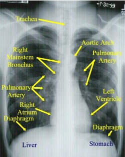
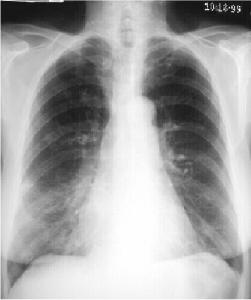
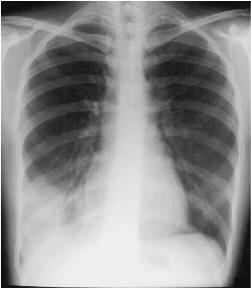
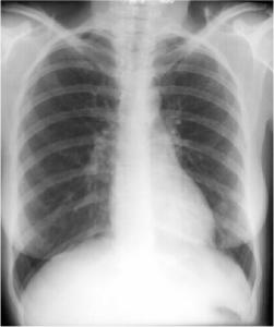
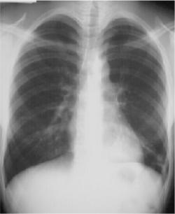
 Normal Posterior to Anterior (PA) Chest X-ray. Normally a PA and Lateral View are obtained. By convention on the PA View, the x-rays enter the patient posteriorly and exit anteriorly (with the patients chest on the film cassette), therefore minimizing the cardiac magnification. On the lateral view, the patients left side is against the film, therefore the right side would be magnified.
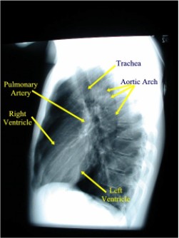
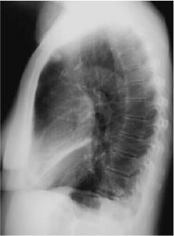
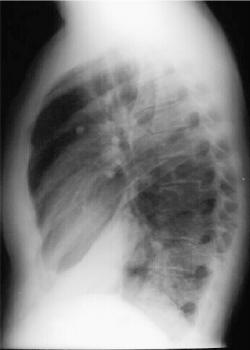
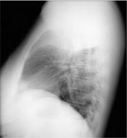
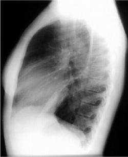
 Normal Lateral Chest X-ray
HOW TO READ THE CHEST X-RAY
- Get a mental image of the patient:
- Demographics
- Gender
- Size
- Shape
- Position of patient
- Approximate age
- Lines & tubes (position, course, complications)
- Foreign bodies.
- Evaluate soft tissues systematically: Don't forget:
- Neck
-
Shoulders
- Diaphragm (the right diaphragm usually is 2-3 cm higher than the left)
- Abdomen
- Breast tissue
-
Evaluate
the lungs (Interstitium, airways and Pleura):
-
Inflation
status
-
Pleural
margins
-
Abnormal
densities/lucencies
-
Masses
-
Infiltrates
-
Calcifications
-
Fissure
locations and thickness. The RUL Bronchus is always higher than the LUL bronchus.
- Change
your attention to the blood vessels:
-
The
size, location and distribution (the left pulmonary artery usually is
higher the left).
-
Don't
forget to check the lateral as this is the best way to look at the
posterior costophrenic recess, anterior/posterior mediastinum, and help
you localize lesions suspected on the frontal view.
-
Note
the "Special Interest" and often missed areas twice:
-
Apices
(esp. RUL- where most
cancer lives)
-
Peripheral
lung margins
-
Hilar,
retrocardiac, cardiophrenic and costophrenic angles.
-
Focus
attention now to the Mediastinum:
Evaluate Size, shape, position in both views PA/LAT.
Attention to the mediastinal lines
-
Check both PA/LAT views. Size,
shape, and silhouette. Look for any chamber enlargement.
Evaluate course of Aorta and position of arch, Pulmonary
Arteries.
-
of SVC (frontal View).
-
Paratracheal Stripe (normal is <5mm, usually 2-3mm), which terminates
at the azygous vein (this portion should be 1.0cm or less). Never
extends below the right bronchus.
-
Left
Subclavian Stripe: Normally 1.0-1.5 cm.
-
On the lat view, the posterior tracheal wall if seen should measure no more
than 4mm
-
Paraesophageal
line: seen only on the PA view. (interface between right lower lobe and
mediastinal edge along the esophagus/azygous vein â€" also called the
azygoesophageal line.) It should be straight, bulging could indicate a
node or mass (90% of all localized paraspinal masses are neurogenic
tumors (particularly neruofibromas and ganglioneuromas.)
-
Aorticopulmonary
window: Seen on
frontal view formed by overlap of the Aortic arch and left pulmonary
artery. Space should be
clear as the left upper lobe fills in this area. It should also be
concave, any bulge could signify nodes or mediastinal mass.
-
Bones:
-
Chest wall
-
Bony thorax
including spine.
-
Look for abnormal
joints, bony lytic/blastic or soft tissue lesions,
and free air, etc
Several signs help evaluate processes:
- Silhouette sign:
Silhouette sign is extremely
useful in localizing lung lesions.
(e.g. loss of right heart border in RML pneumonia)
- Air Bronchogram:
As the bronchial tree branches, the cartilaginous rings become thinner and
eventually disappear in respiratory bronchioles. The lumen of bronchus
contains air as well as the surrounding alveoli. Thus usually there is no
contrast to visualize bronchi.
If you see branching
radiolucent columns of aircorresponding to bronchi
, this usually means air-space (alveolar) disease. Usually one of these: blood, pus, mucous, cells, protein.
- Extra pleural sign:
Signifies Chest Wall disease. Peripheral
location with concave edges.
-
Anatomic landmarks
- Anterior & Posterior
junction lines: respectively,
the anterior and posterior conjunction of the right and left visceral
and parietal pleural layers at the midline of the thorax.
- 2mm linear line projecting
over the trachea. Note the posterior junction line extends above the
clavicles
Back to Top
PNEUMONIA
Pneumonia (consolidation)
Infection of the air spaces (air
bronchograms) and/or interstitium of the lung.
Finding:
- Depending upon the amount and
distribution of the airspaces involved, this may present as confluent
parenchymal (lobar or segmental) opacity or merely patchy opacity.
- If the Interstitium is
predominantly involved, it may appear as a reticulonodular pattern.
- Air bronchograms would confirm an
alveolar process.
- The lung volume should not be
lost (may even be increased).
- Usually all radiographic
abnormalities should disappear after 6 weeks of appropriate antibiotic
therapy. However, pneumonia may
be complicated by abscess or empyema formation.
Examples of Pneumonias and how to
determine location. (look for the silhouette sign…loss of usual visualized
borders.)
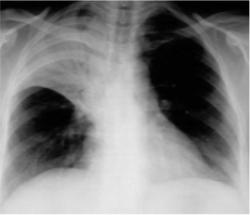
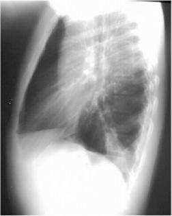
CONSOLIDATION
 Right Middle Lobe Consolidation
 Right Middle Lobe Pneumonia
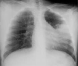
 Right Lower Lobe Pneumonia
Right Lower Lobe Pneumonia, Anterior Segment
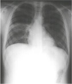
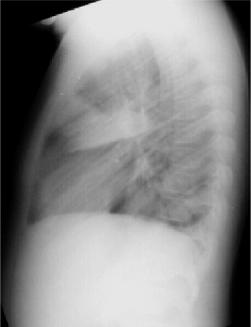 Right Lower Lobe Pneumonia, Superior Segment
 Right Upper Lobe Pneumonia
 Left Lingular Pneumonia
 Left Lower Lobe Pneumonia, Anterior Segment
 Left Lower Lobe Pneumonia, Posterior Segment
Back to Top
ROUND PNEUMONIA
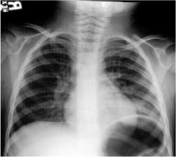
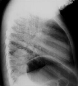 Round Pneumonia.
Round Pneumonias are found typically in the child.
Most often the organism is pneumococcus. The pneumonia appears round because
of poorly developed collateral pathways (pores of Kohn and channels of
Lambert). Over time though initially round, it develops into a more
consolidative pattern.
Round Pneumonia.
Round Pneumonias are found typically in the child.
Most often the organism is pneumococcus. The pneumonia appears round because
of poorly developed collateral pathways (pores of Kohn and channels of
Lambert). Over time though initially round, it develops into a more
consolidative pattern.
Back to Top
|
