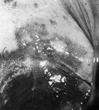
Chapter 10
Benign Neoplasms of the Vagina
James E. Delmore and Douglas V. Horbelt
Main Menu  

Search

James E. Delmore, MD
Division of Gynecologic Oncology, Department of Obstetrics and Gynecology, University of Kansas School of Medicine, Wichita, Kansas (Vol 1, Chaps 9, 10)

Douglas V. Horbelt, MD
Division of Gynecologic Oncology, Department of Obstetrics and Gynecology, University of Kansas School of Medicine, Wichita, Kansas (Vol 1, Chaps 9, 10)

INTRODUCTION
CYSTIC TUMORS
SOLID TUMORS
RELATED CONDITIONS
REFERENCES

INTRODUCTION

Benign or malignant neoplasms of the vagina are uncommon. The frequency of benign lesions ranges from rare to very rare. Neoplasms that may develop in other locations within the genital tract may also be found in the vagina. Most vaginal tumors produce no symptoms until significant size is reached. Symptoms and signs may include a sensation of pressure, dyspareunia, obstruction of the vagina or urethra, or vaginal bleeding. However, most lesions will be detected during a routine exam in the asymptomatic patient. Vaginal neoplasms may be divided into cystic or solid lesions and a third category best described as related conditions. As is true for any neoplasm, biopsy provides a definitive diagnosis.

Back to Top
CYSTIC TUMORS

Gartner's Duct Cyst

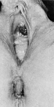
Gartner's duct cysts develop as a result of incomplete regression of the mesonephric or wolffian duct during fetal development (Fig. 1). In the male, these ducts form the epididymis. When present, these cysts may be multiple, and are located submucosally along the lateral aspects of the upper vagina. Histologic evaluation reveals nonsecretory, columnar epithelium. If these cysts are small, asymptomatic, and located in the lateral aspects of the upper vagina, no treatment is indicated. If the diagnosis is in question, or there is a history of antenatal exposure to synthetic hormones, adenosis of the vagina must be considered. The presence of mucosa, which stains normally with Lugol's solution, helps to exclude the diagnosis of adenosis. Regardless of size, biopsies should be performed on symptomatic cysts or they should be excised. Larger cysts in the vaginal fornix may extend to the lateral aspects of the cervix and require excision in the operating room.

Fig. 1. Genital tract in 10-week fetus. Remnants of wolffian duct result in Gartner's duct cysts.(Figures 1–3 and 5–10 are courtesy of ML Smith, MD and JC Scott, Jr, MD)

Paramesonephric Duct Cyst

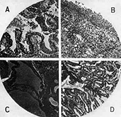
In contrast to Gartner's duct cysts, paramesonephric duct cysts are lined with secretory epithelium resembling endocervix or fallopian tube, suggesting mu¨llerian origin. These cysts may be found anywhere in the vagina and frequently contain mucus. Vaginal adenosis is excluded by staining with Lugol's solution. The diagnosis is established with an excisional biopsy if the cyst is large, symptomatic, or only recently identified.
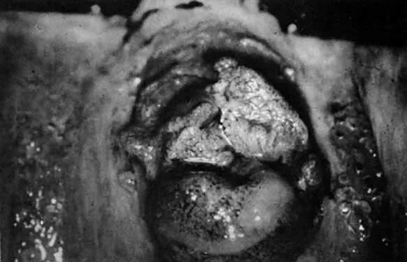
Inclusion Cyst

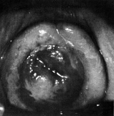
Inclusion cysts of the vagina result from mucosa trapped in the submucosal area by surgical procedures such as episiotomy, colporrhaphy, or trauma including childbirth (Fig. 2). As the cysts enlarge, symptoms may develop. These cysts are lined with squamous epithelium and contain keratin and squamous debris. Foreign-body reaction and inflammation surround the cyst. Treatment involves excision of the intact cyst and approximation of normal epithelium.
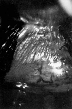
Fig. 2. Vaginal cyst that is typical of larger squamous inclusion cyst.

Endometriosis

Endometriosis in the vagina may develop at the site of a previous operation or as primary implants. Nodularity of the posterior vaginal fornix may represent endometriotic implants of the posterior cul-de-sac and may eventually erode or grow into the vaginal mucosa. When visualized colposcopically, these implants may appear dark blue or brown. If associated with fibrosis, the submucosal implants may appear white. Biopsy may yield chocolate-colored material representing old hemorrhage and dense fibrosis. Endometrial glands and stroma are usually identified histologically although the presence of both are not required to make the diagnosis. The diagnosis is made by biopsy unless endometriosis is identified in other parts of the pelvis. Small, symptomatic lesions are treated by excision or laser vaporization. Large lesions arising in the posterior cul-de-sac and extending into the posterior vaginal fornix may require laparotomy to accomplish excision. Preoperative therapy with gonadotropin releasing hormone analogs may greatly reduce the size of the implants, therefore, reducing the extent of excision or vaporization. We recommend outpatient mechanical bowel preparation prior to surgery. For the symptomatic patient who does not wish surgical excision of the lesion, gonadotropin releasing hormone analog therapy followed by suppression with oral contraceptives may be beneficial.

Back to Top
SOLID TUMORS

Leiomyoma

Vaginal leiomyomas or fibromyomas are rare lesions usually located in the anterior vaginal wall (Fig. 3). Between 250 and 300 cases have been reported in the world literature.1 These lesions are benign smooth muscle neoplasms, usually solitary and in many cases asymptomatic. Histologically, they resemble leiomyoma of other origins. Proposed sites of origin include vaginal smooth muscle, local arterial musculature, or smooth muscle of the bladder or urethra. As is true of uterine leiomyomata, the vaginal lesions are estrogen dependent. Malignant conversion is extremely rare. When large, symptoms can include vaginal discharge or bleeding, dyspareunia, or urinary retention. The differential diagnosis of a midline anterior vaginal mass includes urethral diverticulum, fibroepithelial polyp, cystocele, Skene duct abscess, or vaginal malignancy. Therapy involves excision in the symptomatic patient. Recurrence is uncommon but reported.2,3

Fig. 3. Leiomyoma of anterior vaginal wall, with pedicle.

Fibroepithelial Polyp

Fibroepithelial polyps of the vagina are uncommon and usually asymptomatic. In infants and young girls, sarcoma botryoides must be ruled out. Fibroepithelial polyps of the vagina are usually small and may be multiple. During pregnancy, these lesions may become enlarged, very edematous, and bizarre in appearance. Histologically, the polyps are composed of a squamous epithelial surface with a fibrovascular stalk and edematous stroma.4,5 Proposed etiologies include stromal proliferation or granulation tissue reaction as a result of local injury. Therapy involves excision of the polyp and stalk in the symptomatic patient or the patient with a large polyp.

Condyloma Acuminatum

Condyloma acuminatum represents the clinical manifestation of human papillomavirus infection.There are currently more than 70 human papillomavirus types identified. These lesions may be associated with condylomata of the cervix and vulva or appear only as vaginal lesions. Histologic evaluation confirms the diagnosis and rules out a dysplastic lesion. The microscopic description is similar to that for condyloma in other locations. Hybridization techniques to identify DNA type are currently of little help to the clinician. Clinical management includes topical therapy with carefully applied bichloroacetic or trichloroacetic acid. For large or multiple lesions, excision, cauterization, laser vaporization, or loop electrical excision may be required. Cryotherapy may be helpful for small lesions; however, depth of thermal injury may be difficult to control. The entire genital tract should be evaluated and any obvious lesions treated simultaneously. Overly aggressive treatment, especially with laser or cautery may result in significant distortion and scarring of the vagina and should be avoided.

Rare Lesions

Prolapse of the fallopian tube into the vagina following hysterectomy is uncommon; however, it may be alarming as the edematous fimbria may appear very much like a well-differentiated adenocarcinoma to the unsuspecting.6 Hemangiopericytoma, neurofibromas, mixed cell tumors, granular cell myoblastoma, myxoma, rhabdomyoma, and benign cystic teratoma are rare neoplasms found in the vagina.7,8,9,10,11,12 Excisional biopsy is required to make the diagnosis.

Back to Top
RELATED CONDITIONS

Urethral Caruncle

Urethral caruncles present as localized, red, friable lesions at the urethral meatus (Fig. 4, Fig. 5). They are generally seen in the postmenopausal woman and are thought to result from a localized area of prolapse of the urethral mucosa with secondary inflammatory changes. They can be confused with acute circumferential prolapse of the urethral mucosa, a condition usually seen in young girls. Urethral carcinoma must be excluded in patients with larger urethral caruncles. There may be pain, dysuria, and bleeding. Small asymptomatic urethral caruncles may not require any treatment. Larger or symptomatic lesions can be treated by topical application of estrogen. To establish the diagnosis, small biopsies may be performed under local anesthesia. Large or persistent lesions may require excision and reapproximation with fine absorbable suture.

Fig. 4. Urethral caruncle.(Courtesy of Dr. J. Merrill)

Fig. 5. Microscopic fields from single large urethral caruncle. A. Papillomatous changes. B. Granulomatous changes. C. Angiomatous changes. D. Adenomatous changes.

Diethylstilbestrol Associated Changes of the Vagina

As many as 2 million pregnant women may have received synthetic estrogen compounds, predominantly diethylstilbestrol (DES), from the early 1940s through the early 1970s for prevention of miscarriage and other complications of pregnancy. In 1970, Herbst and Scully reported an alarming number of new cases of clear cell adenocarcinoma of the vagina.13 By 1971, Herbst and co-workers had associated these new cases of adenocarcinoma of the vagina with in utero exposure to DES.14 In 1987, Melnick and colleagues reported on 519 registered cases of clear cell adenocarcinoma of the cervix and vagina, with 60% of those patients exposed to DES in utero.15 Fortunately, the anticipated epidemic of adenocarcinoma of the cervix and vagina resulting from DES exposure did not develop. It is estimated that the risk of developing adenocarcinoma of the cervix or vagina as a result of DES exposure is 1 per 1000 exposed women.15 Nonmalignant structural changes of the cervix and vagina including cockscomb, cervical collar, cervical hood, pseudopolyp, and hypoplastic cervix have been described in 25% to 48% of in utero DES exposed women16 (Fig. 6). Vaginal epithelial changes, predominantly adenosis has been reported in 34% to 65% of in utero exposed women17 (Fig. 7, Fig. 8). Adenosis may be described as the retention of mu¨llerian-derived glandular epithelium in the vagina after vaginal embryogenesis is completed18 (Fig. 9). The evaluation of the DES exposed patient includes careful palpation of the entire vagina searching for any submucosal nodularity which would lead to biopsy. This is followed by careful colposcopic examination of the entire vagina with special attention to the upper third, looking for characteristic changes of columnar epithelium. Cytologic samples are taken from the cervix and vagina. Structural changes of the cervix and vagina are noted. Finally, the entire vagina is stained with Lugol's solution and re-examined to search for any nonstaining areas (Fig. 10). Biopsy is performed on any nonstaining areas, palpable nodules, and colposcopically detected columnar epithelium in the vagina. Biopsies confirm the presence of adenosis and exclude more significant lesions. This extensive evaluation is performed on the first visit and does not need to be repeated. Annual exams including cytologic evaluation and palpation of the cervix and vagina are proposed to the patient with a negative initial evaluation. Once diagnosed, adenosis is observed without therapy. In most cases, adenosis will resolve as a result of squamous metaplasia. Recently, vaginal adenosis has been reported as a result of laser therapy of the vagina for condyloma or dysplasia.19 This should not be confused with DES related adenosis.

Fig. 6. Cervical-vaginal hood in patient with antenatal exposure to synthetic estrogen.

Fig. 7. Vaginal and cervical adenosis. Glandular epithelium onectocervix and vagina. Note cocks-comb appearance of anterior cervical area.

Fig. 8. Adenosis in anterior vaginal fornix.

Fig. 9. Urogenital tract in 14-week fetus. Replacement of müllerian epithelium with specialized urogenital sinus epithelium.

Fig. 10. Adenosis in anterior vagina stained with Lugol's stain. Note absence of stain in involved areas.

Back to Top
REFERENCES

1. Liu MM: Fibromyoma of the vagina. Eur J Obstet Gynecol Reprod Biol 29: 321, 1988

2. Dhaliwal LK, Das I, Goplan S: Recurrent leiomyoma of the vagina. Int Gynecol Obstet 37: 281, 1992

3. Young SB, Rose PG: Vaginal fibromyomata: Two cases with preoperative assessment, resection, and reconstruction. Obstet Gynecol 78: 972, 1991

4. Mucitelli DR, Charles EZ, Kraus FT: Vulvovaginal polyps: Histologic appearance, ultrastructure, immunocytochemical characteristics and clinicopathologic correlations. Int J Gynecol Pathol 9: 20, 1990

5. Halvorsen TB, Johannesen E: Fibroepithelial polyps of the vagina: Are they old granulation tissue polyps? J Clin Pathol 45: 235, 1992

6. Wheelock JB, Schneider V, Goplerud DR: Prolapsed fallopian tube masquerading as adenocarcinoma of the vagina in a postmenopausal woman. Gynecol Oncol 21: 369, 1985

7. Buscema J, Rosenchein NB, Fowzia T, Woodruff JD: Vaginal hemangiopericytoma: A histopathologic and ultrastructural evaluation. Obstet Gynecol 66: 82S, 1985

8. Gersell DJ, Fulling KH: Localized neurofibromatosis of the female genitourinary tract. Am J Surg Pathol 13 (10): 873, 1989

9. Dekel A, Avidan D, Bar-Ziv J et al: Neurofibroma of the vagina presenting with urinary retention. Review of the literature and report of a case. Obstet Gynecol Surv 43 (6): 325, 1988

10. Wright RG, Buntine DW, Forbes KL: Case report: Recurrent benign mixed tumor of the vagina. Gynecol Oncol 40: 84, 1991

11. Egley CC, Fox JS: Vaginal myxoma presenting as acute urinary retention. Obstet Gynecol 73: 882, 1989

12. Vilela DS, Pizarro AG, Suarez M: Vaginal rhabdomyoma and adenosis. Histopathology 16: 393, 1990

13. Herbst AL, Scully RE: Adenocarcinoma of the vagina in adolescence: A case report of 7 cases including 6 clear-cell carcinomas. Cancer 25: 745, 1970

14. Herbst AL, Ulfelder H, Poskanzer DC: Adenocarcinoma of the vagina: Association of maternal stilbestrol therapy with tumor appearance in young women. N Engl J Med 284: 878, 1971

15. Melnick D, Cole P, Anderson D, Horbst A: Rates and risks of diethylstilbestrol-related clear cell adenocarcinoma of the vagina and cervix. N Engl J Med 316: 514, 1987

16. Jefferies JA, Robboy SJ, O'Brien PC: Structural anomalies of the cervix and vagina in women enrolled in the Diethylstilbestrol Adenosis (DESAD) Project. Am J Obstet Gynecol 148: 59, 1984

17. O'Brien PC, Noller KL, Robboy SJ: Vaginal epithelial changes in young women enrolled in the National Cooperative Diethylstilbestrol Adenosis (DESAD) Project. Obstet Gynecol 53: 300, 1979

18. Fenoglio CM, Ferenczy A, Richart RM: Scanning and transmission electron microscopic studies of vaginal adenosis and the cervical transformation zone in progeny exposed in utero to diethylstilbestrol. Am J Obstet Gynecol 126: 170, 1976

19. Sedlacek TV, Riva JM, Magen AB et al: Vaginal and vulvar adenosis: An unsuspected side effect of CO2 laser vaporization. J Reprod Med 35 (11): 995, 1990

Back to Top