Chapter 26
Breast Cancer and the Obstetrician-Gynecologist
William H. Hindle
Main Menu   Table Of Contents

Search

William H. Hindle, MD
Professor Emeritus, Department of Obstetrics and Gynecology, University of Southern California Keck School of Medicine, Founder, Breast Diagnostic Center, Women's and Children's Hospital, LAC+USC Medical Center, Los Angeles, California (Vol 1, Chaps 26, 27)

 
RISK FACTORS
DIAGNOSIS: INVASIVE CANCER AND IN SITU CARCINOMA
REFERRAL AND CONTINUING FOLLOW-UP
TREATMENT OPTIONS
RADIATION THERAPY
ADJUVANT SYSTEMIC THERAPY
DUCTAL CARCINOMA IN SITU
CHEMOPREVENTION AND SURVEILLANCE
ESTROGEN AND ESTROGEN/PROGESTIN THERAPY
CONCLUSION
COLOR PLATES
REFERENCES

Breast cancer is the most common invasive (nonskin) cancer of women. For 2003, it is estimated that there will have been 211,300 invasive breast cancers diagnosed in women and 39,800 female deaths from breast cancer.1 In addition, 55,700 new cases of in situ breast cancer are estimated to be diagnosed in women in 2003.2

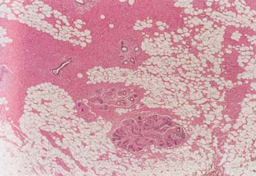
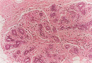
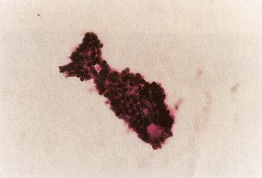
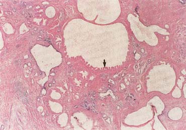
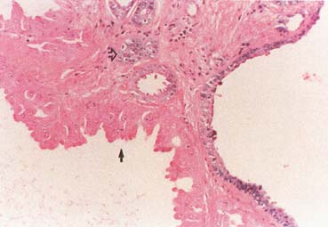
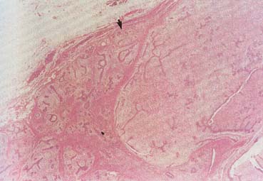
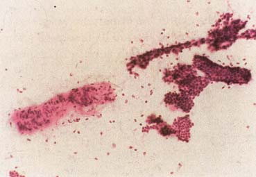
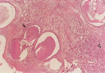
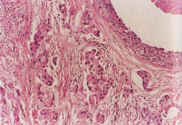
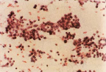
The American Joint Committee on Cancer lists 24 different codable histologic types of breast malignancies (Table 1), of which seven are in situ and 17 are invasive (infiltrating).3 Each specific histologic type has its own unique characteristics, biologic behavior, and prognosis when treated. In addition, the optimum treatment varies with each specific histologic type. However, for invasive and in situ breast cancer, as many as 80% of the cases are of the ductal type. Color plates 1 through 16 show typical examples of the histology and cytology of normal breast tissue, fibrocystic changes, fibroadenoma, and invasive ductal carcinoma under low- and high-power magnification. (Color plates appear at the end of the chapter.)


Table 1. Codable Histologic Breast Malignancies


In Situ
 Carcinoma in situ, NOS*
 Comedocarcinoma, noninfiltrating
 Cribriform carcinoma in situ
 Intraductal carcinoma and lobular carcinoma in situ
 Intraductal carcinoma, noninfiltrating, NOS
 Lobular carcinoma in situ, NOS
 Noninfiltrating intraductal papillary adenocarcinoma
 Paget's disease and intraductal carcinoma of breast
 Paget's disease, mammary
Invasive
 Adenoid cystic carcinoma
 Carcinoma, NOS
 Carcinoma undifferentiated, NOS
 Carcinosarcoma, NOS
 Cribriform carcinoma, NOS
 Infiltrating duct carcinoma, NOS
 Inflammatory carcinoma
 Lobular carcinoma, NOS
 Medullary carcinoma, NOS
 Mucinous adenocarcinoma
 Paget's disease and infiltrating duct carcinoma of breast
 Phyllodes tumor, malignant
 Secretory carcinoma of breast
 Squamous cell carcinoma, NOS
 Tubular adenocarcinoma

*NOS = not otherwise specified.
(Adapted from American Joint Committee on Cancer: AJCC Cancer Staging Manual, p 236. 6th ed. New York, Springer, 2002.)


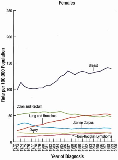
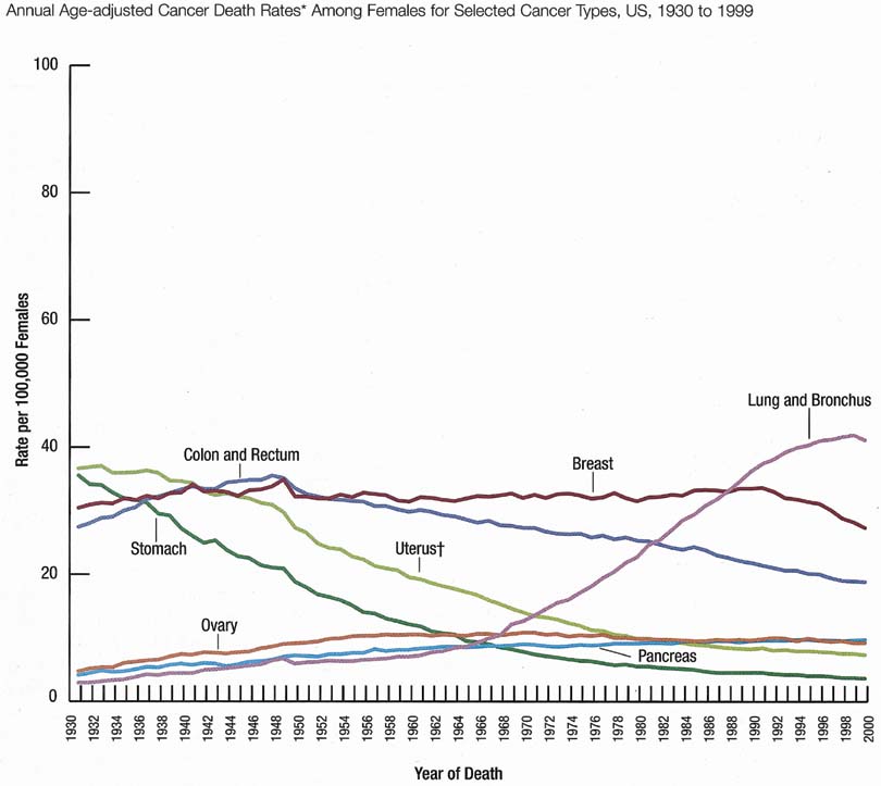
The incidence rate for breast cancer in women has increased steadily since 1977, as shown in comparison with other major invasive cancers in Figure 1.1 However, the incidence rate appears to have peaked in 1997, with a slight decrease since then. Figure 2 graphically depicts the contrasting mortality rates of the major invasive cancers.1 Possibly related to improved methods of treatment, the mortality rate for breast cancer has shown a continuous slightly downward trend of from 1940 to 1990. Probably caused by the widespread use of screening mammography, the mortality rate has decreased from 1990 to 1999. Hopefully, this trend of clinically meaningful decreasing mortality rates will continue as data for the years after 1999 become available. The 5-year survival rates by extent of breast cancer at the time of diagnosis are 86% for all stages, 97% for localized cancer, 78% for cancer with regional involvement, and 23% for metastatic cancer.2

Fig. 1. Trends of incidence rates of the major invasive cancers of women in the United States (1972 to 1999). (Jemal A, Murray T, Samuels A et al: Cancer statistics 2003. CA Cancer J for Clin 53:5–26, 2003; p 13 with permission.)

Fig. 2. Trends of mortality rates of the major invasive cancers of women in the United States (1930 to 1999). (Jemal A, Murray T, Samuels A et al: Cancer statistics 2003. CA Cancer J for Clin 53: 5–26, 2003; p 15 with permission.)

Obstetrician-gynecologists should strive to bring about the diagnosis of breast cancer before the cancer is palpable. This goal can be effectively achieved by annual screening mammography for women beginning at age 40 years.4 Mammography can perceive small (1- to 9-mm) nonpalpable breast cancers that have the optimum curability, with more than 90% 20-year disease-specific survival using current methods of treatment (Table 2).5 Palpable cancers have a less favorable outcome. Generally, survival from palpable breast cancer correlates inversely with increasing size.6 For example, 10-to 29-mm cancers have been reported to have 67% 15-year survival (Table 3).7


Table 2. 20-Year Breast Cancer-Specific Survival: Mammographic Appearance vs. Percentage Survival


Mammographic Appearance Percentage Survival
Stellate mass without calcifications 100
Noncasting calcifications 96
Circular mass without calcifications 93
Casting calcifications 60
All 1–9-mm tumors 93

(Adapted by Patricia T. Kelly, Ph.D., from Tabar L, Vitak B, Chen HH, et al: The Swedish Two-County Trail twenty years later. Updated mortality results and new insights from long-term follow-up. Radiol Clin North Am 38:625–651, 2000.)
Tumors 1–9-mm without nodal involvement.


Table 3. 15-Year Breast Cancer Survival: Correlation With Tumor Size


Tumor Size Percentage Survival
10–14 mm 86
15–19 mm 72
20–29 mm 67
30–39 mm 46

(Adapted by Patricia T. Kelly, Ph.D., from Michaelson JS, Silverstein M, Wyatt J, et al: Predicting the survival of patients with breast carcinoma using tumor size. Cancer 95:713–723, 2002.)


Unfortunately, some cancers large enough to be palpable are not initially perceived on mammography. This probably occurs in less than 10% of cases.8 Increased mammographic density (Table 4) can obscure both nonpalpable and palpable breast cancers. Thus, women should have an annual clinical breast examination in addition to screening mammography. Ideally, the examination is performed before the mammogram so that the results of the examination are known by the radiologist/mammographer.


Table 4. The BI-RADS Descriptive Patterns of Mammographic Breast Density


The breast is almost entirely fat
There are scattered fibroglandular densities that could obscure a lesion on mammography
The breast tissue is heterogeneously dense
This may lower the sensitivity of mammography
The breast tissue is extremely dense, which lowers the sensitivity of mammography

(American College of Radiology (ACR): Illustrated Breast Imaging Reporting and Data System (BI-RADS™). 3rd ed. Reston, VA, American College of Radiology, 1998.)


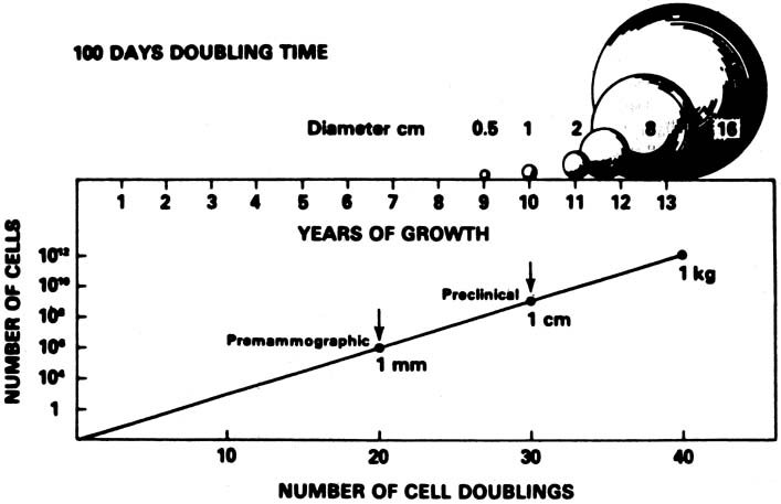
A conceptual mathematical model of the growth of a typical invasive ductal breast cancer is depicted in Figure 3.9 The growth of the cancer is based on a constant 100-day cell mass doubling time, which is the generally accepted mean rate of growth. However, estimated breast cancer doubling times have been reported in the range of 30 to 300 days and are not constant, but vary dependent on the specific cancer characteristics and host interaction. Nevertheless, the concept depicted is sound. There is a premammographic phase when the cancer is too small to be detected by any method currently available, followed by a preclinical phase when the cancer is too small to be palpable. Exactly when breast cancers metastasize is unknown, but the 90% 20-year disease-free survival of treated 1- to 9-mm mammographically detected cancers indicates that few had metastasized before the cancers reached a size of 1 cm. This concept underscores the importance of annual screening mammography particularly in the 40- to 49-year-old group in which, clinically, some of the breast cancers grow aggressively with apparently shorter doubling times.

Fig. 3. Mathematical representation of the theoretical growth (size) of breast cancer plotted against time in years based on a constant cellular doubling time of 100 days. (Wertheimer MD, Costanza ME, Dodson TF et al: Increasing the effort toward breast cancer detection. JAMA 255: 1311 –1315, 1986; with permission.)

The American Cancer Society's 2003 guidelines for the early detection of breast cancer in asymptomatic women are annual mammography and clinical breast examination beginning at age 40 and clinical breast examination every 3 years for women ages 20 to 39.10 If a clinically suspicious breast lesion remains undiagnosed, the patient should be urgently referred to a breast specialist. If the patient has diagnosed breast cancer, optimum treatment is usually obtained at a dedicated breast center with a multidisciplinary treatment planning team (Table 5).


Table 5. A Multidisciplinary Treatment Planning Team Should Include the Following Oncologic Breast Specialists


Imaging
Medicine
Nursing
Pathology
Plastic surgery
Radiation therapy
Social service
Surgery


Back to Top
RISK FACTORS

Most women are concerned about the risk of breast cancer. Numerous epidemiologic studies have been published, but it is often difficult for patients, and even for physicians, to understand and appreciate the pertinent clinical meaning. Typically, a woman wants to know if she will get breast cancer, and that question is not answerable. An obstetrician-gynecologist can recommend that the patient read a clear understandable book, such as Assess Your True Risk of Breast Cancer by Patricia Kelly, PhD (An Owl Book, Henry Holt and Company, New York, NY, 2000). This book is also useful reading for obstetrician-gynecologists for insight into epidemiologic studies, statistics, relative risk, absolute risk, and statistical significance.

The clinically relevant risk factors for breast cancer are: (1) being a woman; (2) growing older; (3) gene mutations; (4) ductal carcinoma in situ; (5) lobular carcinoma in situ (more accurately called lobular neoplasia, a tumor marker); and (6) atypical epithelial hyperplasia. Other epidemiologic risk factors are: (1) birth of first child after age 30; (2) consumption of an alcoholic beverage one or more times per day; (3) early onset of menstruation (menarche); (4) family history of breast cancer; (5) having had breast cancer; (6) increased mammographic density; (7) late ending of menstruation (menopause); (8) nulliparity; (9) postmenopausal estrogen and progestin therapy; (10) postmenopausal obesity; and (11) recent use of oral contraceptives. However, none of these epidemiologic risk factors alters clinical management. A summary of the published breast cancer relative risk data is in Table 6. Relative risk of less than 2 to 3 is an epidemiologic weak association without evidence of causality and, clinically, is likely to be a chance association, perhaps related to the multifactorial natural history and biologic behavior of breast cancers.


Table 6. Breast Cancer Relative Risk Factors Grouped by Magnitude


Relative Risk < 2 Relative Risk 2–4 Relative Risk > 4
Early menarche Age > 35 first birth Gene mutation
Late menarche 1st degree relative with breast cancer Lobular carcinoma in situ
Nulliparity Radiation exposure Ductal carcinoma in situ
Proliferative benign disease Previous breast cancer Atypical hyperplasia
Obesity    
Alcohol use    
Hormone replacement    

Summary of published levels of relative risks for breast cancer.
(Brinkmann E, Morrow M: The surgeon's role in breast cancer chemoprevention. Breast Disease 12:103–112, 2001 [Table 1. p 105].)


Routine vigorous physical activity and maintenance of healthy body weight are associated with epidemiologic decreased risk of breast cancer.2

Back to Top
DIAGNOSIS: INVASIVE CANCER AND IN SITU CARCINOMA

Ductal carcinoma is the most common histologic type of breast cancer, accounting for as many as 80% of breast malignancies. Nonpalpable invasive ductal carcinoma is usually perceived on mammography. The typical findings are stellate solid mass or casting-type calcifications. However, a malignant solid mass may be circular and the calcifications may be noncasting. Ultrasound can be helpful in defining a malignant solid mass, particularly in a young woman or in any woman with mammographically dense breasts, but ultrasound is not effective in evaluating calcifications that are often not perceived on ultrasound. The definitive diagnosis of nonpalpable cancer is established by image-guided tissue core-needle biopsy, commonly with vacuum-assisted technique, or by imaging needle localization and open surgical biopsy for histologic diagnosis.

It is the patient herself who usually notices, often coincidentally, what she perceives as a mass. Then she presents to her physician, often an obstetrician-gynecologist, for confirmation that it is a palpable dominant mass and for diagnosis. A palpable dominant breast mass is defined as a three-dimensional distinct mass that is different from the remainder of the breast tissue and from the tissue of the other breast. The definitive diagnosis of the mass can be established by fine-needle aspiration cytology, tissue core-needle biopsy histology, or open surgical biopsy histology.

Ductal carcinoma in situ (DCIS) is most commonly nonpalpable and perceived on screening mammography as malignant calcifications, usually of the casting type. The definitive diagnosis is established by open surgical biopsy with imaging needle localization. Tissue histology, usually of the entire lesion, is essential to be certain that there is not associated invasive ductal carcinoma.

Invasive lobular carcinoma spreads diffusely with a typical histologic Indian file pattern. Thus, invasive lobular carcinoma is often not apparent, either by palpation or by imaging, until the cancer is at an advanced stage. Lobular carcinoma in situ is thought to be a tumor marker with associated increased risk of eventual invasive carcinoma that usually is of the ductal type when it does occur and may even occur in the contralateral breast.

Back to Top
REFERRAL AND CONTINUING FOLLOW-UP

If an obstetrician-gynecologist is not trained and experienced in an appropriate diagnostic technique, the patient with a palpable dominant breast mass or with mammography findings suspicious for malignancy (BI-RADS categories 4 and 5; Table 7)11 should be expeditiously referred to a breast center or a breast specialist if a breast center is not available. It is important to continue to follow-up the patient to be certain she is seen in referral and that a definitive diagnosis is established. If the patient delays or refuses the referral, her noncompliance should be clearly documented in her medical record and the patient informed, by certified mail if necessary, of the potentially life-threatening seriousness of her condition and inaction.


Table 7. The BI-RADS Mammographic Assessment Categories


Category 0Need additional imaging evaluation
Category 1Negative
Category 2Benign finding
Category 3Probably benign finding: short interval follow-up suggested
Category 4Suspicious abnormality: biopsy should be considered
Category 5Highly suggestive of malignancy: appropriate action should be taken

(American College of Radiology (ACR): Illustrated Breast Imaging Reporting and Data System (BI-RADS™). 3rd ed. Reston, VA, American College of Radiology, 1998.)


As a primary health care provider for women, the obstetrician-gynecologist should continue to be available to the patient, her spouse/partner, and her family for counseling, compassionate explanations of the diagnostic and treatment options, and caring and understandable answers to their questions and concerns.

Back to Top
TREATMENT OPTIONS

Equivalent long-term breast cancer survivals can be achieved by either breast-conserving therapy (lumpectomy, axillary lymph node dissection, and irradiation) or modified radical mastectomy.12 Radical mastectomy, which was The Standard for more than 70 years, is no longer indicated.13,14

Breast-Conserving Therapy

Breast-conserving therapy is the preferred treatment for stages I and II (up to a size of 5 cm) breast cancer.15 Table 8 describes the American Joint Committee on Cancer staging of breast cancer.3 The absolute and relative contraindications for breast conserving therapy are listed in Table 9. The most common reason for selecting modified radical mastectomy instead of breast conserving therapy for stages I and II breast cancers is the patient's own choice. The medical contradictions are uncommon. Perhaps as many as 10% of women with stage I and 30% with stage II are not candidates for breast-conserving therapy.16 However, some of these unfavorable candidates and some women with cancers larger than stage II can benefit from neoadjuvant chemotherapy, before surgery, which can potentially decrease the size of the cancer and make a recommendation for breast-conserving therapy appropriate.17,18


Table 8. TNM Staging of Breast Cancer 2002


DEFINITION OF TNM
Primary Tumor (T)
Definitions for classifying the primary tumor (T) are the same for clinical and for pathologic classification. If the measurement is made by physical examination, the examiner will use the major headings (T1, T2, or T3). If other measurements, such as mammographic or pathologic measurements, are used, the subsets of T1 can be used. Tumors should be measured to the nearest 0.1 cm increment.
TXPrimary tumor cannot be assessed
T0No evidence of primary tumor
TisCarcinoma in situ
Tis (DCIS)Ductal carcinoma in situ
Tis (LCIS)Lobular carcinoma in situ
Tis (Paget's)Paget's disease of the nipple with no tumor

Note: Paget's disease associated with a tumor is classified according to the size of the tumor.

T1Tumor 2 cm or less in greatest dimension
T1micMicroinvasion 0.1 cm or less in greatest dimension
T1aTumor more than 0.1 cm but not more than 0.5 cm in greatest dimension
T1bTumor more than 0.5 cm but not more than 1 cm in greatest dimension
T1cTumor more than 1 cm but not more than 2 cm in greatest dimension
T2Tumor more than 2 cm but not more than 5 cm in greatest dimension
T3Tumor more than 5 cm in greatest dimension
T4Tumor of any size with direct extension to (a) chest wall or (b) skin, only as described below
T4aExtension to chest wall, not including pectoralis muscle
T4bEdema (including peau d'orange) or ulceration of the skin of the breast, or satellite skin nodules confined to the same breast
T4cBoth T4a and T4b
T4dInflammatory carcinoma

Regional Lymph Nodes (N)
Clinical
NXRegional lymph nodes cannot be assessed (e.g., previously removed)
N0No regional lymph node metastasis
N1Metastasis to movable ipsilateral axillary lymph node(s)
N2Metastases in ipsilateral axillary lymph nodes fixed or matted, or in clinically apparent* ipsilateral internal mammary nodes in the absence of clinically evident axillary lymph node metastasis
N2aMetastasis in ipsilateral axillary lymph nodes fixed to one another (matted) or to other structures
N2bMetastasis only in clinically apparent* ipsilateral internal mammary nodes and in the absence of clinically evident axillary lymph node metastasis
N3Metastasis in ipsilateral infraclavicular lymph node(s) with or without axillary lymph node involvement, or in clinically apparent* ipsilateral internal mammary lymph node(s) and in the presence of clinically evident axillary lymph node metastasis; or metastasis in ipsilateral supraclavicular lymph node(s) with or without axillary or internal mammary lymph node involvement
N3aMetastasis in ipsilateral infraclavicular lymph node(s)
N3bMetastasis in ipsilateral internal mammary lymph node(s) and axillary lymph node(s)
N3cMetastasis in ipsilateral supraclavicular lymph node(s)
Pathologic (pN)a
pNXRegional lymph nodes cannot be assessed (e.g., previously removed, or not removed for pathologic study)
pN0No regional lymph node metastasis histologically, no additional examination for isolated tumor cells (ITC)

Note: Isolated tumor cells (ITC) are defined as single tumor cells or small cell clusters not greater than 0.2 mm, usually detected only by immunohistochemical (IHC) or molecular methods but which may be verified on H&E stains. ITCs do not usually show evidence of malignant activity e.g., proliferation or stromal reaction.

pN0(i−)No regional lymph node metastasis histologically, negative IHC
pN0(i+)No regional lymph node metastasis histologically, positive IHC, no IHC cluster greater than 0.2 mm
pN0(mol−)No regional lymph node metastasis histologically, negative molecular findings (RT-PCR)b
pN0(mol+)No regional lymph node metastasis histologically, positive molecular findings (RT-PCR)b
pN1Metastasis in 1 to 3 axillary lymph nodes, and/or in internal mammary nodes with microscopic disease detected by sentinel lymph node dissection but not clinically apparent**
pN1miMicrometastasis (greater than 0.2 mm, none greater than 2.0 mm)
pN1aMetastasis in 1 to 3 axillary lymph nodes
pN1bMetastasis in internal mammary nodes with microscopic disease detected by sentinel lymph node dissection but not clinically apparent**
pN1cMetastasis in 1 to 3 axillary lymph nodes and in internal mammary lymph nodes with microscopic disease detected by sentinel lymph node dissection but not clinically apparent.** (If associated with greater than 3 positive axillary lymph nodes, the internal mammary nodes are classified as pN3b to reflect increased tumor burden)
pN2Metastasis in 4 to 9 axillary lymph nodes, or in clinically apparent* internal mammary lymph nodes in the absence of axillary lymph node metastasis
pN2aMetastasis in 4 to 9 axillary lymph nodes (at least one tumor deposit greater than 2.0 mm)
pN2bMetastasis in clinically apparent* internal mammary lymph nodes in the absence of axillary lymph node metastasis
pN3Metastasis in 10 or more axillary lymph nodes, or in infraclavicular lymph nodes, or in clinically apparent* ipsilateral internal mammary lymph nodes in the presence of 1 or more positive axillary lymph nodes; or in more than 3 axillary lymph nodes with clinically negative microscopic metastasis in internal mammary lymph nodes; or in ipsilateral supraclavicular lymph nodes
pN3aMetastasis in 10 or more axillary lymph nodes (at least one tumor deposit greater than 2.0 mm), or metastasis to the infraclavicular lymph nodes
pN3bMetastasis in clinically apparent* ipsilateral internal mammary lymph nodes in the presence of 1 or more positive axillary lymph nodes; or in more than 3 axillary lymph nodes and in internal mammary lymph nodes with microscopic disease detected by sentinel lymph node dissection but not clinically apparent**
pN3cMetastasis in ipsilateral supraclavicular lymph nodes

Distant Metastasis (M)
MXDistant metastasis cannot be assessed
M0No distant metastasis
M1Distant metastasis

STAGE GROUPING
Stage 0TisN0M0Stage IIBT2N1M0Stage IIIBT4N0M0
Stage IT1*N0M0 T3N0M0 T4N1M0
Stage IIAT0N1M0Stage IIIAT0N2M0 T4N2M0
 T1*N1M0 T1*N2M0Stage IIICAny TN3M0
 T2N0M0 T2N2M0Stage IVAny TAny NM1
     T3N1M0    
     T3N2M0    

* Clinically apparent is defined as detected by imaging studies (excluding lymphoscintigraphy) or by clinical examination or grossly visible pathologically.
a Classification is based on axillary lymph node dissection with or without sentinel lymph node dissection. Classification based solely on sentinel lymph node dissection without subsequent axillary lymph node dissection is designated (sn) for “sentinel node,” e.g., pN0(i+) (sn).
b RT-PCR: reverse transcriptase/polymerase chain reaction.
* Clinically apparent is defined as detected by imaging studies (excluding lymphoscintigraphy) or by clinical examination.
** Not clinically apparent is defined as not detected by imaging studies (excluding lymphoscintigraphy) or by clinical examination.
* T1 includes T1mic
Note: Stage designation may be changed if post-surgical imaging studies reveal the presence of distant metastases, provided that the studies are carried out within 4 months of diagnosis in the absence of disease progression and provided that the patient has not received neoadjuvant therapy.
(American Joint Committee on Cancer: AJCC Cancer Staging Manual, pp 227–228. 6th ed. New York, Springer, 2002.)


Table 9. Medical Contraindications to Breast-Conserving Therapy


Absolute
 Pregnancy, because of contraindicated radiation therapy
 Two or more cancers in different breast quadrants
 Diffuse “malignant” microcalcifications
 Prior radiation therapy to the breast area
 Persistent surgical margins involved with cancer
Relative
 Collagen vascular disease
 Multiple gross cancers in one quadrant
 Diffuse indeterminate calcifications
 A large cancer in a small breast
 Large pendulous breast, because of difficulties with radiation therapy

(Adapted from Morrow M, Strom EA, Bassett LW, et al: Standard for breast conserving therapy in the management of invasive breast carcinoma. CA Cancer J for Clin 52:277–300, 2002.)


An open surgical biopsy of a suspicious dominant mass should be performed with the same careful technique as lumpectomy for known cancer.19 Otherwise, re-excision of the area of the tumor bed is usually necessary.20 As many as 50% of women referred to a cancer center who had their cancer diagnosed by open surgical biopsy required re-excision of the area of the biopsy because the biopsy margins were positive (cut across cancer).20 Definitive diagnosis of cancer by fine-needle aspiration cytology or tissue core-needle biopsy histology should eliminate the need for most re-excisions if the open surgical biopsy follows National Surgical Adjuvant Breast and Bowel Project (NSAPB) technique for lumpectomy.19

Axillary lymph node dissection involves removal of the levels I and II lymph nodes (Table 10 shows the description of the levels of axillary lymph nodes). If the patient has known invasive cancer, the axillary lymph node dissection can be performed at the same time as her lumpectomy. Sentinel node biopsy is becoming an option to routine axillary lymph node dissection. The technique of sentinel node biopsy requires special equipment and a learning curve of experience for the surgeon. Although the technique is well-established,21,22,23 sentinel node biopsy is currently available in a limited number of breast centers. However, the technique has the potential of avoiding lymphedema, which is a major complication of axillary lymph node dissection that requires prolonged treatment, often with limited success. However, in 2003, axillary lymph node dissection remains the standard technique in breast-conserving therapy.24


Table 10. Description of the Surgical Levels of Axillary Lymph Nodes


Level I:lateral to the lateral border of the pectoralis minor muscle
Level II:under the insertion of the pectoralis minor, between the medial and lateral borders of the pectoralis minor muscle and the interpectoral (Rotter's) node
Level III:medial to the medial border of the pectoralis minor muscle, including the nodes designated as apical

(Adapted from American Joint Committee on Cancer: AJCC Cancer Staging Manual, p 224. 6th ed. New York, Springer, 2002.)


Radiation therapy (irradiation) is an essential component of breast-conserving therapy and is usually performed in an oncologic radiation therapy center. The oncologic radiologist directs the technique and dosage of the therapy, which is individualized to the particular medical circumstances of each breast cancer patient. Usually a total dose of whole breast irradiation of 4500 to 5000 centigrays is administered in fractions using opposed tangential fields. The course of irradiation is usually administered in daily fractions (5 days per week) for 6 weeks. A boost dose of irradiation to the tumor bed increases the target dosage to 6000 to 6500 centigrays. Careful individualized radiation therapy planning and modern equipment minimize the adverse effects of irradiation, particularly the uncommon but devastating long-term sequellae.25 Radiation therapy has been demonstrated to control the local recurrence of invasive breast cancer surgically treated with lumpectomy.14,26

A state-of-the-art (as of 2002) comprehensive clinical review of breast-conserving therapy for invasive breast cancer, authored by multidisciplinary authorities, has been published.24 This work provides details and documentation for obstetrician-gynecologists in clinical practice who seek further information about breast-conserving therapy.

Modified Radical Mastectomy

Modified radical mastectomy (total mastectomy and axillary lymph node dissection) removes the entire breast, including the overlying skin and the axillary lymph nodes, usually levels I and II, en bloc. The major modification is the preservation of the pectoralis major muscle, which facilitates improved wound healing and, potentially, allows reconstruction. Modified radical mastectomy is performed for stages I and II breast cancer for which breast-conserving therapy is contraindicated (see Table 9), for selected cancers larger than stage II, and for patient preference when equal therapeutic results and breast cancer survival can be achieved with either breast-conserving therapy or modified radical mastectomy.

Breast Reconstruction

Most women who have a modified radical mastectomy are candidates for breast reconstruction.16 This option should be presented to the patient when she is making her treatment choice. If she is interested in reconstruction, she should be referred, before scheduling her definitive surgery, to a plastic surgeon experienced in the techniques of both prosthetic reconstruction and autologous tissue reconstruction. The transverse rectus abdominis myocutaneous (TRAM) flap is the most common autologous tissue reconstruction. Both prosthetic and autologous tissue reconstructions can be immediate (during the same operation as the modified racial mastectomy) or delayed (performed at a later date). Data from 1994 to 1995 reveal that only 8.3% of mastectomy patients had breast reconstruction.16 This low utilization is influenced by: (1) patient age; (2) income; (3) geographic location; (4) type of hospital; and (5) tumor size (for example, women aged 50 or younger had a more than four-fold higher rate of reconstruction than older women).16

Back to Top
RADIATION THERAPY

Radiation therapy is an essential component of breast-conserving therapy and has been described. Although radiation therapy is effective in local control of breast cancer recurrences, the long term (10-year and 20-year) sequelae are a concern, particularly vascular mortality.25

Radiation therapy, or irradiation, is a common component of the treatment of stages III and IV, often called advanced breast cancer. The exact irradiation technique and dosage is individualized to each case. Irradiation of the axilla was used for involved, so-called positive axillary lymph nodes but complications, often of a long-term disabling type, now restrict this treatment of the axilla to a limited number of special situations. Radiation therapy can be used for recurrent breast cancer and for metastasis, such as in the bones.

Back to Top
ADJUVANT SYSTEMIC THERAPY

Adjuvant systemic therapy for breast cancer began with various surgical hormonal ablations, such as oophorectomy and adrenalectomy. In the 1960s, various single-agent chemotherapies were used and reported. Clinical trails were performed in the 1970s. Multidrug chemotherapy, often called polychemotherapy, was found to be more effective than the single-drug therapies.17,27,28

Chemotherapy

In 1992, the Early Breast Cancer Trialists Collaborative Group updated their meta-analysis of 133 randomized clinical trials of multidrug therapies.29 Combinations of cyclophosphamide, methotrexate, and 5-fluorouracil (CMF), and later of cyclophosphamide, doxorubicin (adriamycin is the proprietary name of doxorubicin), and 5-fluorouracil (CAF) achieved 25% to 35% reductions in recurrent breast cancer risk in clinical trials.

Table 11 lists classifications of systemic therapeutic agents with examples. The recommendations for adjuvant therapy for node-negative breast cancer are given in Table 12. The recommendations for adjuvant therapy for node-positive breast cancer are given in Table 13.


Table 11. Systemic Chemotherapeutic Agents in use for Breast Cancer With Examples


Antimetabolites
 Methotrexate
 5-Fluorouracil
Alkylating Agents
 Cyclophosphamide
Antitumor Antibiotics
 Doxorubicin (an anthracycline)
Mitotic Spindle Inhibitors (Taxanes)
 Paclitaxel
 Docetaxel

(Adapted from Russell CR: Adjuvant systemic therapy for breast cancer. In Hindle WH, (ed): Breast Care, a Clinical Guidebook for Women's Health Care providers. New York, Springer, 1999.)


Table 12. Recommendations for Adjuvant Therapy for Node-Negative Breast Cancer


Condition Standard Therapy Under Consideration
<1 cm, ER+, low histologic grade None Tamoxifen
1–2 cm, ER+, low histologic grade    
Premenopausal Tamoxifen or chemotherapy Ovarian ablation
Postmenopausal Tamoxifen  
>2 cm or ER−    
Premenopausal    
 ER+ Chemotherapy Tamoxifen, ovarian ablation
 ER− Chemotherapy  
Postmenopausal    
 ER+ Tamoxifen Addition of chemotherapy
 ER− Chemotherapy Addition of tamoxifen

(Russell CR: Adjuvant systemic therapy for breast cancer. In Hindle WH (ed): Breast Care, a Clinical Guidebook for Women's Health Care Providers. New York, Springer, 1999.)


Table 13. Recommendations for Adjuvant Therapy for Node-Positive Breast Cancer


Estrogen Receptor States Standard Therapy Consideration
Premenopausal    
 ER+ Chemotherapy or ovarian ablation Addition of tamoxifen
 ER− Chemotherapy  
Postmenopausal    
 ER+ Tamoxifen Addition of doxorubicin-based chemotherapy
 ER− Chemotherapy Addition of tamoxifen

(Russell CR: Adjuvant systemic therapy for breast cancer. In Hindle WH, (ed): Breast Care, a Clinical Guidebook for Women's Health Care Providers. New York, Springer, 1999.)


Breast cancer chemotherapy is complex and is being continuously updated. It is critical that the chemotherapy be individualized and adapted to each patient's particular circumstances. The choice of specific medications used and the dosage and treatment schedule are the purview of the patient's medical oncologist. The actual treatment plan varies with the clinical experience and familiarity of the medical oncologist with a multitude of different chemotherapy options. Furthermore, if the patient's medical situation qualifies, then current chemotherapy clinical trials are often available under local, regional, or national institutional guidelines. The role of the obstetrician-gynecologist is that of supportive follow-up and availability for the patient's consultation.

Hormonal Therapy

Tamoxifen is the most commonly used hormonal therapy. Unless the patient has had a hysterectomy, an obstetrician-gynecologist is directly involved in the follow-up of a woman using tamoxifen. Multiple studies have shown that tamoxifen has an estrogenic effect on the endometrium, with more than a two-fold increased incidence of endometrial carcinoma in women using tamoxifen. Therefore, women using tamoxifen need to be followed-up by the same guidelines as women using unopposed estrogen therapy. Any abnormal uterine bleeding should urgently be investigated and an endometrial biopsy performed for diagnosis. If there is no abnormal uterine bleeding, a woman using tamoxifen should be followed-up in the routine manner with annual mammograms, clinical breast examinations, and pelvic examinations. It is important that a woman using tamoxifen know that the occurrence of any abnormal uterine bleeding is a critical indication for an urgent appointment with her obstetrician-gynecologist.

Oncologists for women with DCIS, node-negative invasive carcinoma, node-positive invasive carcinoma, and metastatic invasive carcinoma often prescribe tamoxifen 20 mg daily as a single or divided dose.30,31 Tamoxifen is most commonly used for postmenopausal women with estrogen receptor-positive cancer. The therapy is usually continued for 5 years, because there is evidence of no further benefit after 5 years of treatment.30,32 Tamoxifen is also administered for prevention of breast cancer for selected women at high risk, such as women with identified BRCA-1 and/or BRCA-2 genetic mutations.33,34,35 However, BRCA gene mutations probably account for only 5% of all breast cancers.36

A woman with biopsy-proven atypical epithelial hyperplasia, lobular carcinoma in situ or DCIS, or estrogen receptor-positive invasive breast cancer is most likely to benefit from tamoxifen therapy.31,33,34,37

The nonneoplastic side effects of tamoxifen compared with placebo therapy are given in Table 14. Tamoxifen therapy has additional benefits of reducing postmenopausal osteoporotic bone loss, reduction of hip, spine, and wrist fractures, and reduction in cholesterol and low-density lipoproteins.


Table 14. Side Effects of Tamoxifen Compared with Placebo Therapy


Percent of Patients Reporting the Symptom
Toxicity Placebo (n = 1439) Tamoxifen (n = 1422)
Hot flashes 46.2 62.4*
Fluid retention 28.6 30.9*
Vaginal discharge 14.2 28.4*
Irregular menses 17.6 23.6*
Nausea 22.6 23.3*
Skin rash 14.3 17.7*
Diarrhea 12.9 9.8*
Vascular thrombi .2 1.5*

*Statistically significant difference.
(Russell CR: Adjuvant systemic therapy for breast cancer. In Hindle WH, (ed): Breast care, a Clinical Guidebook for Women's Health Care Providers. New York, Springer, 1999.)


Tamoxifen belongs to a group of medications called selective estrogen receptor modulators (SERMs). Other SERMs undergoing investigation are droloxifene, idoxifene, raloxifene, tibolone, and toremifene. Only tamoxifen is Food and Drug Administration-approved (FDA-approved) for the treatment of breast cancer. Raloxifene is FDA-approved for the prevention of osteoporosis. Preliminary investigations suggest that raloxifene is as effective as tamoxifen for the prevention of both DCIS and invasive breast cancer. In addition, it appears that raloxifene does not have an estrogenic effect on the endometrium, as does tamoxifen. Breast cancer clinical trials of raloxifene are underway, and if the results are as conclusive as those for tamoxifen, then FDA approval of raloxifene for the treatment of breast cancer will be sought.

Back to Top
DUCTAL CARCINOMA IN SITU

Before the advent and use of mammography, DCIS accounted for approximately 2% of the diagnosed breast malignancies. In 2003, the incidence of diagnosed DCIS is estimated to be somewhat more than 26% of that of invasive carcinoma.2 In a breast center with a large volume of screening mammography, DCIS can account for more than 30% of the diagnosed malignancies.

Until the acceptance of breast conserving therapy for invasive cancer, the treatment of DCIS was total mastectomy without axillary node dissection. Adjuvant chemotherapy is not indicated for DCIS. Currently, when breast-conserving surgery is performed, postoperative radiation therapy is usually administered to the breast in an effort to control local recurrences.12,26 Tamoxifen is often indicated for the same reason.34,37 Without irradiation and/or tamoxifen, the risk of recurrence is approximately 1% per year; however, approximately 50% of the recurrences are invasive ductal carcinoma. There is no consensus as to the optimum treatment of DCIS, particularly for lesions less than 1 cm and those with clear surgical margins. The details and specific treatment of the spectrum of DCIS continues to be debated by authorities in the field. Similar to the article on the treatment of invasive cancer, a state-of-the-art (as of 2002), comprehensive, clinical review of the management of DCIS authored by multidisciplinary authorities has been published.38 Both of these review articles are available online at http://CAonline.AmCancerSoc.org and are readable resources for obstetrician-gynecologists and their patients.

Back to Top
CHEMOPREVENTION AND SURVEILLANCE

Based on an almost 50% decreased incidence of contralateral breast cancer during the study period demonstrated in the NSABP clinical trial,31 a prevention trial for women at high risk for breast cancer was undertaken. The results of the NSABP P-1 prevention trial documented a 45% reduction in clinically diagnosed breast cancer for women treated with tamoxifen 20 mg daily for 5 years.35 The incidence of estrogen receptor-positive cancers was reduced by 67%.33 Long-term clinical trails, such as for 15 to 20 years, will be required to confirm that the cancers were indeed prevented; that is, they did not occur. It is possible that the cancers did not become large enough to be diagnosable during the 5 years of tamoxifen therapy. The fact that most breast cancers are present for many years before the time of mammographic and clinical diagnosis (see Fig. 3) makes the expected outcome of long-term studies uncertain. Knowing the endpoint of disease-specific survival would be the most informative.

There are data to support the importance of routine annual obstetrician-gynecologists follow-up, including mammography, clinical breast examination, and pelvic examination, for women with breast cancer. Postmenopausal uterine bleeding should be expeditiously evaluated because, particularly for women using tamoxifen, the possibility of endometrial cancer is a significant concern.

Back to Top
ESTROGEN AND ESTROGEN/PROGESTIN THERAPY

Because the breasts are a component of the female reproductive system and their development and physiologic function (lactation) requires the coordinated and balanced action of female (and other) hormones, controversy continues as to the role of estrogen and progesterone in the initiation and promotion of breast cancer. Furthermore, with women's fear of breast cancer and the media's dramatization of breast cancer-related news, obstetrician-gynecologists will be called on to respond and put seemingly urgent latest news in perspective.

Human estrogen was identified in 192939 and used clinically in 1938.40 Conjugated equine estrogen (CEE), which is the most commonly prescribed estrogen in the United States, was marketed in 1942; subsequently, more than 3700 studies have reported on CEE. Figure 4 graphically summarizes the risk estimates of breast cancer for ever-users compared with never-users of estrogen replacement therapy (ERT).41 Figure 5 is a similar graph of risk estimates of breast cancer comparing ever-users with never-users of hormone replacement therapy (HRT).41 Figure 6 summarizes the relative risks for breast cancer deaths with HRT.42 Although there are occasional outliers, most of the mean values have wide confidence intervals, and few reach epidemiologic statistical significance. No obvious trends are apparent. Granted, none of the studies includes a prospective, randomized, clinical trial. By empirical logic, it would seem that if there were a strong clinical association between ERT or HRT and breast cancer, it would be apparent by now. Furthermore, although multifactorial and complex, the tumorigenesis of breast cancer is basically genetic.43 However, the controversy will undoubtedly continue, such as between the Women's Health Initiative and the possible association of HRT and breast cancer risk.44,45

Fig. 4. Risk estimates for incident breast cancer: ever-users compared with never-users of estrogen replacement therapy (unopposed estrogen). (Bush TL, Whiteman M, Flaws JA: Hormone replacement therapy and breast cancer: A qualitative review. Obstet Gynecol 98: 498–508, 2001; with permission.)

Fig. 5. Risk estimates for incident breast cancer: ever-users compared with never-users of hormone replacement therapy (estrogen plus progestin). (Bush TL, Whiteman M, Flaws JA: Hormone replacement therapy and breast cancer: A qualitative review. Obstet Gynecol 98: 498–508, 2001; with permission.)

Fig. 6. Relative risks for breast cancer death with hormone replacement therapy use (estrogen plus progestin). (Nanda K, Bastian LA, Schulz K: Hormone replacement therapy and the risk of death from breast cancer: A systematic review. Am J Obstet Gynecol 186: 325–334, 2002; with permission.)


Back to Top
CONCLUSION

Obstetrician-gynecologists play an important role in the detection of breast abnormalities, the diagnosis of breast cancer, and the continuing follow-up of women treated for breast cancer. They should see that breast cancer screening for women aged 40 years and older includes annual mammograms and clinical breast examinations. Furthermore, obstetrician-gynecologists should be a readily available source to their patients of accurate information and caring counsel about breast cancer and breast-related concerns.

If a woman has a dominant breast mass that remains undiagnosed by cytology or histology, she should be expeditiously referred to a breast specialist. The American College of Obstetricians and Gynecologists guidelines for referral are: (1) explain to the patient that she needs further care; (2) provide names of qualified physicians from whom the patient can receive care; (3) answer the patient's questions; and (4) document these steps and include a detailed description of the clinical findings in the medical record.46 This is sound advice.

Furthermore, if a woman with a breast lesion is not responding to treatment or if she or her obstetrician-gynecologist is doubtful about or uncomfortable with the management of her breast problem, she should be referred to a breast specialist. In addition, the obstetrician-gynecologist should continue to follow-up the patient.

Back to Top
COLOR PLATES

Plate 1. Normal breast histology, low power (original magnification (40). Normal breast components are present, including terminal ductal lobular units imbedded in fibrous and adipose tissue. Fat cells compose the majority of the breast tissue volume. (Florentine B, Felix JC: Fine-needle aspiration cytology of the breast and Felix JC. Pathology of the female breast. In Hindle WH, (ed): Breast Care, A Clinical Guidebook for Women's Health Care Providers. New York, Springer, 1999; with permission; between pages 116 and 117: photographically enlarged ×20%.)

Plate 2. Normal breast histology, high power (original magnification (200). A single layer of ductal cells sparsely encircled by elongated myoepithelial cells forms the acini within surrounding fibrous tissue. A well-demarcated terminal duct lobular unit is seen on the left. (Florentine B, Felix JC: Fine-needle aspiration cytology of the breast and Felix JC. Pathology of the female breast. In Hindle WH, (ed): Breast Care, A Clinical Guidebook for Women's Health Care Providers. New York, Springer, 1999; with permission; between pages 116 and 117: photographically enlarged ×20%.)

Plate 3. Normal breast cytology, low power (original magnification (40). The smear is characteristically hypocellular with small cohesive clusters of adipose tissue (center right) and of ductal cells (center left). (Florentine B, Felix JC: Fine-needle aspiration cytology of the breast and Felix JC. Pathology of the female breast. In Hindle WH, (ed): Breast Care, A Clinical Guidebook for Women's Health Care Providers. New York, Springer, 1999; with permission; between pages 116 and 117: photographically enlarged ×20%.)

Plate 4. Normal breast cytology, high power (original magnification (200). The benign ductal cells are compact, uniform, and cohesive in a tight cluster. (Florentine B, Felix JC: Fine-needle aspiration cytology of the breast and Felix JC. Pathology of the female breast. In Hindle WH, (ed): Breast Care, A Clinical Guidebook for Women's Health Care Providers. New York, Springer, 1999; with permission; between pages 116 and 117: photographically enlarged ×20%.)

Plate 5. Fibrocystic changes, histology, low power (original magnification (40). Microcysts and mild degrees of epithelial ductal hyperplasia are seen with increased fibrosis in the surrounding stroma. Many of the microcysts are lined by the typical apocrine metaplasia of ductal cells (solid arrow). (Florentine B, Felix JC: Fine-needle aspiration cytology of the breast and Felix JC. Pathology of the female breast. In Hindle WH, (ed): Breast Care, A Clinical Guidebook for Women's Health Care Providers. New York, Springer, 1999; with permission; between pages 116 and 117: photographically enlarged ×20%.)

Plate 6. Fibrocystic changes, histology, high power (original magnification (200). Apocrine metaplasia (solid arrow) of ductal cells is prominent with characteristic abundant eosinophilic cytoplasm (on the left). A central duct (hollow arrow) shows mild epithelial hyperplasia. A dilated duct or microcyst lined with the usual ductal epithelium is seen on the right. The surrounding stroma is fibrotic. (Florentine B, Felix JC: Fine-needle aspiration cytology of the breast and Felix JC. Pathology of the female breast. In Hindle WH, (ed): Breast Care, A Clinical Guidebook for Women's Health Care Providers. New York, Springer, 1999; with permission; between pages 116 and 117: photographically enlarged ×20%.)

Plate 7. Fibrocystic changes, cytology, low power (original magnification (40). Scattered clusters of dense fibrous tissue and of apocrine (ductal) cells are seen. Large histiocytes (foam cells) are present in a clear background. (Florentine B, Felix JC: Fine-needle aspiration cytology of the breast and Felix JC. Pathology of the female breast. In Hindle WH, (ed): Breast Care, A Clinical Guidebook for Women's Health Care Providers. New York, Springer, 1999; with permission; between pages 116 and 117: photographically enlarged ×20%.)

Plate 8. Fibrocystic changes, cytology, high power (original magnification (400). Dense fibrous tissue often predominates in a smear. Foamy histiocytes (at the right) are frequently seen. Benign ductal cells should be identified elsewhere in the smear to confirm the benign nature of the lesion. In this picture, there are numerous, small, brown red blood cells in the background. (Florentine B, Felix JC: Fine-needle aspiration cytology of the breast and Felix JC. Pathology of the female breast. In Hindle WH, (ed): Breast Care, A Clinical Guidebook for Women's Health Care Providers. New York, Springer, 1999; with permission; between pages 116 and 117: photographically enlarged ×20%.)

Plate 9. Fibroadenoma, histology, low power (original magnification (40). There is a well-demarcated border (solid arrow) separating the solid fibroadenoma from the normal breast tissue. The fibroadenoma has the characteristic elongated branching glandular pattern of ductal cells surrounded by pale hypocellular stroma. (Florentine B, Felix JC: Fine-needle aspiration cytology of the breast and Felix JC. Pathology of the female breast. In Hindle WH, (ed): Breast Care, A Clinical Guidebook for Women's Health Care Providers. New York, Springer, 1999; with permission; between pages 116 and 117: photographically enlarged ×20%.)

Plate 10. Fibroadenoma, histology, high power (original magnification (400). The elongated slit-like glands contain a single layer of uniform ductal epithelial cells with occasional outer small (compared with ductal cells) elongated myoepithelial cells. The surrounding stroma is formed of finely arranged collagen bundles with interspersed small (compared with ductal cells) fibrocytes. (Florentine B, Felix JC: Fine-needle aspiration cytology of the breast and Felix JC. Pathology of the female breast. In Hindle WH, (ed): Breast Care, A Clinical Guidebook for Women's Health Care Providers. New York, Springer, 1999; with permission; between pages 116 and 117: photographically enlarged ×20%.)

Plate 11. Fibroadenoma, cytology, low power (original magnification (40). There are numerous monolayer clusters of extremely cohesive ductal epithelial cells with antler-like formations. The background is filled with scattered, single, small, naked (without cytoplasm), elongated (bipolar) nuclei. (Florentine B, Felix JC: Fine-needle aspiration cytology of the breast and Felix JC. Pathology of the female breast. In Hindle WH, (ed): Breast Care, A Clinical Guidebook for Women's Health Care Providers. New York, Springer, 1999; with permission; between pages 116 and 117: photographically enlarged ×20%.)

Plate 12. Fibroadenoma, cytology, high power (original magnification (200). The epithelial monolayer clusters are composed of uniform, small, evenly spaced ductal cells that are delineated sharply at the cluster edge. A fragment of characteristically cellular stroma and numerous scattered myoepithelial cell naked nuclei (bipolar/elongated shape without attached cytoplasm) are present. (Florentine B, Felix JC: Fine-needle aspiration cytology of the breast and Felix JC. Pathology of the female breast. In Hindle WH, (ed): Breast Care, A Clinical Guidebook for Women's Health Care Providers. New York, Springer, 1999; with permission; between pages 116 and 117: photographically enlarged ×20%.)

Plate 13. Invasive ductal carcinoma, histology, low power (original magnification (40). This tumor is composed of infiltrating ductal carcinoma and areas of in situ ductal carcinoma (solid arrow) with typical necrotic centers within the intact duct. Dilated ducts lined by proliferating epithelial cells, which are delimited by the basement membrane, characterize the in situ component. The invasive component (hollow arrow) presents as small aggregates of malignant cells and single malignant cells infiltrating through the stroma. (Florentine B, Felix JC: Fine-needle aspiration cytology of the breast and Felix JC. Pathology of the female breast. In Hindle WH, (ed): Breast Care, A Clinical Guidebook for Women's Health Care Providers. New York, Springer, 1999; with permission; between pages 116 and 117: photographically enlarged ×20%.)

Plate 14. Invasive ductal carcinoma, histology, high power (original magnification (200). Invasive clusters of enlarged, irregularly shaped, hyperchromatic ductal epithelial cells are infiltrating the benign stroma. A portion of a duct containing in situ ductal carcinoma is present in the upper right corner of the picture. (Florentine B, Felix JC: Fine-needle aspiration cytology of the breast and Felix JC. Pathology of the female breast. In Hindle WH, (ed): Breast Care, A Clinical Guidebook for Women's Health Care Providers. New York, Springer, 1999; with permission; between pages 116 and 117: photographically enlarged ×20%.)

Plate 15. Invasive ductal carcinoma, cytology, low power (original magnification (40). The entire smear is covered with abundant, poorly cohesive, malignant ductal cells with a dirty background of blood and necrotic debris. (Florentine B, Felix JC: Fine-needle aspiration cytology of the breast and Felix JC. Pathology of the female breast. In Hindle WH, (ed): Breast Care, A Clinical Guidebook for Women's Health Care Providers. New York, Springer, 1999; with permission; between pages 116 and 117: photographically enlarged ×20%.)

Plate 16. Invasive ductal carcinoma, cytology, high power (original magnification (200). Numerous poorly cohesive (dyshesive) clusters of malignant ductal cells as well as abundant, characteristic single, malignant cells are present. The malignant ductal cell nuclei are enlarged and irregularly sized with prominent macronucleoli. (Florentine B, Felix JC: Fine-needle aspiration cytology of the breast and Felix JC. Pathology of the female breast. In Hindle WH, (ed): Breast Care, A Clinical Guidebook for Women's Health Care Providers. New York, Springer, 1999; with permission; between pages 116 and 117: photographically enlarged ×20%.)

Back to Top
REFERENCES

1. Jemal A, Murray T, Samuels A et al: Cancer statistics 2003. CA Cancer J Clin 53:5-26, 2003

2. American Cancer Society. Cancer Facts and Figures 2003 Atlanta, GA, American Cancer Society2003

3. American Joint Committee on Cancer. AJCC Cancer Staging Manual. Breast. pp 223-240, 6th ed.. New York, Springer, 2002

4. Michaelson JS, Halpern E, Kopans DB: Breast cancer: Computer simulation method for estimating optimal intervals for screening. Radiology 212:551-560, 1999

5. Tabar L, Vitak B, Chen HH et al: The Swedish Two-County Trial twenty years later. Updated mortality results and new insights from long-term follow-up Radiol Clin North Am 38:625-651, 2000

6. Ries L, Henson D, Harras A: Survival from breast cancer according to tumor size and nodal status. Surg Oncol Clin North Am 3:35-50, 1994

7. Michaelson JS, Silverstein M, Wyatt J et al: Predicting the survival of patients with breast carcinoma using tumor size. Cancer 95:713-723, 2002

8. Baker LH: Breast cancer detection demonstration project: Five-year summary report. Cancer 32:194-225, 1982

9. Wertheimer MD, Costanza ME, Dodson TF et al: Increasing the effort toward breast cancer detection. JAMA 255:1311-1315, 1986

10. Smith RA, Cokkinides V, Eyre HJ: American Cancer Society guidelines for the early detection of cancer, 2003. CA Cancer J Clin 53:27-43, 2003

11. American College of Radiology (ACR). Illustrated Breast Imaging Reporting and Data System (BI-RADStm). 3rd ed.. Reston, VA, American College of Radiology, 1998

12. Fisher B, Bauer M, Margolese R et al: Five-year results of a randomized clinical trial comparing total mastectomy and segmental mastectomy with and without radiation in the treatment of breast cancer [NSABP B-06]. N Engl J Med 312:665-673, 1985

13. Fisher B, Jeong J-H, Anderson S et al: Twenty-five-year follow-up of a randomized trial comparing radical mastectomy, total mastectomy, and total mastectomy followed by irradiation [NSABP B-04]. N Engl J Med 347:567-575, 2002

14. Fisher B, Redmond C, Fisher ER et al: Ten-year results of a randomized clinical trial comparing radical mastectomy and total mastectomy with and without radiation [NASBP B-04]. N Engl J Med 312:674-681, 1985

15. NIH Consensus Conference. Treatment of early-stage breast cancer JAMA 265:391, 1991

16. Morrow M, Scott SK, Menck HR et al: Factors influencing the use of breast reconstruction postmastectomy: a national cancer database study. J Am Coll Surg 192:1-8, 2001

17. Russell CA: Adjunctive systemic therapy for breast cancer. In: Hindle W (ed): Breast Care, A Clinical Guidebook for Women's Primary Health Care Providers. New York, Springer, 1999

18. Fisher B, Brown A, Mamounas E et al: Effect of preoperative chemotherapy on local regional disease in women with operable breast cancer: Findings from National Surgical Adjuvant Breast and Bowel Project B-18. J Clin Oncol 15:2483-2493, 1997

19. Margolese R, Poisson R, Shibata H et al: The technique of segmental mastectomy (lumpectomy) and axillary dissection: A syllabus from the National Surgical Adjuvant Breast Project workshops. Surgery 102:828-34, 1987

20. Cox CE, Reintgen DS, Nicosia SV et al: Analysis of residual cancer after diagnostic breast biopsy: An argument for fine-needle aspiration cytology. Ann Surg Oncol 2:201-206, 1995

21. Tafra L, Lannin DR, Swanson MS et al: Multicenter trial of sentinel node biopsy for breast cancer using both technetium sulfur colloid and isosulfan blue dye. Ann Surg 233:51-59, 2001

22. Krag D, Weaver D, Asikaga T et al: The sentinel node in breast cancer--A multicenter validation study. N Engl J Med 339:941-946, 1998

23. Giuliano AE, Jones RC, Brennan M et al: Sentinel lymphadenectomy in breast cancer. J Clin Oncol 15:2345-2350, 1997

24. Morrow M, Strom EA, Bassett LW et al: Standard for breast conserving therapy in the management of invasive breast carcinoma. CA Cancer J Clin 52:277-300, 2002

25. Early Breast Cancer Trialists' Collaborative Group. Favourable and unfavourable effects on long-term survival of radiotherapy for early breast cancer: An overview of the randomised trials Lancet 355:1757-1770, 2000

26. Fisher B, Anderson S, Redmond CK et al: Reanalysis and results after 12 years of follow-up in a randomized clinical trial comparing total mastectomy with lumpectomy with or without irradiation in the treatment of breast cancer [NSABP B-06]. N Engl J Med 333:1456-1461, 1995

27. Cole BF, Gelber RD, Gelber S et al: Polychemotherapy for early breast cancer: an overview of the randomised clinical trials with quality-adjusted survival analysis. Lancet 358:277-286, 2001

28. 5th International Conference on Adjuvant Therapy of Primary Breast Cancer: St. Gallen, Switzerland, Anticancer Drugs. 6:1-96, 1995

29. Early Breast Cancer Trialists' Collaborative Group: Systemic treatment of early breast cancer by hormonal, cytotoxic, or immune therapy: 133 randomised trials involving 31,000 recurrences and 24,000 deaths along 75,000 women. Lancet 339:1-15, 71-85, 1992

30. Fisher B, Dignam J, Bryant J et al: Five versus more than five years of tamoxifen therapy for breast cancer patients with negative lymph nodes and estrogen receptor-positive tumors [NSABP B-14]. J Natl Cancer Inst 88:1529-1542, 1996

31. Fisher B, Costantino J, Redmond C et al: A randomized clinical trial evaluating tamoxifen in the treatment of patients with node-negative breast cancer who have estrogen-receptor positive tumors. N Engl J Med 320:479-484, 1989

32. Fisher B, Dignam J, Bryant J et al: Five versus more than five years of tamoxifen for lymph node-negative breast cancer: Updated findings from the National Surgical Adjuvant Breast and Bowel project B-14 randomized trial. J Natl Cancer Inst 93:684-690, 2001

33. Fabian CJ, Kimler BF: Chemoprevention for high-risk women: Tamoxifen and beyond. Breast J 7:311-320, 2001

34. Fisher B, Land S, Mamounas E et al: Prevention of invasive breast cancer in women with ductal carcinoma in situ: An update of the National Surgical Adjuvant Breast and Bowel Project [NSABP B-24] experience. Semin Oncol 28:400-418, 2001

35. Fisher B, Costantino JP, Wickerham DL et al: Tamoxifen for prevention of breast cancer: Report of the National Surgical Adjuvant Breast and Bowel Project P-1 study. J Natl Cancer Inst 90:1371-1388, 1998

36. Easton DF, Bishop DT, Ford D et al: Genetic linkage analysis in familial breast and ovarian cancer: results from 214 families. Am J Hum Genet 52:678-701, 1993

37. Fisher B, Dignam J, Wolmark N et al: Tamoxifen in treatment of intraductal breast cancer: National Surgical Adjuvant Breast and Bowel Project B-24 randomised controlled trial. Lancet 353:1993-2000, 1999

38. Morrow M, Strom EA, Bassett LW et al: Standard for the management of ductal carcinoma in situ of the breast (DCIS). CA Cancer J Clin 52:256-276, 2002

39. Marrian GF: The chemistry of oestrin. 1. Preparation from urine and separation from an unidentified solid alcohol Biochem J 23:1090, 1929

40. Dodds EC, Goldberg L, Lawson W et al: Oestrogenic activity of alkylated stilboestrols. Nature (London) 142:34, 1938

41. Bush TL, Whiteman M, Flaws JA: Hormone replacement therapy and breast cancer: a qualitative review. Obstet Gynecol 98:498-508, 2001

42. Nanda K, Bastian LA, Schulz K: Hormone replacement therapy and the risk of death from breast cancer: a systematic review. Am J Obstet Gynecol 186:325-334, 2002

43. Singletary SE: A working model for the time sequence of genetic changes in breast tumorigenesis. J Am Coll Surg 194:202-216, 2002

44. Grimes DA, Lobo RA: Perspectives on the Women's Health Initiative Trail of Hormone Replacement Therapy. Obstet Gynecol 100:1344-1353, 2002

45. Weiss LK, Burkman RT, Cushing KL et al: Hormone replacement therapy regimens and breast cancer risk. Obstet Gynecol 100:1148-1158, 2002

46. American College of Obstetricians and Gynecologists: ACOG Committee on Gynecologic Practice, Committee Opinion. Role of the obstetrician-gynecologist in the diagnosis and treatment of breast disease American College of Obstetricians and Gynecologists, Washington DC, No. 186, September 1997

Back to Top