Chapter 39
Contemporary Use of the Pessary
David Scott Miller
Main Menu   Table Of Contents

Search

David Scott Miller, MD
Director and Dallas Foundation Chair in Gynecologic Oncology, Associate Professor of Obstetrics and Gynecology, University of Texas Southwestern Medical Center at Dallas, Dallas, Texas (Vol 1, Chap 39)

INTRODUCTION
HISTORY
DEVICES
INDICATIONS
MANAGEMENT
COMPLICATIONS
CONTRAINDICATIONS
REFERENCES

INTRODUCTION

The anatomy of the human female pelvis reflects two divergent evolutionary forces. A rigid, narrow platform is necessary for efficient bipedal locomotion, whereas a wide, flexible outlet is needed to accommodate the delivery of a well-developed neonate for propagation of the species. The compromise is a rigid, wide pelvic outlet with the abdominal contents restrained and contained by barriers of fascia and muscle that are stretched and often torn during birth, resulting in the defects of uterine prolapse and procidentia, rectocele, and cystocele. In an attempt to correct these deformities and alleviate the accompanying suffering, pessaries have been developed and refined over the centuries. In this chapter, the development of pessaries, the types of devices in common use, and their indications, management, complications, and contraindications are discussed.

Back to Top
HISTORY

The Egyptians were the first to describe prolapse of the genital organs. The pessary was a known treatment.1 The word pessary frequently appears in both Greek and Latin literature, but in most instances it refers to a mechanical device in no way like the modern one. It usually meant a tampon, a ball of wool or lint soaked in drugs. However, Hippocrates mentioned the use of half of a pomegranate to be introduced into the vagina in instances of prolapse. Soranus likewise suggested the use of this fruit as a pessary and reports that Diocles was in the habit of supporting a prolapsed uterus by the introduction of half of a pomegranate previously treated with vinegar.2 Aurelius Cornelius Celsus (27 B.C.-A.D. 50) wrote of the use of pessaries in De Medicina.3 A bronze cone-shaped vaginal pessary with a perforated circular plate at its widest end was found at Pompeii. Supposedly, a band was attached to these openings and tied around the body to keep the device in place.2

About A.D. 326, Oribasius discussed pessaries, which he considered singularly useful for diseases of the uterus. There were three types of tampons: the emollient, the astringent, and the aperient. The astringent tampons were used for prolapse and were made of drug-saturated pledgets of cotton tied to string. These were placed snugly against the cervix.2

Paulas Aegina around A.D. 600 was touted as the first male midwife. He also used astringent pessaries made of wool dipped in medicine; he applied these to the mouth of the womb to restrain the female discharge, contract the womb when it was open, and impel it upward when it was prolapsed.3

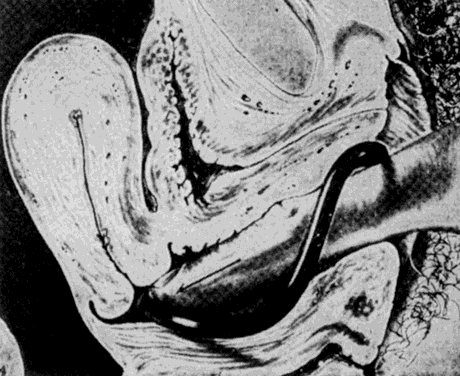
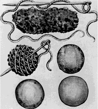
Trotula, the wife of Joannes Platearious, was the first recorded female practitioner of gynecology around A.D. 1050. She originated the use of a ball pessary that was made of strips of linen and filled the vagina in cases of prolapse.1 To complete the therapy, a T binder was applied. Caspar Stromayr of Lindau, Germany, recommended in 1559 that a sponge tightly rolled and bound with string, dipped in wax, and covered with oil or butter be substituted for a pomegranate as a pessary4 (Fig. 1 and Fig. 2).

Fig. 1. Early pessaries.(Stromayr C: Die Handschrift des Schnitt-und Augenarztes Caspar Stromayr. Berlin, Brunn, 1925)

Fig. 2. Insertion of a wax ball pessary.(Stromayr C: Die Handschrift des Schnitt-und Augenarztes Caspar Stromayr. Berlin, Brunn, 1925)

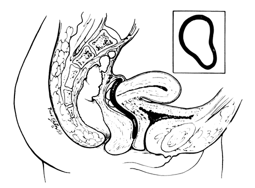
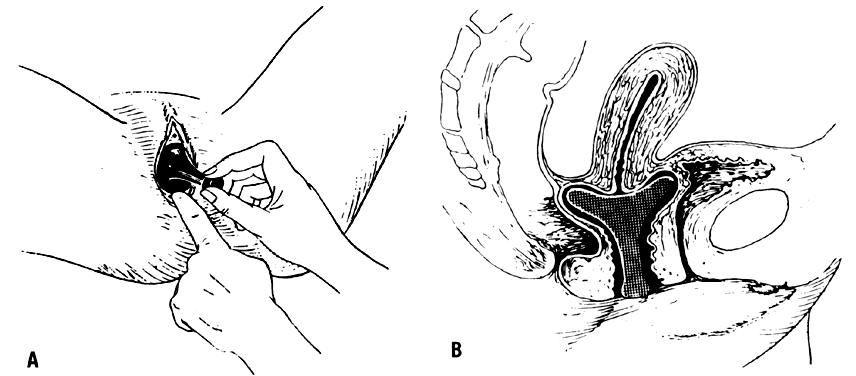
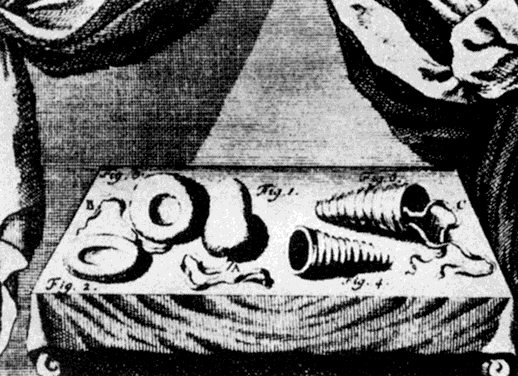
Ambrose Pare, late in the 16th century, devised ingenious oval-shaped pessaries of hammered brass and waxed cork for uterine prolapse. He made an apparatus of gold, silver, or brass that was kept in place by a belt around the waist. He also designed pear-shaped and ring pessaries5 (Fig. 3). William Harvey was an exponent of the conservative school that treated prolapse with pessaries, replacement, and other such means unless carcinoma was present, when surgery was occasionally suggested. In his book, Anatomical Exercitations Concerning the Generation of Living Creatures (1653), he reported an enlarging and completely prolapsed uterus that contained a dead fetus.3

Fig. 3. Spiral, oval, and doughnut-shaped pessaries used for prolapse during the 18th century.(von Haller A: Disp Chirg 3:23, 1755)

Hendrik Van Roonhuyse, born in 1622, was particularly interested in the diseases of women and made a remarkable contribution to 17th century gynecology when in 1663 he published Heelkonstige Aanmerkkingen Betreffende de Grebrecken der Vrouwen. This book often has been referred to as the first textbook on operative gynecology. Van Roonhuyse discussed the etiology and treatment of prolapse. An accepted procedure for treatment of prolapse was the use of cork with a hole in it to allow passage of discharges, plus wax pessaries or cork dipped in wax. He described one of the first complications and its resolution—his removal of one of these putrefied wax pessaries that had obstructed a patient's lochia.3 As noted in 1684 by Thomas Willis, “When at any time a sickness happens in a woman's body of an unusual manner so that its cause lies hid, and the curatory indication is altogether uncertain, presently we accuse the evil influence of the womb.”6

By the late 1700s, Thomas Simson, Professor of Medicine at the University of St. Andrew (Scotland), devised a metal spring that kept a cork ball pessary in place. Most pessaries had string attachments to facilitate removal. In instances of extensive perineal lacerations, pessaries were kept in place by T binders. These devices proved very unsatisfactory because friction injured the labia. 2 Conversely, John Leake had no use for pessaries, believing that rest and astringent applications were in every repect preferable to the use of those painful and indelicate instruments called pessaries. He advocated the use of sponges as pessaries to avoid vaginal wall injuries, which he noticed resulted from pessaries made of hard material. He extracted a pessary from the rectum of a patient at St. Thomas' Hospital.2 Mòrgagni recorded the autopsy of a woman with a pessary in situ.2 To avoid such complications, Jean Juville in 1783 introduced a soft rubber pessary. In devising this new type of pessary, Juville stated that it facilitated introduction by the patient. Juville's pessary resembled the contraceptive cup currently used. In the center of the cup was a perforated gold tip to permit the escape of cervical secretions. When he first introduced this new device, Juville was unable to state whether rubber was injurious to the vaginal tissue. Jean Astruc stated that the pessary had the same effect as a truss for a rupture. He believed that if the pessary were worn long enough and the patient grew fat, a cure would follow.2

Gynecologic faddism enjoyed its heyday throughout most of the 19th century. During the early decades, most of the ills of womankind were attributed to inflammations of the uterus. Toward the middle years, these views were supplanted by the Galenic doctrine that displacements of that organ were the principal cause of women's complaints. The pessary school of gynecology began to flourish. It was said that fortunes were to be made by two groups of gynecologists—those who inserted pessaries and those who removed them.7,8

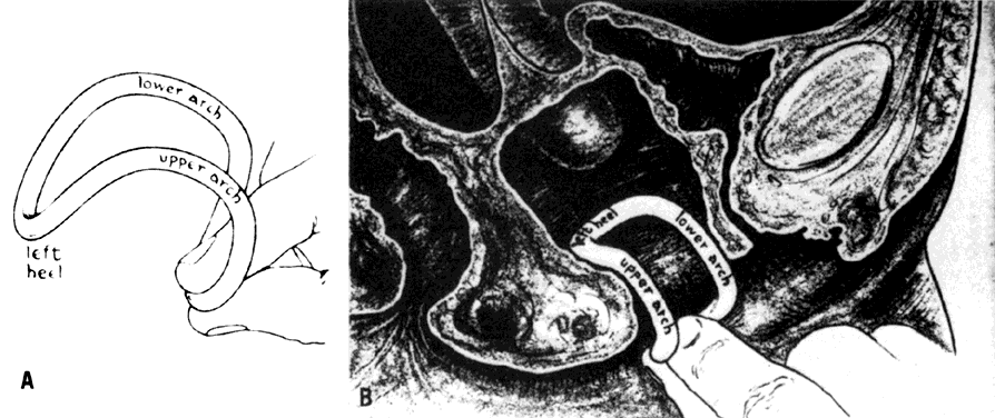
Goodyear's discovery of the vulcanization of rubber was a boon to the purveyors and wearers of pessaries and greatly enhanced the popularity of the devices. Hugh Lenox Hodge, Professor of Gynecology at the University of Pennsylvania, was dissatisfied with the shapes of the existing pessaries. He set out to design a new one using Goodyear's newly patented material. The lever pessary was designed especially for cases of uterine retroversion. Hodge explained in 1860:

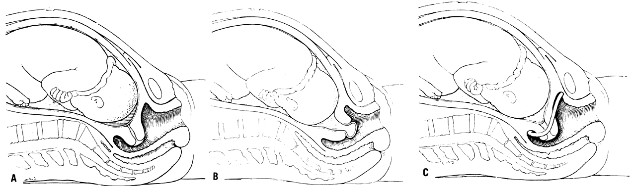
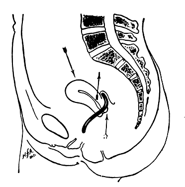
The important modification consists in making a ring oblong, instead of circular, and curved so as to correspond to the curvature of the vagina. Great advantages result from this form; the convexity of the curve being in contact with the posterior wall of the vagina, corresponds, with more or less accuracy, to properly arrange, there is no pressure against the rectum; and the higher the instrument rises, the superior extremity, instead of impinging against the rectum, passes upward and behind the uterus—between this organ and the intestine—giving a proper position to the womb, and yet allowing its natural pendulum-like motion to remain unrestrained9,10 (Fig. 4).

Fig. 4. Mechanism of action of the lever pessary. The posterior vaginal wall stretched over the upper transverse bar of the pessary pulls the cervix upward and backward, thus tilting the fundus forward. The pessary imitates the action of the uterosacral ligaments.

Albert Smith of Philadelphia later narrowed the anterior portion and widened the posterior end of the Hodge pessary.6

With the advent of asepsis and anesthesia, gynecologists' fascination with pessaries abated as uterine support deficiencies were addressed by surgery. Pessaries were used mainly for inoperable patients or as temporizing measures. Little progress was made in the development of new devices. Minor changes typically were made to existing pessaries and a new eponym attached. The most significant advance in the art and science of pessaries was the replacement of hard rubber by polystyrene plastics in the 1950s and recently by silicone-based materials.11

Back to Top
DEVICES

More than 200 different types of pessaries have been invented, but only a few are currently in clinical use.

Ring

The ring pessary in its modern version appears similar to a contraceptive diaphragm (Fig. 5I and Fig. 5J). It is useful in first- and second-degree uterine prolapse. The ring with the support membrane is beneficial for a mild cystocele accompanying the prolapse. This pessary is fitted like a contraceptive diaphragm. The length of the vaginal canal can be measured with the examining finger and the size of pessary approximated. The ring pessary is folded so that the arc points downward and is directed past the cervix into the posterior fornix.

Fig. 5. A. Smith; B. Hodge without Support; C. Hodge with Support; D. Gehrung with Support; E. Risser; F. Incontinence Dish without Support; G. Incontinence Dish with Support; H. Incontinence Ring; I. Ring with Support; J. Ring without Support; K. Cube; L. Tandem-Cube; M. Shaatz; N. Gellhorn; O. Gellhorn; P. Gellhorn; Q. Inflatoball; R. Doughnut.(Courtesy of MILEX Products, Inc., Chicago, IL)

Lever

The lever pessaries are variations on the design by Hodge.9 They are intended to treat uterine retroversion by posteriorly displacing the cervix and anteverting the uterus.11,12 The Smith pessary has a narrower anterior limb for application in a patient with a narrow pubic arch (Fig. 5A). The somewhat broader anterior limb of the Hodge pessary prevents the pessary from turning and precludes pressure on the urethra (Fig. 5B and Fig. 5C). The Risser pessary is a further modification, for an even flatter pubic arch13 (Fig. 5E). Although originally designed to treat uterine retroversion, these devices currently are used for the treatment of an incompetent cervix in pregnancy, for mild uterine prolapse with retroversion, and as a diagnostic maneuver in evaluation of patients with large cystoceles or urinary stress incontinence. The pessary is inserted by first manually elevating a retrodisplaced uterus. The pessary is then folded and pushed into the vagina by the index finger, pressing on the posterior bar until it is behind the cervix, with the anterior bar behind the pubic notch14 (Fig. 6 and Fig. 7).

Fig. 6. Introducing a lever pessary. The index finger is passed to the top of the posterior end, which is then depressed until it can be pushed past the cervix.(Crossen RJ: Disease of Women, 10th ed. St. Louis, CV Mosby, 1953)

Fig. 7. Lever pessary in place after the uterus has been brought forward; the inner end behind the cervix holding it up and back, and the outer end resting on the muscles of the pelvic floor. Anteriorly it gets its support from the soft tissue between it and the pubic arch.(Crossen RJ: Disease of Women, 10th ed. St. Louis, CV Mosby, 1953)

Gehrung

The Gehrung pessary provides support in cases of cystocele and rectocele (Fig. 5D). It provides support to the anterior vaginal wall, with its lateral bars resting on the levator sling. These bars tend to flatten out a rectocele. Because of its unusual shape, most practitioners are unfamiliar with it and have difficulty inserting it. However, insertion is easily accomplished by introducing the lateral bars of the pessary over the perineum completely into the vagina while the device is on its side (Fig. 8). The pessary is then rotated so that the curved bars point with their convexity toward the anterior wall of the vagina14 (Fig. 9 and Fig. 10).

Fig. 8. Introducing the Gehrung pessary. A. How the pessary is held. B. First step in the introduction.(Crossen RJ: Disease of Women, 10th ed. St. Louis, CV Mosby, 1953)

Fig. 9. Introducing the Gehrung pessary. A. Swinging the right heel to the right side, which carries the left heel under the cervix to the left side and brings up the upper arch, which was below. B. Pushing the pessary around the vaginal wall in back of the cervix to get the right heel within the vagina. C. Further progress in the same direction.(Crossen RJ: Disease of Women, 10th ed. St. Louis, CV Mosby, 1953)

Fig. 10. Introducing the Gehrung pessary. A. The right heel within the vagina and being carried to its position on the right side. B. Two heels situated symmetrically on each side. The arches are still too low. C. The arches pushed up into place behind the symphysis.(Crossen RJ: Disease of Women, 10th ed. St. Louis, CV Mosby, 1953)

Gellhorn

The Gellhorn pessary provides support of a third-degree uterine prolapse or procidentia (Fig. 5N, Fig. 5L, Fig. 5M, Fig. 5N, Fig. 5O and Fig. 5P).15 The cervix rests behind the flat base of the pessary, and the stem prevents turning the support within the vagina, making it possible to keep even a large prolapse in place by means of a comparatively small pessary. It requires a relatively capacious vagina so that the base is broad enough to rest above the levators. It is inserted by turning the disk portion parallel to the introitus; then the base is rotated over the perineal body and into position16 (Fig. 11).

Fig. 11. The Gellhorn pessary. A. Fitting the pessary. It is well lubricated and inserted edgewise and in an oblique direction to avoid the urethral opening while the perineum is pushed strongly downward. It is introduced into the vagina by a corkscrew-like motion. Once within the vaginal lumen, the pessary is pushed upward until only the extremity of the stem shows in the vaginal entrance. The appliance then lies transversely beneath the cervix, as in ( B ).(Gellhorn G: A new pessary for the treatment of inoperable prolapse of the uterus. Am J Obstet Gynecol 29:737, 1935)

Doughnut

Probably the most popular pessary in the past was the doughnut pessary and its modifications. It is simply a doughnut-shaped ring that is solid or can be inflated by a bulb and valve assembly (Fig. 5Q and Fig. 5R). It works by occluding the upper vagina to support a uterine prolapse. To remain in place, the mass of the inflated pessary must be greater than the defect in the levator sling. The Inflatoball pessary can be removed easily and replaced by a patient for vaginal hygiene.

Cube

An effective pessary in patients with third-degree prolapse—especially very elderly patients with complete procidentia, rectocele, or cystocele—is the cube pessary (Fig. 5K). On each of the six sides of the cube are suction cups that adhere to the vaginal walls. A cube pessary is often useful in a very elderly patient with significant atrophy and complete prolapse.17,18 However, this pessary is difficult for a patient to remove and thus requires close monitoring. It is inserted by merely compressing the cube and placing it as high up in the vagina as possible.

Back to Top
INDICATIONS

Neonatal Uterine Prolapse

Neonatal uterine prolapse is a rare condition. 19,20,21,22 Most cases occur in association with meningomyelocele or other defects of the central nervous system. It typically occurs during the first few days of life, presenting as a mass at the introitus. Satisfactory treatment has been reported using 1- to 2-cm doughnut-shaped pessaries constructed from small Penrose drains. These devices were applied for days to months and then removed. Prolapse did not recur.20,21,22,23

Prolapse in Pregnancy

Two hundred fifty-six cases of uterine prolapse during pregnancy have been reported, including eight patients who died of septicemia or uterine rupture.24,25,26,27,28 Fortunately, no fatalities have been reported since 1925. It occurs in multiparous patients who have noted a degree of pre-existent uterine prolapse that is often asymptomatic. However, the weight of the gravid uterus causes the uterine prolapse to descend farther. If prolapse is not treated early, the cervix becomes ulcerated and infected and undergoes hypertrophy to the extent that it continues to prolapse even as the uterus rises out of the pelvis. Complications have varied from minor cervical ulcerations and infection to uterine rupture with fetal and maternal fatalities. In their review of the literature, Piver and Spezia recommend prompt treatment with bed rest in Trendelenburg's position to reduce edema and allow reduction of the uterus. The infected cervix can be treated with local antiseptics. After reduction is accomplished, a large doughnut or lever pessary should be inserted and left in place until the onset of labor.24 Theirs and subsequent case reports of patients treated with pessaries have shown an excellent maternal and fetal outcome.24,25,26,27,28

Incarcerated Uterus

Retrodisplacement of the uterus in early pregnancy occurs in 15% of pregnancies. It almost always is self-limited because the enlarging uterus rises into the abdomen. Incarceration occurs rarely when a retroverted pregnant uterus becomes entrapped in the sacral hollow, probably as a result of adhesions or endometriosis. The uterus presses against the rectum and fails to rise into the abdomen as it enlarges. Patients present after 12 weeks of gestation with pain and urinary and rectal complaints. Examination shows a posterior cul-de-sac mass with the cervix rotated anteriorly high up in the vagina behind the pubic symphysis, impinging on the bladder trigone.29 Gibbons and Paley reported that patients who were treated with uterine repositioning and pessary support before 15 weeks did well but that those beyond that gestation aborted despite treatment.30 The appropriate lever pessary, typically the Hodge, displaces the cervix posteriorly, anteverting the uterus and allowing it to rise out of the pelvis (see Fig. 4). It has been used successfully to treat mobilized incarceration and to prevent recurrences in subsequent pregnancies.30,31

Incompetent Cervix with Premature Labor

The diagnosis of incompetent cervical os as a cause of midtrimester pregnancy loss is difficult to document. Nonetheless, in this country, it usually is treated with an invasive surgical procedure fraught with well-described complications. The use of the pessary as a nonsurgical alternative to cervical cerclage is well reported but has not yet been accepted in the United States. Cross, of the Rotunda Hospital in Dublin, was the first to report the use of Bakelite ring pessaries in 13 patients with a history of only seven term deliveries in 54 pregnancies (13%) before treatment. After application of the pessary, the patients delivered 11 of 13 viable infants (85%).32 In the United States, Vitsky studied 21 patients with a pregnancy history of only 23% viable deliveries; this improved to 87% with the use of a Smith pessary.33,34,35 Oster and Javert later reported a similar experience in 29 patients using the Hodge pessary.36,37 Multiple European reports have confirmed these findings.38,39,40,41,42,43,44,45,46,47,48,49,50,51,52,53 In the United States, the Hodge pessary has been used successfully to treat cervical incompetence due to defective connective tissue in Ehlers-Danlos syndrome.54 The incompetent cervix points anteriorly in the axis of the vagina (Fig. 12). The lever pessary directs the cervix to point posteriorly, as in a normal pregnancy. Direct pressure on the cervical os is reduced and brought to bear on the lower uterine segment.36 Although a randomized prospective placebo-controlled study evaluating pessaries for the treatment of incompetent cervical os has yet to be accomplished, a randomized prospective study comparing cervical cerclage versus the Mayer ring pessary showed no difference in the efficacies of these treatments.50 A controlled study recently showed no increase in pathologic vaginal flora or puerperal febrile morbidity in patients who had a pessary in place during pregnancy versus a control group.47,49 It has never been shown that any treatment method affects the subsequent pregnancy outcome of women who have recurrent pregnancy losses.55,56,57,58 This recently was confirmed by Harger, who found that these patients had a 70% to 80% chance of a viable pregnancy regardless of whether a defect was identified or treated.59 In fact, two prospective randomized studies failed to show any significant advantage of cervical cerclage over no treatment in this group of patients.60,61 Further study of this problem clearly is required. Pending clarification, a pessary is certainly a less invasive treatment option.

Fig. 12. A. Normal cervix usually points posterior in the last trimester of pregnancy. B. An incompetent cervix usually points anteriorly in the mid and last trimesters of pregnancy, allowing the membranes to herniate. C. The lever pessary “cradles” an incompetent cervix, keeping it pointing posteriorly in the last trimester, preventing herniation of membranes.(Javert CT: Further follow-up on habitual abortion patients. Am J Obstet Gynecol 84:1149, 1962)

Stress Urinary Incontinence

In the modern therapy of stress urinary incontinence, pessaries originally were used merely as platforms for electrodes to stimulate the pudendal nerve to strengthen pelvic muscles—an involuntary Kegel exercise.62,63 Richardson and colleagues elucidated the mechanism of paradoxical continence in patients with significant uterovaginal prolapse (procidentia).64 Using a doughnut pessary to replace the prolapse, the patients became incontinent. Urethrovesical pressure dynamic studies showed that the mechanism of continence in significant uterovaginal prolapse was urethral obstruction. Reducing the prolapse with a doughnut pessary could differentiate patients who also require urethrovesical neck suspension as part of their surgical treatment.64,65,66 The “doughnut” pessary merely replaces a prolapsed uterus but offers no support to the proximal urethra. The lever pessaries are applied behind the pubic arch and support the proximal urethra. Using detailed urodynamic studies, Bhatia and colleagues showed that Smith and Hodge pessaries promoted consistent and significant increases in urethral functional length and closing pressure without causing obstruction. These pessaries restored continence by stabilizing the urethra and urethrovesical junction to allow proper pressure transmission and actively increasing urethral resistance to the escape of urine under resting and stressful conditions. They proposed the use of the lever pessary as a simple prognostic test to identify patients with stress urinary incontinence that would benefit from urethrovesical neck suspension procedures.67,68,69,70 A recent study showed that a Hodge pessary with support was successful in preventing incontinence in exercising women.71 Lever pessaries are effective nonsurgical and nonpharmacologic options for the elderly woman with stress incontinence.72,73,74

Preoperative Preparation

Surgery, the most effective long-term therapy for uterovaginal prolapse, often cannot be immediately undertaken because of complications of the prolapse or underlying medical problems.15,75 Typical examples are patients who have procidentia and have developed cervical or vaginal decubiti surrounded by inflamed edematous but atrophic vaginal mucosa. This inflammation and ulceration make the necessary surgical maneuvers difficult and obviously increase the risk of local infection. Reduction of the prolapse and retention with an appropriately fitted pessary allow the inflammation to subside and ulcers to heal. A temporizing pessary allows for better vaginal hygiene than a vaginal pack. The application of systemic or topical estrogens in patients without contraindications promotes healing, matures vaginal epithelium, and subjectively improves surgical planes. Rarely, uterovaginal prolapse causes ureteral obstruction and hydronephrosis, and patients may present with flank pain and uremia. Pessary application has been shown to relieve obstruction and improve renal function so that patients can undergo surgical correction.76,77,78 The most common indication for the temporizing use of pessaries is to alleviate the suffering of a patient while she awaits medical and anesthesia clearance for surgery. Such use illustrates to the patient the relief she might expect from the operation.15

Poor Operative Risk Patients

The most common use for pessaries in modern gynecology has been for the treatment of press continence uterovaginal prolapse in patients deemed to be a poor risk for a vaginal procedure.72,73,74,79,80,81,82,83 The advent of modern monitoring and regional anesthesia techniques should limit the number of these patients. If necessary, colpocleisis can be done under local anesthesia; because it is in this group of patients—the aged, the frail, and the disabled—that the complications of a pessary are most often encountered. These patients, deemed to have a limited life expectancy, often live long enough to develop embedment, incarceration, and, rarely, fistulization of their poorly fitted, infrequently monitored, or forgotten pessaries.64,65,66,67,68,69,70,71,72,73,74,75,76,77,78,79,80,81,82,83,84,85,86,87,88,89,90,91,92,93,94 However, with proper care as outlined subsequently, uterovaginal deformities can be supported and suffering alleviated by pessaries in this poor-risk group.

Back to Top
MANAGEMENT

Selecting a Pessary

Selecting the most appropriate pessary requires an understanding of the devices and their indications.79,80 However, the pessary should be fitted to the patient. Different devices often are used to treat the same process in different patients. Those with uterine prolapse should be fitted first with a ring. The Gellhorn is also useful if the patient has an intact perineal body.15 If results are less than satisfactory, the doughnut or cube may be used in patients with complete procidentia or poor perineal bodies. Stress urinary incontinence with or without prolapse is best approached with one of the lever pessaries. Symptomatic cystocele and rectocele are well supported by the Gehrung pessary. Prolapse of the vagina after hysterectomy usually is best treated with a doughnut or cube pessary.

Most pessaries currently on the market are made of inert polystyrene plastics or silicone rubber.11 Only these inert materials should be used. Old Bakelite or hard rubber pessaries should be discarded.

Fitting a Pessary

In general, the largest pessary that a patient can comfortably accommodate should be fitted. Ring and lever pessaries should fit snugly behind the pubic symphysis and posterior to the cervix. As when fitting a contraceptive diaphragm, the examiner's finger should easily pass between the vagina and the circumference of the pessary. After the pessary has been inserted, separate the labia; have the patient perform a Valsalva's maneuver. A properly fitted pessary descends to where the lower bar is visible and then ascends with relaxation.13 The patient should be able to stand, sit, squat, urinate, and defecate comfortably without dislodging the device. Coitus is possible with the ring, lever, and Gehrung pessaries in place.13,79,80

Follow-Up

Physicians must consider a patient as being under their active care as long as she wears the device. The patient and her family should be instructed about the management plan with the pessary. After placement of the pessary, the patient should return in 1 week for re-evaluation consisting of palpation of the pessary in place; removing it; inspecting the vagina for excoriation, laceration, or ulceration; cleaning and inspecting the pessary; and refitting it. These maneuvers should occur subsequently at 1 month and then every 3 months. Although most pessaries are made of inert materials, they still are foreign bodies, and physicians placing a pessary must assume responsibility for it and assure themselves of the patient's continued follow-up. More dexterous and highly motivated patients may remove the pessary at night and replace it on arising. This usually can be accomplished only by women in early pregnancy or those awaiting corrective surgery. A patient who is a poor operative risk is likely to wear the pessary for the rest of her life and usually is not capable of removing and replacing the device. Thus, a physician must assume responsibility for maintaining the device with adequate follow-up to avoid complications. Patients without contraindications should be treated with systemic or topical estrogen therapy, which matures the vaginal epithelium, increases the pliability of submucosal connective tissue, and improves perineal muscular tone. These patients often require resizing of their pessaries.

Back to Top
COMPLICATIONS

The most common complaint of patients wearing pessaries is an increase in vaginal discharge and odor. Cytologic atypia in the form of severe inflammatory changes and prominent superficial maturation of squamous cells, atypical metaplasia, on a background of superficial squamous maturation have been reported on vaginal cytology.95 These complaints usually are resolved by proper sizing and fitting of the pessary and the use of estrogen therapy. Dilute vinegar or hydrogen peroxide douches also provide relief. Poorly fitted or neglected pessaries can ulcerate the walls of the vagina or become embedded.86 Ulceration should be managed by pessary removal with treatment of any infection and topical sucralfate, a basic aluminum salt of sucrose octasulfate that binds preferentially to ulcerated areas and promotes healing.96 General anesthesia and surgical procedures have been required to remove embedded pessaries.84,86,87,88,91,93 In the heyday of pessaries, a pessariotome was used to fracture and extract these devices, thus obviating the advantage of pessaries in patients who are supposedly poor operative risks. Incarceration can occur when the cervix and uterus or intestines herniate through the center of a poorly fitted ring pessary and become strangulated.85,93 Patients with embedded or incarcerated pessaries sometimes can have them removed on an outpatient basis, using improved vaginal hygienic measures and topical estrogens to mature epithelium and diminish inflammation.89 Surprisingly, fistula formation secondary to pessaries is quite unusual.2,86,90,93 Uncontrolled studies of vaginal cancer implicated pessaries as an etiologic factor.86,97 This cause has not been confirmed in population-based studies.98,99 Because physical contact or irritation has never been shown to cause any cancer, the possible relationship between pessaries and vaginal cancer may have been due to the composition of the devices rather than their mere presence.

Back to Top
CONTRAINDICATIONS

The contraindications to pessary placement include primary vaginitis, active pelvic inflammatory disease, endometriosis, a noncompliant patient, and lack of assured follow-up.

Back to Top
REFERENCES

1. Morice P, Josset P, Colau JC: Gynecology and obstetrics in ancient Egypt. J Gynecol Obstet Biol Reprod (Paris) 23: 131, 1994

2. Ricci JV: Genealogy of Gynecology, p. 350. Philadelphia, Blakiston, 1950

3. Cianfrani T: Short History of Obstetrics and Gynecology. Springfield, IL, Charles C. Thomas, 1960

4. Stromayr C: Die Handschrift des Schnitt-und Augenarztes Caspar Stromayr. Berlin, Brunn, 1925

5. Emge LA, Durfee RB: Pelvic organ prolapse, four thousand years of treatment. Clin Obstet Gynecol 9: 997, 1966

6. Speert H: Obstetrics and Gynecology in America: A History. Baltimore, Waverly Press, 1980

7. Bantock GG: Use and Abuse of Pessaries. London, HK Lewis, 1884

8. Haller JS: Trends in American gynecology, 1800–1910: A short history. NY State J Med 5: 278, 1989

9. Hodge HL: On Disease Peculiar to Women, Including Displacements of the Uterus. Philadelphia, Blanchard & Lea, 1860

10. Speert H: Obstetrics and Gynecologic Milestones. New York, Macmillan, 1958

11. Robertson DNS, Stoff FD: Advantages of “polythene” ring pessaries. Br Med J 2: 30, 1958

12. Smith AA: A golden opportunity. J Med Assoc GA 53: 349, 1964

13. Risser JA: Symptomatic Descensus Uteri (Prolapse Syndrome). Chicago, Milex Products, 1957

14. Crossen RJ: Disease of Women, 10th ed. St. Louis, CV Mosby, 1953

15. Sulak PJ, Kuehl TJ, Shull BL: Vaginal pessaries and their use in pelvic relaxation. J Reprod Med 38: 919, 1993

16. Gellhorn G: A new pessary for the treatment of inoperable prolapse of the uterus. Am J Obstet Gynecol 29: 737, 1935

17. Hutter CG: Bee-cell pessary. West J Surg 57: 481, 1949

18. Seyffarth K, Arabin H: Treatment of uterine descent and prolapse with the cube pessary. Zentralbl Gynakol 112: 1181, 1990

19. Findley P: Prolapse of the uterus in nulliparous women. Am J Obstet Dis Wom 75: 12, 1917

20. Dixon RE, Acosta AA, Young RL: Penrose pessary management of neonatal genital prolapse. Am J Obstet Gynecol 119: 855, 1974

21. Johnson A, Unger SW, Rodgers BM: Uterine prolapse in the neonate. J Pediatr Surg 19: 210, 1984

22. Shuwarger D, Young RL: Management of neonatal genital prolapse: Case reports and historic review. Obstet Gynecol (Suppl) 66: 61S, 1985

23. de Mola JR, Carpenter SE: Management of genital prolapse in neonates and young women. Obstet Gynecol Surv 51: 253, 1996

24. Piver MS, Spezia J: Uterine prolapse during pregnancy. Obstet Gynecol 32: 765, 1968

25. Bluett D: Uterine prolapse in pregnancy: Case report and description of pessary. Am J Obstet Gynecol 101: 574, 1968

26. Suzuki Y, Shane JM: Uterine prolapse in the pregnant primigravida. Am J Obstet Gynecol 112: 303, 1972

27. Schinfeld JS: Prolapse of the uterus during pregnancy: A report of two cases and review of management. Am J Obstet Gynecol 129: 587, 1977

28. Hill PS: Uterine prolapse complicating pregnancy: A case report. J Reprod Med 29: 631, 1984

29. Weinberger MW, Julian TM: Voiding dysfunction and incontinence caused by uterine retroversion: A case report. J Reprod Med 40: 387, 1995

30. Gibbons JM Jr, Paley WB: The incarcerated gravid uterus. Obstet Gynecol 33: 842, 1969

31. McGann KP, Griffin WT: Recurrent classical sacculation of the pregnant uterus. J Fam Pract 26: 339, 1988

32. Cross RG: Treatment of habitual abortion due to cervical incompetence. Lancet 2: 127, 1959

33. Vitsky M: Simple treatment of the incompetent cervical os. Am J Obstet Gynecol 81: 1194, 1961

34. Vitsky M: The incompetent cervical os and the pessary. Am J Obstet Gynecol 87: 144, 1963

35. Vitsky M: Pessary treatment of the incompetent cervical os. Obstet Gynecol 31: 732, 1968

36. Javert CT: Further follow-up on habitual abortion patients. Am J Obstet Gynecol 84: 1149, 1962

37. Oster S, Javert CT: Treatment of the incompetent cervix with the Hodge pessary. Obstet Gynecol 28: 206, 1966

38. Jiratko K: Mayer pessary as prevention of premature labor. Cesk Gynecol 39: 537, 1974

39. Jiratko K, Baran P, Zabransky F: Useful treatment of imminent precocious delivery: Confrontation of results gained by insertion of pessary. Cesk Gynekol 41: 184, 1976

40. Jiratko K: Our experience with Mayer-ring pessary in the prevention of premature labor. Zentralbl Gynakol 99: 220, 1977

41. Bayer H: Various new aspects for prevention and therapy for impending premature birth. Zentralbl Gynakol 99: 547, 1977

42. Bechnini E: Treatment of cervix incompetence in pregnant women by means of Mayer's pessary. Cesk Gynekol 42: 205, 1977

43. Jorde A: A cubical support pessary within the scope of complex therapy of threatening prematurity. Zentralbl Gynakol 100: 235, 1978

44. Seyffarth VK: Non-invasive cerclage using supportive pessaries for prevention and therapy of premature birth. Zentralbl Gynakol 100: 1566, 1978

45. Dahl J: Prevention of premature labor by means of supporting pessaries (1st experiences). Z Arztl Fortbild (Jena) 73: 1010, 1979

46. Bayer H: New aspects in the prevention and therapy of threatened labor. Akush Ginekol (Mosk) 7: 18, 1980

47. Jorde VA, Kastli Y, Hamann B et al: Detection of changes of the vaginal flora due to supporting pessary treatment during pregnancy. Zentralbl Gynakol 105: 855, 1983

48. Roberts PJ, Liston WA: Cervico-vaginal fistula and recurrent premature delivery: Successful treatment with a Portex ring pessary. J Obstet Gynecol 5: 101, 1984

49. Havlik I, Stasek K, Franek B et al: Vaginal flora during supportive therapy using a pessary in pregnancy. Cesk Gynekol 51: 258, 1986

50. Forster F, During R, Schwarzlos G: Therapy of cervix insufficiency—cerclage or support pessary?. Zentralbl Gynakol 108: 230, 1986

51. Quaas L, Hillemanns HG, du Bois A, Schillinger H: The Arabian cerclage pessary—An alternative to surgical cerclage. Geburtshilfe Frauenheilkd 50: 429, 1990

52. Moser G, Girardi F, Mayer HO et al: The support pessary—A therapeutic possibility in premature opening of the uterine cervix. Gynakol Rundsch 2: 117, 1991

53. Bechinie E: Use of pessary for bloodless cerclage for cervical insufficiency in pregnant women—25 years' experience. Cesk Gynekol 57: 217, 1992

54. Leduc L, Wasserstrum N: Successful treatment with the Smith-Hodge pessary of cervical incompetence due to defective connective tissue in Ehlers-Danlos syndrome. Am J Perinatol 9: 25, 1992

55. Cousins L: Cervical incompetence, 1980: A time for reappraisal. Clin Obstet Gynecol 23: 467, 1980

56. Thomason JL, Sampson MB, Beckmann CR et al: The incompetent cervix: A 1982 update. J Reprod Med 27: 187, 1982

57. Harris BA Jr: The incompetent cervix. Compr Ther 11: 46, 1985

58. Shortle BM: Correcting cervical incompetence after habitual abortions. Contemp Obstet Gynecol 27: 147, 1986

59. Harger JH, Archer DF, Marchese SG et al: Etiology of recurrent pregnancy losses and outcome of subsequent pregnancies. Obstet Gynecol 62: 574, 1993

60. Rush RW, Isaacs S, McPherson K et al: A randomized controlled test of cervical cerclage in women at high risk of spontaneous preterm delivery. Br J Obstet Gynaecol 91: 724, 1984

61. Lazar P, Gueguen S, Dreyfus J et al: Multicentered controlled trial of cervical cerclage in women at moderate risk of preterm delivery. Br J Obstet Gynaecol 91: 724, 1984

62. Alexander S, Rowan D, Millar W et al: Treatment of urinary incontinence by electric pessary: A report of 18 patients. Br J Urol 42: 184, 1970

63. Paterson PJ, Harrison NW: Electronic techniques in the treatment of female urinary incontinence. J Obstet Gynaecol Br Commonw 77: 732, 1970

64. Richardson DA, Bent AE, Ostergard DR: The Effect of uterovaginal prolapse on urethrovesical pressure dynamics. Am J Obstet Gynecol 146: 901, 1983

65. Bergman A, Koonings PP, Ballard CA: Predicting postoperative urinary incontinence development in women undergoing operation for genitourinary prolapse. Am J Obstet Gynecol 158: 1171, 1988

66. Rosenberg BA, Pushkin S, Blumenfeld D, Bhatia NN: Prevalence of abnormal urodynamic test results in continent women with severe genitourinary prolapse. Obstet Gynecol 79: 539, 1992

67. Bhatia NN, Bergman A, Gunning JE: Urodynamic effects of a vaginal pessary in women with stress urinary incontinence. Am J Obstet Gynecol 147: 876, 1983

68. Bergman A, Bhatia NN: Pessary test: Simple prognostic test in women with stress urinary incontinence. Urology 24: 109, 1984

69. Bhatia NN, Bergman A: Pessary test in women with urinary incontinence. Obstet Gynecol 65: 220, 1985

70. Richardson DA: Investigative procedures in urogynecology. Curr Opin Obstet Gynecol 3: 513, 1991

71. Nygaard I: Prevention of exercise incontinence with mechanical devices. J Reprod Med 40: 89, 1995

72. Rousseau P, Fuentevilla-Clifton A: Urinary incontinence in the aged, part 2: Management strategies. Geriatrics 47: 37, 1992

73. Zeitlin MP, Lebherz TB: Pessaries in the geriatric patient. J Am Geriatr Soc 40: 635, 1992

74. Walters MD, Realini JP, Dougherty M: Nonsurgical treatment of urinary incontinence. Curr Opin Obstet Gynecol 4: 554, 1992

75. Puddington IE, Guiou NM: Support for posthysterectomy prolapse. Can Med Assoc J 114: 409, 1976

76. Hildreth TA, Stewart W, Cass AS: Uterine prolapse and upper urinary tract obstruction. Minn Med 59: 450, 1976

77. Jurascheck J, Salti A, Fernandex R et al: Bladder retention and blockage of urinary tract caused by prolapse. Acta Urol Belg 54: 391, 1986

78. Jay GD, Kinkead T, Hopkins T, Wollin M: Obstructive uropathy from uterine prolapse: a preventable problem in the elderly. J Am Geriatr Soc 40: 1156, 1992

79. Melody GF: The use and abuse of pessaries. West J Surg 55: 544, 1947

80. Colmer WM: Use of the pessary. Am J Obstet Gynecol 65: 170, 1953

81. Williams B: The modern pessary treatment of vaginal prolapse. Practitioner 201: 782, 1968

82. Rodenberg TA: Pessaries and prolapse. J Fla Med Assoc 66: 895, 1981

83. Meinhardt W, Schuitemaker NW, Smeets MJ, Venema PL: Bilateral hydronephrosis with urosepsis due to neglected pessary: Case report. Scand J Urol Nephrol 27: 419, 1993

84. Pommerenke WT: The neglected pessary. Obstet Gynecol 1: 226, 1953

85. McElin TW, Paalman RJ: Pessary complications in the management of uterine prolapse. Am J Obstet Gynecol 78: 643, 1959

86. Russell JK: The dangerous vaginal pessary. Br Med J 2: 1595, 1961

87. Pritchard JG: A watchspring pessary retained for 62 years. Lancet 1: 172, 1961

88. Thornton CA, Harrison RF: Brief report: Unusually rapid incarceration of a polyvinyl ring pessary. Ir J Med Sci 146: 116, 1977

89. Poma PA: Management of incarcerated vaginal pessaries. J Am Geriatr Soc 29: 325, 1981

90. Knight DG, Kernohan N: The “forgotten” vaginal ring pessary—An unusual cause of rupture of the urinary bladder. Br Med J 292: 992, 1986

91. Sivasuriya M: Cervical entrapment of a polythene vaginal ring pessary—A clinical curiosity. Aust NZ J Obstet Gynaecol 27: 168, 1987

92. Goldstein I, Wise GJ, Tancer ML: A vesicovaginal fistula and intravesical foreign body: A rare case of the neglected pessary. Am J Obstet Gynecol 163: 589, 1990

93. Summers JL, Ford ML: The forgotten pessary: A medical oddity. Am J Obstet Gynecol 111: 307, 1991

94. Ott R, Richter H, Behr J, Scheele J: Small bowel prolapse and incarceration caused by a vaginal ring pessary. Br J Surg 80: 1157, 1993

95. Christ ML, Haja J: Cytologic changes associated with vaginal pessary use with special references to the presence of actinomyces. Acta Cytol 22: 146, 1978

96. Lentz SS, Barrett RJ, Homsley HD: Topical sucralfate in the treatment of vaginal ulceration. Obstet Gynecol 81: 869, 1993

97. Schraub S, Sun XS, Maingon P et al: Cervical and vaginal cancer associated with pessary use. Cancer 68: 2505, 1992

98. Rutledge F: Cancer of the vagina. Am J Obstet Gynecol 97: 635, 1967

99. Underwood PB Jr, Smith RT: Carcinoma of the vagina. JAMA 217: 46, 1971

Back to Top