Chapter 69
Ectopic Pregnancy
Alan H. DeCherney and William O. Agel
Main Menu   Table Of Contents

Search

Alan H. DeCherney, MD
Executive Chair, University of California, Los Angeles, School of Medicine, Department of Obstetrics and Gynecology, Los Angeles, California (Vol 1, Chaps 58, 69)

William O. Agel, MD
Assistant Professor, University of Massachusetts Medical School, Worcester, Massachusetts (Vol 1, Chap 69)

INTRODUCTION
INCIDENCE AND RISK FACTORS
MORTALITY
PATHOPHYSIOLOGY
PATHOLOGIC AND ANATOMIC CONSIDERATIONS
SIGNS AND SYMPTOMS
DIAGNOSIS
SURGICAL MANAGEMENT
ISTHMIC AND INFUNDIBULAR ECTOPIC PREGNANCY AND TUBAL ABORTIONS
LAPAROSCOPIC TREATMENT OF ECTOPIC PREGNANCY
ADJUNCTIVE THERAPY
REPRODUCTIVE PERFORMANCE AFTER CONSERVATIVE SURGERY FOR A TUBAL ECTOPIC PREGNANCY
NONOPERATIVE MANAGEMENT
MEDICAL THERAPY FOR ECTOPIC PREGNANCY
CONCLUSION
REFERENCES

INTRODUCTION

Lawson Tait,1 the father of gynecologic surgery, reported the first successful operation for ectopic pregnancy before the turn of this century. His main difficulty lay in establishing the diagnosis.

Until a little more than decade ago, little change had occurred in the diagnosis and management of ectopic pregnancy. The clinical use of sensitive pregnancy testing, ultrasonography, and diagnostic laparoscopy has had a major impact on the preoperative diagnosis of this condition. The rate of ectopic rupture has declined, and the option of conservative surgical management of an unruptured fallopian tube is now a viable alternative. This chapter briefly reviews the epidemiology of the disease and then focuses on the new diagnostic modalities, options, and therapeutic operations.

Back to Top
INCIDENCE AND RISK FACTORS

The reported incidence of ectopic gestation increased from a total of 17,800 cases in 1970 to 70,000 cases in 1984.2 Whether the higher reported incidence reflects a true increase in the prevalence of the disease or is secondary to improved diagnostic techniques is unknown. Both factors probably play a role. Factors implicated in the increased incidence of ectopic gestation include widespread use of intrauterine devices (IUDs), higher prevalence of pelvic inflammatory disease (PID), complications of infections, including therapeutic abortions, the wide clinical use of reconstructive tubal surgery, exposure to diethylstilbestrol, and the conservative surgical treatment of ectopic pregnancy itself (Table 1).3,4,5,6 A critical review of the relative contributions of these factors is pertinent. It is widely accepted that when pregnancy occurs in a woman using an IUD, there is an increased likelihood of an ectopic pregnancy. Indeed, the ratio of ectopic pregnancy to intrauterine pregnancy has been reported to have increased sevenfold.7,8

TABLE 1. Factors Contributing to Ectopic Pregnancy

  Previous pelvic surgery
  Contraception
  Transperitoneal migration
  Diethylstilbestrol exposure
  Endometriosis
  Infection
  Infertility
  Maternal age
  Maternal race
  Hormonal factors
  Previous ectopic pregnancy
  Previous spontaneous abortion
  Alterations in tubal physiology
  Assisted reproductive technology
  Uterine curettage

The observation that use of an IUD for longer than 2 years increases the risk of an ectopic pregnancy has strengthened the opinion that IUD use increases the overall risks of ectopic pregnancy.9,10 Lehfeld and colleagues7 reported the findings in 1459 accidental pregnancies occurring in 30,000 women using IUDs. By calculating the incidence of expected intrauterine and ectopic pregnancies, they concluded that IUD use reduced the occurrence of intrauterine pregnancies by 99.5%, tubal pregnancies by 95%, and ovarian implantations not at all.

Ory11 reported the following results of a case-controlled study (the Women's Health Study) regarding the health risks associated with IUD use:

  The risk of having an ectopic pregnancy was the same in women with a history of IUD use as in women who had never used this contraceptive device.
  The risk was reduced in women who used any form of contraception, including IUDs versus women not using contraception.
  The risk was reduced threefold in women who used oral contraceptives compared with users of IUDs and barrier contraception.
  The risk triples after 2 years of IUD use but still remains similar to that for women who have never used an IUD.
  The risk was independent of the composition of the device (e.g., copper or plastic).

Therefore, it can be concluded that an IUD affords some protection against ectopic gestation for 2 years of use, after which the risk approaches that for women who are not currently using contraception. When accidental pregnancy occurs in a woman using an IUD, there is an increased likelihood that the pregnancy will be an ectopic one.

PID is considered a major etiologic factor in ectopic pregnancy. Because the incidence of PID has risen dramatically over the past two decades, it has been suggested that part of the increased incidence of ectopic gestation can be attributed to this disease. Indeed, women with laparoscopic-proven PID have a sixfold increase of ectopic pregnancy (1:24) over a controlled group undergoing laparoscopy for abdominal pain but without PID (1:146).3 If these figures are applied to the estimate of 500,000 new cases of PID annually in the United States, an expected 13,500 ectopic pregnancies will occur the first year the patients try to conceive, and 25,000 ectopic pregnancies if they try to conceive twice.12 Therefore, PID and its increased incidence are important epidemiologic risk factors for the occurrence of ectopic gestation.

The use of progesterone-only pills has been implicated as a causative factor in ectopic gestation.13 However, when one compares the risk in women using progesterone-only pills (0.6 to 2.0 per 100 women-years) versus the risk in women using no contraception (0.8 to 1.2 per 100 women-years) as calculated by Tietze,14 one can conclude that there is no difference in risk for women using progesterone-only pills. There is evidence, however, to suggest a sixfold to eightfold increase in the incidence of ectopic pregnancy in women using progestogen-releasing IUDs compared with that in women using non - progesterone-releasing IUDs, and a threefold increase compared with that in women using no contraception.15,16

A case-control study conducted in Athens, where abortion is illegal, suggested a 10-fold increased risk of ectopic pregnancy in patients having therapeutic abortions.17 Another study confirmed the increased risk of ectopic pregnancy in patients in whom a therapeutic abortion was associated with a postabortal infection.4

The most important risk factor for ectopic pregnancy is a history of a previous ectopic gestation. This event confers a 10-fold increase in the likelihood of another ectopic pregnancy.18,19

Back to Top
MORTALITY

Despite the increased frequency of ectopic pregnancy, the case mortality rate has declined from 3.5 deaths per 1000 ectopic pregnancies in 1970 to 0.4 deaths per 1000 ectopic pregnancies in 1985. Although ectopic pregnancies accounted for only 1.5% of the total gestations in 1984 and 1985, they accounted for 14% of maternal deaths in 1984 and 11% in 1985.20

Ectopic pregnancy confers a greater risk of maternal mortality than either childbirth or legal abortion. An extrauterine gestation is 50 times more likely to result in a maternal death than a first-trimester abortion and 10 times more likely than delivery in the third trimester.21

A study of the clinical aspects of ectopic pregnancy mortality in the United States has shown that the most frequent direct causes of death are hemorrhage, infection, and anesthetic complications. Of women dying from hemorrhage, 70% did not undergo surgery. In 50% of women, the condition was misdiagnosed or confused with other pathology such as gastrointestinal disorders, intrauterine pregnancies, spontaneous abortions, PID, sequelae of induced abortion, and psychiatric disorders. In 70% of cases, patients had either called or visited a physician within 1 week of development of symptoms. Diagnostic delay could be ascribed to physician delay in 53% of cases and to combined patient and physician delay in 8% of cases. In 5% of cases, the physician made the diagnosis but did not act promptly enough to prevent maternal death. The site of implantation was also important, as interstitial and nontubal ectopic pregnancies account for only 5% to 10% of all ectopic pregnancies, but for 20% of all fatalities.22

Back to Top
PATHOPHYSIOLOGY

The mechanisms responsible for ectopic implantation are unknown. The four main possibilities are an anatomic obstruction to the passage of the zygote, an abnormal conceptus, abnormalities in the mechanisms responsible for tubal motility, and transperitoneal migration of the zygote.

Anatomic distortion and obstruction of the fallopian tube are widely believed to be responsible for most ectopic implantations. Obstruction could result from PID, salpingitis isthmica nodosa,5 tubal endometriosis, or postsurgical fibrosis.23 Scarring of the endosalpinx could lead to diverticuli formation, in which the zygote could be trapped, or to simple obstruction of the tubal passage. Support for the contribution of an anatomic cause is the demonstration of histologic and gross evidence of past infection in 30% to 50% of cases of ectopic gestation.3,24 Other researchers studying the isthmic portion of the fallopian tube, however, have failed to demonstrate significant pathology (e.g., fibrosis, chronic inflammation of endometriosis) associated with ectopic pregnancy.25 This observation suggests the contribution of nonanatomic factors to the etiology of ectopic pregnancy. Functional causes could include a defective conceptus, abnormalities in the motility of the fallopian tube, or transperitoneal migration.

An abnormal conceptus could theoretically result in defective migration of premature implantation in an ectopic site. This possibility has been investigated by examining the chromosomal constitution of ectopic gestations. Elias and co-workers26 found that the incidence of chromosomal abnormalities in ectopic pregnancies is no different from that in intrauterine pregnancies. The bias with this type of study, however, is that a significant proportion of ectopic pregnancies cannot be adequately karyotyped because of the nonviability of their cells in culture. Similarly, Fedele and colleagues,27 in a case-control study, reported the risk of ectopic pregnancy (after adjustment for maternal age and parity) to be fourfold greater in women with a history of recurrent spontaneous abortion.

Tubal motility seems to be influenced by the hormonal milieu. The suspicion that some cases of ectopic pregnancy may be due to endocrine abnormalities stems from clinical observations that have suggested an association in patients using a progesterone-only pill, an IUD,13,15,16 or human menopausal gonadotropins for ovulation induction.28,29 It has been suggested that high estrogen levels noted in cases of hyperstimulation with human menopausal gonadotropins interfere with tubal transport. An alternative explanation is that an increased number of eggs are released (superovulation) in hyperstimulated patients, resulting in an increased risk of ectopic implantation. In contrast, subnormal estrogen levels subsequent to vigorous exercise and dietary fads have been hypothesized to contribute to increased ectopic rates in today's more health-conscious society.30

Korenga and Kadota31 have demonstrated the importance of luteal-phase progesterone in circular muscular activity of the fallopian tube. Abnormal progesterone levels in the luteal phase of the cycle could theoretically lead to impaired motility. Laufer and associates32 have shown differences in the secretions of the cumulus cell mass surrounding the ovum in cleaved versus noncleaved eggs (Fig. 1).

Fig. 1. Steroid secretion in culture of intermediate and mature oocyte-corona-cumulus complexes that yielded cleaved and noncleaved oocytes.(Laufer N, Tarlatzis BC, DeCherney AH et al: Steroid secretion by human oocyte-corona-cumulus complexes associated with conceptions following in vitro fertilization. J In Vitro Fert Embryo Transfer 2:159, 1985)

Ectopic pregnancy occurs in as many as 10% of recipients of embryo transfer during in vitro fertilization.33 Indeed, the first pregnancy reported in humans with this technique was an ectopic pregnancy.34 Postulated mechanisms include the inadvertent injection of embryos into the fallopian tube, uterine irritability stimulating the contractile portion of the tube secondary to cervical and uterine manipulation, and a gravitational effect. It has been suggested that this inadvertent injection or migration of embryos into the fallopian tubes occurs more often than realized. A diseased tube is less likely than a normal tube to propel the embryo back into the uterus.35 In addition, infertile women undergoing gamete and zygote intrafallopian transfer should be counseled regarding at least a theoretic increased risk of tubal gestation.36

Back to Top
PATHOLOGIC AND ANATOMIC CONSIDERATIONS

Of all ectopic pregnancies, 97% occur in the fallopian tube (tubal), 2.5% in the uterine cornu, and the remaining 2% in various other locations including the cervix, abdomen, and ovary (Fig. 2). The majority of tubal pregnancies are located in the ampullary portion of the tube. Coexistence of an intrauterine and a tubal pregnancy (heterotopic) was initially reported to occur in 1:30,000 ectopic pregnancies, and the occurrence of bilateral ectopic pregnancy in the same cycle is even rarer.37,38,39 With the increased use of ovulation induction, the reported incidence of heterotopic pregnancy has increased to reports of 1:2600 in Columbus, Ohio, and 1:6778 in Israel.40,41

Fig. 2. Sites and incidence of ectopic pregnancy.(Maheux R: Ectopic pregnancy. In DeCherney AH, Polan ML (eds): Reproductive Surgery. Chicago, Year Book Medical Publishers, 1986)

Cervical pregnancy is a rare cause of mortality today.42 Causes include an endometrial lining that may be unreceptive to implantation (as with infection), uterine fibroids, an atrophic endometrium, a septate uterus, current IUD use, endometrial scarring, and use of oral contraceptives. Ovarian pregnancies make up 0.5% of all extrauterine gestations.43,44,45 There is a higher proportion of these ectopic pregnancies in patients who use IUDs.45 To be classified as an ovarian pregnancy, a pregnancy must fulfill Spiegelberg's criteria:

  The tube on the affected side must be intact.
  The fetal sac must occupy a position in the ovary.
  The ovary must be connected to the uterus by the ovarian ligament
  Decidual ovarian tissue must be found in the sac wall.46

Anatomically, tubal pregnancies can be divided into two distinct types based on the anatomy of the tube in the specific location in which the pregnancy occurs. The first to be considered is an ectopic pregnancy occurring in the ampullary portion. The ampullary portion of the fallopian tube is made up of lining epithelium, loose connective tissue, and an ill-defined muscularis and serosa. This reflects the function of this portion of the tube. It is the incubator where fertilization and early cleavage occur. Therefore, ectopic pregnancies in this area rapidly erode through the tubal epithelium into this loose adventitious area surrounding the lumen; propagation then occurs in the space between the serosa and the tubal lumen, rendering the lining of the tube only partially damaged. This phenomenon was first described by Budowick and co-workers.47 The zygote also usually spends a very brief time in the isthmic portion of the tube. This portion has a very well-defined and functional muscularis, and its primary function is to squeeze the zygote into the waiting endometrial cavity. This compact muscularis causes the embryo to grow within the lumen of the tube, because this is the path of least resistance. The growth of the ectopic pregnancy in this portion of the tube destroys the surrounding luminal epithelium. These differences in the location and growth characteristics of ectopic pregnancies dictate the surgical technique used to correct these specific lesions.

Back to Top
SIGNS AND SYMPTOMS

Schermers48 reviewed the symptoms in 3970 ectopic pregnancies reported in the literature and found pain to be the most common presenting symptom, occurring in 96.3% of the patients; irregular bleeding was second, occurring in 74.1% of the patients. Other symptoms included shoulder pain, gastrointestinal symptoms, and syncope. Also reported as the second most common sign was adnexal tenderness, occurring in 85% to 95% of patients. The irregular vaginal bleeding might not be a result only of a breakdown of the endometrium, but of blood flowing from the fallopian tube into the uterine cavity and out the cervical os as well.

Other findings also include the Arias-Stella phenomenon and decidual casts (Fig. 3). These are rare occurrences and are more a curiosity than a helpful adjunct in diagnosing an ectopic pregnancy. Physical findings, although helpful, are inconsistent other than in the case of hemorrhagic shock, and a pelvic mass is palpable in only approximately 50% of cases.49 This leads to a need for more objective criteria.

Fig. 3. Arias-Stella phenomenon.

Back to Top
DIAGNOSIS

Patients clinically suspected of having ectopic pregnancy fall into two major categories: those who have an acute abdomen and in whom immediate surgery is indicated, and those who are clinically stable and in whom adjunctive diagnostic procedures can be performed.

Patients with a surgical abdomen are evaluated in the emergency room with rapid urine pregnancy tests and a culdocentesis. A positive culdocentesis in a patient with a positive pregnancy test result has been reported to correspond with ectopic pregnancy in 99.2% of cases.50

The results of a culdocentesis can be classified as negative, positive, or nondiagnostic. A negative culdocentesis is indicated by the presence of clear fluid. A positive result refers to the free flow of nonclotting blood. If no fluid is obtained, the test is considered nondiagnostic. When bloody fluid is obtained, a hematocrit of the aspirate is helpful. Ectopic pregnancies are generally associated with hematocrits of more than 15%. Lower hematocrits frequently indicate the presence of cystic fluid. Traditionally, culdocentesis was considered useful in 
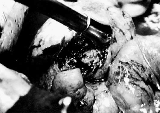
the detection of a hemoperitoneum associated with a ruptured ectopic pregnancy. We have found that most of the positive culdocenteses in our institution are associated with an unruptured ectopic pregnancy. In such cases, bleeding through the distal end of the fallopian tube results in a hemoperitoneum.

In our series, 86% of all positive culdocenteses performed in the emergency room were associated with an ectopic pregnancy.50 Other causes of positive culdocentesis included ruptured ovarian cysts, retrograde menstruation, endometriosis, torsion of the fallopian tube, and bleeding of unknown etiology. It should be emphasized that a nondiagnostic culdocentesis should not lower the suspicion of an ectopic pregnancy. In our series, 16% of ectopic pregnancies had nondiagnostic culdocentesis, and one quarter of these were ruptured at the time of operation.50

Patients who will benefit most from a culdocentesis are those in whom a clinical suspicion of ectopic pregnancy exists, and who present at a time when expeditious diagnosis is desired and when sophisticated diagnostic modalities, such as ultrasonography and sensitive human chorionic gonadotropin (hCG) assays, cannot be obtained without significant delay. Under these circumstances, culdocentesis is an inexpensive, rapid, and easily performed means of patient evaluation that often provides the impetus for immediate intervention.

In clinically stable patients, a culdocentesis may be indicated when fluid is visualized in the cul-de-sac at the time of ultrasound examination of the pelvis. A positive culdocentesis dictates the need for a surgical diagnosis. The procedure should not be overlooked in patients without signs of peritoneal irritation. We have found that 45% of our patients with a positive culdocentesis did not have rebound tenderness.50

The modern approach to the evaluation of clinically stable patients suspected of having an ectopic pregnancy is based on the combined use of sensitive pregnancy testing (or hCG testing), ultrasound examination, and laparoscopy. hCG testing is used to screen for pregnancy, and ultrasonography is employed to locate it.51

Evaluation of the patients begins with a sensitive and rapid pregnancy test. Blood and urine pregnancy tests have been used to screen for ectopic pregnancy. Traditional slide pregnancy tests have been found to have a 15% to 50% false-negative rate in ectopic pregnancy, and for this reason they are unsuitable for screening.52 Advances in technologies of enzyme-linked immunoassay and monoclonal antibodies to the β subunit have promoted the evolution of rapid, inexpensive, and similar qualitative urinary pregnancy tests. They are expected to improve the accuracy of the old urine pregnancy tests. Meanwhile, as the information for these tests becomes available, we prefer to use the sensitive blood pregnancy testing in the form of a radioimmunoassay against β-hCG.

We recommend an assay with a sensitivity of 2.5 mIU/mL because with this system the false-negative rate is 0.5%. The radioreceptor assay, with a sensitivity of 200 mIU/mL, is not suitable for screening because its false-negative rate ranges between 6% and 12%, which is unacceptably high for a life-threatening condition.53,54 Other investigators using a urine hCG test with a stated sensitivity of 50 IU/L support this test's clinical application in detecting ectopic pregnancy in emergent conditions.55

A negative blood pregnancy test virtually rules out pregnancy and thus an ectopic gestation. A positive result requires further investigation, requiring a determination of the hCG titer and an ultrasonographic examination of the pelvis.

A positive diagnosis of ectopic pregnancy can be made if fetal motion is demonstrated outside the uterus. Unfortunately, this is a rare and late finding, and awaiting its appearance would delay the diagnosis and conceivably increase the risk of tubal rupture. In practice, ultrasonography is used to identify an intrauterine pregnancy, which would render the simultaneous presence of an ectopic pregnancy extremely unlikely (1:30,000). Confirmation of the presence of an intrauterine pregnancy can be made by identifying either a gestational sac or a fetal pole within the endometrial cavity.

The traditional ultrasonographic criterion used for the diagnosis of ectopic pregnancy is failure to visualize a gestational sac in the uterus of patients with more than 6 weeks of amenorrhea. The problem with this criterion is that one third of patients with ectopic pregnancy do not know the date of their last menstrual period, and in others irregular bleeding makes interpretation of the menstrual history difficult.

It has been established that the sac of a normal intrauterine pregnancy becomes visible with ultrasonography when the hCG titer is greater than 6500 mIU/mL. When levels are higher than this, the absence of a sac is associated with an ectopic pregnancy in 86% of cases. This criterion has a sensitivity of 100%, a specificity of 96%, and a negative predictive value of 100%. The clinical application of this criterion includes the fact that approximately 40% of women with an ectopic pregnancy possess titers in excess of 6500 mIU/mL.50 The absence of a sac at levels less than 6000 mIU/mL is a nondiagnostic finding and should not lower or raise the suspicion of ectopic pregnancy.56,57 Management of these patients depends on the clinical situation. If a patient remains clinically stable, serial hCG determinations are used for the assessment of such a patient and for deciding whether to proceed to diagnostic laparoscopy. The presence of a sac below an hCG titer of 6000 mIU/mL can be explained if the pregnancy develops beyond the level of sac formation (above an hCG level of 6500 mIU/mL) and then stops secreting hCG. A sac will be detected by ultrasonography when the hCG titer has dropped below 6000 mIU/mL. Some normal intrauterine pregnancies produce lower levels of hCG and therefore may show a gestational sac at lower hCG concentrations. This may account for Nyberg and associates'58 detection of gestational sacs in 13 patients in whom the hCG titer was less than 6000 mIU/mL. In addition, this discrepancy may be confounded by the common use of two distinct hCG standards in the evaluation of ectopic pregnancy. In 1974, the World Health Organization introduced a new standard that involved the use of a more highly purified preparation of hCG and subsequently established the International Reference Preparation (IRP). A decade earlier, the second International Standard had been introduced, but because of heterogeneity, it was often unsuitable for use in many immunoassays.59 In comparing the two quantitative standards, clinicians should note that the serum hCG titers determined by the IRP approximately double the value calculated by the second International Standard. The hCG values stated in this review are based on the IRP standard unless otherwise specifically noted. Finally, ectopic pregnancies can show a single-ring sac due to the presence of blood within the endometrial cavity in association with a significant decidual reaction. This appearance has been shown to occur in 10% to 20% of all cases (Fig. 4).60,61,62,63,64,65,66,67,68

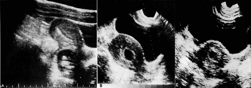
Fig. 4. A. Segmental scan of the pelvis in an ectopic pregnancy. A fetus is visualized in the cul-de-sac. B. Gestational sac of two normal pregnancies. There is a double ring given by the separation of the decidual capsularis and the decidual parietalis. C. Pseudogestational sac in an ectopic pregnancy. There is a single ring.

Nyberg and colleagues69 and Bradley and associates70 have proposed morphologic criteria to distinguish the pseudogestational sac of ectopic pregnancy from the gestational sac of a normal intrauterine pregnancy. They have described the normal intrauterine gestational sac as having a double contour produced by the decidua capsularis and the decidua parietalis. The pseudogestational sac of an ectopic pregnancy has only a single ring. The researchers have reported that 98.3% of all patients with a double-ring sac had an intrauterine pregnancy and 64 of 68 patients with a single-ring sac had ectopic pregnancies.

Management of patients with an hCG titer less than 6000 mIU/mL is based mainly on adnexal ultrasonographic findings and clinical suspicion. Romero and colleagues71 prospectively evaluated 220 patients who were suspected of having ectopic pregnancy and who had hCG titers less than 6000 mIU/mL and abdominal ultrasonographic adnexal findings. The demonstration of a noncystic mass, alone or associated with cul-de-sac fluid, was an indication for diagnostic laparoscopy.

The value of transvaginal ultrasonography in the detection of ectopic pregnancy, especially in cases of hCG titers less than 6000 mIU/mL, is now recognized. Proximity of the ultrasound transducer to both the adnexa and the cul-de-sac allows increased resolution and image quality with the detection of an intrauterine pregnancy up to 7 days earlier than 
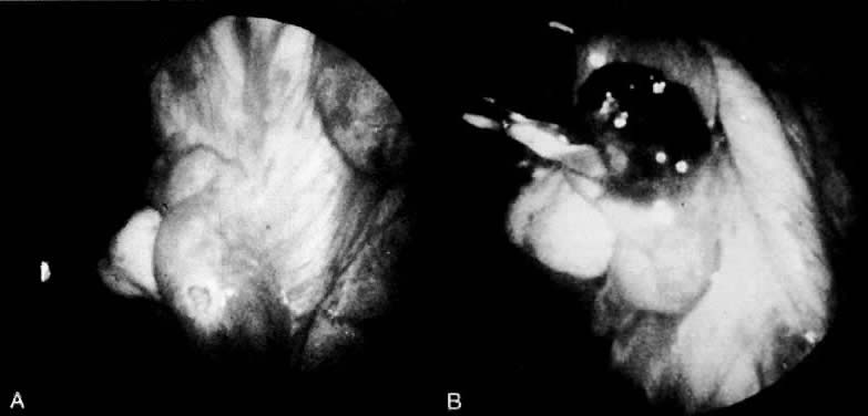
with the classic transabdominal approach (Fig. 5).

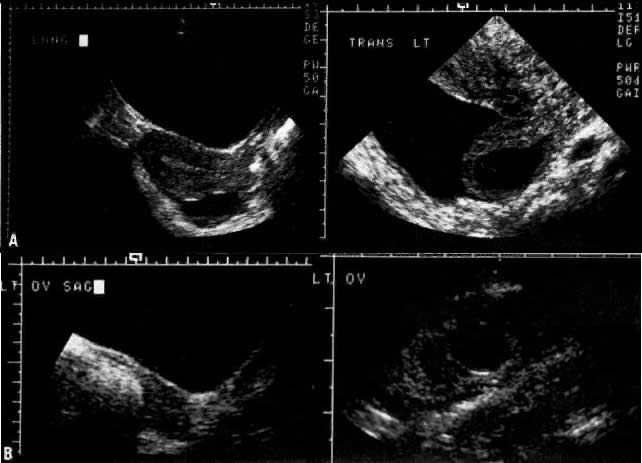
Fig. 5. The increased sensitivity of transvaginal (right) versus transabdominal (left) ultrasonography in detecting an ectopic pregnancy. A. Proximity to the cul-de-sac allowed detection of this ectopic gestation (hCG titer 880 mIU/mL) by means of transvaginal ultrasonography in this patient. B. Transvaginal ultrasound permitted verification of a left infundibular pregnancy missed on abdominal scanning in another woman (hCG titer 4146 mIU/mL)

Vaginal scanning has proved to be more accurate than abdominal scanning in detecting ectopic pregnancies (90% vs 80%) and cul-de-sac fluid (77% vs 46%) and in discerning whether the tubal pregnancy has ruptured (76% vs 50%).72 In the transvaginal ultrasonographic evaluation of pregnancy, Bernaschek and associates,73 using a 5-MHz transducer, proposed a “discriminatory zone” of an hCG titer of 750 mIU/mL (second International Standard) for the detection of an intrauterine gestational sac. Unfortunately, using this proposed criterion and similar ultrasonographic equipment, Fossum and associates74 may have inadvertently surgically investigated several pregnancies that proved to be normal intrauterine gestations (Fig. 6). If an hCG titer exceeds 2000 mIU/mL, we now expect to detect an intrauterine gestational sac using transvaginal ultrasonography.

Fig. 6. Relationship between transvaginal ultrasonographic findings and serum hCG levels (range and mean ± SEM) using two World Health Organization standards: the IRP and the second International Standard.(Fossum GT, Davajan V, Kletzky OA: Early detection of pregnancy with transvaginal ultrasound. Fertil Steril 49:788, 1988. Reprinted with permission of the publisher, The American Fertility Society)

Even with the advent of transvaginal ultrasonography, the value of serial hCG determinations cannot be overemphasized. If hCG titers increase with time, Table 2 can be used to evaluate the normality of the increase. As a rule of thumb, the hCG titer should increase by at least 66% of the initial titer in a 48-hour period of observation.75 On the other hand, in asymptomatic women, declining hCG levels may be indicative of a nonviable, nondetectable intrauterine gestation. Serum progesterone values may prove a useful adjunct to hCG titers in distinguishing a viable intrauterine pregnancy from that of an ectopic or missed abortion. Accordingly, in two separate reports on 99 women, a progesterone value less than 15 ng/mL was always predictive either of an ectopic pregnancy or a nonviable intrauterine gestation.76,77

TABLE 2. Lower Limits of Percentage Increase of Serum hCG


Sampling Interval (Days)

Increase in hCG (%)

1

29

2

66

3

114

4

175

5

225

(Kadar N, Caldwell B, Romero R, A method of screening for ectopic pregnancy and its indications. Obstet Gynecol 58:162, 1980)

It should be emphasized that early intrauterine pregnancy failures may not have enough tissue to be detectable by routine pathologic examination, and therefore some patients with declining hCG titers will ultimately prove to have intrauterine pregnancy failures.

We recommend laparoscopy for patients with subnormal quantitative increases of hCG. Normal increments in hCG are monitored until hCG titers exceed either 2000 mIU/mL or 6500 mIU/mL, at which time either transvaginal or transabdominal ultrasonography, respectively, may be performed. The presence or absence of a gestational sac above these hCG levels should be supportive either of an ectopic or an intrauterine gestation. Finally, if hCG titers plateau, we perform a diagnostic laparoscopy because the pattern is often characteristic of an ectopic pregnancy (Fig. 7).

Fig. 7. An algorithm for the management of an ectopic pregnancy.

Back to Top
SURGICAL MANAGEMENT

The ultimate decision regarding surgical management depends on a patient's desire for future fertility. If a patient is not interested in future fertility, the appropriate surgical procedure is salpingectomy. If a patient does desire future fertility, however, much data from the past few years support performing conservative surgery in a majority of these cases. One must always keep in mind and also inform the patient, though, that whatever procedure is performed, the pregnancy rate after an ectopic pregnancy is decreased by 40% to 70%.46 Conservative options range from expression of a tubal abortion through the distal end of the tube to segmental resection and secondary anastomosis of an isthmic ectopic pregnancy.

Salpingectomy Versus Salpingo-oophorectomy

A majority of ectopic pregnancies are associated with contralateral tubal disease, such as salpingitis. Proper evaluation of the contralateral tube is imperative before embarking on a surgical procedure. The fact that clinical assessment and evaluation of the contralateral tube during the initial surgery correlates poorly with postoperative hysterosalpingographic findings underscores the importance of intraoperative conservative management whenever feasible.78 It is not recommended that a test of tubal patency be done during the operation for an ectopic pregnancy in order to evaluate the status of the contralateral tube. The information provided can be erroneous because decidual reaction around the cornual portion might give a false impression of an occluded tube. The possibility of introducing infection into an already compromised pelvis is also an important consideration.

In 1955, Jeffcoate79 suggested that in conjunction with a salpingectomy, which is still the most commonly employed surgical treatment for ectopic pregnancy, an oophorectomy on the ipsilateral side be done as well. The theory behind this is that all ovulations would be into the good tube; this discounts the importance of transmigration. There are no data that justify this conjecture. Several reports have shown that there is no advantage in performing a salpingo-oophorectomy compared with salpingectomy in terms of pregnancy rates and repeat ectopic pregnancies in those patients.80 Considering the successful pregnancy rates with in vitro fertilization and embryo transfer, every effort must be maintained to conserve as much ovarian tissue as possible in any eventuality.

Salpingectomy Versus Salpingostomy (Conservative Surgery)

In a stable patient who desires future childbearing, a salpingostomy (conservative surgery) is the treatment of choice. The controversy surrounding salpingectomy versus salpingostomy was based on two assumptions. The rationale for salpingostomy lay in the fact that the contralateral tube was abnormal in more than one half of the women treated for ectopic pregnancy. Those in support of salpingectomy feared (1) that patients undergoing salpingostomy would be more likely to have a repeat ectopic pregnancy, and (2) that salpingostomy is a tedious surgical procedure because tubal hemorrhage during surgery is difficult to control. Neither of these assumptions has proved correct, and in fact, intrauterine pregnancy rates have actually increased with the use of conservative surgical techniques (Table 3),81 primarily as a result of the application of modern microsurgical techniques to acute tubal pathology. These results represent the same change in outcome noted in the surgical management of chronic tubal disease when these modalities are properly employed.

TABLE 3. Choice of Surgical Treatment and Subsequent Fertility among 151 Patients with Ectopic Pregnancy


No. of Subsquent Pregnancies


(% in parentheses)

Incidence of Sterility

Surgical Treatment

No. of Patients

Intrauterine

Repeat Ectopic

(% in parentheses)

Conservative

47

39 (83)

3 (6.4)

175

Radical

104

75 (72.1)

6 (5.8)

225

(Sherman D, Lunger R, Sadovsky G: Improved fertility following ectopic pregnancy. Fertil Steril 37:497, 1982. Reproduced with permission of the publisher, The American Fertility Society)

Timonen and Nieminen82 reported a 21% repeat ectopic pregnancy rate after conservative surgery versus a 12.5% repeat rate after salpingectomy. Since this initial report, however, a number of studies have demonstrated a lower repeat ectopic pregnancy rate in conservatively operated patients.83,84,85 There are no substantial data to support the role of cornual resection in conjunction with a salpingectomy. The theoretic rationale is to prevent interstitial implantation. A review of the literature by Kalchman and Meltzer86 demonstrated only 75 cases of interstitial pregnancy following a salpingectomy, leading to the conclusion that this is a rare occurrence and should not dictate surgical technique. Hallatt has suggested that the procedure has some inherent risks secondary to increased bleeding at the time of the original surgery and the possibility of a rupture in the course of a subsequent intrauterine pregnancy.80 Uterotubal fistula formation after this procedure is also a possibility. Therefore, for aesthetic reasons, only a small, shallow cornual resection should be performed.

Salpingostomy Versus Salpingotomy

The primary difference between a salpingotomy and a salpingostomy is that, in the former, the fallopian tube is closed by primary intention; in the latter, the tube is allowed to close by secondary intention after hemostasis is achieved. Stromme87 was the first to describe salpingotomy. This procedure involves an antimesenteric incision over the ectopic pregnancy, excising the products of conception and closing the tube in either one or two layers with fine suture material after hemostasis is achieved. The sutures used should be interrupted. Other reports have modified this technique but still espouse primary closure of the tube.81,84 Anecdotal reports of tuboperitoneal fistulas subsequent to salpingostomy, but not salpingotomy, support performance of the latter procedure.88

A salpingostomy can be performed based on the evidence describing the propagation of ampullary ectopic pregnancies outside of the lumen itself in the adventitious space between the serosa and the tubal epithelium. Salpingostomy may be associated also with an increased incidence (43%) of recurrent ectopic pregnancy, decreased chances of complete trophoblast removal, and delayed postoperative hemorrhage.89,90,91,92

Salpingostomy seems to have no adverse effect on a tube's anatomic and physiologic future. This is a simpler procedure that can be used by all gynecologic surgeons. In 1951, Israel93 performed total linear salpingostomies in eight patients for PID. The procedures were done between 1935 and 1949. Seven of the eight were done for gonorrheal salpingitis and one for tuberculous salpingitis. Five of the eight patients went on to conceive. The procedure was then tried on six dogs. The dogs were operated on at 2-, 4-, and 8-week intervals. At 2 weeks, the raw edges were separated from each other and epithelialized. At 4 weeks, the gaping incision was beginning to be bridged by fibrous tissue, and at 8 weeks the organs appeared totally healed, with “fusion of the tunica propria as well as a conjunction of the circular muscle layer. Its salient feature is the absence of a visible scar.”93

The incision for a salpingostomy is made on the antimesenteric border of the tube over the area of maximal bulge; as small an incision as possible is made, but a surgeon must make sure that the products of conception are removed entirely. It is also important not to debride the base or to treat the base too vigorously to achieve hemostasis. If too much dissection is done at the implantation site, bleeding will ensue that is difficult to stop, and if suture material or cautery is used extensively in this area, nothing has been gained by the conservative treatment. Blood testing demonstrates the disappearance of β-hCG levels over time (Fig. 8.)94 There seems to be no quantitative difference between use of a laser, scalpel, or electrocautery to make the incision. These incisions in the tube seem to heal well, as documented in follow-up surgery and hysterosalpingogram. Hemostasis is critical. Preincisional serosal surface injection of vasopressive agents often reduces incisional bleeding, but may distort the area of maximal tubal bulge. If bleeding continues, vessels in the broad ligament can be ligated with fine suture material (4-0), but remember that this ligation devascularizes the tube and may cause some ischemia in the tube that may lead to decreased reproductive function and adhesion formation.95

Fig. 8. Disappearance curve of hCG in patients treated by salpingectomy versus conservative surgical treatment for ectopic pregnancy. This similarity demonstrates the clearance of the hormone rather than the persistence of viable trophoblastic tissue.(Kamrava MM, Taymor ML, Berger MJ et al: Disappearance of human chorionic gonadotropin following removal of ectopic pregnancy. Obstet Gynecol 62:484, 1983)

A number of these patients have been observed postoperatively, and there has been no evidence of stenosis or fistula formation.

Back to Top
ISTHMIC AND INFUNDIBULAR ECTOPIC PREGNANCY AND TUBAL ABORTIONS

Ectopic pregnancies that occur in the isthmic portion of the tube present a different entity entirely. A recent histopathologic comparison of ampullary and isthmic ectopic pregnancies noted preservation of the ampullary muscularis in 85% of the former cases and only a 43% preservation rate of the muscularis in isthmic pregnancies. The disruption of the tubal wall was also more extensive in isthmic pregnancies.96 This is why patients with isthmic ectopic pregnancies are believed to be the only ectopic pregnancy patients who develop a tuboperitoneal fistula after linear salpingostomy (Fig. 9). Therefore, segmental resection is most commonly recommended.97

Fig. 9. Total peritoneal fistula following salpingostomy for an isthmic ectopic pregnancy.

The option then becomes to close the tube by primary intention, which has been done successfully by Stangel and colleagues,98 or to perform a second procedure and do an anastomosis at that time. The major advantage of doing an anastomosis at the time of resection of the ectopic pregnancy is that the wide lumen of the tube facilitates the anastomosis. However, edema and the presence of blood may increase the chance of infection. Normal intrauterine pregnancies are achieved regardless of the mode of anastomosis employed. It is recommended that patients waiting for a secondary anastomosis be placed on birth control pills so that they do not form an ectopic pregnancy in the blind distal portion of the tube that has been created (Fig. 10).99 In either event, the anastomosis is performed in two layers using the operating microscope. Four to six 8-0 polyglycolic acid or polydioxanone sutures are placed approximating the muscularis. The serosa is then loosely opposed with running 6-0 Vicryl or PDS sutures (Fig. 11).

Fig. 10. Transmigration of sperm to the contralateral horn of the tube, resulting in an ectopic pregnancy.(Cartwright PS, Entman SS: Repeat ipsilateral tubal pregnancy following partial salpingectomy: A case report. Fertil Steril 42:647, 1984. Reproduced with permission of the publisher, The American Fertility Society)

Fig. 11. Isthmic anastomosis following segmental resection for an ectopic pregnancy.

With the move toward laparoscopic conservative surgery (see later), it is of interest to note Smith and colleagues'100 similar postoperative tubal patency rates when microsurgical anastomosis was compared with linear salpingostomy in the treatment of isthmic ectopic gestation at the time of laparotomy. Pouly and associates101 accordingly treated 22 isthmic ectopic pregnancies by laparoscopic linear salpingostomy, yielding a 54.5% subsequent intrauterine pregnancy rate. DeCherney and Boyers,102 however, showed that treating isthmic pregnancy with linear salpingostomy by laparotomy resulted in subsequent occlusion of the tube in three of four patients.

A rare form of ectopic pregnancy occurs in the infundibular portion of the tube between the fimbria and the ampulla. These are usually treated by “milking or squeezing” the pregnancy out through the distal end. Schermers48 reported a higher incidence of delayed hemorrhage after the milking-out procedure. This experience has been substantiated by Bruhat.103 Success rates are also lowest with this procedure; therefore, these patients also should have a linear salpingostomy rather than “milking out.”

This entity is different from a tubal abortion, in which the products of conception are already partially or completely extruded from the fimbriated end of the fallopian tube. This process can be completed, if necessary, simply by removing the remaining products of conception.

Back to Top
LAPAROSCOPIC TREATMENT OF ECTOPIC PREGNANCY

Salpingostomy

A number of early studies documented the appropriateness of laparoscopic treatment of ectopic pregnancies.103,104,105 Rates of conception of an intrauterine pregnancy after the procedure were as high as 70% in these cases. Pouly and associates101 reported on 321 women with ectopic pregnancies who underwent conservative laparoscopic treatment. Of the women who did not have a history of infertility or a previous ectopic pregnancy, 86% had a subsequent intrauterine pregnancy.

The advantages of laparoscopic removal of an ectopic pregnancy are a shortened operating time, convalescence, and hospital stay. It is imperative, however, that proper case selection be exercised.106 If laparoscopic therapy is to be warranted, the first criterion is the expertise of the operator in performance of a laparoscopic surgical procedure. Patients must be stable without evidence of a significant hematoperitoneum. Optimally, the ectopic pregnancy should be confined to the ampullary portion of the tube and should not be greater than 3 cm in size. The tube must be easily accessible.

The operative technique is precisely the same as for a linear salpingostomy done at laparotomy. The incision is made on the antimesenteric side of the fallopian tube using electrocautery or a laser, and the products of conception are removed with duckbill forceps (Fig. 12).105 No studies assessing clinical utility of electrocautery versus any of the laser modalities for the salpingostomy incision have yet been conducted. Pregnancy rates and repeat ectopic pregnancy rates are the same as those for a linear salpingostomy performed at the time of laparotomy for an ampullary ectopic pregnancy.103,105

Fig. 12. Laparoscopic removal of an ectopic pregnancy. A. Initial salpingostomy incision was made with the argon laser. B. Expulsion of products of conception after the incision is made.

Interestingly, laparoscopic intratubal and corpus luteal injections of prostaglandin F have proved quite effective in the treatment of ectopic pregnancy.107

Salpingectomy

The option to remove the fallopian tube during laparoscopy allows a radical surgical procedure to be performed in a conservative manner. In addition, tubal salvage often is not feasible or recommended in cases of repeat or severe rupture of the tube, and also in cases of failed salpingostomy. Laparoscopic salpingectomy, partial or complete, offers many advantages over laparotomy. Although snare electrocautery initially was used in the procedure, forceps electrocautery and the use of pre-tied 0 chromic catgut (Endoloop, Ethicon Corp., Sommerville, NJ) suturing facilitate performance of the procedure.108 This is demonstrated best in a report of 100 women treated accordingly; only 3 required laparotomy to complete the salpingectomy.109

Back to Top
ADJUNCTIVE THERAPY

The therapy used is the same as with reproductive surgery for chronic tubal disease. It includes prophylactic antibiotics, such as doxycycline 100 mg twice a day, and 200 mL of Hyskon hysteroscopy fluid (32% w/v dextran 70 in dextrose) instilled into the abdominal cavity either through the laparoscopic port or at the time the laparotomy incision is closed to prevent adhesion formation. Rh-negative patients should receive Rh0 (D) immune globulin (RhoGAM).110

Postoperatively, patients should be followed with a hysterosalpingogram 4 months after surgery and laparoscopy 1 year after surgery if pregnancy has not occurred.

Back to Top
REPRODUCTIVE PERFORMANCE AFTER CONSERVATIVE SURGERY FOR A TUBAL ECTOPIC PREGNANCY

Conservative treatment can be carried out without lowering the chance of a subsequent intrauterine pregnancy or raising the incidence of an extrauterine pregnancy. A review of the literature summarizing 467 cases operated on for ectopic pregnancy is summarized in Table 4.111 Of these patients, 46% had a subsequent intrauterine pregnancy and 12% had a repeat ectopic pregnancy. These results do not differ greatly from older statistics based on the results after radical surgery. DeCherney and Oelsner111 reviewed 1630 cases treated radically and found a 41% intrauterine pregnancy rate and a 14% repeat extrauterine pregnancy rate. It is hoped that over time, with the use of modern microsurgical techniques, the intrauterine pregnancy rate will increase and the repeat ectopic pregnancy rate will decrease in these patients.

TABLE 4. Results of Radical and Conservative Surgery for Extrauterine Pregnancy


Women

Women with

Women with


Desiring

Intrauterine

Extrauterine


Pregnancy

Pregnancy

Pregnancy

Radical sugery

1630

667 (41%)

231 (14%)

Conservative surgery

467

215 (46%)

54 (12%)

(Adapted from DeCherney AH, Oelsner G: Ectopic pregnancy. In Garcia C-R, Mikuta JJ, Rosenblum NG (eds): Current Therapy in Surgical Gynecology. Toronto, BC Decker, 1986)

Based on the similar findings in radically and conservatively treated patients, an important question is whether salpingostomy works at all. This begs the question that perhaps those patients who conceive are using the tube that was not operated on. This possibility has been reviewed, and the intrauterine pregnancy rate and the repeat ectopic pregnancy rate seem to be the same in patients who had salpingostomies performed on only one fallopian tube with an ectopic pregnancy for both the group receiving radical treatment and the group receiving conservative treatment.46

These statistics do not hold, however, for patients who have had two or more ectopic pregnancies. In these patients, the incidence of repeat ectopic pregnancy approximates that of a subsequent intrauterine gestation.112,113 An unanswered question at this point remains: How many conservative procedures for an ectopic pregnancy can a patient undergo before her reproductive future is compromised to the point where in vitro fertilization is the only viable alternative (Table 5)?112,113

TABLE 5. Reproductive Outcome of 47 Patients After Two Ectopic Pregnancies


Number of Patients (% in parentheses)

Outcome

DeCherney et al N = 23

Tulandi N = 24

Sterilized (unable to conceive)

6 (26.1)

4 (16.7)

At least one tube present

17 (73.9)

20 (83.3)

Using contraception

4 (23.5)

4 (20)

Trying to conceive

13 (76.5)

16 (80)

Intrauterine pregnancy

4 (30.8)

8 (50.0)

 Term

4 (30.8)

5 (31.3)

 Spontaneous abortion

0 (0.0)

3 (18.8)

Third ectopic gestation

1 (20.0)

3 (27.3)

 (% of those trying to conceive)

(7.7)

(18.8)

Not able to conceive

8 (61.5)

5 (31.3)

(Adapted from DeCherney AT, Silidker JS, Mezer HC et al: Reproductive outcome following two ectopic pregnancies. Fertil Steril 43:82, 1985; Tulandi T: Reproductive performance of women after two ectopic pregnancies. Fertil Steril 50:164, 1988, Reproduced with permission of the publisher, The American Fertility Society)
Back to Top
NONOPERATIVE MANAGEMENT

Nonoperative management can be divided into observation/expectant care and medical therapy. As early as 1955, Lund114 reported on a series of 119 women with ectopic pregnancies treated with observation. In this series, 55% of the cases resolved without intervention; the remainder required laparotomy. More recently, several investigators have reported their experience with expectant management of unruptured ectopic pregnancy. In Maymon's115 review of 8 series of patients (total of 81 patients) managed expectantly, 76% resolved spontaneously, 23% required laparotomy for rupture, and 79% demonstrated ipsilateral tubal patency on follow-up hysterosalpingogram. The subsequent pregnancy rate, however, was only 16%. In all these studies, patients were rigorously selected, with documented unruptured ectopic pregnancies at laparoscopy, with no active bleeding. The only advantage seen in these reports was the avoidance of surgery. Disadvantages included an increased risk of tubal rupture, a possible increase in pelvic adhesions, and a protracted course of illness.

At this time, expectant management should be considered an option only for patients with extreme surgical risk, falling hCG titers, or in a research setting.

Back to Top
MEDICAL THERAPY FOR ECTOPIC PREGNANCY

The use of drug therapy for ectopic pregnancy was first reported in 1982, in a patient with an interstitial pregnancy who refused surgery.116 Since then, many investigators have reported the successful treatment of selected patients using a variety of agents, including parenteral methotrexate, intratubal or intra-amniotic methotrexate, and intratubal osmotic agents.117,118,119,120,121 To date, there have not been a significant number of reports of successful treatment of ectopic pregnancy using mefipristone (RU-486).

At this time, parenteral methotrexate seems to be the best studied and most accepted agent for the medical treatment of ectopic pregnancy. In 1993, Stovall117 reported a prospective study of 120 women treated with a single dose (50 mg/m2) of intramuscular methotrexate. In this series, the author reported a success rate of 94%, with subsequent tubal patency rates similar to those quoted for conservative surgical treatment. Pregnancy rates were nearly 80%, with approximately 88% of these being intrauterine. There were no adverse effects of methotrexate therapy reported in this series; however, there have been recent reports of tubal rupture and hematosalpinx after methotrexate therapy.122,123 Overall, the outcome of medical treatment of ectopic pregnancy closely approximates results obtained surgically, and there is evidence to support a significant improvement in cost-effectiveness.124

In summary, although surgical therapy will for now remain the mainstay of treatment for ectopic pregnancy, current evidence would support a shift toward treating carefully selected patients medically, using the single-dose methotrexate regimen reported by Stovall.

Back to Top
CONCLUSION

During the past decade, the incidence of ectopic pregnancy has noticeably increased. Although 50% of ectopic pregnancies are attributable to infection, the remainder remain unexplained. Clinical findings such as adnexal tenderness, irregular bleeding, and abdominal pain still represent reliable but imperfect clues that a patient has an ectopic pregnancy. It is the advent of newer diagnostic techniques, including laparoscopy, serial β-hCG testing, and ultrasonography, that has allowed for the earlier diagnosis of ectopic pregnancy. Previously, 85% of ectopic pregnancies were diagnosed as ruptured and 15% as unruptured.125 Today, this ratio is reversed. The diagnosis of many cases of small, unruptured ectopic pregnancies has led to a revival of conservative reconstructive surgery for tubal ectopic pregnancy. This trend has been based, in part, on success with reconstructive surgery and microsurgical technique in chronic tubal disease. The hallmark of improved diagnostic management remains the β-hCG test, with failure to achieve a normal slope of increase during the early stages of gestation heralding an ectopic pregnancy or an inevitable abortion.126

Back to Top
REFERENCES

1. Tait RL: Five cases of extrauterine pregnancy operated upon at the time of pregnancy. Br Med J 1: 1250, 1884

2. Centers of Disease Control: Current trends: Ectopic pregnancies-United States, 1979 - 1980. MMWR 33:201, 1984

3. Westrom L, Bengtsson LPH, Mardh PA: Incidence trends, and risks of ectopic pregnancy in a population of women. Br Med J (Clin Res) 282: 15, 1981

4. Chung CS, Smith RG, Steinhoff PG et al: Induced abortion and ectopic pregnancy in subsequent pregnancies. Am J Epidemiol 115: 879, 1982

5. Majumdar BH, Henderson PH, Semple L: Salpingitis isthmica nodosa: A high-risk factor for tubal pregnancy. Obstet Gynecol 62: 73, 1983

6. Wolf GC, Thompson NJ: Female sterilization and subsequent ectopic pregnancy. Obstet Gynecol 55: 17, 1980

7. Lehfeld H, Tietze C, Gorstein F: Ovarian pregnancy and the intrauterine device. Am J Obstet Gynecol 108: 1005, 1970

8. Vessey MP, Johnson B, Doll R et al: Outcome of pregnancy in women using an intrauterine device. Lancet 1: 495, 1974

9. Tatum JH, Schmidt FM, Jain AK: Management and outcome of pregnancies associated with the copper T intrauterine contraceptive device. Am J Obstet Gynecol 126: 869, 1976

10. Sivin I: Copper 1 IUD use and ectopic pregnancy rates in the United States. Contraception 19: 151, 1979

11. Ory HW: Ectopic pregnancy and intrauterine contraceptive devices: New perspectives. Obstet Gynecol 57: 137, 1981

12. Urquhart J: Effect of the venereal disease epidemic on the incidence of ectopic pregnancy: Implications for the evaluation of contraceptives. Contraception 19: 455, 1979

13. Bonnar J: Progesterone-only contraception and tubal pregnancies. Br Med J 1: 287, 1974

14. Tietze C: Extrauterine pregnancy and intrauterine devices. Br Med J 2: 302, 1966

15. Snowden RF: The Progestasert and ectopic pregnancy. Br Med J 4: 1600, 1977

16. Diaz S, Croxatto HB, Pavez M et al: Ectopic pregnancy associated with low dose progesterone-releasing IUD's. Contraception 22: 259, 1980

17. Panayotou PP, Kaskarelis DB, Mietinnen OS et al: Induced abortion and ectopic pregnancy. Am J Obstet Gynecol 114: 507, 1972

18. DeCherney A, Minkin MJ, Spangler S: Contemporary management of ectopic pregnancy. J Reprod Med 26: 519, 1981

19. Douglas ES, Singleton HM, Crist T: Surgical management of tubal pregnancy: Effect on subsequent fertility. South Med J 62: 954, 1969

20. Lawson HW, Atrash HK, Saftlas AF et al: Ectopic pregnancy surveillance, United States 1970 - 1985. MMWR 37: 9, 1988

21. Dorfman SF: Deaths from ectopic pregnancy. United States, 1979 to 1980. Obstet Gynecol 64: 386, 1984

22. Dorfman SF: Ectopic pregnancy mortality, United States, 1979 to 1980: Clinical aspects. Obstet Gynecol 64: 386, 1984

23. Siegler AM: Salpingotomy and salpingectomy: indications and techniques for tubal pregnancy. In Sciarra JJ (ed): Gynecology and Obstetrics. Philadelphia, JB Lippincott, 1991

24. Hallatt JG: Ectopic pregnancy associated with the intrauterine device: A study of seventy cases. Am J Obstet Gynecol 106: 227, 1976

25. Fortier KJ, Haney AF: The pathologic spectrum of uterotubal junction obstruction. Obstet Gynecol 65: 93, 1985

26. Elias S, LeBeau M, Simpson JL et al: Chromosome analysis of ectopic human conceptuses. Am J Obstet Gynecol 141: 698, 1981

27. Fedele L, Acaia B, Parazzini F et al: Ectopic pregnancy and recurrent spontaneous abortion: Two associated reproductive failures. Obstet Gynecol 73: 206, 1989

28. Gemzell G, Guillome J, Wang CF: Ectopic pregnancy following treatment with human gonadotropins. Am J Obstet Gynecol 143: 761, 1982

29. McBain JC, Evans JH, Pepperell RJ et al: An unexpectedly high rate of ectopic pregnancy following the induction of ovulation with human pituitary and chorionic gonadotrophin. Br J Obstet Gynaecol 87: 5, 1980

30. James WH: A hypothesis on the increasing rates of ectopic pregnancy. Paediatr Perinat Epidemiol 3: 189, 1989

31. Korenga M, Kadota T: Changes in mechanical properties of the circular muscle of the isthmus of the human fallopian tube in relation to hormonal domination and post-ovulatory time. Fertil Steril 36: 343, 1981

32. Laufer N, Tarlatzis BC, DeCherney AH et al: Steroid secretion by human oocyte-corona-cumulus complexes associated with conceptions following in vitro fertilization. J In Vitro Fert Embryo Transfer 2: 156, 1985

33. Dor J, Radak E, Mashiach S et al: Unilateral tubal twin pregnancy following in vitro fertilization and embryo transfer. Fertil Steril 42: 297, 1984

34. Steptoe PC, Edwards RG: Reimplantation of the human embryo with subsequent tubal pregnancy. Lancet 1: 880, 1976

35. Correy JF, Watkins RA, Bradfield GF et al: Spontaneous pregnancies and pregnancies as a result of treatment on an in vitro fertilization program terminating in ectopic pregnancies or spontaneous abortions. Fertil Steril 50: 85, 1988

36. Olive DL, Taylor N, Cothran GE et al: Gamete intrafallopian transfer (GIFT) complicated by bilateral ectopic pregnancy. Fertil Steril 49: 719, 1988

37. Maheux R, DeCherney AH: Surgical management of tubal pregnancy. In DeCherney AH, Polan ML (eds): Seminars in Reproductive Endocrinology, Vol 2, pp 175 - 185. New York, Thieme-Stratton, 1984

38. Winer AE, Bergman WD, Fields C: Combined intra- and extrauterine pregnancy. Am J Obstet Gynecol 74: 1701, 1957

39. Shah Y, Zevallos H, Moody L: Combined intra- and extrauterine pregnancy. J Reprod Med 25: 290, 1980

40. Richards SR, Stempel LE, Carlton BD: Heterotopic pregnancy: Reappraisal of incidence. Am J Obstet Gynecol 142: 928, 1982

41. Hann LE, Bachman DM, McArdle CR: Coexistent intrauterine and ectopic pregnancy: A re-evaluation. Radiology 152: 151, 1984

42. Parenta JT, Ou CS, Levy J et al: Cervical pregnancy analysis: A review and report of five cases. Obstet Gynecol 62: 79, 1983

43. Bobrow ML, Winelstein LB: Intrafollicular ovarian pregnancy. Am J Surg 91: 991, 1956

44. Hallatt JG: Primary ovarian pregnancy: A report of twenty-five cases. Am J Obstet Gynecol 143: 55, 1982

45. Groycol Ruffolo EH: Ovarian pregnancy associated with intrauterine contraceptive devices. Am J Obstet Gynecol 132: 134, 1978

46. DeChemey AH, Maheux R: Modern management of tubal pregnancy. Am Prob Obstet Gynecol 6: 1, 1983

47. Budowick M, Johnson TR Jr, Genadry R et al: The histopathology of the developing tubal ectopic pregnancy. Fertil Steril 34: 169, 1980

48. Schermers JA: Ectopic Pregnancy: A Morphological and Endocrine Study. Amsterdam, Boek-en Offsetdrukkerij Los-Naarden, 1984

49. Kallenberger DA, Ronk DA, Jimerson GK: Ectopic pregnancy: A 15-year review of 160 cases. South Med J 71: 758, 1978

50. Romero R, Copel JA, Kadar N et al: Value of culdocentesis in the diagnosis of ectopic pregnancy. Obstet Gynecol 65: 519, 1985

51. Romero R, Taylor KW Jr, Kadar N et al: The diagnosis of ectopic pregnancy. In Steril WB, Cochrine WJ (eds): Clinics in Diagnostic Ultrasound: Gynecologic Ultrasound, p 123. New York, Churchill-Livingstone, 1984

52. Glass RH, Iesurum HM: Immunologic pregnancy test in ectopic pregnancy. Obstet Gynecol 27: 66, 1966

53. Berry CM, Thompson JD, Hatcher R: The radioreceptor assay for hCG in ectopic pregnancy. Obstet Gynecol 54: 43, 1973

54. Romero R, Kadar N, Copel JA et al: The value of serial human chorionic gonadotropin testing as a diagnostic tool in ectopic pregnancy. Am J Obstet Gynecol 155: 392, 1986

55. Emancipator K, Cadoff EM, Burke MD: Analytical versus clinical sensitivity and specificity in pregnancy testing. Am J Obstet Gynecol 158: 613, 1988

56. Kadar N, DeVore G, Romero R: The discriminatory hCG zone: Its use in sonographic evaluation for ectopic pregnancy. Obstet Gynecol 50: 156, 1980

57. Romero R, Kadar N, Jeaty P et al: The value of the discriminatory hCG zone in the diagnosis of ectopic pregnancy. Obstet Gynecol 66: 357, 1985

58. Nyberg DA, Filly RA, Mahony BS et al: Early gestation: Correlation of hCG levels and sonographic identification. AJR 144: 951, 1985

59. Bangham DR, Storring PL: Standardisation of human chorionic gonadotrophin, hCG subunits, and pregnancy tests. Lancet 1: 8268, 1982

60. Kadar N, Taylor KW Jr, Rosenfield A et al: The sonographic appearance of the uterus in ectopic pregnancy. Am J Roentgenol 141: 609, 1980

61. Mueller CE: Intrauterine pseudogestational sac in ectopic pregnancy. J Clin Ultrasound 7: 133, 1979

62. Spirt BA, O'Hare KR, Gordon L: Pseudogestational sac in ectopic pregnancy: Sonographic and pathologic correlation. J Clin Ultrasound 9: 338, 1981

63. Weiner CP: The pseudogestational sac in ectopic pregnancy. Am J Obstet Gynecol 1139: 959, 1981

64. Mantoni M, Pedersen JF: Massive pseudogestational sac in ectopic pregnancy. J Clin Ultrasound 11: 29, 1983

65. Abramovici H, Auslender R, Lewin A et al: Gestational pseudogestational sac: A new ultrasonic criterion for differential diagnosis. Am J Obstet Gynecol 145: 377, 1983

66. Marks WM, Filly RA, Callen PW et al: The decidual case of ectopic pregnancy: A confusing ultrasonographic appearance. Radiology 133: 451, 1979

67. O'Neill AGB, Hammond JG, Reid SE: Problems in the diagnosis of ectopic pregnancy: The pseudogestational sac. Aust NZ J Obstet Gynaecol 22: 94, 1982

68. Dunne MG: Shed decidual cast simulating an intrauterine fetus: Case reports. Am J Roentgenol 139: 591, 1982

69. Nyberg DA, Laing FC, Filly et al: Ultrasonographic differentiation of the gestation sac of early intrauterine pregnancy from the pseudogestational sac of early intrauterine pregnancy from the pseudogestational sac of ectopic pregnancy. Radiology 146: 755, 1983

70. Bradley EG, Fiske CE, Filly RA: The double ring sac sign of early intrauterine pregnancy: Use in exclusion of ectopic pregnancy. Radiology 143: 223, 1982

71. Romero R, Kadar N, Castro D et al: The value of adnexal sonographic findings in the diagnosis of ectopic pregnancy. Am J Obstet Gynecol 158: 52, 1988

72. Cacciatore B, Stenman UH, Ylostalo P: Comparison of abdominal and vaginal sonography in suspected ectopic pregnancy. Obstet Gynecol 73: 770, 1989

73. Bernaschek G, Rudelstorfer R, Csaicsich P: Vaginal sonography versus serum human chorionic gonadotrophin in early detection of pregnancy. Am J Obstet Gynecol 158: 608, 1988

74. Fossum GT, Davajan V, Kletzky OA: Early detection of pregnancy with transvaginal ultrasound. Fertil Steril 49: 788, 1988

75. Kadar N, Caldwell B, Romero R: A method of screening for ectopic pregnancy and its indications. Obstet Gynecol 58: 162, 1980

76. Yeko TR, Gorrill MJ, Hughes LH et al: Timely diagnosis of early ectopic pregnancy using a single blood progesterone measurement. Fertil Steril 48: 1048, 1987

77. Matthew CP, Coulson PB, Wild RA: Serum progesterone levels as an aid in the diagnosis of ectopic pregnancy. Obstet Gynecol 68: 390, 1986

78. Mitchell DE, McSwain HF, McCarthy JA et al: Hysterosalpingographic evaluation of tubal patency after ectopic pregnancy. Am J Obstet Gynecol 157: 618, 1987

79. Jeffcoate TNA: Salpingectomy or salpingo-oophorectomy. Br J Obstet Gynaecol 135: 74, 1955

80. Hallatt JG: Repeat ectopic pregnancy: A study of 123 consecutive cases. Am J Obstet Gynecol 122: 520, 1975

81. Sherman D, Langer R, Sadovsky G: Improved fertility following ectopic pregnancy. Fertil Steril 37: 497, 1982

82. Timonen S, Nieminen U: Tubal pregnancy, choice of operative method of treatment. Acta Obstet Gynecol Scand 46: 327, 1967

83. DeCherney AH, Kase N: The conservative surgical management of unruptured ectopic pregnancy. Obstet Gynecol 54: 451, 1979

84. Langer R, Bukovsky I, Herman A et al: Conservative surgery for tubal pregnancy. Fertil Steril 38: 427, 1982

85. DeCherney AH, Polan ML, Kort H et al: Microsurgical technique in the management of tubal ectopic pregnancy. Fertil Steril 34: 324, 1980

86. Kalchman CG, Meltzer RM: Interstitial pregnancy following homolateral salpingectomy. Am J Obstet Gynecol 96: 1139, 1966

87. Stromme WB: Salpingotomy for tubal pregnancy. Obstet Gynecol 1: 472, 1953

88. Semm K: Operative Manual for Endoscopic Abdominal Surgery, p 192. Friedrich ER (trans). Chicago, Year Book, 1982

89. Oelsner G, Rabinovitch O, Movad J et al: Reproductive outcome after microsurgical treatment of tubal pregnancy in women with a single fallopian tube. J Reprod Med 31: 483, 1986

90. Richards BC: Persistent trophoblast following conservative operations for ectopic pregnancy. Am J Obstet Gynecol 150: 100, 1984

91. Kelly RW, Martin SA, Strickler RC: Delayed hemorrhage in conservative surgery for ectopic pregnancy. Am J Obstet Gynecol 133: 225, 1979

92. Johnson TRB, Sanborn JR, Wagner KS et al: Gonadotrophin surveillance following conservative surgery for ectopic pregnancy. Fertil Steril 33: 207, 1980

93. Israel SL: Total linear salpingostomy: Clinical and experimental observations. Fertil Steril 2: 505, 1951

94. Kamrava MM, Taymor ML, Berger MJ et al: Disappearance of human chorionic gonadotrophin following removal of ectopic pregnancy. Obstet Gynecol 62: 484, 1983

95. Schinfeld JS, Reedy G: Mesosalpingeal vessel ligation for conservative treatment of ectopic pregnancy. J Reprod Med 28: 823, 1983

96. Senterman M, Jibodh R, Tulandi T: Histopathologic study of ampullary and isthmic tubal ectopic pregnancy. Am J Obstet Gynecol 159: 939, 1988

97. DeCherney AH, Naftolin F, Graebe R: Isthmic ectopic pregnancy: Segmental resection and anastomosis to conserve fertility. Fertil Steril 41: 458, 1984

98. Stangel JJ, Reyniak JV, Stone ML: Conservative surgical management of tubal pregnancy. Obstet Gynecol 48: 241, 1976

99. Cartwright PS, Entman SS: Repeat ipsilateral tubal pregnancy following partial salpingectomy: A case report. Fertil Steril 42: 647, 1984

100. Smith HO, Toledo AA, Thompson JD: Conservative surgical management of isthmic ectopic pregnancies. Am J Obstet Gynecol 157: 604, 1987

101. Pouly JL, Mahnes H, Mage G et al: Conservative laparoscopic treatment of 321 ectopic pregnancies. Fertil Steril 46: 1093, 1986

102. DeCherney AH, Boyers SP: Isthmic ectopic pregnancy: Segmental resection as the treatment of choice. Fertil Steril 44: 307, 1985

103. Bruhat MA, Manhes H, Mage G et al: Treatment of ectopic pregnancy by means of laparoscopy. Fertil Steril 33: 411, 1980

104. Shapiro HI, Adler DLH: Excision of an ectopic pregnancy through the laparoscope. Am J Obstet Gynecol 117: 290, 1973

105. DeCherney AH, Romero R, Naftolin F: Surgical management of unruptured ectopic pregnancy. Fertil Steril 35: 21, 1981

106. Brumsted J, Kessler C, Cison C et al: A comparison of laparoscopy and laparotomy for the treatment of ectopic pregnancy. Obstet Gynecol 71: 889, 1988

107. Lindblom B, Hahlin M, Källfelt B et al: Local prostaglandin Finjection for termination of ectopic pregnancy. Lancet 8536: 776, 1987

108. Tarasconi JC: Endoscopic salpingectomy. J Reprod Med 26: 541, 1981

109. Dubuisson JB, Aubriot FX, Cardone V: Laparoscopic salpingectomy for tubal pregnancy. Fertil Steril 47: 225, 1987

110. Grimes DA: Rh immunoglobulin utilization after ectopic pregnancy. Am J Obstet Gynecol 140: 246, 1981

111. DeCherney AH, Oelsner G: Ectopic pregnancy. In Garcia C-R, Mikuta JJ, Rosenblum NG (eds): Current Therapy in Surgical Gynecology. Toronto, BC Decker, 1986

112. DeCherney AH, Silidker JS, Mezer HC et al: Reproductive outcome following two ectopic pregnancies. Fertil Steril 43: 82, 1985

113. Tulandi T: Reproductive performance of women after two tubal ectopic pregnancies. Fertil Steril 50: 164, 1988

114. Lund J: Early ectopic pregnancy. Obstet Gynaecol J Br Empire 62: 70, 1955

115. Maymon R: The new gynecological epidemic disease: Review of the modern work up of ectopic pregnancy and the non surgical treatment option. Int J Fertil 37 (3): 146, 1992

116. Tanaka T, Hayashi H, Kutsuzawa T et al: Treatment of interstitial ectopic pregnancy with methotrexate: Report of a successful case. Fertil Steril 37: 851, 1982

117. Stovall T: Single dose methotrexate: An expanded clinical trial. Am J Obstet Gynecol 168: 6, 1993

118. Yeko T: A prospective series of unruptured ectopic pregnancies treated by tubal injection of hyperosmolar glucose. Obstet Gynecol 85: 2, 1995

119. Sauer MV, Gorrill MJ, Rodi IA et al: Nonsurgical management of unruptured ectopic pregnancy: An extended clinical trial. Fertil Steril 48: 752, 1987

120. Rodi IA, Sauer MV, Gorrill MJ et al: The medical treatment of unruptured ectopic pregnancy with methotrexate and citrovorum rescue: Preliminary experience. Fertil Steril 46: 811, 1986

121. Ichinoe K, Wake N, Shinkai N: Nonsurgical therapy to preserve oviduct function in patients with tubal pregnancies. Am J Obstet Gynecol 156: 484, 1987

122. Frishman G, Seifer D: Hematosalpinx after methotrexate treatment of unruptured ectopic pregnancy. Fertil Steril 60: 3, 1993

123. Abbot J: Ruptured ectopic pregnancy after medical management: Current management strategies. Am J Emerg Med 11: 5, 1993

124. Creinin M, Washington A: Cost of ectopic treatment: Surgery vs. methotrexate. Fertil Steril 60: 6, 1993

125. Breen JL: A 21 year survey of 654 ectopic pregnancies. Am J Obstet Gynecol 106: 1004, 1970

126. DeCherney AH, Romero R, Polan ML: Ultrasound in reproductive endocrinology. Fertil Steril 37: 323, 1982

Back to Top