Chapter 85
Stress Urinary Incontinence
Alfred E. Bent and Andrew W. McBride
Main Menu   Table Of Contents

Search

Alfred E. Bent, MD
Head, Division of Urogynecology/Reconstructive Pelvic Surgery, Greater Baltimore Medical Center, Baltimore, Maryland (Vol 1, Chap 85)

Andrew W. McBride, MD
Department of Obstetrics/Gynecology, Greater Baltimore Medical Center, Baltimore, Maryland (Vol 1, Chap 85)

 
DEFINITIONS
ETIOLOGY
PATHOPHYSIOLOGY
DIAGNOSIS
NONSURGICAL MANAGEMENT
SURGICAL INTERVENTIONS FOR SUI
COMPLICATIONS OF INCONTINENCE SURGERY
SUMMARY
REFERENCES

Urinary incontinence is a condition that impacts approximately 30% to 40% of older American women,1 with the majority afflicted with stress urinary incontinence.2 This is a socially disruptive condition that has a significant impact on health care costs, with one population study suggesting that an American woman has up to an 11% lifetime chance of undergoing a procedure for correction of incontinence or prolapse.3 This chapter addresses the etiology, diagnosis, pretreatment evaluation, and treatment of stress urinary incontinence. Also reviewed are the new terminology related to this condition, evolving medical and surgical therapies, and complications of these therapies.

Back to Top
DEFINITIONS

Stress urinary incontinence (SUI) is the symptom of any involuntary leakage of urine on effort or exertion, or on sneezing or coughing.4 The sign of SUI is the involuntary leakage observed from the urethra seen at the exact time of exertion/effort, or sneezing or coughing.4 The International Continence Society (ICS) has replaced the term genuine stress incontinence with urodynamic stress incontinence. This condition is defined as the involuntary leakage of urine observed during urodynamics with increased abdominal pressure in the absence of a detrusor contraction. Previously, stress incontinence had been divided into hypermobile stress incontinence caused by anatomic defects, and intrinsic sphincter deficiency (ISD) with incontinence resulting from a poorly functioning urethra. This separation, although sometimes still made, is not always distinct in patients presenting with incontinence. The loss of urinary continence in patients may result from a wide spectrum of varying degrees of disruption of normal anatomy causing hypermobility and abnormal urethral function.

Back to Top
ETIOLOGY

There are several factors believed to cause or promote SUI or lead to decompensation resulting in SUI (Table 1). The primary factors resulting in SUI are believed to be related to the effects of childbearing, aging, genetics, conditions causing chronic straining or coughing, iatrogenic causes, and obesity. Injury to the pudendal and perineal nerves, musculofascial attachments, and increased pelvic relaxation as measured by the pelvic organ prolapse quantitation (POP-Q) examination has been documented to occur after parturition, even if the delivery was uneventful and without excess overt perineal damage.5,6,7 One review by Thom and Brown8 demonstrated an odds ratio (OR) range of 1.3 to 4.6 for any incontinence (by varied definitions) for parous patients as compared with nulliparous controls in a series of retrospective reviews. A triphasic occurrence of incontinence after parturition can be seen in some patients. Most pregnancy-induced and puerperal incontinence will resolve within months of delivery as a functional recovery ensues in the younger patient. At some point, the effects of age-related nerve degeneration, in combination with the original nerve damage, tissue damage, and other factors, results in loss of compensatory mechanisms and stress incontinence.


Table 1. Factors Contributing to Stress Urinary Incontinence


Factor Possible Mechanisms
Childbirth Disruption of normal anatomy, pelvic nerve injury
Aging Estrogen deficiency, peripheral neuropathy
Iatrogenic after surgery Disruption of normal anatomy, pelvic nerve injury, loss of tissue elasticity, loss of vascular cushion
COPD, chronic heavy lifting, chronic constipation Straining causing stresses on normal anatomy and pelvic nerve injury
Iatrogenic- medication side effect Loss of urethral tone
Pelvic radiation Peripheral neuropathy, loss of tissue elasticity, loss of vascular cushion
Obesity Increased intra-abdominal pressure causing stresses on normal anatomy and pelvic nerve injury
Neurogenic disease Loss of normal reflexes, pelvic floor tone, and urethral tone
Congenitally poor tissues: Poor anatomic integrity
 Connective tissue disorder  
 Genetic predisposition  


Back to Top
PATHOPHYSIOLOGY

The continence mechanism of the lower urinary tract relies on a complex interaction of anatomy, tissue integrity, and nerve reflexes. The purpose of the urethra is to provide a conduit from the bladder to the outside. Essential anatomic elements to continence include proper mucosal apposition, constant urethral tone, and maintenance of the bladder neck/proximal urethra in the retropubic position. The mucosa and submucosa of the urethra contain an extensive submucosal venous plexus that forms a cushion to keep the urethra closed at rest (Fig. 1). Two layers of smooth muscle, an inner longitudinal and outer circular layer, surround the mucosa. The longitudinal layer serves primarily to shorten the urethra during micturition, but the circular layer adds further to the urethral tone. The striated urethral sphincter, also termed the urogenital sphincter, surrounds the inner layers and is composed of three elements located in the mid part of the urethra: the rhabdosphincter, the compressor urethrae, and the urethrovaginal sphincter (Fig. 2). These striated muscles are under voluntary control but are subject to reflex arcs, with the three portions acting as a functional unit. This sphincter contributes to the resting tone of the urethra as well as to continence during stress, acting primarily as a back-up mechanism in those with hypermobility of the urethrovesical junction. With a sudden increase in intra-abdominal pressure (i.e., coughing, sneezing), reflex contraction of this sphincter aids in continence. Pelvic floor training (Kegel exercises) can increase the efficacy of these muscles both in strength and duration of contraction.

Fig. 1. A. Sagittal section of urethra. B. Cross-section of the mid urethra. Components of the internal urethral sphincter include the urethral mucosal lining, the submucosal venous plexus, collagen and elastic components of the submucosal layer, and the two layers of smooth muscle. (Asmussen M, Miller A: Clinical Gynaecological Urology, pp 7–19. Oxford, Blackwell Scientific, 1983.)

Fig. 2. Components of striated urethral sphincter (inset) and proximal urethral support. The upper left portion of the figure shows the muscular and fascial (vaginal musculofascial) attachments of the proximal urethra to the pelvic diaphragm (levator ani) and arcus tendineus fascia pelvis. The inset at the lower right shows the mid portion of urethra with compressor urethrae, urethrovaginal sphincter, and sphincter urethrae muscles. The vaginal musculofascial attachments in the proximal urethra respond with contraction of the levator ani muscles by elevating the anterior vagina, contributing to bladder neck closure. (Walters MD: Anatomy of the lower urinary tract and pelvic floor. In Walters MD, Karram MM, (eds): Clinical Urogynecology, pp 3–16. St. Louis, Mosby-Year Book, 1993, reprinted with permission from Elsevier Science.)

The muscular and fascial attachments of the urethrovaginal junction (UVJ) or bladder neck keep this crucial area in an intra-abdominal position. The proximal two thirds of the urethra and the UVJ are fused to the anterior vagina, with the underlying endopelvic fascia forming a hammock of support. This hammock under the UVJ helps proximal urethral closure during increased abdominal pressure and extends laterally to the arcus tendineus fascia pelvis and the levator muscles.9 The tone of the levator ani pelvic muscles maintains the UVJ in the retropubic intra-abdominal position at rest and allows descent and funneling during relaxation for micturition. Contraction of the levator ani during stress prevents movement of the supportive hammock which acts as a backstop for urethral closure (see Fig. 2).

Alterations in pelvic innervation can affect the internal smooth muscle sphincter and the striated sphincter, as well as the levator tone and normal reflexes. Disruption of the normal anatomic supports of the hammock can allow excess rotation of the UVJ with physical stress, resulting in impaired transmission of pressure to this area. The increased pressure on fluid in the bladder overcomes the lower pressure in the urethra, allowing urine to escape (Fig. 3). Additionally, abnormal anatomy and hypermobility may compromise the effectiveness of compensatory mechanisms such as the striated urethral sphincter and/or put further stretch on branches of the pudendal nerve, further impairing function.

Fig. 3. Normal pressure transmission to the bladder neck and urethra. The increased intra-abdominal pressure generated by actions such as coughing is transmitted to the bladder and to the normally supported urethra. Reflex contraction of periurethral striated muscles also helps compress the urethra. (Walters MD, Newton ER: Pathophysiology and obstetric issues of genuine stress incontinence. In Walters MD, Karram MM, (eds): Clinical Urogynecology, pp. 151–162. St.Louis, Mosby-Year Book, 1993, reprinted with permission from Elsevier Science.)

A poorly functioning urethra can also contribute to or be the primary cause of SUI in patients. Patients with this condition were previously described as having intrinsic sphincteric deficiency (ISD) or type III incontinence. An atrophic urethral mucosa, a scarred or fibrotic submucosal layer, a poor venous plexus, and/or impaired smooth muscle function can prevent normal apposition of the urethral mucosa. This loss of the normal tissue elastic properties from multiple surgeries, aging, estrogen deficiency, or radiation can impair normal coaptation and pressure transmission in the urethra, resulting in urine loss with minimal exertion. Importantly, patients may have some degree of a poorly functioning urethra working in concert with deranged anatomy or nerve injury, with all factors contributing to stress incontinence. When these urethral characteristics manifest in a severe form, descriptive terms such as low-pressure urethra, lead-pipe urethra, or patulous urethra have been used. Although the condition of ISD with defining diagnostic criteria is still debated, a urethra with these characteristics but with intact support (i.e., immobile) helps determine specific therapies as discussed later.

Back to Top
DIAGNOSIS

The Agency for Health Care Policy and Research (AHCPR) published its consensus guidelines for the evaluation and management of urinary incontinence in 1996.10 These recommendations from a panel of experts include the following: a thorough history (including voiding diary), physical examination, postvoid residual, and urinalysis. Described here are these basic elements with other potential tools that may also help determine the best mode of treatment.

History

Patients with stress urinary incontinence have a history of urine loss during physical stress such as coughing, laughing, sneezing, running, and lifting. Other features important to ascertain include the time of onset, progression or remission of symptoms (especially in relation to parturition), and frequency of leakage. Indications of severity can be garnered from number of pads used per day and interference or cessation of both physical and social activities. Determination of urinary frequency, nocturia, and presence or absence of urgency, urge incontinence, and nocturnal enuresis are essential to exclude overactive bladder or mixed incontinence. Previous surgeries, medical conditions, obstetric history, and a list of medications should be elucidated. Completion of a 24-hour or, more ideally, a 72-hour urolog (urinary diary) before the appointment gives extremely valuable information validating symptoms. This urinary diary should contain micturition frequency, leak episodes, pad use, voided volume, and volume of fluid intake (Fig. 4).

Fig. 4. A 24-hour voiding diary. A. Instructions. B. Patient chart for recording the information during the next 24 hours.

Fig. 4. Continued.

Examination

The general physical examination, including pelvic examination, may not confirm the diagnosis of SUI but is important in the assessment of specific pelvic support defects and prolapse severity. After the patient empties her bladder, a general abdominal examination and focused neurologic examination of the lower extremities should be performed. The neurologic assessment of the sacral reflex arc is accomplished by stroking over the labia majora parallel to the labia minora or tapping the clitoris with a cotton swab. A normal response is contraction of the anal sphincter. During pelvic examination, a Sims' speculum or the posterior half of a Graves's speculum may be used for direct observation of the anterior vaginal wall, and then reversed for examination of the posterior vaginal wall. The POP-Q can be performed at this point to assess severity of support defects.11 Palpation of the anterior vaginal wall can help assess for a suburethral mass such as a urethral diverticulum. Before bimanual examination, a vaginal examination to assess voluntary pelvic floor muscle strength (pubococcygeus squeeze) is performed by asking the patient to hold her urine or squeeze her rectum.

Before assessment of bladder neck mobility, asking the patient to cough and strain down will evaluate the empty supine stress test. A positive test may indicate severe incontinence. Checking a postvoid residual is also prudent to help exclude chronic retention or overflow incontinence. A standard sterile cotton swab lubricated with anesthetic jelly is passed through the urethra just to the level of the bladder neck. The axis of the applicator is observed at rest and with coughing or Valsalva maneuver. A positive straining angle of 30 degrees or greater from the horizontal indicates UVJ hypermobility (Fig. 5). A straining angle of less than 30 degrees indicates adequate UVJ support.

Fig. 5. Cotton swab test. The straining angle of the cotton swab compared with the horizontal is a measure of urethral mobility. (Adapted with permission from Crystle CD, Charme LS, Copeland WE: Q-tip test in stress urinary incontinence. Obstet Gynecol 38:313, 1971.)

Ancillary Testing

A provocative cough stress test or office cystometry can be performed as an extension to the basic examination to confirm the diagnosis of SUI. Leakage of urine/water from the urethra coincident with coughing or straining after retrograde filling of the bladder with a known amount (usually 250 to 300 mL) of fluid demonstrates a positive stress test. This can be attempted first in the lithotomy position, but standing may be necessary to demonstrate leakage. Bladder filling can take the form of office cystometry, which should be performed in the standing or upright sitting position after placement of a catheter. Placement of the catheter can also drain the bladder for determination of postvoid residual. Attachment of a 60-mL catheter tip syringe to the catheter aids in filling, which should be performed in 50- to 60-mL increments with sterile water. First sensation, fullness, and maximum cystometric capacity can be approximately determined during the filling phase. The syringe should be elevated 10 to 15 cm above the symphysis pubis, with close observation of the meniscus. Rising of the meniscus, sometimes associated with the patient's urge to void or loss of fluid around the catheter from the urethra, indicates a detrusor contraction, confirming urge incontinence. After filling to maximum capacity, the catheter is removed and a cough test is performed to assess for SUI.

Multichannel cystometry can also be performed with pressure transducer catheters in the bladder to detect bladder pressure and in the vagina or rectum to assess intra-abdominal pressure. The subtraction of the two values (bladder pressure minus abdominal pressure) reveals the true detrusor pressure and may show contractions in patients with detrusor overactivity (formerly termed detrusor instability).4 This more exact study allows calculation of Valsalva leak-point pressures (VLLP). During the filling phase, at predetermined volumes (usually 150 and/or 200 mL), the patient is asked to gradually strain down and the point of fluid leakage is marked. The VLLP is calculated by subtracting the bladder pressure value at the moment of leakage from the baseline bladder pressure. Assessment of urethral closure pressures may also be performed as part of the urodynamic evaluation. The urethral pressure catheter, which may have dual leads for the bladder and urethra, is slowly withdrawn from the bladder by a puller. In addition to assessing the maximum urethral closure pressure (MUCP), functional urethral length, evaluation of the continence zone, and pressure transmission profiles can be obtained.

Multichannel urodynamics can be helpful in the exclusion of detrusor overactivity incontinence, determination of normal bladder capacity (usually more than 250 to 300 mL) and evaluation of voiding dysfunction before any planned procedure for suspected SUI. Occult incontinence in patients with significant POP can be determined by cystometry or stress test. Reduction of prolapse with tampons, a pessary, or speculum can straighten out a previously kinked urethra that may now show fluid loss with stress. Additionally, determinations of VLPP and MUCP in patients with SUI have helped guide therapy, especially in patients suspected of having a poorly functioning urethra. Specifically, a MUCP of less than 20 cm H2O has been correlated with an increased risk of failure of retropubic suspension in patients with SUI.12,13 When this value has been used as an indicator for sling surgery in SUI patients, the success rates have been excellent.12,14 Other researchers have proposed that a VLPP of less than 60 cm H2O is indicative of a weak sphincter15 and mandates sling surgery for repair. There continues to be debate within the research community regarding the diagnosis of a poorly functioning sphincter and labeling patients with the condition of ISD. Using a combination of historic and clinical criteria appears to be the most prudent when evaluating the urethra. Table 2 lists historic and clinical characteristics for classic incontinence caused by hypermobility in contrast to incontinence caused by a poorly functioning urethra.


Table 2. Clinical and Diagnostic Findings Commonly Seen in Hypermobile SUI and ISD


Hypermobile SUI Poorly Functioning Urethra (ISD)
Incontinence history Urine loss with activity, after child-bearing age, usually parous Severe urine loss with minimal activity, elderly, may be nulliparous
Surgical history No previous surgery Failed previous continence surgery, previous radical pelvic surgery or radiation
Examination Normal anterior vagina, positive cotton >30 deg ± positive ESST Scarred anterior vagina, cotton swab may be <30 deg or >30 deg, positive ESST
Urodynamics Normal CMG, VLPP One >60 cm H2O, MUCP >20 cm H2O Normal CMG, VLPP <60 cm H2O, MUCP <20 cm H2O

SUI, stress urinary incontinence; ISD, intrinsic sphincteric deficiency; ESST, empty supine stress test; CMG, cystometrogram; VLPP, valsalva leak point pressure; MUCP, maximum urethral closure pressure.


Perineal pad testing is another means of assessing the severity of incontinence, and it has been used as an objective measure of improvement or cure.16,17 A 20-minute pad test is performed by ensuring that the patient has a relatively full bladder (or placing a known quantity of fluid in the bladder by catheter) and then asking her to perform a series of exercises intended to produce stress incontinence. A preweighed pad is placed in position and weighed immediately after the exercises. An increase of 2 g or more is considered significant. The test may also be performed as a 12-hour pad test at home. The patient is given six preweighed, sequentially numbered pads to wear for 2 hours each. During this time, she keeps a voiding diary to indicate amount of fluid intake, time of intake, amount voided, activities performed, and leak episodes. The test is clearer if she also administers phenazopyridine tablets three times during the day to stain the urine dark orange. Each pad is contained within a sealed plastic storage bag, on which is written the number of the pad and the weight. The patient removes the backing from the pad and places the backing back in the bag; after wearing the pad for 2 hours, she places it in the bag and seals it. All bags are kept refrigerated overnight and returned to the clinician for weighing the next day. A significant loss is greater than a 2-g increase during any 2-hour period, or 1 g per hour. This is an excellent test for determining severity of leakage as viewed by the patient. Not infrequently, there are very small losses that the patient may consider a major problem. This patient should be counseled regarding expectations for any therapeutic intervention.

Back to Top
NONSURGICAL MANAGEMENT

Conservative treatment should always be considered and offered first to patients presenting with symptoms of SUI. Most patients who present with uncomplicated SUI can be evaluated by the guidelines delineated by the AHCPR: a thorough history with voiding diary, general physical and pelvic examination, postvoid residual determination, and urinalysis. Primary treatments should center on lifestyle alterations, pelvic muscle rehabilitation, pessaries, and, perhaps, trials of pharmacologic therapy before any more advanced work-up or referral to a specialist.

Lifestyle Alterations

Most patients have already altered many of their usual activities before seeking medical help. Obvious alterations include having the patient empty her bladder before physical activity, regulation of caffeinated beverage and fluid intake, and wearing protective pads. Job or activity alteration is seldom a reasonable avenue, because the patient usually has discontinued the activity or has considered and rejected this option.

Patients with chronic obstructive pulmonary disease, asthma, and other related lung diseases should seek optimal control of these conditions. Although one study of more than 600 women, most referred to a specialist for lower urinary tract symptoms, did show an OR of 2.2 for SUI in former and current smokers;18 no studies have been performed to demonstrate that cessation of smoking will alleviate symptoms. Needless to say, cessation of smoking and weight loss should always be encouraged, but reliance on these changes can lead to frustration. Of note, though, two small studies did show subjective improvement in SUI in women after weight reduction induced by bariatric surgery for morbid obesity.19,20

Mechanical Devices

Numerous devices aimed at nonsurgical correction of stress urinary incontinence have been developed over the years. Almost all of them have been removed from the United States market for various reasons, usually infectious or compliance issues. The primary devices used now for correction of SUI are pessaries (see Fig. 6), some of which have been designed with added incontinence knobs. These devices are fitted in the office but can be removed and replaced by patients when they expect to be in situations that aggravate or result in incontinence. Alternatively, in the elderly or less agile patient, the pessary can be fitted and the patient may return periodically for removal and cleaning (i.e., every 3 to 4 months). Great vigilance to assess for urinary retention should be taken during the initial trial period if the patient is not removing it herself. These devices primarily act to help support the bladder neck or partially occlude the urethra during times of increased physical stress, acting as a mechanical object restricting excess mobility and keeping pelvic anatomy in a normal position.

Fig. 6. A variety of pessaries may be tried in a patient with a hypermobile bladder neck (with or without significant prolapse) to aid in continence during daily activity, or placed just for strenuous activities. Top row (left to right): incontinence dish with knob, incontinence ring, cube, Hodge with knob. Middle: Hodge, and Hodge with support and knob. Bottom: incontinence dish with support, Hodge with support, Gehrung with knob, Ring with support and knob. (Photos courtesy of Milex).

Pelvic Floor Rehabilitation

Kegel described a pelvic muscle exercise program in 1948 that has formed the basis for many techniques of exercising the appropriate muscles.21,22 Pelvic floor muscle training (PFMT) is best taught by a therapist who can instruct the patient on which muscles to exercise. Many formulas have been used for instruction, but the successful ones include use of maximum strength contractions for 10 seconds with 15 seconds of rest between contractions. One program starts with 15 contractions per day, gradually increasing to 45 contractions per day. The patient should keep a record of the number of times the exercises are performed, and she should be seen for follow-up weekly as part of pelvic rehabilitation program. Maximum effect should be seen at 12 weeks, although benefit may continue to increase up to 24 weeks, especially in the elderly. Of patients who adhere to a pelvic muscle exercise program, approximately one-quarter at most will be cured,; however, importantly, 40% to 60% achieve significant improvement in symptoms and may avoid surgery. Other methods of patient instruction include pressure-monitoring devices (perineometers), which show the patient a response based on the effectiveness of her contraction and may include auditory signals. Vaginal weighted cones (StepFree; Timm Medical Technologies, www.timmmedical.com; Kegel Exersize Kones; Milex, www.milexproducts.com) may also be used to strengthen the pelvic floor to aid in continence. The patient must contract the pelvic floor to retain the cone, which is used twice daily for 15 minutes. As pelvic floor strength increases, higher weighted cones may be used in stepwise fashion from 20 100 g.23 A recent Cochrane Database review reaffirmed the usefulness of PFMT for SUI with some suggestion that it may be more effective than electrical stimulation (discussed later) or vaginal cones.24

Functional Electrical Stimulation

Functional electrical stimulation (FES) is another form of conservative therapy that results in a passive Kegel contraction. FES has been used in Europe for more than 30 years for both stress and urge incontinence, with some reporting improvement rates as high as 60%, a figure confirmed by a double-blinded placebo controlled trial in the United States.25 FES is delivered to the patient through a vaginal probe or conducting pads connected to a small, portable, battery-operated unit (Fig. 7). The unit delivers an electrical charge to the nerves, which innervate the surrounding pelvic muscles; these muscles, in turn, contract in response to the stimulus. The settings are in the range of 65 milliamperes, 5 seconds on and 5 to 10 seconds off, 50-Hz frequency, and duration of 15 to 30 minutes twice per day. Treatment is continued for 3 to 6 months to achieve maximum benefit.

Fig. 7. Innova functional electrical stimulation device. (Photo courtesy of Empi).

Pharmacologic Treatment for Stress Incontinence

Drug therapy for SUI has shown poor results in the past. Because of the historical link of increasing incontinence after menopause, estrogen has been proposed as a possible treatment. Some studies have shown estrogen to increase urethral blood supply26 and improvement in some urodynamic parameters in baboons,27 but the only large randomized clinical trial did not show improvement in SUI symptoms in menopausal women receiving estrogen replacement as compared with those not using the therapy.28 Although not indicated for treatment of SUI, estrogen, administered locally 2 to 3 times per week or systemically in those women with no contraindications, may nonetheless help some women with lower urinary tract symptoms.

Drugs with alpha-adrenergic action (phenylpropanolamine, phenylephrine, ephedrine, pseudoephedrine) have been used in the past for the treatment of mild SUI. This off-label use to increase the tone of the rhabdosphincter has decreased because of cardiovascular and cerebrovascular concerns. Because of the lack of specific action on the urethra, elevation in blood pressure, headaches, and tremors can occur with effective doses of these drugs. These and mores serious concerns, such as stroke and heart attacks, have moved the FDA to recommend removal of phenylpropanolamine from the market. Imipramine, a tricyclic antidepressant with anticholinergic and alpha-adrenergic properties, has been shown to improve SUI symptoms in a few small, nonplacebo, controlled trials.29,30 The usual dose for SUI is 25 mg one to three times per day. It is notable that the drug does have an indication for nocturnal enuresis in children at doses up to 25 mg at night (maximum 2.5 mg/kg per day).

Newer pharmacologic studies have focused on the central nervous system's influence on the bladder and continence mechanism. Onuf's nucleus, located in the ventral (anterior) horn of the sacral spinal column, contains the nuclei of the lower motor neurons (LMNs) that feed the external striated sphincter of the urethra. These cell bodies in Onuf's nucleus can be influenced not only by upper motor neuron pathways but also from input from the hypothalamus and autonomic regions. Stimulation of these specialized LMN nuclei can occur from these surrounding norepinephrine (NE) and serotonin (5-HT) nerve terminals. The impulse is carried through the pudendal nerve resulting in contraction of the striated urethral sphincter. The novel drug, duloxetine, is a 5-HT and NE re-uptake inhibitor aimed to promote continence by acting at Onuf's nucleus, causing increased impulses along this pathway (Fig. 8). In the double-blind phase III clinical trial, duloxetine demonstrated a significant reduction in incontinence episodes (64% compared with 41%) and increase in quality of life as compared with placebo.31

Fig. 8. Norepinephrine and serotonin effects at Onuf's nucleus causing stimulation of the striated external sphincter. (Adapted from Thor KB, Katofiasc MA: Effects of duloxetine, a combined serotonin and norepinephrine reuptake inhibitor, on central neural control of lower urinary tract function in the chlorase-anesthetized female cat. J Pharm Exper Ther 274: 1014–1024, 1995.)

Back to Top
SURGICAL INTERVENTIONS FOR SUI

The general principles of any surgical procedure are to relieve suffering, restore normal anatomy, and maintain function. The function of the lower urinary tract is to store urine until volitional emptying can occur. Hence, relief of suffering by resolving untimely and socially embarrassing urine loss by procedures that compensate for abnormal anatomy should not overcorrect, causing impairment of storage (diminished bladder capacity or urge incontinence) or release of urine (retention or voiding dysfunction). Proper counseling and preoperative evaluation can be crucial in the avoidance of these problems.

The American College of Obstetricians and Gynecologists (ACOG) has (Table 3) recommended that a number of steps be taken before surgical intervention, much of which has been outlined.32 The presence of stress incontinence and absence of urge incontinence symptoms should be confirmed in the history. The transient causes of urinary incontinence, including delirium, infection, hypoestrogenism, pharmaceutical causes, psychological causes, excessive urine production, restricted mobility, and stool impaction, should be identified and managed before other intervention considered. The basic preoperative evaluation should include a history and physical examination, local neurologic examination, pelvic examination, 24-hour voiding diary, stress test demonstrating leakage, cotton swab test to confirm urethral hypermobility, residual urine determination, urinalysis or urine culture, and a screening cystometrogram.


Table 3. ACOG Guidelines for Primary Surgery for SUI


Confirmation of Indication Actions to the Procedure
Documentation of stress incontinence Document normal voiding habits
Identify and manage transient causes of stress incontinence Document normal neurological examination
Demonstrate stress loss and confirm low-residual urine Document absence of previous incontinence or radical surgery
  Document absence of pregnancy
  Counsel patient regarding alternative therapy


More than 200 procedures have been described in the literature for the treatment of stress incontinence. This extraordinary number reflects a combination of the alteration of techniques and approaches of established and effective procedures, the introduction of newer technologies and materials, as well as the discarding of procedures shown to be less efficacious. In today's surgical practice, it is well-established that performing an anterior repair or Kelly plication for the treatment of SUI is substandard compared to more effective procedures.33 Because of significant recurrence rates at even 1 and 2 years of follow-up, long-needle procedures such as the Peyera, Stamey, or Raz procedures, and their other modifications have also decreased in popularity over the recent years. Currently, the two primary forms of surgery for SUI remain the retropubic urethropexy and sling procedures. Injection of urethral bulking agents also is performed as a less invasive intervention for SUI in certain patients.

Retropubic Urethropexy

The gold standard surgical treatment of SUI in patients with a hypermobile bladder neck but otherwise normally functioning urethra has been accomplished through a retropubic approach (Fig. 9). The two procedures most studied are the Burch retropubic urethropexy and the Marshall-Marchetti-Krantz (MMK) procedure. In a Burch procedure, permanent sutures are placed lateral to both sides of the mid urethra and UVJ into the fibromuscular layer of the vagina (avoiding full thickness) and fixed to the ipsilateral Cooper's ligament for a total of four sutures. Attachment to the fibrocartilage of the symphysis pubis is performed for the MMK. Placement of these sutures is aimed at reestablishing the intra-abdominal location of the proximal urethra and UVJ and to minimize descent, thus allowing normal pressure transmission to this crucial area during times of stress. Cure rates for these procedures range from 85% to 90% at 1 to 5 years, and most 10-year data 
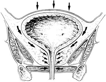
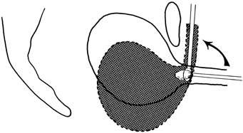
with more than a 70% cure rate.34,35

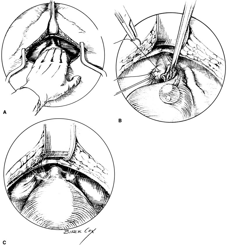
Fig. 9. Burch colposuspension. A. The space of Retzius is entered and a hand placed over the bladder. The bladder neck is just distal to the fingers, and the vaginal tissues and pubocervical fascia are located on either side of the urethra. B. The pubocervical fascia is sutured approximately 2 cm lateral to the bladder neck with a double bite of permanent suture. The sponge stick keeps the bladder retracted medially and superiorly away from the site of suture placement in the pubocervical fascia. C. Each arm of each suture is brought up through Cooper's ligament and tied over the top of the ligament, leaving banjo strings but satisfactorily elevating the bladder neck.

Traditionally, this procedure can be performed in concert with an abdominal hysterectomy or alone, through a retropubic approach without opening the peritoneal cavity. With the increased trend toward minimally invasive surgery, methods for laparoscopic urethropexy have been developed. Besides either an intraabdominal or an extraperitoneal approach, numerous modifications have been suggested including using only two sutures, substituting mesh, or using tacks, anchors, and other tools to elevate the bladder neck. These variations significantly lower cure rates as compared with traditional open urethropexy. Placement of four permanent sutures identical to an open procedure, though, has yielded similar 1- and 2-year cure rates as an open Burch (93% and 89%, respectively),36 although no prospective trial between abdominal and laparoscopic Burch procedures has been published. Currently, it is our practice to place four sutures identical to the open procedure when using a laparoscopic approach for optimal results. Transvaginal placement of urethropexy sutures has also been reported in small numbers, but no RCTs demonstrating comparable efficacy have been performed.37

Suburethral Slings

Placement of a sling under the bladder neck and proximal urethra had traditionally been used for patients with severe incontinence and poorly functioning urethras, and for those with recurrent stress incontinence. More often, though, suburethral slings (SUS) are being used as treatment for primary stress incontinence, an approach advocated by some urologists.38,39 The debated increase in morbidity, specifically urinary retention and urge incontinence, as compared with urethropexy is the point of contention regarding primary use of this procedure for treatment of SUI.40 Intermediate (1 to 2 years) and long-term (2 to 10 years) cure rates of 85% to 90% and 70% to 85%, respectively, are cited in the literature for most slings.39,41

In general, SUS can be considered full-length slings extending from rectus fascia all the way around the bladder neck and back up to the contralateral rectus fascia, or patch slings that use suture bridges that are tied over or sutured to the fascia. Full-length autologous slings can be obtained by harvesting fascia either from the rectus sheath or from the fascia lata of the leg. The sling is usually sutured into place with delayed absorbable sutures under the bladder neck. The tails of the full-length slings are sutured to the rectus fascia directly with good care to keep the sling loose. Similarly, the permanent suture bridge of patch slings is tied over the fascia or to the fascia loosely to protect against overcorrection and outlet occlusion.

The two slings described are the most common types of autologous slings cited in the literature. Other less-proven autologous materials include vaginal wall slings and rectus abdominus slings. Cadaveric fascia lata, dermal allograft, porcine dermis, and porcine small intestinal submucosa are other sources of nonsynthetic sling material. Synthetic slings, used as either full-length or patches, include Mersilene, Marlex, Prolene, Gore-tex, and silastic bands. Some of these novel materials have used bone anchors or other minimally invasive means to place a sling under the bladder neck, although long-term data regarding efficacy of these modifications, as compared with traditional slings, remain lacking.

A variation of the classic sling was developed in Europe by Ulmsten and colleagues using a Prolene mesh placed under the mid urethra (Fig. 10).42 The increasingly popular tension-free vaginal tape (TVT) and its subsequent competitors (Sparc, Uretex) were developed as a minimally invasive outpatient procedure that can be performed under intravenous sedation and local anesthesia. Placement of the mesh is accomplished through a small 1- to 2- cm incision under the mid urethra with blind passage of delivery needles up behind the pubic bone and through two small 5-mm supra-pubic skin incisions. The mesh is positioned loosely under the mid urethra, the protective plastic sheath removed, and the tails trimmed, which retract under the skin. These slings are not sutured in place, but rather the friction of the mesh pores keeps it in position initially until ingrowth of fibroblasts scars the sling in place. Most studies report an 85% cure rate with an additional 5% to 10% significantly improved.42,43,44 Because the needles are passed blindly, common complications of bladder perforation (4%–9%) and, less commonly, hematoma formation (1%) can occur.42,43,44,45 The low incidence of postoperative urge incontinence and obstructive voiding as well as the quick return to normal voiding has been attributed to the minimal peri-urethral dissection and loose application of the sling as directed by the developers. Although a lower incidence of erosion (< 1%) has been seen with this synthetic mesh compared with historical numbers for synthetic slings, the blind passage of the needles has caused some notable trauma to bowel, iliac vessels, and epigastric vessels, resulting in life-threatening
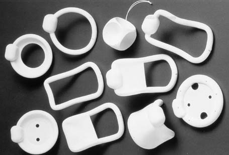
 situations.

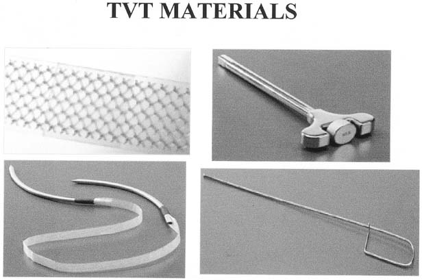
Fig. 10. The tension-free vaginal tape (TVT) device. Top left: polypropylene mesh tape in plastic sheath . Top right: introducer handle. Bottom left: mesh tape attached to introducer needles. Bottom right: rigid catheter guide used to deflect urethra and bladder away from side of tape placement (Reproduced with permission from Gynecare, Somerville, NJ).

Injectable Treatments for Stress Incontinence

Injectable agents designed to bulk the proximal urethra and bladder neck have been developed as a minimally invasive, office-based procedure for the treatment of SUI. The numerous agents now in use and in development are primarily aimed for the treatment of hypomobile stress incontinence in patients with poorly functioning urethral sphincters. Cystoscopically guided placement of these agents, either transurethrally or periurethrally, under the urethral mucosa aids in proximal urethral and bladder neck closure during times of increased stress (Fig. 11). Although primarily aimed to treat patients with immobile bladder necks, patients with severe comorbidities precluding more extensive procedures for their stress incontinence may benefit from injections despite 
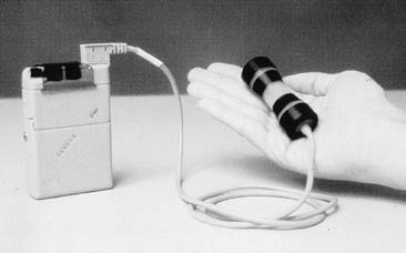
a hypermobile UVJ.46

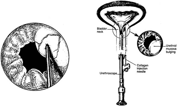
Fig. 11. Collagen injection therapy. On the right is the periurethral injection technique. The collagen material is deposited underneath the urethral mucosal lining while the surgeon observes with a 0-degree lens and scope. On the left is the transurethral technique. The needle is in the urethral mucosa at the 2 o'clock position, ready to inject (Bent AE: Periurethral collagen injections. Oper Tech Gynecol Surg 2: 51 1997.)

Similar to the variations on slings and colposuspension, many agents have been used or are in trials as a urethral bulking substance (Table 4). The many substances reflects the desire for an agent that is easily injectable, is permanent and will not be degraded by the host, and does not migrate or cause a damaging host reaction. The most commonly used agent is sterile bovine dermal collagen that is cross-linked with glutaraldehyde (Contigen; Bard Collagen Implant). A skin test to detect an allergic response is required before injection. Cure rates generally range from 20% to 40% with up to 70% to 80% improved and satisfied at 1 year.47 Degradation of the foreign collagen, which may occur between 3 months and 3 years after placement, is the most common cause necessitating reinjection. Similar results have been seen with trials of carbon beads (Durasphere), the next most commonly used bulking agent, when compared with collagen.


Table 4. Biomaterials and Synthetic Injectable Agents in Use and Development as Urethral Bulking Materials


Human and Xenograft Injectable Agents Synthetic Injectable Agents
Bovine cross-linked collagen Carbon beads
Autologous fat Teflon
Human collagen Ethylene vinyl alcohol copolymer-DMSO
Autologous cartilage Silicone
Protein/elastin admixture Injectable suture material
Bovine elastin/collagen Implantable microballoons
Autologous muscle derived cells Hyaluronic acid and dextranomener microspheres
  Adjustable volume balloons
  Cross-linked hyaluronic acid
  Calcium hydroxyapatite


Other Treatments

Placement of artificial urethral sphincters tends to be performed by urologists in patients who have not had symptoms cured by other aforementioned interventions. Most of these devices involve an occlusive cuff placed around the proximal urethra that is fed by a reservoir situated in the labia majora for patient control. Consensus regarding suitable patients and efficacy is still in question because of small cohort studies and poor standardization. A success rate of between 80% and 90% is most quoted, but a significant rate of revision, explantation, or erosion of 5.9%48 to 17%49 has been seen.

A different, minimally invasive, treatment modality being developed involves radiofrequency thermal energy to cause tissue shrinkage at the bladder neck (SURx; Thermatrx). This procedure, performed as an outpatient procedure by a laparoscopic or vaginal approach, is designed to cause scarring at the bladder neck to restrict mobility. In a single small study of 94 women who underwent the laparoscopic procedure,50 subjective improvement, and cure rates, as well as objective cure (negative stress test) were seen in approximately 80% of patients. Further studies and longer follow-up are obviously needed.

Back to Top
COMPLICATIONS OF INCONTINENCE SURGERY

Urge Incontinence

Although symptoms of urgency and frequency are not uncommon in the immediate postoperative period after any anti-incontinence procedure, patients must be warned before surgery regarding the possibility of persistence or de novo occurrence after any procedure. Furthermore, distinction between new or worsened urge incontinence and voiding dysfunction/outlet obstruction (discussed later) may be difficult in the postoperative period. Between 17% and 33% of patients presenting with SUI may have urge incontinence,51 with a good proportion having resolution of their symptoms after surgery. Unfortunately, approximately 15% (range: 5%–28%) will have de novo detrusor instability after a Burch or sling procedure.34,39,47 Fortunately, if symptoms are not too severe, treatment with anticholinergics and/or pelvic muscle rehabilitation may alleviate or resolve the condition in the majority.

Voiding Dysfunction

Voiding dysfunction may present in the postoperative period as the complete inability to void, only partial emptying, urgency-frequency syndrome, or other irritative voiding symptoms. It is not uncommon for patients to have some symptoms along this spectrum in the immediate postoperative period caused by bruising of tissues, hematomas, and pain. Persistence without improvement beyond 4 to 6 weeks may warrant intervention in patients with complete or almost complete obstruction, severe urgency-frequency or significant irritative symptoms not caused by infection and not responsive to conservative therapy. Urethrolysis is the term used when releasing an anti-incontinence procedure in this situation. This generally involves vaginal transection of a sling and release of periurethral scarring, or, in the case after a retropubic procedure, abdominal dissection of the space of Retzius and removal of sutures if possible. The majority of patients will have improvement or resolution of their symptoms after urethrolysis in the range of 60% to 88%. Unfortunately, there is a small group that will need to continue with self-catheterization, and approximately one-third will have recurrence of stress incontinence.

Surgical Failure

Failure of a surgical procedure usually results from one of four causes: (1) incorrect or incomplete diagnosis; (2) wrong procedure; (3) technical failure; and (4) new diagnosis or condition.

Urge incontinence as a primary diagnosis is unlikely to be cured by a procedure to correct stress incontinence. If a diagnosis of ISD is missed, an incorrect procedure may have been performed. Technical failures include incorrect suture placement, incorrect suture selection, tissue fragility, infection or hematoma, suture pull-through or dislodgment, and damage to surrounding structures. New symptoms may develop postoperatively, such as urge incontinence, detrusor instability, or a sphincter problem resulting in incontinence. To form an exact diagnosis and plan for therapy, a complete evaluation of the patient presenting with surgical failure should be instituted.

Coital Incontinence

Incontinence occurring with intercourse is a result of either stress or urge incontinence. In the former case, the patient has urine loss associated with penetration and/or thrusting. The increased intra-abdominal pressure results in a small but persistent loss of urine. Corrective actions include precoital voiding and altering coital positions. The effect of surgery is variable. Some patients may have the problem after surgery, while others have resolution after surgery. Although there are no studies to support the various theories, there seems to be a higher frequency of this problem after sling surgery than after other surgeries. Persistent problems are managed by pelvic floor muscle exercises and any other symptom-relieving maneuvers.

The second kind of intercourse-related incontinence occurs when detrusor activity is stimulated by physical contact near the bladder or by orgasm, which triggers a detrusor contraction. These patients often empty larger amounts of urine during intercourse compared with those with stress incontinence, and voiding immediately before intercourse only partly relieves the problem. Behavioral intervention and pharmacotherapy do not offer much improvement. It would be prudent to discuss these difficulties before surgical intervention.

Modifiable Risk Factors

Obesity and smoking are two modifiable factors that may put patients at risk for stress incontinence. As mentioned previously, altering these characteristics of the patient may be difficult, and no studies have demonstrated that smoking cessation or moderate weight loss will aid continence. That being said, certain aspects inherent to these patients must be considered when proceeding to surgery. Firstly, there may be an increased risk of venous-thromboembolic events and poor tissue healing with risk of wound breakdown. Increased abdominal pressure from excess coughing or abdominal girth always makes one concerned about suture disruption or pull-through. Because of this last concern, some surgeons prefer to use primary slings in these patients with the thought that they are more durable, although no data support this methodology. Either way, these patients must be counseled regarding increased surgical risk, whether from technical difficulties from excess soft tissue or from possible pulmonary compromise caused by underlying lung disease.

Back to Top
SUMMARY

Although some of the terminology has changed, the basic diagnosis of SUI remains the same. A deeper understanding of the anatomy and neurophysiology of the lower urinary tract has led to viewing sphincter dysfunction and hypermobile bladder neck on an overlapping spectrum of incontinence. Also, new knowledge regarding the neuro-urology connection has led to novel pharmacologic treatments that can be used instead of, or in concert with, other well-established conservative therapies such as pelvic muscle exercise programs and electronic stimulation. After failure of conservative therapy, surgery may be indicated for SUI after the proper work-up as outlined. The choice of a suburethral sling or retropubic Burch for incontinence, both approximately equal in short-term and long-term success, may hinge on obtaining urodynamics on suspected patients and finding a poorly functioning urethral sphincter mechanism. The development of the new mid urethra slings (TVT) may obviate this distinction and serve as a minimally invasive alternative for all stress incontinence with some mobility. In patients with a fixed urethra and SUI, urethral bulking agents continue to evolve from the tested, but temporary, bovine collagen to longer-lasting synthetics and biomaterials.

Stress urinary incontinence is a condition that generalist physicians need be more cognizant of as life expectancy lengthens and the proportion of the elderly within the population increases. Patients suspected of having stress incontinence need to be evaluated with a history and physical, residual urine determination, urinalysis, 24-hour voiding diary, stress test, and cotton swab test. If preliminary evaluation reveals SUI, conservative therapy may be attempted without further testing. Patients who refuse or do not respond to conservative therapy should be evaluated, at the minimum, as dictated by the ACOG guidelines before consideration for surgery. Patients with recurrent stress incontinence or other complicated medical or surgical history, or those suspected of having a weak urethral sphincter, may benefit from ancillary testing such as multichannel urodynamics. All patients should be counseled regarding the risks of anti-incontinence surgery, and those with complications and voiding problems should be identified and evaluated accordingly.

Back to Top
REFERENCES

1. Bump RC, Norton PA: Epidemiology and Natural History of Pelvic Floor Dysfunction. Obstet Gynecol Clin 25:(4):723-746, 1998

2. Cundiff GW, Harris RL, Coates KW et al: Clinical predictors of urinary incontinence in women. Am J Obstet Gynecol 177:262-267, 1997

3. Olsen AL, Smith VJ et al: Epidemiology of Surgically Managed Pelvic Organ Prolapse and Urinary Incontinence. Obstet Gynecol 89:501-506, 1997

4. Abrams P, Cardozo L, Fall M et al: The standardization of terminology of lower urinary tract function: Report from the standardization sub-committee of the international continence society. Am J Obstet Gynecol 187:(1):116-126, 2002

5. Smith ARB, Hosker GL, Warrell DW: The role of patial denervation of the pelvic floor in the aetology of genitourinary prolapse and stress incontinence of urine: A nerurophysiological study. Br J Obstet Gynecol 96:24-28, 1989

6. DeLancey JOL, Kearney R, Chou Q et al: The appearance of levator ani muscle abnormalities in magnetic resonance images after vaginal delivery. Obstet Gynecol 101:46-53, 2003

7. Boyle AL, Woodman PJ, O'Boyle JD et al: Pelvic organ support in nulliparous pregnant and nonpregnant women: a case control study. Am J Obstet Gynecol 187:99-102, 2002

8. Thom DH, Brown JS: Reproductive and Hormonal Risk Factors for Urinary Incontinence in Later Life: A Review of the Clinical and Epidemiologic Literature. J Am Geriatr Soc 46:(11):1411-1417, 1998

9. DeLancey JOL: Structural support of the urethra as it relates to stress urinary incontinence: The hammock hypothesis. Am J Obstet Gynecol 70:1713-1723, 1994

10. Fantl JA, Newman DK, Colling J et al: Urinary incontinence in adults: acute and chronic management. Clinical Practice Guideline, No. 2, 1996 Update. Rockville, MD: U.S. Department of Health and Human Services. Public Health Service, Agency for Health Care Policy and Research. AHCPR Publication No. 96-0682 March 1996

11. Bump RC, Mattiasson A, Bo K et al: The standardization of terminology of femal pelvic organ prolapse and pelvic floor dysfunction. Am J Obstet. Gynecol 175:10-17, 1996

12. Sand PK, Bowen LW, Panganiban R et al: The low pressure urethra as a factor in failed retropubic urethropexy. Obstet Gynecol 69:399, 1987

13. Koonings PP, Bergman A, Ballard CA: Low pressure urethra and stress urinary incontinence in women: Risk factor for failed retropubic surgical procedure. Urology 36:245, 1990

14. Horbach NS, Blanco JS, Ostergard DR et al: A suburethral sling procedure with polytetrafluoroethylene for the treatment of genuine stress incontinence in patients with low urethral pressure. Obstet Gynecol 71:648, 1988

15. McGuire EJ, Fitzpatrick CC, Wan J et al: Clinical assessment of urethral sphincter function. J Urol 150:1452-1454, 1993

16. Sutherst J, Brown M, Shawer M: Assessing the severity of urinary incontinence in women by weighing perineal pads. Lancet 23:1128, 1981

17. Thind P, Gerstenberg TC: One-hour ward test vs 24-hour home pad weighing test in the diagnosis of urinary incontinence. Neurourol Urodyn 10:241, 1991

18. Bump RC, McClish DK: Cigarette smoking and urinary incontinence. Am J Obstet Gynecol 17:1213-1218, 1992

19. Deitel M, Stone E, Kassam HA et al: Gynecologic-obstetric changes after loss of massive excess weight following bariatric surgery. J Am Coll Nutr 7:147-153, 1988

20. Bump RC, Sugerman HJ, Fantl JA et al: Obesity and lower urinary tract function in women: Effect of surgically induced weight loss. Am J Obstet Gynecol 166:392-329, 1992

21. Kegel AH: Progressive resistance exercises in the functional restoration of the perineal muscles. Am J Obstet Gynecol 56:238, 1948

22. Wells TJ, Brink CA, Diokno AC et al: Pelvic muscle exercise for stress urinary incontinence in elderly women. J Am Geriatr Soc 39:785, 1991

23. Peattie A, Plevnik S, Stanton S: Vaginal cones: A conservative method of treating genuine stress incontinence. Br J Obstet Gynecol 95:1049, 1988

24. Hay-Smith EJ, Herbison P, Morkved S: Physical therapies for prevention of incontinence in adults. Cochrane Protocol. 2001

25. Sand PK, Richardson DA, Staskin DR et al: Pelvic floor electrical stimulation in the treatment of genuine stress incontinence: A multicenter, placebo controlled trial. Am J Obstet Gynecol 173:72-79, 1995

26. Tsai EM, Yang CH, Chen HS et al: Bladder neck circulation by Doppler ultrasonography in postmenopausal women with urinary stress incontinence. Obstet Gynecol 98:52-56, 2001

27. Bump RC, Friedman CI: Intraluminal urethral pressure measurements in the female baboon: effects of hormonal manipulation. J Urol 136:508-511, 1986

28. Fantl JA, Bump RC, Elser DM et al: Efficacy of estrogen supplementation in the treatment of urinary incontinence. Obstet Gynecol 88:745-749, 1996

29. Gilgja I, Radeh M, Kovacic et al: Conservative treatment of female stress incontinence. J Urol 132:909, 1984

30. Lin HH, Sheu BC, Lo MC et al: Comparison of treatment outcomes for imipramine for female genuine stress incontinence. Br J Obstet Gynaecol 106:1089, 1999

31. Norton PA, Zinner NR, Yalcin I et al: Duloxetine versus placebo in the treatment of stress urinary incontinence. Am J Obstet Gynecol 187:40-48, 2002

32. American College of Obstetricians and Gynecologists, Committee on Quality Assessment, ACOG Criteria Set: Surgery for Genuine Stress Incontinence Due to Urethral Hypermobility. No. 4 1995

33. Glazner CM: Anterior vaginal repair for urinary incontinence in women. Cochrane Database Syst Rev. January 2000

34. Jarvis GJ: Surgery for genuine stress incontinence. Br J Obstet Gynecol 101:371-374, 1994

35. Alcalay M, Monga A, Stanton S: Burch colposuspension: a 10–20 year follow up. Br J Obstet Gynaecol 102:740-745, 1995

36. Ross JW: Multichannel urodynamic evaluation of laparoscopic Burch colposuspension for genuine stress incontinence. Obstet Gynecol 91:55-59, 1998

37. Nguyen JK: Transvaginal bladder neck suspension to Cooper's ligament: A review of the literature. Int Urogynecol J Pelvic Floor Dysfunct 11:(5):320-324, 2000

38. Appell RA: Primary slings for everyone with genuine stress incontinence? The argument for Int Urogynecol J 9:249-251, 1998

39. Leach GE, Dmochowski RR, Appell RA et al: Female stress urinary incontinence clinical guidelines panel summary report on surgical management of female stress urinary incontienence. J Urol 158:875-880, 1997

40. Ostergard DR: Primary slings for everyone with genuine stress incontinence? The argument against Int Urogynecol J 8:321-322, 1998

41. Morgan TO, Westney OL, McGuire EJ: Pubovaginal sling: 4 year outcome analysis and quality of life assessment. J Urol 163:1845-1848, 2000

42. Ulmsten U, Henricksson L, Johnson P et al: An ambulatory surgical procedure under local anesthesia for treatment of female urinary incontinence. Int Urogynecol J 7:81-86, 1996

43. Ulmsten U, Falconer C, Johnson P et al: A mulicenter study of tension-free vaginal tape (TVT) for surgical treatment of stress urinary incontinence. Int Urogynecol J 9:210-213, 1998

44. Olsson I, Kroon U: A three-year postoperative evaluation of tension-free vaginal tape. Gynecol Obstet Invest 48:267-269, 1999

45. Meschia M, Pifarotti P, Bernasconi F et al: Tension-free vaginal tape: analysis of outcomes and complications in 404 stress incontinent women. Int Urogynecol J Suppl 2:S24-S27, 2001

46. Bent AE, Foote J, Siegle S et al: Collagen implant for treating stress urinary incontinence in women with urethral hypermobility. J Urol 166:1354-1357, 2001

47. Smith T, Daneshgari F, Dmochowski R et al: Surgical Treatment of Incontinence in Women. In: Abrams P, Cardozo L, Khoury S, et al (eds): Incontinence. pp 825-863, Plymouth, Health Publications Ltd, 2002

48. Costa P, Mottet N, Rabut B et al: The use of an artificial urinary sphincter in women with type III incontinence and a negative Marshall test. J Urol 165:1172-1176, 2001

49. Webster GD, Perez LM, Khoury JM et al: Management of type III stress urinary incontinence using artificial urinary sphincter. Urology 39:499, 1992

50. Fulmer BR, Sakamoto K, Turk TMT et al: Acute and long-term outcomes of radio frequency bladder neck suspension. J Urol 167:141-145, 2002

51. Bent AE: Etiology and management of detrusor instability and mixed incontinence. Obstet Gynecol Clin North Am 16:853, 1989

Back to Top