Chapter 103
Biologic Effects of Nuclear Magnetic Resonance
Thomas L. Chenevert, Paul L. Carson and Louis K. Wagner
Main Menu   Table Of Contents

Search

Thomas L. Chenevert, PhD
Assistant Professor of Radiology, University of Michigan, Ann Arbor, Michigan (Vol 2, Chaps 101, 103)

Paul L. Carson, PhD
Professor of Radiology and Director, Division of Radiologic Physics and Engineering, University of Michigan Medical Center, Ann Arbor, Michigan (Vol 2, Chaps 101, 103)

Louis K. Wagner, PhD
Associate Professor of Radiology, University of Texas Medical School at Houston, Houston, Texas (Vol 2, Chaps 101, 103)

INTRODUCTION
BACKGROUND
EFFECTS OF STATIC MAGNETIC FIELDS
TIME-VARYING MAGNETIC FIELDS
RADIOFREQUENCY FIELDS
GUIDELINES FOR OPERATION OF CLINICAL NMR DEVICES
OPINIONS OF RISK TO THE CONCEPTUS IMPOSED BY MRI
REFERENCES

INTRODUCTION

The intent of this chapter is to acquaint the reader with current opinions concerning bioeffects of nuclear magnetic resonance (NMR). NMR has the desirable feature of not involving ionizing radiation; therefore, at first glance it may appear entirely safe. However, a variety of interaction mechanisms exist between magnetic and electromagnetic fields and tissue that should be considered in evaluating safety. These effects are categorized and discussed in terms of observable thresholds. It is emphasized that while numerous bioeffects have been reported in the literature, studies were often contradictory or inconclusive in establishing the nature of the effect. For this reason, we will discuss only those effects that are reasonably well established or are believed to have the greatest potential of biohazard. In addition, safety guidelines established by government agencies are reviewed, and emphasis is placed on how these guidelines impact routine clinical magnetic resonance imaging (MRI).

Back to Top
BACKGROUND

It is beneficial to review fundamentals of MRI to introduce which aspects of the procedure have the potential of adverse effects.

At the heart of all MRI systems is a strong static magnetic field. When placed in the magnetic field, hydrogen nuclei (protons), which themselves have a magnetic property, will align parallel or antiparallel with the magnetic field direction. The slight excess of protons aligned parallel to the field cumulatively produces a macroscopically measurable quantity known as magnetization. A means to measure magnetization is by the NMR process wherein a radiofrequency (rf) pulse is applied causing the magnetization to be tipped away from the direction defined by the static magnetic field. Once the radiofrequency wave is turned off, the magnetization will relax back to its original orientation, releasing energy in the form of an NMR signal. The minute magnetic resonance signal is detected by a radiofrequency pickup coil (usually the same coil used for transmission of the radiofrequency pulse), digitized, and processed by a computer.

Contained within the NMR signal is a wealth of information about the tissue. Signal strength is dependent on concentration of protons, frequency content of the signal is dependent on the chemical environment of the protons (e.g., protons in water versus fat have a slightly different signal frequency), and signal persistence and recovery rate yield information about the physical environment (e.g., bound versus free water). Typically, MRI systems forfeit chemical information to generate tomographic maps of the protons' signal intensity emanating from the body. Spatial encoding of the source of the magnetic resonance signal involves rapid alterations of the magnetic field strengths across the body as the radiofrequency pulses prepare the magnetization to yield its signal. These field gradient transients alter the resonant frequency of the protons as a function of position in the body. For our interest in bioeffects, greater detail of the imaging sequence is not required.1 As a preface to our discussion of bioeffects, it is sufficient to say imaging requires exposure of the patient to (1) a strong static magnetic field, (2) radiofrequency pulses, and (3) time-varying magnetic field gradients. Possible biohazards are usually grouped within these three categories.

Back to Top
EFFECTS OF STATIC MAGNETIC FIELDS

MRI systems operate at magnetic field strengths of 0.1 tesla (T) to 2 T. For a reference, the earth's magnetic field is approximately 50 microtesla (10-6). Whole-body magnets designed specifically to maintain a high field strength over the large volume of the body are either permanent magnets, resistive electromagnets, or, most commonly, superconductive magnets. Optimal field strength for MRI remains an open issue; however, systems operating at 0.15 T to 0.5 T and 1.5 T are popular. High field strength units may have advantages in signal to noise over low field strength systems and offer the potential of spectroscopy. However, as field strength increases so does radiofrequency power deposition in the body,2 as we shall see later.

It is believed there are no direct adverse effects of static magnetic field strengths up to 2 T.3,4,5 This is based on studies in which behavioral and important physiological processes of mammals exposed to fields up to 2 T were monitored. An indirect, but very real, danger associated with high magnetic fields is the attractive force on ferromagnetic objects, which may become projectiles causing injury or death as they are drawn to the bore of the magnet. Also, ferromagnetic implants, protheses, heart valves, and surgical and aneurysm clips are a concern since they may become displaced under the pull of the magnet.6

Persons with cardiac pacemaker implants should not be allowed near the magnet.7 Some of these devices contain a reed relay switch that can be activated externally by a relatively low magnetic field. Tests have demonstrated that fields as low as 1.7 mT can activate the relay, causing a change in pace mode from synchronous to asynchronous. The 0.5-mT limit has been established as a conservative perimeter within which persons with pacemakers and uncontrolled traffic should not be allowed.

Another observable effect of high magnetic fields is the force on ionic currents flowing in blood.8 This force is perpendicular to the direction of flow, which causes a separation of charge across the blood vessel diameter. The resulting electric potential is measurable on the electrocardiogram signal in animal studies. but it appears to be completely reversible and to have no adverse physiological effect.9

Back to Top
TIME-VARYING MAGNETIC FIELDS

As mentioned, pulsed magnetic field gradients are essential in an imaging sequence for spatial localization of the NMR signal within the body. The peak additional magnetic field strength provided by the gradients is several orders of magnitude weaker than the static magnetic field and therefore presents no additional risk in terms of magnetic field strength. However, the temporal rate of change of the magnetic field is high.10 Gradient pulses may have field changes as high as 3 T/sec, although they are generally in the range of 0.5 T to 1 T/sec for conventional MRI. The response of any conductive body in the presence of changing magnetic fields is to oppose the field change with internal electric currents. using the conductivity of tissue and a 10-cm-diameter current path, the current density induced by a 1-T/sec field change would be 1 microamp/cm2.11

An observable effect of induced current densities of this magnitude is the visual sensation of flashing lights known as magnetophosphenes.12 Studies indicate that duration and frequency of the magnetic field change play an important role in the threshold at which this sensation is induced. For example, 1.3-T/sec, 2-msec pulses will invoke magnetophosphenes, yet much higher field rate changes will not if the pulse duration is short.13

Another bioeffect observed in animal studies of tissue currents induced by time-varying magnetic fields is direct neuromuscular stimulation.14,15 It is emphasized, however, that tissue current densities required to involve neuromuscular stimulation are far beyond those generated in commercial MRI systems. For example, it is estimated 200 microamp/cm2 is the threshold current density to stimulate ventricular fibrillation, which is orders of magnitude greater than those induced in the human heart by conventional imaging gradients.

Back to Top
RADIOFREQUENCY FIELDS

The patient is exposed to short radiofrequency fields to stimulate magnetized protons to yield an NMR signal. A by-product of this process is deposition of radiofrequency energy in the body as heat.16 In addition to transmitted radiofrequency power level, several factors influence the amount of tissue heating; these include radiofrequency coil design, electric properties of tissue, pulse sequence, and size and shape of the body part being scanned. The body's ability to dispose of additional heat is an important consideration in evaluating safety. Tissues having low thermoregulatory blood supply, such as the testes and lens of the eye, may be more susceptible to damage by radiofrequency power deposition. Excessive localized heating may also occur in areas near conductive metals such as metallic implants and in constricted current paths such as the armpits and groin.

When a radiofrequency pulse is applied, the body experiences an oscillating magnetic field. Just as with a pulsed magnetic field gradient, the body responds to the oscillating magnetic field with internal electric currents that generate heat by resistive losses. The measure of power deposition is the specific absorption rate (SAR), defined as the amount of power deposited per kilogram of tissue.16 It can be shown that power deposition in whole-body NMR increases with current path radius (body size), frequency of the radiofrequency pulses, and pulse duty cycle.2 In NMR the frequency of radiofrequency pulses is proportional to the static magnetic field strength. Consequently, tissue heating is a greater concern in high field MRI systems. This is particularly true in body imaging, in which high-frequency radiofrequency penetrates less deeply into the body so high-powered radiofrequency pulses are required.

The total radiofrequency power absorbed by the body can be determined empirically using the following equation:5

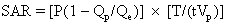
where P is the root mean square power input to the radiofrequency coil, Qp is the coil quality factor with the patient in the coil, Qe is the quality factor with the coil empty, T is the duration of the radio-frequency pulse, and t-1 is the number of radiofrequency pulses per second. For the body average SAR, Vp represents the total body mass, or as a more conservative and accurate measure Vp is only the tissue mass contained within the coil. The quality factor is an electrical quantity indicating the ratio of inductance to resistance. The quantity (1 - Qp/Qe) represents the fraction of total resistive heat losses that is deposited in the patient.

Usually an imaging sequence requires more than one type of radiofrequency pulse. For example, the popular two-echo spin-echo sequence requires two of the more powerful 180° radiofrequency pulses for each 90° radiofrequency pulse. The total SAR is the sum of contributions from each type of pulse. An SAR calculation for a medium-field-strength two-echo spin-echo sequence is shown below:

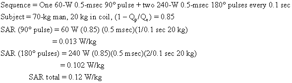
As we shall see, this body average SAR is well below guideline limits.

The power requirements for head scanning is one tenth or less that of body scanning; therefore, it is not limited by tissue heating.

Peak SAR refers to the maximum power deposited per kilogram averaged over any 1 g of tissue. Estimating peak SAR involves models of tissue geometry and electric characteristics.2 For cylindrical and spherical models peak SAR (at the skin surface) is two to three times the average SAR. It is emphasized, however, that actual peak SAR may be significantly higher depending on body geometry and actual electric properties.

The temperature rise of tissue for given SAR and exposure time (texp) is given by the equation

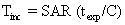
where C is the heat capacity of tissue (0.83 kcal/kg °C).5 Tissue exposed to 2 W/kg for 20 minutes would be expected to rise in temperature by 0.7°C. Temperature increases of this magnitude are not observed clinically owing to the body's thermoregulatory system (acquisition time for MRI sequences varies from subminute to several tens of minutes).

Currently, static magnetic field strength and time-varying magnetic field gradients do not place limits on routine NMR procedures for reasons of patient safety. Field strengths in excess of 2 T are technically difficult to achieve on whole-body NMR systems and may not offer advantages in image quality or diagnostic value. Also, magnetic field gradient pulses currently used are believed to be within safe limits and are adequate for virtually all magnetic resonance procedures. However, as new techniques are developed that require stronger gradient pulses, such as rapid scanning, further careful investigation of safety of time-varying magnetic fields is warranted. The remaining issue of radiofrequency power deposition does restrict the type of pulse sequences that are usable in body scanning at high magnetic field strengths. Considerable effort has gone into radiofrequency coil design and modes of radiofrequency polarization to minimize the percentage of protocols that exceed safety guidelines. In the event the selected protocol exceeds radiofrequency power deposition limits, the examination may be partitioned into two or more acquisition sequences that individually do not exceed power deposition limits. The body's ability to continually transfer heat to the environment allows many pulse sequences to be applied sequentially as long as no one sequence exceeds the guidelines. This is a fundamental distinction between radiofrequency power deposition in MRI and ionizing radiation dosage in x-ray procedures, in which the total examination dose is the primary concern.

Back to Top
GUIDELINES FOR OPERATION OF CLINICAL NMR DEVICES

Table 1 summarizes the limits of static magnetic field, time-varying fields, and SAR values as determined by the Bureau of Radiological Health (BRH) of the US Food and Drug Administration3 and the National Radiation Protection Board (NRPB) of the United Kingdom.4

TABLE 1. Established Guidelines for Clinical NMR


Static

Time-varying

SAR

SAR


Magnetic

Magnetic

Average

Peak

Agency

Field (Tesla)

Field (Tesla/Sec.)

(W/kg)

(W/kg)

BRH of US FDA

2.0

3

0.4

2

NRPB of UK

2.5

20*

0.4

4


* Root mean square (rms) maximum for periods of change exceeding 10 msec. For periods, t less than 10 msec the rms maximum in T/sec is permitted to increase to the numerical value of 2/ √t, where t is expressed in seconds.

The BRH guidelines have been established to determine whether a particular clinical NMR procedure represents a “significant risk” to the subject. That is, the BRH believes any NMR study involving exposures below these levels probably does not present an acceptable risk of harm to the subject. These guidelines are considered interim, since they are based on the current body of information on bioeffects, which the BRH admits is not yet definitive. As bioeffects research continues, these guidelines will be revised. The BRH value of 0.4 W/kg body average SAR is derived from the recommended limit in the American National Standards Institute (ANSI) standard.16 In the ANSI standard the value of 4 W/kg is cited as the limit above which there exists reliable evidence for hazardous effects. A factor of 10 was proposed as the safety margin to define the current 0.4-W/kg whole-body average SAR guideline. This value represents about half the basal metabolic rate for adults and should not result in significant elevations in body temperature. The ANSI standard also recommends a limit of 8 W/kg peak SAR averaged over any I g of tissue, which is a factor of 4 greater than the more conservative BRH guideline.

Back to Top
OPINIONS OF RISK TO THE CONCEPTUS IMPOSED BY MRI

As is true of other imaging modalities, risk to the human embryo or fetus as imposed by NMR procedures is an important issue that must be considered by physicians ordering examinations. It is the policy of many MRI facilities not to image pregnant women. This conservative posture is not based on scientific evidence of biohazards, but rather is a precautionary measure. Institutions that do perform MRI of the pregnant abdomen usually do not do so during the first trimester. The NRPB recommends that women in their first trimester be excluded from MRI even though there is no evidence of damage to the embryo.4

Recent animal studies of the effects of NMR fields on the developing fetus did not demonstrate adverse bioeffects17,18 unless very high power deposition levels were used.19 These results, coupled with growing experience with MRI, leads one to conclude that routine NMR procedures are probably safe for humans and their unborn.

Back to Top
REFERENCES

1. Pykett IL: NMR imaging in medicine. Sci Am 246 (6): 78, 1982

2. Bottomley PA, Edelstein WA: Power deposition in whole-body NMR imaging. Med Phys 8 (4): 510, 1981

3. Bureau of Radiological Health: Guidelines for evaluating electromagnetic exposure risk for trials of clinical NMR systems. Department of Health and Human Services, US Public Health Service, Food and Drug Administration, 1982

4. National Radiological Protection Board: Revised guidance on acceptable limits of exposure during nuclear magnetic resonance clinical imaging. Br J Radiol 56:974, 1983

5. Budinger TF: Nuclear magnetic resonance (NMR) in vivo studies: Known thresholds for health effects. J Comput Assist Tomogr 5 (6): 800, 1981

6. New PFJ, Rosen BR, Brady TJ et al: Potential hazards and artifacts of ferromagnetic and nonferromagnetic surgical and dental materials and devices in nuclear magnetic resonance imaging. Radiology 147: 139, 1983

7. Pavlicek W, Geisinger M, et al: The effects of nuclear magnetic resonance on patients with cardiac pacemakers. Radiology 147: 149, 1983

8. Kolin A: Improved apparatus and technique for electromagnetic determination of blood flow. Rev Sci Instrum 23: 236, 1952

9. Beischer DE: Vectorcardiogram and aortic blood flow of squirrel monkeys (Saimiri sciureus) in a strong superconductive electromagnet. In Barnothy MF (ed): Biological Effects of Magnetic Fields, Vol 2. New York, Plenum Press, 1969

10. Gore JC, McDonnell MJ, Pennock JM, Stanbrock HS: An assessment of the safety of rapidly changing magnetic fields in the rabbit: Implications for NMR imaging. Magn Reson Imag 1: 191, 1982

11. Budinger TF: Potential medical effects and hazards of human NMR studies. In Kaufman L, Crooks LE, Margulls AR (eds): Nuclear Magnetic Resonance Imaging in Medicine, pp 207–231. New York, Igaku-Shoin, 1981

12. Barlow HB, Kohn HI, Walsh EG: Visual sensation aroused by magnetic fields. Am J Physiol 148: 372, 1947

13. Budinger TF, Cullander C, Bordaw R: Switched magnetic field thresholds for the induction of magnetophosphenes (abstr). Proceedings of the 3rd Annual Meeting of the Society of Magnetic Resonance in Medicine, pp 118 and 119, 1984

14. Watson AB, Wright JS, Joughman J: Electrical thresholds for ventricular fibrillation in man. Med J Aust 1: 1179, 1973

15. Roy OZ: Summary of cardiac fibrillation for 60 Hz currents and voltages applied directly to the heart. Med Biol Eng Cornput 18: 657, 1980

16. American National Standard Safety Levels with Respect to Human Exposure to Radio Frequency Electromagnetic Fields, 300 kHz to 100 kHz (C95.1–1982). ANSI, New York, 1982

17. McRobbie D, Foster MA: Pulsed magnetic field exposure during pregnancy and implication for NMR foetal imaging: A study with mice. Magn Resort Imag 3: 231, 1985

18. Heinrichs WL, Fong P, Moseley ME et al: Analysis of teratogenesis and reproductive toxicity in Balb/c mice after midpregnancy MRI or MRS exposure (abstr). Proceedings of the 4th Annual Meeting of the Society of Magnetic Resonance in Medicine, p 922, 1985

19. O'Conner ME: Mammalian teratogenesis and radiofrequency fields. Proc IEEE 68: 56– 60, 1980

Back to Top