Chapter 1
Care of the High-Risk Mother
Richard Depp
Main Menu   Table Of Contents

Search

Richard Depp, MD
Professor and Chairman, Department of Obstetrics and Gynecology, Thomas Jefferson Medical College, Philadelphia, Pennsylvania (Vol 3, Chaps 1, 76)

INTRODUCTION
THE INITIAL VISIT
HIGH-RISK CARE: THE FIRST 20 WEEKS
HIGH-RISK CARE: THE LAST 20 WEEKS
INFANT FOLLOW-UP
PERINATAL DEATH
OVERVIEW
REFERENCES

INTRODUCTION

A high-risk pregnancy is one in which either the mother or the fetus has a high risk of death or disability as a consequence of one or more conditions that complicate the normal pregnancy process. Most notable of these conditions are preterm labor, pregnancy-induced hypertension, maternal diabetes, maternal-fetal infections, congenital anomalies, and labors complicated by fetal-neonatal hypoxia. All processes frequently leave their surviving victims disabled for life. Table 1 and Table 2 compare maternal and infant mortality experience in the United States by cause for the years 1970 and 19781. In the past 20 years since I entered obstetrics, maternal mortality has been virtually eliminated as a major problem. In the brief interval from 1970 to 1978, reduction in overall maternal, infant, and neonatal mortality has been dramatic (Table 3). In large part, this improvement is attributable to widespread clinical application of information previously developed in reproductive and perinatal research. Most notable are more widespread availability of family planning and abortion services, new techniques for the early pregnancy diagnosis of congenital malformations or genetic disorders, widespread postpartum administration of Rh immune globulin to prevent sensitization, availability of techniques to assess fetal pulmonary maturity and health status, improved intrapartum surveillance, and specialized neonatal care. In view of the dramatic reduction in perinatal mortality, we are becoming increasingly close to our ultimate goal, which is to ensure that the genetic potential established at conception is maintained at least through the neonatal period. The latter goal is attainable only through knowledge of maternal and fetal-placental physiology and pathophysiology; biologic processes that impinge on embryonic development and fetal growth; as well as some understanding of environmental factors that may affect either the fetus or the mother. Knowledge of the former is necessary to foster a rational selective implementation of antenatal and intrapartum diagnostic procedures. More remotely, it is necessary to eventually establish precise causation for congenital malformations, preterm labor, and abnormal fetal growth.

TABLE 1. Maternal Deaths and Mortality Rates, United States, 1970 and 1978


Maternal


Mortality Rate (Deaths

_Rank

Cause of

No. of Maternal Deaths

per 100,000 Live Births)

(1978)

Death

1970

1978

1970

1978

1

Toxemia

142

62

3.8

1.9

2

Sepsis

144

61

3.9

1.8

3

Ectopic

63

37

1.7

1.1


 pregnancy*


4

Hemorrhage*

86

36

2.3

1.1

5

Abortion

128

16

3.4

0.5

6

Other

240

109

6.4

3.2


Total

803

321

21.5

9.6


*Many of the deaths from ectopic pregnancy could be combined with those from hemorrhage, which is the usual cause of death in ectopic pregnancy.
(Compiled from National Center for Health Statistics data)

TABLE 2.Deaths Under 1 Year and Infant Mortality Rates, United States, 1970 and 1978


Infant Mortality Rate


(Deaths per 100,000)

_Rank


No. of Infant Deaths

Live Births

(1978)

Cause of Death

1970

1978

1970

1978

1

Congenital anomalies

11,259

8,404

301.7

252.1

2

Sudden infant death syndrome

Not coded

4,963

Not coded

148.9

3

Immaturity, unqualified

8,752

3,677

234.6

110.3

4

Respiratory distress syndrome

4,459

3,324

119.6

99.7

5

Asphyxia of newborn

9,438

2,955

252.9

88.7

6

Hyaline membrane disease

5,304

2,667

142.1

80.0

7

Birth injury

2,150

1,851

57.6

55.5

8

Influenza and pneumonia

6,303

1,533

168.9

46.0

9

Accidents

2,294

1,262

61.5

37.9

10

Septicemia

865

1,093

23.2

32.8

11

Conditions of placenta

2,281

768

61.1

23.0

12

All other

21,562

13,448

577.9

403.5


Total

74,667

45,945

20.01

13.78

(Compiled from National Center for Health Statistics data)

TABLE 3. Patterns of Change in Maternal, Infant, and Neonatal Mortality*


1970

1978

Net Change

Maternal mortality*

21.5

9.6

-55%

Infant mortality†

20.0

13.8‡

-31%

Neonatal mortality†

15.1

9.5

-37%


* Per 100,00 live births.
† Per 1,000 live births.
‡ 1980 estimate rate: 12.5%
(Compiled from National Center for Health Statistics data)

Although the past 20 years have been marked by great advances in technology, infant mortality in the United States remains excessive when compared with other Western countries. The major reason for this discrepancy is the high incidence of prematurity in the United States. Four of the six leading causes of infant mortality listed in Table 2 are directly related to prematurity and are responsible for approximately one quarter of all infant deaths. With the exception of accidents, the remainder of the leading causes of infant mortality are also more common in the premature period. Consequently, it is not surprising that at least 60% of all infant mortality follows delivery of newborns weighing less than 2500 g. More surprising, however, is the observation that although newborns weighing less than 1500 g constitute only 1% of all live births, they are responsible for a markedly disproportionate two thirds of neonatal deaths. Further, it is clear that morbidity, particularly neurologic morbidity, increases as birth weight decreases. Fitzharding has noted that at least 10% of survivors with birth weights less than 1500 g have handicaps.2 Kitchen and associates have observed that the incidence of handicaps may be as high as 80% at 8 years.3 It is because of these data that this chapter emphasizes the importance of not only preventing preterm delivery but also avoiding unnecessarily early intentional intervention in high-risk pregnancies that may result for instance in the delivery of a 1300-g rather than an 1800-g newborn. The difference in birth weight achieved may be the result of aggressive tocolytic therapy in a perinatal center or more experience and confidence with the implementation and interpretation of antenatal biochemical or biophysical testing in high-risk pregnancies.

Back to Top
THE INITIAL VISIT

At the time of the initial consultation, the primary problem is patient anxiety. There may have been prior perinatal mortality or morbidity. The expectant mother may require reassurance that both she and her previous physician expended every reasonable effort in prior pregnancies and that any prior morbidity or mortality was essentially nonpreventable. The clinician must consciously attempt to avoid appearing astounded by the care rendered by the prior or referring physician. Such behavior is not professional, absolutely unnecessary, and often quite disturbing to the patient. However, reassurance regarding past performance does not limit the statement of additional positive steps that will be taken to increase the likelihood of favorable outcome in the current pregnancy. The collection of a detailed history alone often reassures the patient that no stone will be left unturned. A historical format such as the Hobel or Popras form is a good example. Once this information is recorded, the significance, relative risk, and specific game plan for each problem detected should be discussed with the patient. In our practice, we complete a simplified problem list (Fig. 1), which is placed at the very front of the outpatient chart. An intended set of actions for each problem is listed on the form while discussing the short-term future with the patient. Completion of the problem-plan listing while discussing future care with the patient accomplishes the primary objective of patient education while at the same time ensuring communication with other caregivers. It is important that the patient view the actions of all caregivers as planned and coordinated. The fewer inconsistencies she perceives, the more likely she is to feel confident in the care given. The development of a simplified statement of problems and plans readily accessible to all who deal with the patient is essential in any group practice to ensure this appearance of coordination. It also provides rapid access to prior outpatient problems and management for nurses/residents at the time of admission to the hospital. This is of special importance should an emergency occur.

Fig. 1. Obstetric problem, plan, pertinent data sheet.

Since patient denial is a commonly encountered problem, we encourage the patient to have her husband attend all counseling sessions. The husband may provide necessary emotional support; moreover, he may play an integral role in the couple's understanding of future management, as the expectant mother's anxiety over prior reproductive failure frequently prevents immediate retention of the information provided. The patient should also be offered routine childbirth education classes and in many instances can successfully achieve vaginal delivery and postpartum breast feeding and participate in many of the practices common to the normal parturient. Provision of a flexible attitude on the part of the physicians in areas that do not impinge on overall outcome may increase patient cooperation and trust. In caring for the high-risk mother, efforts should focus on avoidance or early detection of preterm delivery, intrauterine growth retardation (IUGR), and lethal congenital anomalies, which contribute so heavily to perinatal morbidity and mortality. Reduction of the relative impact of the former two entities is dependent on early patient risk assessment and identification, accurate assignment of gestational age, evaluation of fetal-placental function, and fetal organ maturity. Early access to aggressive to colytic therapy through risk scoring and periodic pelvic examinations is a short-term answer to the problem of preterm labor. The ability to make selective decisions that minimize unnecessarily early intervention, iatrogenic prematurity, and third-trimester fetal death is largely responsible for recent obstetric improvements in perinatal morbidity and mortality. The clinician must not only address the patient's high-risk characteristics but also avoid the introduction of unnecessary interventions, which further complicate matters for the patient and her family. Although such a concept appears self-evident, the stresses to both family and caregivers attendant to the provision of such care can cloud judgment and dull sensitivity. Finally, the importance of patient education cannot be overemphasized. A feeling of participation on the part of the patient and her family and the consequent feeling of some control over the future contribute to the patient reassurance.

Risk Scoring

Although we do not formally employ the risk-scoring format as advocated by Hobel (Chapter 2), we do use a detailed data base checklist and frequently use weighted scores to explain the concept of relative risk to the patient. As alluded to above, we do believe that early collection of a detailed data base is essential, so that effective problem-oriented planning of patient care can be established. Ideally, this should be accomplished prior to 20 weeks' gestation to increase diagnostic and therapeutic options. Undoubtedly, more widespread access to computerized medical records will increase utilization of formalized risk scoring. Once the time-consuming nature of present systems is overcome, we may achieve greater specificity in our identification of the occasional patient, not presently recognized, that may require specialized and expensive care. However, the risk identification scheme eventually employed must be relatively simple and, more importantly, allow identification of a population of patients that generates the vast majority of overall perinatal morbidity and mortality. It is also important that the population so identified be of a size that it is easily manageable and not contain a percentage of normal outcomes that is so large as to make the utilization of expensive antenatal assessment tools economically unjustifiable.

Several semiobjective scoring systems have been developed to identify the patient at risk. Such systems usually consider the impact of five factors: (1) chronic maternal disease; (2) maternal disease or complications limited to pregnancy; (3) socioeconomic status as measured by income, husband's occupation or education, race, and so forth; (4) genetic factors; and (5) maternal biologic characterization, such as height, weight, and age. Unfortunately, the interaction of the risk factors is not simple; they may have either cumulative or opposing effects in terms of modifying perinatal outcome. Future application of more sophisticated statistical techniques, such as the analysis of covariance or multiple regression techniques as described in Chapter 2, may be necessary. As an example, the interaction of associated risk factors such as nonwhite, lower socioeconomic class, minimal education, out-of-wedlock or teenage pregnancies, poor nutrition, extremes of maternal height and weight, stress, fatigue, need to work, and decreased utilization of contraception is complex and difficult to assess.

Nesbitt and Aubry have developed a semiobjective scoring system that assigns a relative score of 0, 5, 10, 15, 20, 30 to a number of risk factors.4 The total score is the result of subtracting the weighted risk of each identified factor from a perfect score of 100. A score of less than 70 indicates considerable risk. Risk factors with a value of 30 or more points are listed below.

  Abortions (three or more)
  Fetal death (two or more)
  Neonatal death (two or more)
  Syphilis at term
  Diabetes (all)
  Hypertension (severe chronic)
  Hypertension (nephritis)
  Heart disease (class III, IV)
  Adrenal, pituitary, thyroid disorder
  Rh sensitization
  Severe obesity
  Prior cesarean section
  Submucous fibroid
  Contracted pelvic plane

The actual outcome of pregnancies in the low-risk versus high-risk groups is depicted in Figure 2.

Fig. 2. Pregnancy outcome according to estimate of vulnerability based on index score; 100% is average incidence for the total group in each category. (Modified from Nesbitt REL, Aubry RH: High-risk obstetrics. Am J Obstet Gynecol 103:972, 1969)

Hobel has devised an expanded scoring system, which includes two risk scores.5, 6, 7 The first, the antenatal score, focuses on problems detected in the history or antenatal period. This score can be continuously updated or repeated at 32 weeks. The second, the intrapartum score, overlaps with the antenatal score but largely focuses on problems encountered late in pregnancy or labor as well as placental factors such as placenta previa or placental abruption and fetal factors such as premature labor, fetal monitor abnormality, or abnormal presentation. Assessment of risk in both antenatal and intrapartum periods yields four possible risk groups (low-low, low-high, high-low, high-high). This system has the theoretical advantage of (1) assessing the risk of synergistic antenatal, intrapartum, and neonatai factors that predict perinatal morbidity and mortality and (2) identifying the patient who requires specialized care in a regionalized system. Outcome accordiing to perinatal morbidity and mortality is shown in Table 4.6 The total score may be used to determine the level of care and supervision that is appropriate for the patient under consideration.

TABLE 4. Risk Group Characteristics for 1417 Patients Studied Prospectively Between 1969 and 1972*


High-Risk


Neonatal Morbidity

Perinatal Mortality

Risk Groups

No. of

% of

No. of

% of

No. of

% of

Prenatal

Intrapartum

Cases

Group

Cases

Group

Cases

Group

I. Low

Low

642

45

39

6.1

1

0.2

II. High

Low

233

16

29

12.5

3

1.3

III. Low

High

320

23

72

23.2

17

5.3

IV. High

High

222

16

83

39.9

25

11.3

Total


1417

100

223

16% of 1417

46

3.2% of 1417


births


births


* Neonatal morbidity differences between groups are significantly different (p = <0.005); perinatal mortality differences between groups are significantly different (p = <0.0025).
(Hobel CJ: Recognition of the high risk pregnant woman. In Spellacy WN (ed): Management of the High-Risk Pregnancy. Baltimore, University Park Press, 1976)

If one reanalyzes Hobel's data in Table 4, the 455 patients at high risk during the prenatal period have a 1.3 (49%: 33%), 2.2 (25%: 11.5%), and 3.3 (6.2%: 1.9%) relative increase in risk over the 962 cases in the low-risk group of becoming high risk in labor, developing neonatai morbidity, or having perinatal mortality, respectively. The ability to predict increases somewhat in the 542 patients noted to be high risk at the onset of labor when compared with the 875 low-risk patients. The increased relative risk for neonatal morbidity and perinatal mortality were 3.8 (29%:7.7%) and 15.4 (7.7%:0.5%), respectively. Although the scoring system does identify a population subset requiring special attention, its application remains somewhat limited, since its predictive ability is limited until the onset of labor. Fifty percent of total neonatal morbidity and 39% of perinatal mortality arise from the prenatal low-risk population (Table 5). One can easily understand the limitation of selective rather than routine intrapartum monitoring. If risk status is reassessed at the onset of the intrapartum period, the percent of total fetal morbidity and mortality contributed by the low-risk population drops to 30% and 9%, respectively. However, these critical observations should not detract from the application of risk scoring. The importance of failure to implement such a system has been emphasized in a review of 973 perinatal deaths.8 In that series, 25% of deaths were considered preventable. Additional avoidable factors were found in 20% of the nonpreventable deaths, and multiple errors in management were found in 25% of the cases. Inaccurate assessment of gestational age, failure to detect poor fetal growth, and limited antenatal fetal evaluation of fetal pulmonary maturity and uteroplacental function were common features in the series.

TABLE 5. Low Risk Vs. High Risk: Relative Contribution to Total Neonatal Morbidity and Perinatal Mortality


Neonatal Morbidity


Period

Risk

No.

(% Total)

Perinatal Mortality

Prenatal

Low

962

111 (50)

18 (39)


High

455

112 (50)

28 (61)


Intrapartum

Low

875

68 (30)

4 (9)


High

542

155 (70)

42

(91)


Total


1417

223

46


(Data from Hobel CJ: Recognition of the high risk pregnant woman. In Spellacy WN (ed): Management of the High-Risk Pregnancy. Baltimore, University Park Press, 1976)

In summary, the present risk scoring systems, although highly sensitive, are not specific.. To some extent, the lack of specificity results from the positive actions made by obstetricians in response to risk factors, thereby preventing morbidity and mortality that might otherwise occur. In the future, a high risk score could conceivably be associated with no morbidity and mortality, reflecting the ability of a system to respond to any need. As health-care capabilities increase, it is important that ongoing retrospective analysis of risk factor versus outcome data be performed to avoid making the screening system overly sensitive, thereby reducing its efficiency.7

Risk Scoring for Preterm Delivery

It is important to emphasize that premature delivery does not equal preterm delivery.9,10,11 A pregnancy is designated preterm if delivery occurs prior to 37 completed weeks. Prematurity refers to newborns of less than 2500 g; a more appropriate term is low birth weight (LBW). A significant fraction of LBW newborns, estimated as being one third, are small for gestational age (SGA).12 Thus, many LBW babies are actually not preterm. Although perinatal mortality has dropped dramatically over the past 25 years as a function of more sophisticated intrapartum and neonatal care, the LBW (less than 2500 g) rate continues unchanged and, if anything, may actually be increasing.13 The United States ranks 16th among industrialized nations in perinatal mortality, primarily as a function of our high LBW incidence. It should be obvious that if we are to substantially improve our ranking, we must lower the rate of preterm and LBW delivery.14 The potential processes by which the incidence of preterm delivery 2454may be reduced are limited to two: prevention of preterm labor onset and inhibition once labor is initiated. The former is presently precluded by our lack of understanding of the physiology of the initiation of preterm labor. At present, with few exceptions, we are forced to rely on our ability to inhibit labor once the diagnosis is made. The efficacy of the latter approach is limited by the inability of our health-care system to diagnose and attack preterm labor in its incipient phases of development. Risk scoring for pre-term labor offers considerable promise in this area.

Recently, Creasy and colleagues have developed a formalized system of scoring for preterm labor.15,16 Risk factors with relative weighted scores of 1 to 10 points are summarized in Table 6. A past history of uterine anomaly, second-trimester abortion, or diethylstilbestrol (DES) exposure has a weighted score of 5; a prior preterm delivery or history of repeated second-trimester abortions scores 10 points. Current pregnancy risk factors with scores of 5 or more points include placenta previa (5), hydramnios (5), twins (10), and abdominal surgery (10). In the initial trial conducted by Creasy's group in New Zealand, approximately 9% of patients (4% of primigravidas and 11% of multigravidas) were identified as high risk (10 or more points:). Only one third of high-risk patients (9% of primigravidas and 32% of multigravidas) delivered prior to 37 completed weeks. Nonetheless, the results were encouraging, particularly in the multigravida, in that the high-risk group accounted for approximately two thirds of preterm deliveries. A subsequent evaluation of a preterm prevention program employing the risk scoring system is encouraging.16 Patients were initially screened in early pregnancy, then rescreened at 26 to 28 weeks (Figure 3 and Figure 4

TABLE 6. Risk of Preterm Delivery*


Points

Socioeconomic Status

Past History

Daily Habits

Current Pregnancy

1

Two children at home;

One abortion; less than 1

Work outside home

Unusual fatigue


low socioeconomic

year since last birth


status


2

Younger than 20 years;

Two abortions

More than 10 cigarettes

Less than 13-kg gain by 32


older than 40 years;


per day

weeks’ gestation; albumin-


single parent


uria; hypertension; bacteriuria

3

Very low socioeconomic

Three abortions

Heavy work; long tiring

Breech at 32 weeks; weight


status; shorter than


trip

loss of 2 kg; head engaged;


150 cm; lighter


febrile illness


than 45 kg


4

Younger than 18 years

Pyelonephritis


Metrorrhagia after 12 weeks’ gestation; effacement; dilatation; uterine irritability


Placenta previa; hydramnios

5


Uterine anomaly; second-trimester abortion; diethylstilbestrol exposure; premature delivery


10


Repeated second-trimester


Twins; abdominal surgery abortion

(Creasy RK, Gummer BA, Liggins GC: System for predicting spontaneous preterm birth. Obstet Gynecol 55:692, 1980)
*Score is computed by addition of the number of points given any item.0–5 = low risk; 6–9 = medium risk; 10 = high risk.

Fig. 3. Preterm labor risk scoring: distribution into high-risk and low-risk categories and initial and repeat screening. (Herron M.A., Katz M.D., Creasy R.K.: Evaluation of a preterm birth prevention program: Preliminary report. Obstet Gynecol 59:452, 1982)

Fig. 4. Treatment and outcome of high-risk and low-risk patients with preterm labor. ( Rx, tocolytic therapy; PTL, preterm labor; PTD, preterm delivery) (Herron MA, Katz MD, Creasy RK: Evaluation of a preterm birth prevention program: Preliminary report. Obstet Gynecol 59:452, 1982)

Patient Counseling

Ideally, counseling should begin prior to conception, when actions to be taken are less imminent and diagnostic and planning options more flexible. As an example, avoidance of substances with potential adverse effects on pregnancy, such as tobacco,17 ethanol,18 amphetamines,19 and certain insecticides,20 should be stressed. In addition, it may be worthwhile to discuss the risk of TORCH infections (toxoplasmosis, rubella, cytomegalic inclusion disease, and herpes), as well as radiation exposure. Where appropriate, the need for genetic counseling and amniocentesis should be discussed (see elsewhere in these volumes). Should the patient have long-standing insulin-dependent diabetes, it may be important to ensure tight metabolic control prior to or during the first trimester. Baseline assessment of organ function is best done prior to conception, independent of pregnancy alterations. Finally, it may be necessary for the patient to restrict her activities during pregnancy; planning for such alterations may require considerable lag time, which cannot be accomplished once the pregnancy has advanced significantly.

Since preterm labor contributes heavily to perinatal morbidity and mortality, initial patient counseling should also focus on this issue to a considerable extent. The approach to its prevention is largely empirical, since the mechanism of the initiation of labor is unknown. Bed rest, initiated at approximately 20 weeks' gestation, is commonly employed, particularly when twin gestation is diagnosed or when there is a history of prior preterm labor or significant fetal growth retardation. Whether bed rest offers any benefit to the prevention of preterm labor in multiple gestation has not been established conclusively (Table 7).21 Several studies have found that bed rest does not prolong the duration of pregnancy but apparently does increase birth weight and perinatal survival.

TABLE 7. Twin Perinatal Mortality and the Effectiveness of Bed Rest


PND/1000 Labor


No. of Twins

Live Births

<36 Wk (%)

Author

Place

Years

Bed

Active

Bed

Active

Bed

Active

Barter et al

Washington

1954–1964

25

225

80

217

35

25

Robertson

Scotland

1956–1962

152

237

75

206


Laursen

Denmark

1958–1970

79

107

32

85

13

45

Misenhimer et al

Baltimore

1964–1975

70

161

7

55

57

48

Jeffrey et al

Denver

1968–1973

41

31

61

229


Jouppila et al

Finland

1971–1973

117

161

31

78


Weekes et al

England

1973–1977

60

36

66

55

23

22

Perrson et al

Sweden

1973–1977

86

24

6

105


( PND, perinatal deaths; bed, patients treated with bed rest; active, bed rest not instituted)(Hawrylyshyn PA, Barkin M, Bernstein MD et al: Twin pregnancies –a continuing perinatal challenge; Obstet Gynecol 59:463, 1982)

Current Medications

One of the most troublesome initial questions in the management of the high-risk expectant mother with chronic disease is the determination of whether current medications should be continued. The issue is a very complex one that focuses on the classic issue of benefit versus risk. In large part, the decision to discontinue or continue the medication will depend on the current status and severity of maternal illness, the amount of medication necessary to control the disease, and the period in gestation when the patient presents, as well as the risk of the particular drug to fetal growth and development. Inherent in any decision is the basic understanding that virtually all drugs, regardless of their physical characteristics, eventually achieve an equilibrium between the maternal and fetal departments.22 In some instances, drugs with potentially harmful fetal effects must be continued; the fetus cannot survive in the absence of a reasonably well-controlled mother. The determination should include consultation with relevant subspecialists who have more in-depth understanding of the subtleties to be considered in the management of complex disease processes. With the exception of clinical research protocols, it is best to rely on drugs with proved efficacy, avoiding, where possible, drugs with significant potential side-effects. Although this chapter cannot discuss all relevant issues, a brief overview will be made of four common problem areas: maternal hypertension, seizure disorders, thyroid dysfunction, and diabetes. Controversy regarding continuation of therapy is probably most controversial in hypertension and least so in diabetes.

HYPERTENSION.

The majority of patients with essential hypertension do not require any hypertensive medications in pregnancy. Only rarely should it be necessary to initiate antihypertensive medications in the first half of pregnancy. A non-pregnant or first-trimester diastolic blood pressure of 100 mm Hg to 105 mm Hg or more is accepted by most clinicians as a reasonable threshold to initiate drug therapy. Home blood pressure monitoring by the patient's husband or friend twice to three times daily is often useful in the decision process. The primary issue regards the continuation of preestablished medications, particularly thiazide diuretics or β-blockers such as propranolol. Concerns include overcorrection of hypertension to relatively hypotensive ranges, maternal intravascular volume depletion, and potential neonatal morbidity.

Thiazide (Diuril, Esidrix) diuretics are the first-line therapy for essential hypertension in the non-pregnant state. Although it has been shown that they have no value in the prevention of pregnancy-induced hypertension, there have been no prospective control studies to evaluate the impact of long-term use on pregnant patients; with essential hypertension. Potential risks attendant to their use include electrolyte disturbances, neonatal throm-bocytopenia, alteration of maternal carbohydrate metabolism, hypokalemia, hyperuricemia, pancreatitis, and possible fetal congenital anomalies. Some would argue that thiazide diuretics offer no benefit in the presence of significant potential risks, the most significant being depletion of the essential mineral, sodium, and blood volume reduction. However, the risks secondary to chronic use are in most part theoretical and not well documented in a controlled fashion. The hypovolemic effect is observed only in the first 2 weeks of use. Further, early control of pressure may be important, since the incidence of preeclampsia with underlying chronic hypertension is increased threefold to fivefold over that of normal counterparts. At this time, there are no studies demonstrating that diazides given for chronic hypertension prior to conception and throughout early pregnancy have any adverse side-effects. All reports to date concern patients treated acutely in the third trimester with thiazides for superimposed toxemia. Consequently, it is difficult to justify their discontinuation in early pregnancy. Only a few case reports document thrombocytopenia in the newborn, a complication that may be associated with other neonatal problems, such as IUGR. Although some patients may conceivably develop hypokalemia or even pancreatitis as a consequence of thiazides, hypokalemia can be treated with potassium chloride. Fortunately, pancreatitis is a rare complication. The argument that discontinuation of the thiazides will allow physiologic adjustment seems tenuous, since a significant percentage of such patients will eventually develop hypertension requiring reinstitution of therapy. Admittedly, there is a group of mildly hypertensive patients who do not require continuing thiazides. In some instances, the blood pressure goes down in the second trimester and continues in that manner throughout the pregnancy. In others, the pressure does not go down and, in many such cases, the patient develops problems at the end of pregnancy with worsening hypertension. Finally, although observation of pressure greater than 140/90 in a patient presenting early in gestation on thiazides does not seem extraordinarily high, it is certainly abnormal in a young, healthy woman, particularly in the first or second trimester when blood pressures ordinarily decrease. Clearly, this controversy will not be resolved until a prospective clinical study is conducted.

In general, furosemide is not indicated in pregnancy, except for congestive heart failure, as it is very potent and may result in significant depletion of total body sodium. Ethacrynic acid (edecrin) is also probably contraindicated because of its oxytocic effects.

Oral hypertensive agents such as hydralazine or methyldopa should probably be continued during pregnancy if started in the nonpregnant state for a reasonable indication. This is particularly so if either is being used in combination with thiazide, since this is indicative of earlier failure to respond to thiazide alone. In such case, both should be continued.

The use of β-blockers such as propranolol (Inderal) is almost as controversial as that of thiazides. They are indicated for hypertension, particularly when complicated by cardiac arrhythmias, and are particularly useful in patients subject to palpitations or chest pain. Potential risks include IUGR and fetal bradycardia. It must be emphasized, however, that it is likely that IUGR is associated with the disease process rather than the drug itself, even though propranolol has been reported to elevate uterine tonus. Lieberman and co-workers, for instance, compared two groups of patients and reported that babies whose mothers received propranolol had a worse outcome; however, most of those mothers had more severe disease.23 At present, the use of β-blockers should be limited to either those patients whose pressures are not easily controlled or those who have palpitations or chest pain.24 Essentially, this means that there are few patients for whom it will be useful, except under clinical research protocol conditions.

The final consideration in hypertensive patients regards the continuation of salt restriction. The pregnant patient tends to conserve sodium because of increased needs. It is therefore reasonable to advise the patient to assume a regular diet, but not to use a large amount of salt in cooking, and to avoid obvious foods such as potato chips. The primary admonition should be to avoid the addition of salt at the table.

SEIZURE DISORDERS.

Management of the patient on anticonvulsive medications is frequently difficult. There is good evidence of an increase in mental retardation and fetal developmental abnormalities. The incidence of midfacial defects such as cleft palate and harelip may be as high as 6% if the patient is on phenytoin. There may also be an increased incidence of congenital heart disease. However, it must be remembered that the incidence of congenital malformations is approximately 2% in the general population. Further, there is some question whether the cyclic ididemides and the barbiturates or the disease itself causes the defects, since the incidence of anomalies may be as high as 4% in epileptics not currently on medications.

Should the patient present preconceptually or in the first trimester, the logical approach is to investigate the current status of her disease process. The diagnosis may have been erroneous. The frequency of seizures, their occurrence during menstrual periods, and the interval since the last seizure should be determined. Many patients who have been seizure free for several years can be withdrawn from medications or controlled with phenobarbital. However, even if she has not had a seizure for a number of years, the decision can be a difficult one. Should she have a seizure while driving a car, the repercussions are numerous. Appropriate consultation with a neurologist should be obtained prior to either discontinuing the drug or relying solely on phenobarbital.

Should the patient present at or after 12 weeks, the decision regarding continued use is less difficult. One could consider that the teratogenic potential has already been largely tested. Clearly if the patient presents after 12 weeks, there is no reason to discontinue the drug. The emphasis should be on the reassurance that she has at least a 90% chance of normal outcome. For further information, the American Academy of Pediatrics has released guidelines for the use of anticonvulsants.25

THYROID DISEASE.

Management of the occasional hypothyroid patient currently on thyroxine (T4) or triiodothyronine (T3) does not present a major problem. In general, the drug of choice is L-thyroxine for chronic hypothyroidism. Exogenous T4 crosses the placenta slowly, if at all, and has no known contraindications. The adequacy of the dose is reliably tested by serum thyroid-stimulating hormone (TSH) or T4 and free T4 index determinations.

The management of maternal hyperthyroidism is more troublesome, particularly regarding the use of propylthiouracil (PTU) and methimazole (Tapazole). Both PTU and methimazole act by blocking the production of thyroid hormone; both have been associated with maternal agranulocytosis. However, PTU has a peripheral effect, blocking the conversion of T4 to T3, therefore offering the advantage of acting a little bit more rapidly. In general, although PTU is more widely employed, there is no practical advantage of using one over the other. The major risk of either is fetal goiter; methimazole is also associated with fetal scalp lesions. Although some have advocated the use of thyroid in conjunction with the PTU, the current recommendation is to achieve control with as low a dose as possible and then taper off to an even lower dose.26

Both PTU and methimazole are useful in treating maternal Graves' disease, in that they cross the placenta and therefore can prevent neonatal thyroid storm. However, the newborn must be followed in the immediate neonatal period for occurrence of hyperthyroidism requiring PTU and propranolol.27

DIABETES IN PREGNANCY.

The final consideration is the occasional patient who presents on oral hypoglycemic agents. Should a patient present on tolbutamine, the drug should be discontinued immediately and control achieved by diet or insulin therapy.

Back to Top
HIGH-RISK CARE: THE FIRST 20 WEEKS

The primary objectives of early pregnancy care include avoidance of unnecessary medications, detection where possible of cervical incompetence or other risk factors for preterm delivery, sonographic confirmation of gestational age, and detection of congenital anomalies or genetic disorders through sonography, amniocentesis, or relatively widespread screening as practiced in some centers employing α-fetoproteins. The latter diagnostic objectives are important in increasing patient options to include pregnancy termination at one extreme to fetal surgery for progressive: hydrocephalus or bilateral hydronephrosis at the other.

A number of general risk factors have been shown by various investigators to impact on early pregnancy wastage. If one excludes genetic defects, early pregnancy risk factors that are potentially susceptible to obstetric intervention include occult cervicitis, retained intrauterine device (IUD), and cervical incompetence, as well as uterine anomalies or synechiae. Wherever possible, all intrauterine contraceptive devices should be removed in early pregnancy, since an increased risk of preterm delivery has been associated if the foreign body is left in the uterus.28

Perhaps the most difficult management decision to make regards cervical incompetence. The problems inherent in the diagnosis and choice of surgical therapy have recently been critically reviewed.29, 30 For instance, Cousins has noted that a timely cerclage can increase the fetal salvage rate from 20% to 80% in the patient with true incompetence characterized by silent cervical dilatation. In spite of such testimonials, the issue is far from resolved, as several investigators feel that cerclage may actually be harmful. Bibby and co-workers, for instance, have demonstrated that plasma concentrations of prostaglandin metabolites increase after cervical cerclage, suggesting that the procedure may actually promote uterine contractions.31 Furthermore, Keirse and others could detect no evidence that cervical cerclage reduces the incidence of preterm delivery in a high-risk population32 In a related area, Weekes and co-workers found cervical cerclage ineffective in reducing the incidence of preterm labor in multiple gestation.33 It is thus clear that best results are obtained in the patient with a classic history of cervical incompetence. In the present era, in which many are unwilling to await the evolution of classic criteria (i.e., painless midtrimester loss), the patient should be advised of the diminished expectations and procedural risks that accompany the procedure when done on the basis of “non-classical” indications such as prior preterm labor or multiple gestation.

Occasionally, a patient presents which repeated preterm labors associated with either multiple uterine leiomyomata or congenital uterine malformations. Distortion of the endometrium or inability of a single uterine horn to accommodate fetal growth may be a problem. In such instances, metroplasty may increase fetal salvage.34 We have had two such patients in our practice who have undergone metroplasty followed by near-term deliveries.

Back to Top
HIGH-RISK CARE: THE LAST 20 WEEKS

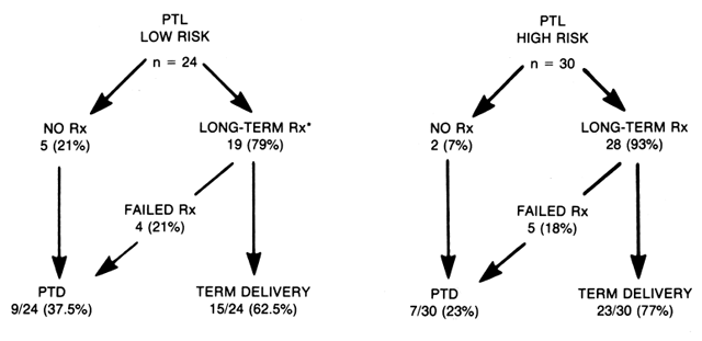
Efforts in the latter half of pregnancy continue to focus on the detection and prevention of pre-term labor. Detection of IUGR or fetal anomalies and the determination of the need, if any, of early pregnancy intervention are new objectives. Foremost of these efforts should be the early detection and prevention of preterm labor. A considerable effort should be extended to detect more subtle warning signs. As mentioned earlier, risk scoring for preterm labor is a tool that offers considerable promise. Clinically obvious factors to consider are a history of preterm labor or multiple gestation in the present pregnancy. Major medical/surgical illness may also result in the delivery of a preterm neonate. Foremost on this list are severe maternal hypertension, cardiac disease, and renal disease. Even severe maternal anemia has been implicated.35

Although the above list includes items that are often beyond the control of the managing clinician, they are nonetheless factors to be considered in managing the patient. Clearly, any patient who has conceived while taking ovulation inducing drugs should be evaluated for multiple gestation. More remotely, multiple gestation should be considered when conception occurs soon after discontinuance of oral contraceptives.36

Although the above theoretical considerations are interesting and may one day impact on the prevention of preterm labor, a more pragmatic approach is required. Should the patient be determined to be at risk for preterm labor, the patient can be seen at frequent intervals after 20 weeks' gestation. At these visits, it may be useful to perform routine cervical examinations and record changes in cervical dilatation, length in centimeters, and position, station of the presenting pan, and presence and frequency of uterine contractions.

Patient education regarding prophylactic measures, such as bed rest, and warning signs of premature labor, such as low back pain, cramps, increased vaginal discharge, or spotting, should be emphasized. It may not be possible to prevent the onset of preterm labor; however, early detection and consequent early employment of labor-inhibiting drugs will undoubtedly have a beneficial effect in a considerable number of patients.

Gestational Age Assignment

Inaccurate assignment of gestational age is often the limiting variable in high-risk pregnancy management. The incidence of patients with “suspect” dates has been estimated to range between 22% and 40%;37, 38 a relatively high number of patients who claim to have a reliable menstrual history may also fall into that category. It is obvious that failure to accurately assign gestational age in patients considered for elective repeat cesarean section or induction at term may result in iatrogenic prematurity.39, 40, 41 In other circumstances, the impact on gestational age assignment errors is more subtle and often not apparent unless obstetric versus pediatric age42 comparisons are made and perinatal statistics by gestational age are carefully evaluated. For instance, significant differences may exist in neonatal morbidity and mortality between a fetus inaccurately estimated to be 29 to 30 weeks' rather than 32 to 33 weeks' gestation. The benefit of accurate fetal age assignment is particularly important in the management of premature labor and premature rupture of the membranes prior to 32 to 34 weeks. In other circumstances, the patient may be spared the unnecessary apprehension attendant to an unnecessary diagnosis of postterm pregnancy if a liberal policy of sonographic estimation of gestational age is employed in early pregnancy. Such a policy may be cost-effective if one balances the cost of ultra-sound against that of unnecessary nonstress or stress testing, long inductions, and cesarean sections performed on the indication, failure to progress in cases falsely labeled postterm. Early detection of multiple gestation and congenital anomalies also has obvious benefit. At the other extreme, early confirmation of dates increases the credibility of routine intervention at a “true” 42 weeks' gestation, especially if there is associated oligohydramnios.

Clinically, a history of regular menses with minimal variation in flow duration or quantity (the last normal menstrual period, LMP) is reassuring regarding the reliability of menstrual data. Close correlation of fundal height with gestational age prior to 20 weeks, prospective maternal recognition and documentation of fetal movement (quickening) at 18 to 19 weeks' menstrual age, and physician detection of fetal heart sounds at 18 to 20 weeks by fetoscope are useful supporting data. However, the detection of fetal heart sounds as a measure of gestational age has limited value unless the patient is seen weekly prior to the anticipated time of detection; otherwise, the critical observation point may pass unobserved. Further, total reliance on these clinical estimators may be hazardous in the management of high-risk pregnancies in which a difference of 7 to 14 days may be critical. Table 8 projects the interval required to ensure 90% certainly (10% uncertainty) that an individual pregnancy is at least 38 weeks' gestation.43 It appears that one cannot make decisions that hinge on an accuracy of ± 1 week even when a menstrual history is designated “reliable.”

TABLE 8. Clinical Prediction of Maturity: Interval Necessary to Ensure 90% Confidence of Pregnancy Being At Least 38 Weeks’ Gestation


Indicator

Interval (Weeks)

Reliable LMP

42

Unreliable LMP

45

First fetal heart sounds

21

Quickening, nulliparous

25

Quickening, multiparous

25

The recent interest and availability of diagnostic ultrasound has dramatically increased the clinician's ability to assess fetai gestational age. Table 9 summarizes the predictive range and confidence limits of crown-rump length, biparietal diameter (BPD), and femur length measurements at various gestational ages. In normal gestation, the rate of growth of BPD or femur length is probably a function of genetic predisposition; the variance about the mean of both measurements increased with advancing gestational age.44, 45, 46, 47, 48, 49, 50, 51 The increasing biologic variation of the BPD with advancing gestational age is graphically displayed in Figure 5.46 Fetuses growing in upper percentlie limits have more rapid growth rates in the third trimester than those growing in the lower percentile ranks. This phenomenon must be considered in the interpretation of all sonographic data.

TABLE 9. Prediction of Gestational Age by Ultrasonic Determination of Crown-Rump Length and Biparietal Diameter


Predictive

Confidence

Author

Interval

Range

Limits

Campbell

Second trimester

±9 days

84%

Varma

Second trimester

±9 days

91%

Sabbagha

20 to 26 weeks

±11 days

90%


27 to 28 weeks

±14 days

90%


29 weeks

±21 days

90%

Sabbagha et al

GASA*

±1 to 3 days

95%

Robinson,

Crown-rump

±1 to 4 days

95%

 Fleming

 7 to 14 weeks


* Paired scans: first at 20 to 26 weeks; second at 31 to 33 weeks.

Fig. 5. Gestatonal age as X coordinate of graph depicting normal growth of the biparietal diameter( BPD ); maximum separation of large versus small BPDs between 30 and 33 weeks’ gestation. (Sabbagha RE, Barton FB, Barton BA: Sonar leiparietal diameter: I. Analysis of percentile growth differences in two normal populations using some methodology. Am J Obstet Gynecol 126:497,1976)

Since there appears to be significant advantage to early application of ultrasound, the clinician must develop a selective plan of management, which includes ultrasonic estimation of gestational age prior to 26 weeks in pregnancies at high risk for gestational age inaccuracy (Table 10), IUGR (Table 11), or prematurity (Table 12). Unfortunately, this scheme does not include all patients who will develop high-risk characteristics in the third trimester. A number of complications in which age assignment is critical, which may appear for the first time in the third trimester, are listed in Table 13 Other useful indications for ultrasound are found in Table 14.

TABLE 10. Indications for Routine Early Cephalometry: High Risk for Gestational Age Assignment Inaccuracy

  Irregular menses
  Recent discontinuation of oral contraceptives
  Fundal height versus LMP age discrepancy
  Late appearance of fetal movement
  Late appearance of fetal heart sounds
  Obesity
  Maternal age greater than 35 years

TABLE 11. High Risk for Altered Fetal Growth: Paired Scans at 20 to 26 and 31 to 33 Weeks (GASA) Plus Probable Serial Third-Trimester Scans

  Prior SGA/intrauterine growth retardation newborn
  First-trimester bleeding
  First-trimester viral infection
  Essential hypertension
  Diabetes
  Family history of hypertension/diabetes

TABLE 12. High Risk for Preterm Delivery: Indications for Routine Early Cephalometry and Possible Second Scan at 31 to 33 Weeks

  Candidate for elective repeat cesarean section
  Candidate for elective induction
  Candidate for “indicated induction”
  Prior preterm labor
  At risk for altered fetal growth
  At risk for multiple gestation

  Family history
  Ovulation induction


TABLE 13. Complications First Noted in the Third Trimester in which Gestational Age or Fetal Growth Assessment Is Important

  Premature labor
  Premature rupture of membranes
  Hydramnios/oligohydramnios
  Poor maternal weight gain*
  Poor fundal height growth*
  Oligohydramnios*
  Hypertension/preeclampsia*


* Fetal growth assessment is indicated.

TABLE 14. Ultrasound: Other Useful Indications

  Confirm normal pregnancy

  Gestational sac (5–6 weeks)
  Fetal viability (7th week)
  Fetal echoes (8th week)


  Prior to amniocentesis

  Locate placenta, umbilical cord, fetal structures
  Confirm viability


  Follow-up of abnormal α-fetoprotein
  Detect fetal anomalies

  Cephalic
  Spina bifida
  Genitourinary
  Limb reduction abnormalities


  Direct fetal surgery

The ability of all single measurement sonographic techniques to predict gestational age or assess growth is limited by both the inherent biologic variation of the part (BPD, femur length, crown-rump length) measured and the precision of the technique. In certain cases at extreme high risk for uteroplacental insufficiency (UPI), it is thus important that an attempt be made to determine the actual fetal growth curve and, where possible, match it to a specific population growth percentile. This is not possible in most instances, unless gestational age has been reliably assigned on the basis of a basal body temperature (BBT) or close correlation of menstrual history and physical findings. Thus, in most cases, one is forced to assign gestational age by matching a single BPD or femur length to a point on an established growth curve arbitrarily at the 50th percentlie. Since biologic variation of the BPD is most marked (see Table 9) after 26 weeks,46, 47 sonography probably should be used with extreme caution to assign gestational age if the first scan cannot be accomplished prior to 26 to 28 weeks. Although it is often acceptable to predict gestational age within a ± 11-day interval, as may be accomplished by a single scan prior to 27 weeks, management of many high-risk problems requires a more accurate estimation.

Fortunately, Sabbagha and co-workers, using serial cephalometry, has demonstrated that approximately 90% of monkey52 and human53 fetuses maintain an apparently predetermined relative cephalic growth pattern or ranking throughout pregnancy. On the basis of this concept, it is possible to assign a growth-adjusted sonographic age (GASA), which predicts gestational age within a ± 5-day (99% confidence limit) range; it also allows division of cephalic growth curves into three basic ranks: (1) BPDs greater than the 75th percentile; (2) BPDs between the 25th and 75th percentile; and (3) BPDs less than the 25th percentile. Variation in the external environment (UPI) may alter the projected course somewhat. However, since cephalic growth is spared until late in the course of most cases of placental insufficiency, this predictable pattern is disrupted in only a few instances prior to 31 to 33 weeks. Early onset of severe symmetrical growth retardation may be an exception to this statement. In such instances, cephalic growth may diminish prior to 31 weeks; as a consequence, fetal age may be overestimated by 1 week, even employing GASA. The issue has minimal clinical relevance, however, as the early failure of cephalic growth will persist and become a prime issue. To some extent, the failure of cephalic growth must then be managed independent of age. A similar error may develop in approximately 5% of cases where there is significant early acceleration of cephalic growth, which will result in underestimation of gestational age.

To obtain a GASA, the clinician must order two cephalic measurements, the first at 18 to 26 weeks and the second at 31 to 33 weeks.48, 53 Should the slope of cephalic growth be in excess of the 75th percentile, the original fetal BPD is assumed to have been large for gestation; in such a case, the gestational age assigned at the time of the first scan is reassigned to a gestational age consistent with a BPD at the 75th percentile (i.e., earlier gestation) at the time of the original scan; to accomplish this, gestational age is reduced up to 7 to 11 days. In contrast, the fetus whose head growth slope is less than the 25th percentile is assumed to have had a BPD at the 25th or lower percentile rather than the 50th percentile at the time of the initial scan and thus the gestational age is increased by 7 to l 1 days. The original assignment is maintained if an average BPD growth slope (25th–75th percentile) is observed.

In practice, we first make an assessment of the reliability of menstrual age. If menstrual age is deemed unreliable, an early scan is ordered (i.e., at 18 weeks). If there is significant discrepancy (greater than 7 days) between menstrual and sonographic age or the pregnancy is at high risk for UPI, GASA is planned and early scheduling of a second scan accomplished. In those cases complicated by preterm labor or preterm premature rupture of membranes at 27 to 34 weeks, in which no early ultrasound has been obtained, menstrual age is generally accepted if menstrual data are felt to be reliable, particularly if the menstrual age falls within the gestational age range predicted by sonography. In such cases, it is common to observe a menstrual age that falls at the upper limit of the gestational age range predicted by the late single scan.

It should be obvious from the previous discussion that the system employed even in perinatal centers with great resources is fraught with many problems regarding prospective fetal age assignment. In the absence of a future economic crisis, it is likely that early-pregnancy ultrasound will become more routine. Certainly, more widespread use of real-time ultrasound should facilitate a dramatic drop in current ultrasound charges. This reduction in patient cost, combined with the obvious benefits to pregnancy management, would include (1) early confirmation of gestational age, (2) early diagnosis of multiple gestation, and (3) exclusion of major congenital anomalies. If performed early in the first trimester, secondary benefits might include early diagnosis of (1) blighted ova (suspect if no gestational sac is noted after 8 weeks), (2) early diagnosis of ectopic pregnancy, and (3) early diagnosis of hydatidiform mole.

Crown-rump length is the most accurate technique to assign gestational age at 8 to 14 weeks.54, 55 There appears to be a transition in the reliability of measurement of both crown-rump and BPD in the 12- to 14-week interval in that the fetus is frequently more curled and a BPD may be more difficult to perform because of the variation in skull calcifications. Femur lengths are of no value in this interval.

Perhaps the best interval to perform routine ultrasound would be in the 14- to 20-week interval. In this interval, both the BPD44, 49 and the femur length50, 51 are very accurate in the assignment of gestational age. If routine sonography is performed at 17 to 20 weeks, the clinician is also provided with the most information regarding structural abnormalities of the cranial ventricular system, spine, abdominal contents, bladder, limbs, and abdominal wall. The determination of these abnormalities in this particular interval is important in increasing the options for future management of the patient. In rare circumstances, such as observed with chromosomal or undiagnosed congenital anomalies, the fetus may be symmetrically small, thus reducing the accuracy of both the fetal femur length and the BPD. However, in most instances, particularly with unusual head configuration or significant discrepancies between menstrual and sonographic age, the femur length is useful in corroborating the date assigned by BPD.

Fetal Evaluation

In the presence of stable maternal status, therapeutic decisions regarding intervention in obstetrics should focus on the issue of benefit (reduced fetal mortality) versus risk (morbidity and mortality of preterm delivery). Ideally, intervention should occur at an instant in time when the risk of intrauterine death (detected by some objective technique) outweighs that of neonatal death, usually from respiratory distress syndrome (RDS). Objective clinical evaluation of fetal health status is a primary goal of obstetric care. Prior to the late 1960s, the management of pregnancies thought to be associated with increased risk for fetal mortality was empiric. Pregnancies were frequently terminated by induction or cesarean section at a gestational age selected by evaluation of published data comparing the risk of intrauterine death with the risk of neonatal death at each week of gestation. For example, all pregnancies complicated by diabetes were interrupted at 37 weeks, the point in gestation at which the cumulative risk of intrauterine and neonatal death was the lowest. Large numbers of otherwise normally developing fetuses were thus delivered prior to full maturation. The benefit was a reduction in fetal mortality in the subset of high-risk patients with true UPI; the cost unfortunately was often unnecessary neonatal morbidity (RDS) or mortality in patients at high risk but not having true UPI. Management of pregnancy complicated by isoimmune disease was another example. Inability to specifically assess the status of the fetus at risk for erythroblastosis fetalis other than by historical data and antibody titers often resulted in the unnecessary premature delivery of an Rh-negative fetus in cases in which the father was heterozygous for the D antigen. The development of amniocentesis to evaluate the pregnancy at risk for isoimmune disease in the early 1960s revolutionized obstetric practice and initiated interest in developing other invasive and noninvasive techniques to selectively evaluate the status of the individual fetus. Since that time, techniques to evaluate fetal gestational age and growth, amniotic fluid content, endocrine products of the fetoplacental unit, instantaneous fetal heart rate antenatally or in labor, and fetal scalp blood samples have become available to the practicing clinician. Most techniques if properly applied allow the physician to delay intervention until maturity is attained. Proper use of instantaneous fetal heart rate and scalp blood sampling should promote more selective use of cesarean delivery for fetal distress. As a result of the availability of new diagnostic tools and superb neonatal care, the measure of clinical success in a practical sense is no longer perinatal mortality, which has been reduced dramatically in many centers, but rather perinatal morbidity.

Decisions to intervene or not to intervene in a high-risk pregnancy usually hinge on evaluation of maternal and fetal health status. In the vast majority of instances in which intervention is necessary, it is for fetal purposes; severe preeclampsia is a notable exception: maternal status deterioration is a more common indication. In all cases, evaluation of gestational age, lung maturity (also indirectly estimated by gestational age), and integrity of the fetoplacental unit function are key variables. Intervention prior to fetal lung maturity should occur only in the presence of documented evidence of UPI or significant maternal health deterioration. Ideally, intervention should occur when all major organ systems, particularly lung and brain, are mature. As a bottom line, it is emphasized that reassuring test results are more predictive than nonreassuring ones.

Fetal Growth Assessment

IUGR may be a function of intrinsic or extrinsic (UPI) factors. Although there are exceptions, IUGR of the intrinsic type tends to be symmetrically small, that is, both cephalic and chest-abdominal measurements are symmetrically two or more standard deviations below the mean for gestational age. In contrast, the extrinsic IUGR tends to be asymmetrically small, that is, cephalic head size is proportionately larger.

At birth, asymmetrical fetal growth retardation is characterized by subcutaneous and organ (especially liver) wasting in the presence of cephalic sparing. Although head growth by population standards may be normal in many cases, it is not known whether final cephalic growth is normal compared with the potential established at conception. We do know, however, that observed fetal growth deviates below that expected on the basis of the observed tendency of the normal population to maintain percentile rankings throughout gestation52, 53 In contrast, early and persistent symmetrical reduction in cephalic and body size is commonly associated with viral (TORCH) infections, multiple congenital anomalies, or significant chromosomal aberrations; many are idiopathic.

The prenatal diagnosis of IUGR is difficult. A high index of suspicion is essential; even then the diagnosis may be missed. More frequently, the prenatal diagnosis is not confirmed until delivery. A history of a prior SGA newborn, maternal failure to gain 2 lb/month in the third trimester, and markedly retarded fundal height growth in a patient with established dates are commonly recognized clinical warning signs. Third-trimester suspicion of oligohydramnios is a particularly significant observation. Maternal vascular disease is the most commonly associated clinical problem, accounting for approximately 20% to 30% of cases. Unfortunately, the remaining 60% to 80% of cases are idiopathic (40%–60%) or are associated with congenital infections (20%).

RISK FACTORS FOR IUGR.

Most risk-scoring systems pertain to risk for general perinatal morbidity and mortality. Few systems address the specific issue of IUGR. Galbraith and associates, for instance, have reviewed risk factors in 395 growth-retarded newborns. In that population, 122 (31%) had no risk factors.56 However, it was noted that patients with “no prenatal risk” had other prominent medical characteristics. Further, Galbraith's group found that risk factors for IUGR are addirive. Significant risk factors according to their time of presentation are shown in Table 15 (obstetrics historical risk factors), Table 16 (medical risk factors), and Table 17 (present pregnancy risk factors) for IUGR. They further found that the incidence of IUGR in pregnancies complicated by multiple gestation and preterm delivery was 21% and 11%, respectively. Evaluation of overall perinatal mortality after excluding: births less than 500 g and those complicated by congenital anomalies revealed a perinatal mortality rate of 16.1 (123:7635) per 1000 in the 7635 non-IUGR newborns. In contrast, the overall perinatal mortality in the IUGR group was 55.7 per 1000 in the 395 IUGR infants. When IUGR newborns were separated into two groups according to the presence of risk factors, it was observed that those with no risk factors had a mortality rate of 32.8 per 1000 (four deaths). This risk increased to 65.9 deaths per 1000 (18 deaths) in the IUGR infants at risk. Thus, there is a fourfold increase in perinatal mortality for the IUGR infant with risk factors as compared with a normal newborn. In evaluating the effect of multiple risk factors, the authors observe a progressive increase in incidence of IUGR from 2.3% in the absence of risk factors to 14.0% with three or more risk factors. Patients with no prenatal risk factors who subsequently delivered IUGR infants were significantly different with regard to decreased maternal weight, decreased maternal weight gain, increased smoking, higher incidence of first pregnancies, and smaller fundal height at term.

TABLE 15. Incidence of IUGR in Pregnancies With Risk Factors in Which There Had Been a Past Gestational Complication*


Total

IUGR

IUGR (%)

IUGR

384

77

20

Recurrent abortion

91

10

11

Fetal death (stillbirths)

106

10

9.4

Neonatal death

64

6

9.4

Preterm

260

23

9

Congenital anomaly

109

8

8


* The factors considered were singly found.
(Galbraith RS, Karchmar EJ, Piercy WN et al: The clinical prediction of intrauterine growth retardation. Am J Obstet Gynecol 133:281, 1979)

TABLE 16. Incidence of IUGR in Pregnancies With a Maternal Medical Complication


Total

IUGR

IUGR (%)

Hypertension


 Mild to moderate

93

11

12.0

 Severe

9

4

44.0

Renal


 Nephritis

24

5

21.0

 Other

79

4

5.0

Urinary tract


 infection

495

39

8.0

Cardiopulmonary

73

11

15.0

(Galbraith RS, Karchmar EJ, Piercy WN et al: The clinical prediction of intrauterine growth retardation. Am J Obstet Gynecol 133:281, 1979

TABLE 17. Incidence of IUGR in Pregnancies With an Obstetric Complication


Total

IUGR

IUGR (%)

Preeclamptic toxemia


 Mild to moderate

363

38

10.5

 Severe

39

12

31

Antepartum hemorrhage


 First trimester

640

51

8

 First trimester with


  recurrence

115

13

11

 Second trimester

250

19

8

 Second trimester with


  recurrence

42

5

12

 Third trimester

378

41

11

(Galbraith RS, Karchmar EJ, Piercy WN et al: The clinical prediction of intrauterine growth retardation. Am J Obstet Gynecol 133:281, 1979)

It is likely that more widespread application of early and serial ultrasound as well as gross evaluation of amniotic fluid volume in the third trimester will increase the likelihood of prenatal diagnosis. Although ultrasound, particularly serial cephalometry, is helpful, it is by no means perfect. We continue to more reliably predict a normal outcome than the reverse. Good judgment and common sense must temper the interpretation of available data. Factors that may limit the accuracy of the prenatal diagnosis include limitations of precision between serial determinations, variation in head shape, and the facts that clinical problems may not become apparent until after 26 weeks' gestation, slowing of cephalic growth may be a late manifestation of IUGR, and the pattern of fetal growth may vary according to IUGR pattern type. If risk factors for impaired fetal growth do not appear until the third trimester and an early BPD measurement is not available, the clinician must accept the ± 14- to 21-day confidence limits for a single scan at 27 or more weeks’ gestation. Subsequent evaluation of cephalic growth is thus limited. Since age is uncertain, growth evaluation is limited to comparison to the mean population growth rate; growth should average at least 2 mm/ week up to 34 weeks and at least 1 mm/week measured over a 2- to 3-week interval even in the lowest percentile ranking. Because of the slow growth rate and limitations in precision (1.0 mm-1.5 mm) of serial measurements, we require three measurements over a 3-week interval (days 0, 7 to 10. and 20 to 21) to assign a “no growth” designation.

Serial cephalometry, although helpful, has a false-abnormal diagnosis rate of at least 28%; these cases with retarded cephalic growth but normal birth weight are thought to be normal.57 In contrast, approximately 9% of cases have normal serial cephalic measurements in the presence of birth weights below the normal range for gestation. These false-normal cases probably represent cephalic sparing. Although frequently designated normal in the prenatal period, they are probably at greater risk than their normal counterparts for intrauterine death secondary to UPI. It is thus obvious that these techniques must be supplemented to increase the sensitivity and specificity of the diagnosis. Reevaluation of Campbell's original series (Table 18), in which he was able to identify 68% of 114 SGAs on the basis of serial cephalometry (but not abdominal circumference), suggests (on the basis of LBW percentile and retarded cephalic growth) that approximately three fourths (77: 105) of the SGAs will demonstrate symmetrical growth retardation.57 Although this seems to be a high ratio of symmetrical IUGR, further evaluation of assessment employing both cephalic and abdominal measurements seems worthwhile.

TABLE 18. Diagnosis of Small-for-Dates Fetus by Serial Ultrasonic Cephalometry


Cephalic Growth Patterns


Weight

Normal

Borderline*

Retarded†


Group

(No./%)

(No./%)

(No./%)

Total

AGA

220 (83)

18 (69)

21 (18)

259

BDRL SGA

22 (8)

4 (15)

16 (14)

42

SGA

‡24 (9)

4 (15)

77 (68)

105

Total

266 (100)

26 (100)

114 (100)

406


* Distribution of birth weights appears close to that of “normal” growth rate category.
† Difference between birth weights of “normal” versus “retarded” categories is highly significant (P < 0.001).
‡ 9% false-normal secondary to cephalic sparing in a population with asymmetrical retardation pattern.
(AGA, appropriate for GA; BDRL, borderline; from Campbell S, Dewhurst CJ: Diagnosis of the small-for-dates fetus by serial ultrasonic cephalometry. Lancet 2:1002, 1971)

Sabbagha has shown that GASA used alone allows the physician to compare a predicted or potential growth to observed cephalic growth rather than simply comparing observed growth to a population mean growth rate. In 
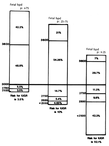
addition, GASA facilitates prediction of birth weight (Fig. 6) and risk of IUGR.48,53 The likelihood of a newborn's birth weight being less than 2750 g is 3.5%, 10%, and 52% for the greater than 75th, 25th to 75th, and less than 25th GASA percentile rankings, respectively. It thus seems reasonable to more vigorously evaluate the fetus in whom cephalic growth is less than the 25th percentlie, or in whom there has been significant deviation in growth subsequent to GASA assignment. Contrarily, it is reasonable to be more concerned regarding macrosomia if BPD growth is at greater than the 751h percentile, since 42% of such newborns will have weights of at least 3600 g.

Fig. 6. Risk for IUGR in fetuses who maintain cephalic growth in one of three percentile ( p) brackets (75th p, 25th–57th p, and 25th p). (Sabbagha RE: Intrauterine growth retardation. In Sabbagha RE (ed): Diagnostic Ultrasound, p. 106. Hagerstown, Harper & Row, 1980)

GASA PERCENTILES AND ABDOMINAL CIRCUMFERENCE.

Since ultrasound offers the possibility of determining symmetry of growth, it is reasonable to selectively assess fetal growth pattern in cases in which cephalic growth has been suboptimal (by GASA or serial third-trimester scans). Measurements of the abdominal circumference at the level of the ductus venosus is ideal, since it reflects fetal liver and subcutaneous tissue volume, both of which are diminished in association with IUGR.9 Comparison of the abdominal circumference and head circumference, although time-consuming and costly, can modify future management in the high-risk group by differentiating the symmetrically growth-retarded fetus from the asymmetrically growth-retarded fetus.

The initial data concerning the ratio of head circumference to abdomen circumference were reported by Campbell37 Classically, the ratio of the cephaloabdominal circumference is greater than 1:1 up to 35 to 36 weeks, after which the ratio is less 1. The transition period may vary about the 35- to 37-week interval, depending on the time of subcutaneous fat and soft tissue accumulation about the fetal abdomen. Campbell and Thoms subsequently determined the mean head-abdominal circumference ratio in 568 normal pregnancies.58 Simply employing the ratio, the authors were able to predict IUGR correctly in 71% of fetuses; all detected cases were asymmetrically growth retarded.

Recently, Sabbagha has described nine fetal growth patterns that depend on the relationship of cephalic versus abdominal circumference percentlies (Figure 7).59 The abdominal circumference percentiles are derived from the data of Tamura and Sabbagha, which provide these percentile measurements (percentiles 2.5 to 97.5) from the 18th to the 41 st week of pregnancy.59, 60 Growth patterns 3 and 6 reflect a symmetrical growth retardation with cephalic sparing, whereas persistent slowing of both cephalic and abdominal growth (pattern 9) two or more standard deviations below the mean increases the risk for symmetrical retardation. Thus, lets use demonstrating patterns 3 and 6 should be followed closely for UPI. Cases in which pattern 9 is observed may require evaluation for viral infection, multiple anomalies, or chromosomal aberration. In those instances in which asymmetrical 1UGR is suspected, determination of total intrauterine volume or relative amniotic fluid volume provides additional useful information.

Fig. 7. Nine fetal growth patterns noted by using both biparietal diameter ( Bpd) and abdominal circumference ( a-c) percentiles ( p ). (Tamura RD, Sabbagha RE: Assessment of fetal weight. In Sabbagha RE (ed): Diagnostic Ultrasound, Chap 9, p 99. Hagerstown, Harper & Row, 1980)

In practice, we perform serial BPD measurements at (1) 18 to 24 weeks, (2) 31 to 33 weeks, and (3) 35 to 38 weeks. GASA is determined on the basis of the first two scans, which also provide relative risk of IUGR. The long interval between the first and second scan increases the likelihood of detection of the small difference in growth of fetal head size of fetuses with large versus small cephalic growth patterns.61 Performance of the second scan at 31 to 33 weeks has only minor limitations in the detection of IUGR, since the majority of IUGR pregnancies do not develop subnormal growth until after 32 weeks' gestation.61 The final reading at 35 to 38 weeks is used to evaluate the trend of growth subsequent to attaining the abdominal circumference at the time of the second scan.

TOTAL INTRAUTERINE VOLUME (TIUV).

Oligohydramnios accompanies and increases the risk of fetal death in many cases of significant IUGR. As a result, uterine volume (fetal and placental mass plus amniotic fluid) assessment, obtained by measuring transverse and longitudinal diameters of the uterus, has been advocated.62 A value more than 1.5 standard deviations below the mean for gestational age is highly suggestive of IUGR; a value of 1.0 to 1.5 standard deviations below the mean for gestation represents a gray zone where the diagnosis is less secure. Recently, clinicians have begun to assess amniotic fluid volume more simply with real-time ultrasound. A pocket of fluid greater than 1 cm in diameter is helpful in reducing the risk of growth retardation. This more simplified approach reduces the necessity to know the precise gestational age or to perform the calculations required for TIUV measurements.

Fetoplacental Function Evaluation

Fetoplacental status may be assessed in the third trimester by biochemical (estriol or placental lactose) or biophysical nonstress test, oxytocin challenge test means. These subjects are eloquently discussed elsewhere in these volumes and are mentioned in overview to reflect some personal bias of the author. In most instances, the anticipated result is patient and physician reassurance that pregnancy can be safely continued. The overwhelming majority of reassuring results are highly specific and are associated with favorable outcome (low false-normal rate); the incidence of false-positive tests (no evidence of fetal or placental disease during labor or at delivery following an abnormal test result) in most instances ranges from 40% to 60%. Consideration to intervene on the basis of a single abnormal result, particularly in the presence of uncertain fetal pulmonary status, should be rare unless maternal status deteriorates.

Fetal assessment tests should be ordered in conditions at risk for UPI. By design, they offer little or no useful information in the evaluation of pregnancies with subsequent morbidity and mortality that are the consequence of trauma, congenital anomalies, or cord accidents. Since most assays/ procedures are expensive, the clinician should consider if the test to be ordered actually provides useful information not detectable by other means. Historically, biochemical assay, particularly urinary estriols, has been commonly used. However, in recent years, biophysical assessment of fetal placental respiratory function-nonstress test (NST) or contraction stress test--tCST) or, more recently, of fetal activity has largely replaced biochemical assays. In general, the NST/CST is considered more reliable, can be performed at less frequent intervals, and is less expensive than currently available biochemical measures of fetoplacental function.

OXYTOCIN CHALLENGE TEST.

Since it is well known that uterine contractions are associated with a reduction in uteroplacental blood flow, it is reasonable to employ spontaneous or oxytocin-induced contractions with a frequency of three in 10 minutes as a standard test of fetoplacental respiratory function.63, 64, 65, 66, 67, 68, 69 Stress of that magnitude has been proved clinically to be used in separating the occasional fetus with suboptimal oxygen reserve from the vast majority of cases with normal reserve.65, 66, 67 This stress does not significantly compromise the normal fetus with adequate reserves. In contrast, a fetus can be presumed to have diminished “fetal reserve” should repetitive late decelerations associated with most contractions of any frequency be noted; the absence of accelerations and baseline variability increases the specificity of the diagnosis. In ordinary circumstances, an oxytocin challenge test (OCT) or CST is initiated in pregnancies at high risk for UPI once the diagnosis is suspected. Circumstances in which the CST or OCT is indicated are as follows:

  Nonreactive NST
  Diabetes mellitus
  Preeclampsia
  Chronic hypertension
  Intrauterine growth retardation
  Post-term pregnancy (42+ weeks)
  History of previous stillbirth
  Narcotic addiction
  Sickle cell hemoglobinopathy
  Chronic pulmonary disease
  Organic heart disease
  Rh isoimmune disease
  Meconium-stained amniotic fluid

Testing may begin as early as 28 to 30 weeks in conditions such as early-onset maternal pre-eclampsia (Figure 8). We routinely begin NST/CST testing at 30 weeks' gestation in class D or greater diabetics. In contrast, we delay initiation of routine testing until 32 weeks in class C, 34 weeks in class B, and 36 to 37 weeks in uncomplicated class A diabetics.

Fig. 8. Scheme for initiation of NST/CST testing according to weeks of gestation. ( PIH, pregnancy-induced hypertension)

In some instances, oxytocin is not required (spontaneous CST); in most cases, however, it is necessary to induce uterine activity by administering intravenous oxytocin using an oxytocin infusion pump. The fetal heart rate is recorded for a baseline (nonstress) period of 15 to 20 minutes. If three contractions per 10 minutes are observed within this time interval, oxytocin need not be administered. If three contractions are not observed, oxytocin is administered beginning at a 0.5 mu/min interval. In most instances, it is not necessary to exceed 20 mu/min. The CST (Figure 9) is negative (normal) if there are no late decelerations associated with a contraction frequency of three in 10 minutes. Such a result is very reassuring; it is associated with a 1 to 2:1000 false-normal rate. In most instances, the procedure is repeated on a weekly basis. However, more frequent testing may be indicated for insulin-dependent diabetics. patients with moderate to severe preeclampsia, or postdate pregnancies, particularly when associated with oligohydramnios.

Fig. 9. Representation of possible contraction stress test ( CST) outcomes. The upper channel of each strip is the fetal heart rate ( FHR ); the lower channel reflects uterine contractions ( UC) in a 10-minute time interval. (Depp R: Assessment of fetal weight. In Sabbagha RE (ed): Diagnostic Ultrasound Applied to Obstetrics and Gynecology. Hagerstown, Harper & Row, 1980)

The presence of consistent and persistent late decelerations with most uterine contractions regardless of their frequency is diagnostic of a positive CST (Figure 10). In many instances, this is associated with decreased variability and absence of fetal heart rate accelerations with fetal movement. Since a positive CST is an indication of UPI, delivery should be accomplished within a relatively short interval if fetal pulmonary maturity is present. It should be remembered, however, that approximately 24% to 60% of such cases are actually false-positive (false-abnormal) when not preceded by a nonreactive NST; if allowed to labor subsequently, such patients will demonstrate no further late decelerations. For this reason, it may be safe to attempt a vaginal delivery if it is possible to rupture the membranes, apply a direct electrode, and allow the patient to labor in a lateral decubitus position. In the absence of pulmonary maturity, evaluation of fetal status can be supplemented by daily plasma or urinary estriols; the pregnancy may be allowed to continue as long as the estriol levels are stable or rising. Although a positive CST result is less predictive (24%–60% false-positive) than a negative one (99% true-negative), the certainty of diagnosis may be increased by evaluating the fetal heart rate baseline for the presence or absence of fetal heart rate accelerations with fetal movement.65 Most cases in which the positive OCT is accompanied by accelerations with a magnitude of at least 15 beats per minute in response to fetal movement will tolerate labor or can tolerate some additional time interval to delivery. If accelerations are absent and baseline variability is poor or absent, it is unlikely that the supplemental use of serial estriols will be helpful in allowing the pregnancy to continue until maturity is obtained and unlikely that labor will be tolerated without further evidence of late decelerations. In those cases in which cesarean is planned after a positive CST, it may be desirable to stop the oxytocin, begin administration of oxygen, and allow time for intrauterine recovery prior to delivery.

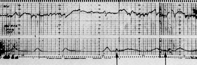
Fig. 10. Reactive (negative) result of contraction stress test using an abdominal electrocardiogram as source. The lower channel reflects uterine contractions of 40- to 60-second duration. Small superimposed peaks represent fetal movement. Fetal heart rate (FHR) baseline (upper channel) is 145 to 150. Note accelerations of FHR in response to fetal movement ( arrows) on lower channel. The estimated magnitude of accelerations is 25 to 30 beats/min. (Depp R: Dynamics of fetal growth. In Sabbagha RE (ed): Diagnostic Ultrasound Applied to Obstetrics and Gynecology. Hagerstown, Harper & Row, 1980)

NONSTRESS TEST.

Although the CST/OCT is highly predictive with a very low false-negative rate, the test is time-consuming, requires intravenous fluids and oxytocin, and in most instances must be performed in a labor unit. As a consequence, the procedure is costly as well as invasive. Early work by Kubli and Rutgers70 and Hammacher71 established the characteristics of the fetal heart rate baseline in normal and abnormal fetuses. At this point in the evolution of antepartum fetal heart rate monitoring, it became obvious that it was rare to observe repetitive late decelerations consistent with a positive CST/OCT following an initial baseline observation period (prior to initiation of oxytocin) characterized by significant accelerations of the fetal heart rate in association with fetal movements. The observation of the importance of the reactive nonstress portion of the CST thus paved the way for development of the nonstress test (NST) as proposed in this country by Lee72, 73, 74 and subsequently employed by Schifrin75 and Evertson76 and their colleagues and numerous other investigators (Figure 9).

It is generally accepted that a reactive (negative or reassuring) NST is characterized by the presence of two or more accelerations of the fetal heart rate observed in 20 or less minutes; the accelerations must be at least 15 beats per minute above the baseline. Apparently it does not matter if the accelerations observed are associated with spontaneous movement or in response to manual stimulation. If the fetus is not reactive within the first 20 minutes, the fetus should be stimulated artificially and observed for an additional 20 minutes before making the designation “nonreactive.” The latter step minimizes the possibility of lack of activity associated with fetal sleep cycles. A non-reactive NST is characterized by accelerations of less than 15 beats per minute or less than the normal CST. Like the normal CST, a reactive NST is very reassuring. The false-negative (false-normal) rate approximates that of a CST; most series report a false-negative rate of 1 to 3:1000. It is currently thought that the unsuspected death is a consequence of unforeseen cord accidents, such as tight nuthal cords, cord knots, or “cord vulnerability” associated with oligohydramnios. Such deaths are currently not preventable, as is probably the case with sublethal insults resulting in cerebral palsy.

There has been some concern that late intervention may occur if the clinician delays until a nonreactive NST followed by a positive CST evolves. This concern does not seem realistic for several reasons: (1) The NST appears to be a very conservative indicator of fetal status in that the nonreactive rate may be as high as 20% to 35% for the NST versus only a 3% positive rate for the CST. This significant increase in the population judged to be at special risk identifies a subset population that may be better served by the CST with its associated lower false-abnormal rates. (2) There appears to be a gradation of non-reactive results (decreasing acceleration magnitude, more obvious loss of variability); clearly the more fiat the base-line, the less favorable the outcome. Since these changes are generally observed over time, a CST can be ordered to evaluate fetal status at a very early time in development of the nonreactive state. (3) The majority of false-positive CSTs are characterized by a baseline that is reactive. (4) There is little if any increase in perinatal mortality when comparing the patient with a reactive NST and one with a nonreactive NST followed by a negative CST. (5) From a practical standpoint, the diminished cost and the patient convenience of the NST allow the clinician to assess certain high-risk pregnancies more frequently and less expensively than is realistically possible if the CST is used as the primary test procedure.

In most cases, an NST is used as a screening test; reactive tests may safely be repeated at weekly intervals; however, some investigators repeat them more frequently in insulindependent diabetics, preeclamptics, and patients at high risk for IUGR or postmaturity. Twenty percent or more of tests are nonreactive; such cases require CST. Ninety percent of subsequent CSTs will be negative. Most of the few who subsequently have a positive CST have good outcome; deaths are associated with prematurity or, occasionally, congenital anomalies. If there is a deficiency in the NST, when compared to the CST, there may be a possible diminished ability to detect variable decelerations, which may be detected in up to 10% of CSTs but in a considerably smaller number of NSTs. The etiology of these variable decelerations has been specifically determined. Primary concerns would be the presence of a nuchal cord (currently not detectable in the prenatal period) or vulnerable cord as a consequence of oligohydramnios. Possible solutions to this potential deficiency of the NST are (1) real-time scanning for adequacy of amniotic fluid volume for patients with nonreactive NSTs or clinical suspicion of diminished amniotic fluid volume on the basis of physical examination or variable deceitrations noted on NST and (2) nipple stimulation to induce uterine activity in all patients, to facilitate physiologically induced uterine activity. The latter approach may or may not be effective, in that uterine hypertonus and hyperactivity may be induced in some patients, while in others the contractile activity generated may not be equivalent to the traditional CST. Further research must be done in this area.

FETAL ACTIVITY.

Assessment of presence or absence of fetal activity has been used on a subjective basis by clinicians for many years.77 Recently, it has been reconfirmed that active fetal movements are an expression of fetal well-being. A sudden increase (cord compression or abruption) or a decrease (chronic UPI) in activity may precede fetal death. Some have suggested that antepartum surveillance of patients with decreased fetal movement would be more beneficial in reducing perinatal morbidity than simply monitoring all classically high-risk patients. Total reliance on such a scheme is unlikely, in that patients tend to observe a decrease in fetal movement as gestation progresses; patient reliability also varies considerably.

ESTRIOLS.

Third-trimester biochemical assessment of the fetoplacental unit is most commonly done using either urinary or plasma estriols.78, 79, 80, 81 Since estriol is the product of fetal and placental compartments, its assay had great theoretical potential.78 Prior to the mid 1970s, serial urinary estriol assays were the only commonly used technique of evaluating fetal health. Since the advent of NST/CST and biophysical profile programs, biochemical surveillance has been largely replaced by biomechanical surveillance. This is a function of potential limitations of the technique where normal values are not strictly related to suboptimal fetal health, as well as the gestational age dependency of normal values.

The clinician who employs estriol determinations in his management must understand the estriol synthesis pathway and the potential for its disruption. For instance, chronically low for gestational age estriol values may be seen in association with anencephaly (presumed failure of central stimulation or absence or aplasia of the adrenal). Further, liver or biliary disease or disturbance of gut flora by oral antibiotics may reduce estriol levels detected in the maternal urine, since approximately half of estriol conjugates are excreted by way of bile into the enterohepatic circulation where they are hydrolyzed by intestinal bacteria to free estriol, which is then reabsorbed. In addition, the clinician must be aware of the complexities of the analysis of plasma or urinary estriols. The first step in analysis is acid or enzyme hydrolysis. There may be significant inconsistent losses of estriol in the assay. Significant reduction of recovery also occurs in the presence of urinary antiseptic methenamine. Many laboratories correct for these recovery losses by using an internal standard of radioactive estriol-16-glucuronide; however, many do not. The clinician should be aware of the laboratory policies in his institution. Although serial estriols are used with considerably less frequency, when used they are probably best employed in the management of pregnancy complicated by diabetes, hypertension, suspected IUGR, or postmaturity. In most instances, serial values should be compared to both a standard curve and to the average of the preceding two or three values. Collection of specimens is usually on a daily basis in diabetes or two to three times per week in hypertension, suspected IUGR, or post-maturity. A single value should virtually never be used for assessment of management. Abnormal patterns may be manifest in one of three patterns: (1) progressive downward slope, (2) rapid fall (35%–40% of baseline established by average of values for preceding 3 days), (3) persistent low values. However, the clinician must be hesitant in acting immediately on the basis of low values. Persistent low values may be seen with failure to collect a true 24-hour specimen, renal function impairment, or overestimation of gestational age. High urinary glucose or methenamine therapy for cystitis may also be associated with false-abnormal values. Assay of 24-hour urinary creatinine excretion in parallel with estriol provides a reasonable internal standard to determine if 24-hour collection is complete. Sudden drops in estriol must be interpreted with caution; adequacy of the collection can be verified by noting 24-hour urine creatinine values; the average patient excretes 12 mg to 15 mg of creatinine per kilogram (approximately 1 g) of body weight per 24 hours in her urine. Intelligent assessment of gestational age and renal function is also helpful.

Some investigators have favored collection of serial short-term urine collections of which an estriol/creatinine ratio is calculated. However, in those institutions still using estriols, the recent trend is to favor plasma over urinary estriol assay. Plasma estriols may be assayed as (1) unconjugated (free) estriol, which comprises 8% to 10% of total estriol;80 (2) total estriol;80 and (3) immunoreactive estriol.81 Patient inconvenience and inability to reliably collect a true 24-hour urinary specimen have provided the impetus to assess plasma estriols. Plasma estriol collection is rapid (obviating delays in obtaining results), simple, and complete. Rising or stable values within normal limits are very reassuring. False-positive results unfortunately are common.

Plasma unconjugated estriol levels are primarily dependent on fetoplacental production and secretion rates; they are therefore less affected by disorders of maternal liver or kidney. Distler and co-workers feel that plasma free urinary estriols are the most useful of the plasma tests in the management of the pregnant diabetic.80 Total plasma estriol includes both the unconjugated and the conjugated (glucuronides and sulfates) fractions. The assay includes solvent extraction or enzyme hydrolysis prior to radioimmunoassay. As a consequence, assay time is 6 to 8 hours in a routine laboratory.

Some advocate the use of immunoreactive estriol, which reflects approximately 40% of total estrio.81 This assay has the advantage of eliminating the extraction process and thus the assay time is considerably less than that of either total or unconjugated estriol. At the present time, clinical utilization of plasma estriols is not widespread.

HUMAN PLACENTAL LACTOGEN.

Human placental lactogen (HPL) is a single-chain polypeptide hormone immunologically similar to human growth hormone, with a molecular weight of approximately 21,000 and half-life of approximately 25 minutes.82, 83 It is produced by the syncytiotrophoblast. Although the precise function of this hormone has not been determined, it does have inherent somatotrophic, lactogenic, mammo-trophic, and luteotrophic activities. As a result, it has also been called human chorionic somatomammotropin.

The clinical significance of HPL is controversial. It was originally thought that HPL levels might be useful in predicting the outcome of patients with threatened abortion. However, many patients with low HPL values do not abort; clinical over assessment of gestational age may be a factor. First-trimester real-time ultrasound evaluation of uterine contents is probably more accurate in predicting eventual outcome than is HPL. At one time, HPL levels were thought to be of value in fetal placental function in hypertensive diseases of pregnancy, diabetes, and postmaturity or in assessing or predicting the presence of IUGR; subsequent studies have not supported this conclusion. Unfortunately, there is a high false-positive and false-negative rate when HPL is used to assess these conditions. Furthermore, HPL levels do not necessarily fall prior to or immediately after intrauterine fetal death. HPL levels correlate best with placental weights; however, this correlation is not consistent. As a consequence, the value of HPL in assessing placental integrity is limited. If there is a clinical application, it may be for high-risk screening.84

Fetal Maturity Assessment

Spontaneous premature labor, premature rupture of the membranes, or high-risk pregnancies requiring premature intervention for maternal-fetal indications account for most newborns delivered prior to functional pulmonary maturity. However, as recently as 1976, 8% to 15% of newborns developing RDS followed failure to use currently available techniques to assess fetal pulmonary maturity or to correctly assess gestational age.39, 40 Although most cases of RDS are not preventable, a significant fraction could probably be eliminated by more specific diagnostic techniques to assess fetal health status or willingness to rely on reassuring results, thus avoiding unnecessarily early intervention.

Fetal lung maturation is marked by biochemical changes of surfactant with increasing gestational age. RDS/hyaline membrane disease (HMD) is a clinical state of respiratory distress/pathologic findings at autopsy of hyaline membrane associated with a deficiency of surfactant. Surfactant is a generic term for a complex mixture of substances that act as a detergent to lower alveolar surface tension (reduce the tendency to collapse). It is possible to assess surfactant in both a quantitative and qualitative sense by performing a number of biochemical assays on amniotic fluid. Eighty to 90% (by weight) of surfactant is lipid; 80% to 90% of the lipid is phospholipid. Lecithin is the major phospholipid (70%–80%)85, 86, 87 Protein (10%–20%) and carbohydrate 11%–2%) compose the remaining 10%–20%. Saturated (palmitic acid at C-1 and C-2) dipalmitoyl lecithin (DPL) accounts for 50% of total lecithin88 Phosphatidylcholine (PC) and phosphatidylglycerol (PG) are two acidic phospholipids that act to stabilize lecithin in the surfactant layer.89, 90 PG seems to be most active in this regard. Sphingomyelin, a sphingolipid, also has surface-active properties.

LECITHIN-SPHINGOMYELIN (L/S) RATIO.

Under ordinary circumstances, surface-active lecithin begins to appear in the amniotic fluid at approximately 24 to 26 weeks. Sphingomyelin appears earlier and in higher concentrations early in the third trimester. Since sphingomyelin concentrations change very little with gestation, it is useful as an “internal standard” for lecithin in performing the L/S ratio.91, 92, 93 In uncomplicated pregnancies, prior to 30 to 32 weeks, the ratio is generally less than 1.5. At 34 to 35 weeks, there is a fourfold rise in lecithin versus sphingomyelin, which remains relatively stable or actually decreases at the time of the iecithin surge. An L/S ratio of 2, ordinarily achieved by 35 to 36 weeks' gestation, is clinically accepted as a reliable indicator of functional pulmonary maturity.

The timing of lung maturation may vary considerably in high-risk pregnancies.91 In many instances, conditions known to modify the timing of suffactant appearance are known only in retrospect; a diagnosis of class A diabetes or IUGR is unfortunately made only after delivery. It is thus not surprising in unselected patient series that the L/S ratio is independent of gestational age and birth weight. As a result, the clinician should not assume the presence of pulmonary maturity unless there is early sonographic confirmation of dates and the patient is at term.94 Consequently, consideration of surfactant activity assessment is important whenever intervention for maternal or fetal indications or inhibition of preterm labor is considered. This consideration may be important very early in the third trimester, since approximately 19% of cases at risk for premature delivery at 28 to 32 weeks and 35% at risk to deliver between 33 and 36 weeks demonstrate an L/S ratio in excess of 2.95

An L/S ratio in excess of 2 correctly predicts maturity in approximately 98% of cases; only 2% of patients develop RDS (false-mature/false normal). Most false-mature results are associated with perinatal asphyxia, maternal diabetes, or Rh isoimmune disease. In contrast, an L/S ratio less than 2 is not as predictive; approximately 35% of newborns delivering within 72 hours of an L/S ratio less than 2 actually develop RDS (true-abnormal).96, 97

LUNG PROFILE.

Recently, Gluck's group has developed a more specific measure of surfactant activity, the lung profile.97, 98 In addition to the L/S ratio, the profile includes assay of the percentage of DPL, PI, and PG (Figure 11). The profile seems to have particular merit in patients with an L/S ratio less than 2 and perhaps in diabetics. Ordinarily, PI increases in parallel with a rise in the L/S ratio up to a value of 2; thereafter, it falls to early third-trimester values. In contrast, PG first appear in normal pregnancies at 35 weeks; its presence in excess of 3% signals the presence of stable mature function of the neonatal lung. In a recent series of 57 cases with an L/S ratio less than 2, the profile correctly predicted maturity in 14 of the 18 cases (78% reduction of the false-immature) in which the L/S ratio was less than 2. Prediction of true outcome thus increased from 68% to 93% in that series.

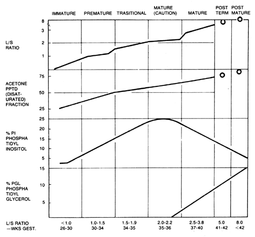
Fig. 11. Form used to report lung profile. L/S ratio, acetone-precipitated desaturated lecithin, phosphatidyl inositol, and phosphatidyl glycerol are recorded at points on curves. Values usually fall within a given grid, and lung maturity may be read off at top of form. (Kulovich MV, Hallman MB, Gluck L: The lung profile: I. Normal pregnancy. Am J Obstet Gynecol 135:57, 1979. Copyright © by the Regents of the University of California, 1977)

SURFACTANT ASSESSMENT IN HIGH-RISK PREGNANCY.

The timing of functional maturation (L/S ratio greater than 2) of the fetal lung may vary according to maternal or fetal placental disease states. It may occur as early as 28 weeks in association with class F and R diabetics, in prolonged rupture of the membranes, and in cases complicated by severe hypertension-proteinuria syndromes. In contrast, maturation is often delayed in class A diabetics.

The issue of surfactant assessment in pregnancies complicated by maternal diabetes is both controversial and worrisome. The infant of the diabetic mother has a six fold increase in risk for developing RDS.99 It is generally accepted that traditional assessment of suffactant using the L/S ratio in diabetics has a false-mature rate of 3% to 5.5%100, 101 versus an expected rate of 2%97 in the normal population. A higher incidence of cesarean section, asphyxia, and premature delivery, all of which increase the risk of RDS, may influence the difference in predictability.102 The diabetic is also prone to gestational age assignment errors. Fortunately, it appears that the presence of PG in excess of 3% ensures greater likelihood of pulmonary maturity.98

Pregnancy complicated by hypertensive disease, particularly when severe and associated with significant proteinuria or placental infarction, may be associated with accelerated pulmonary maturity manifest by early elevation of the L/S ratio.91, 93 In some cases. although the L/S ratio is less than 2, PG may be elevated to a mature 3% level as early as 29 to 30 weeks.98 Advancement of maturation is generally not the case in milder cases or more acute terminal cases.

Although there is some controversy, most investigators feel that prolonged rupture of the membranes is associated with early appearance of pulmonary maturity.103, 104, 105 Most series to date are retrospective and do not control for other factors that may impinge on suffactant induction. However, in cases in which membranes are ruptured longer than 24 hours, the lung profile when corrected for gestational age demonstrates an earlier appearance of PG (by 1.5 weeks), higher mean L/S ratios, and an early appearance of a mature L/S ratio.98

Preparation for Delivery

The obstetrician managing the high-risk mother should make an attempt when possible to plan delivery timing in cooperation with the neonatologist. Except in the case of fetal distress, there are few instances when the obstetrician cannot afford to vary the timing of the delivery according to availability of the neonatologist. Such planning not only is important for increasing the chance of survival of the high-risk newborn but also is very useful in fostering professional interrelationships and a positive relationship between the mother and the neonatologist, which is so vital to perinalal care.

Back to Top
INFANT FOLLOW-UP

Those who perform long-term follow-up of high-risk newborns are often not considered as essential members of the perinatal process. However, this is a mistake of major magnitude. It is critical that a person or group of persons who are willing to devote a considerable portion of their efforts be chosen for this purpose. Ideally, one physician/ group would follow all newborns delivered in an institution. A general pediatric neurologist with no special interest or information data base in newborn neurology will provide less than ideal follow-up. It is important that causal relationships not be stated unless there is objective information. Ideally, cases with poor outcome should be reviewed by a team (the obstetrician, the neonatologist, and the neurologist) for potential insults. Unfortunately, causal relationships commonly decided on are frequently based on second-hand information provided by the mother. In many instances, a low Apgar score is assumed to be equivalent to “birth asphyxia.” This is obviously not always a reasonable conclusion. It is thus very important that the persons performing the follow-up be very knowledgeable in the areas of perinatal physiology, the predictive value of fetal monitoring, and multiple origins of low Apgar scores and, in addition, have in-depth knowledge of the available literature regarding prenatal fetal morbidity and mortality in addition the pathogenesis of newborn neurologic complications such as intraventricular hemorrhage and cerebral palsy. Some of the established information in this area is discussed in Chapter 99.

Back to Top
PERINATAL DEATH

Nothing arouses a wider spectrum of psychological responses than the death of a near-term fetus or newborn. Both the patient and the care-givers are affected. Unfortunately, many care-givers are not prepared to deal with the issue, although this is becoming less common. Medically, there is a tendency for the care-givers to immediately begin a process of determining “what went wrong.” Although this is academically correct, it is often carried on in the wrong location, such as a nurse's station, elevator, or cafeteria. Discussion of such matters is best handled privately among the limited number of persons involved in the care of the patient. It is necessary that some persons, particularly those experiencing a perinatal loss for the first time, talk out their anxieties and be reassured regarding their actions. Frequently, there is a misunderstanding of what has transpired and the possibility of preventability. It is thus important to resolve and separate the emotional response of the care-giver to perinatal death from the more objective discussion of the medical care given.

A more important concern is the counseling of the patient and her family. This issue has been described in detail elsewhere.106, 107 It is important to (1) reassure the patient and family that they bear no responsibility, (2) encourage the acute grieving process, (3) offer referral to parent support groups in the community, and (4) prepare the family to anticipate weeks of emotional lability that are normal during the first few months after the death. The health-care team should present the circumstances of the death in an open, straightforward fashion. Grieving parents grasp limited amounts of information during early conversations with medical personnel. Therefore, explanations may need to be repeated during the first few days after the loss. It is very useful to allow the family members to touch and hold the dead newborn, even when it is abnormal. Parents frequently imagine that the dead baby looks considerably worse than is true in reality. Finally, it is often useful to provide some personal material for retention by the family. Something as simple as an ultrasound picture or snapshot taken after delivery is frequently useful. Other mementos include a footprint sheet, hospital bracelet, baptismal certificate, or lock of hair. Finally, the number of persons who counsel the patient should be limited, to avoid confusing the patient and her family and to avoid the possibility of misinformation.

Back to Top
OVERVIEW

With the exception of a few disease processes, proper management results in a favorable outcome for most patients. Prenatal biochemical and bio-physical testing should be selective and thoughtfully planned, not only to detect incipient problems but also to provide reassurance of normal fetal growth and development. The patient should come to understand that the care given is designed to provide favorable outcome to most, but not necessarily all, women with similar problems. No program eliminates all untoward outcomes. In those cases in which the prognosis is particularly poor, it may be necessary to repeatedly stress realistic expectations. Application of such an approach should not disrupt the development of trust between patient and care-giver, but rather reinforce the seriousness of the matter at hand. This is most important should intervention/nonintervention prior to 36 to 37 weeks be an important consideration. The patient must understand that while each passing day increases the likelihood of neonatal survival and decreases the incidence of prematurity-dependent morbidity, it does so at the risk of fetal death. Pefinatal loss in the neonatal period of a normally developed infant is in many ways worse than a fetal loss. Past practice patterns based on “what ifs” or “what might occur if pre-term delivery is delayed?”, even in the presence of reassuring biophysical testing, are to be condemned; a practice pattern that results in more neonatal deaths from prematurity than fetal deaths secondary to the associated disease process is not acceptable. Prospective prenatal management must thus include a willingness to accept an occasional fetal loss in the hope that such losses will be more than balanced by reduced neonatal morbidity and mortality.

Similar problems arise in the intrapartum period. Prospective management of the laboring patient should encompass traditional fetal heart rate monitoring and interpretation. Much is yet to be determined with regard to monitoring the fetus during labor. In the early 1970s, for instance, we abandoned the use of meconium as a primary indicator of fetal status and now rely on it only as a risk factor. Future research will undoubtedly teach us that our current approaches are not as specific as we assume they are. Even continuous electronic fetal monitoring has limits. In general, continuous intrapartum monitoring is highly predierive of good outcome but very unsatisfactory in predicting poor outcome. Even though it is very efficient at predicting good outcome, occasionally a newborn will be delivered with Apgar scores and umbilical cord gases lower than anticipated on the basis of a fetal monitor tracing. The physician may be tempted to overreact to this inherent inadequacy of our present detection system by being more surgically aggressive. Although such an approach may seem superficially logical, the result may be to inflict greater harm to the overall population through increased cesarean sections, hospital duration, and so forth. Further, it may be that some deaths presumed to be preventable by more aggressive actions may not be so. Rather, the physician should understand that our present fetal monitoring system does not predict or assess all insults. Occasionally, the unpredicted poor outcome may be a function of two or more processes, including a primary insult, such as mild to moderate cord compression (detectable), and a secondary insult, such as unsuspected fetal sepsis (with associated shock, underperfusions, and acidemia) or congenital heart disease (with associated shunting and diminished cardiac outputs). In other situations, the modifier may be diminished fetal tolerance to stresses ordinarily tolerated by the vast majority of fetuses. The obstetrician, for instance, may be dealing with an unsuspected IUGR fetus with diminished glycogen and cardiovascular reserves to withstand stress. Fetal heart rate monitoring is at best an indirect method of assessing neural and cardiovascular responses to stress (such as head or cord compression, myocardial hypoxia). Even scalp blood sampling performed serially during labor will occasionally be insufficient should fetal collapse from secondary causes occur subsequent to the last determination. Perhaps continuous fetal scalp pH will be helpful when it becomes commercially available. Until then, we are limited to being vigilant and avoiding a philosophy of unnecessarily lowering our threshold for intervention simply to avoid the rare poor outcome that follows the traditional approach to labor management. Ideally, prospective control studies should be performed to test the hypothesis that a traditional approach must be changed before implementing the change. The experience with diethylstilbestrol use in high-risk pregnancy should always serve as a reminder to avoid the “easy fix.”

Inherent in the above statements is the need for routine accumulation of problem- or disease-oriented perinatal outcome statistics. Unreasonable fear of malpractice action should not guide care. Although it is true that virtually all untoward outcome is reviewed, it is the planning, implementation, and documentation that are most critically evaluated. In the end, it is the impact of the relative diligence and sensitivity of the care-giver rather than the outcome that makes successful legal action unlikely. Maintenance of system-wide or problem-specific data for comparison with published data is the best way to assess the propriety of care rendered by an institution or even group of high-risk specialists.

Finally, the importance of prematurity or LBW as a cause of perinatal mortality must be emphasized once more. While the incidence of prematurity for the United States (7.1% in 1977) is the lowest ever, it is not remarkably lower than the rate of 7.6% observed in 1950. As indicated in a recent report of the National Institute of Child Health Development (NICHD), the present rate is not a biologic limit.1 This conclusion is based on data presented in a recent study by the World Health Organization.108 The prematurity rate in Sweden is 3.9%, while it is 5.9% in whites in the United States. The differences result from social, economic, and educational factors, as well as access to prenatal and newborn care, rather than from any deficiency in the quality of care available. That the latter is true is indicated by standardized statistics that compare perinatal (fetal and neonatal) survival from births of at least 28 weeks' gestation. Such statistics minimize the influence of social factors, while at the same time define comparisons in such a way as to ensure more reasonable conclusions, since most Western countries employ different statistical methods than the United States. Some do not include losses prior to 28 weeks' gestation in their perinatal statistics. There is tremendous variation even in the United States. For instance, Pennsylvania includes losses at 16 or more weeks' gestation in their perinatal statistics. Even if one compares 1973 perinatal data standardized for birth weight (Table 19), the United States compares favorably with Sweden. Further, birth weight-specific comparisons (Table 20) support the claim that the quality of our perinatal system is the best in the world. For each birthweight group from 500 g to 3500 g, perinatal mortality is lower in the United States than in Sweden or any other Western country.

TABLE 19. Perinatal Mortality Rates,* Observed and Standardized for Birth Weight, United States and Other Countries


Standardized

Country

Crude Rate

Rank

Rank

Rank

Sweden

12.6

1

14.5

2

United States

14.9

2

11.7

1

Japan

17.0

3

18.9

5

New Zealand

17.3

4

17.3

3

Austria

21.4

5

18.2

4


* Perinatal deaths per 1000 live births.
(Quilligan EJ, Little AB, Oh W et al: Pregnancy, Birth, and the Infant. In Child Health and Human Development: An Evaluation and Assessment of the State of the Science. NIH publication No. 82–2304, October 1981)

TABLE 20. Perinatal Mortality Rates* by Birth Weight, United States and Other Countries


Perinatal Mortality Rate

Country

<2500 g

2500 g

Sweden

197.1

5.2

United States

141.9

4.6

Japan

174.7

8.1

New Zealand

201.9

7.0

Austria

243.9

7.8


* Perinatal deaths per 1000 live births.
(Quilligan EJ, Little AB, Oh W et al: Pregnancy, Birth, and the Infant. In Child Health and Human Development: An Evaluation and Assessment of the State of the Science. NIH publication No. 82–2304, October 1981)

It should be obvious that our health-care system must nonetheless address the origin or preterm labor while at the same time fostering early diagnosis and treatment. Until the pathophysiology of preterm labor is established, the system must attempt to provide widespread patient education, earlier access to specialized care through regionalization, and more widespread early consultation with maternal-fetal medicine subspecialists for those mothers at increased risk for preterm labor or deficient fetal growth. In those instances in which consultation referral is not possible, the importance of maternal-fetal transport as opposed to neonatal transport offers the greatest hope for reducing perinatal death or long-term suffering.

Evidence for the potential for regionalization is demonstrated by the study of 973 patients by Harper and co-workers, who reviewed all perinatal deaths in Nassau County, New York, in 1973.8 They concluded that 26% of deaths might have been prevented if currently available medical knowledge and facilities had been available. Preventable factors included a failure to appropriately manage patients at high risk, insufficient neonatal support, and inadequate transfer procedures. They concluded that increased professional education, as well as greater use of available resources for patients at high risk, was mandatory.

Although the perinatal system has become more adept at providing lifesaving intensive care for the preterm newborn, the progress, particularly in the very low birth weight, is primarily limited to those born in level III centers. Hein and Brown, for instance, have noted that 55% of potentially preventable neonatal deaths occur in level I centers and that mortality from asphyxia was 3% at level III centers, versus 23.5% at level 1 units.109 Along similar lines, Paneth observed that only 34% of LBW infants were born in level III units, even though mortality was significantly lower in level III facilities.110 The answers to this complex issue are not easy to achieve. There is definite social pressure to impede regionalization mounted by individuals, local community groups, and even physicians. All fear depersonalization of care, added cost, and loss of control. Further, many patients who require either maternal or neonatal transport do not have adequate third-party reimbursement. As a consequence, receiving units are increasingly being faced with the financial burden that more rightfully should be shared by society at large. It is likely, however, that these issues will be resolved and that early identification and referral of patients at risk for preterm delivery will become reality. In many cases, referral or transport may not be possible. The practitioner must therefore be familiar with modern approaches to high-risk obstetrics as dealt with in this volume. As an example, selective use of aggressive uterine tocolysis and steroid therapy may significantly reduce morbidity and mortality as well as care expense. This has been nicely demonstrated by Johnson and associates, who estimated that patient care cost could be reduced in their unit by $300,000 in 1 year if such therapy had been administered earlier.111 Thus, even though perinatal medicine is undergoing dramatic change, in the end, with few exceptions, it is the practitioner who will determine the quality of future offspring.

Back to Top
REFERENCES

1. Quilligan EJ, Little AB, Oh Wet al: Pregnancy, Birth and the Infant. In Child Health and Human Development: An Evaluation and Assessment of the State of the Science. NIH Publication No. 82–2304, October 1981

2. Fitzharding PM: Follow-up studies on the low birth weight infant. Ciin Perinatol 3: 503, 1976

3. Kitchen WH, Ryan MM, Richards A et al: A longitudal study of very low birth weight infants. Clin Perinatol 3: 503, 1976

4. Nesbitt REL, Aubry RH: High risk obstetrics. Am J Obstet Gynecol 103: 972, 1969

5. Hobel C J, Hyvarinen MA, Okada DM et al: Prenatal and intrapartum high-risk screening. Am J Obstet Gynecol 117: 1, 1973

6. Hobel C J: Recognition of the high risk pregnant woman. In Spellacy WN (ed): Management of the High-Risk Pregnancy. Baltimore, University Park Press, 1976

7. Hobel CJ: Risk assessment in perinatal medicine. Clin Obstet Gynecol 21: 287, 1978

8. Harper RG, Sokal MM, Sokal J et al: The high risk perinatal registry: A systematic approach for reducing perinatal mortality. Obstet Gynecol 50: 264, 1977

9. Gruenwald P: Chronic fetal distress and placental insufficiency. Biol Neonate 5: 215, 1963

10. Yerushalmy J: The classification of newborn infants by birth weight and gestational ag e. J Pediatr 71: 164, 1967

11. Working party to discuss nomenclature based on gestational age and birthweight. Arch Dis Child 45:730, 1970

12. Battaglia FC, Lubchenco LO: A practical classification of newborn infants by weight and gestational age. J Pediatr 71: 159, 1967

13. Hemminki E, Starfield B: Prevention of low birth weight and preterm births: Literature review and suggestions for research policy. Milbank Mem Fund Q 56: 339, 1978

14. Lee KS, Paneth N, Gartner LM et at: Neonatal mortality: An analysis of the recent improvement in the United States. Am J Public Health 70, No. 1: 15, 1980

15. Creasy RK, Gummer BA, Liggins GC: System for predicting spontaneous preterm birth. Obstet Gynecol 55: 692, 1980

16. Herron MA, Katz MD, Creasy RK: Evaluation of a pre-term birth prevention program: Preliminary report. Obstet Gynecol 59: 452, 1982

17. Meyer MB, Jones BS, Tonascia JA: Perinatal event associated with smoking during pregnancy. Am J Epidemiol 103: 467, 1976

18. Rosett HL, Quellette EM, Weiner L et al: Therapy of heavy drinking during pregnancy. Obstet Gynecol 51: 41, 1978

19. Eriksson M, Larsson G: Amphetamine addiction in pregnancy: II. Pregnancy, delivery, and the neonatal period: Sociomedical aspects. Acta Obstet Gynecol Scand 60, No. 3: 253, 1981

20. Saxena MC, Siddiqui MKJ, Seth TD et al: Organochlorine pesticides in specimens from women under going spontaneous abortion, premature or full term delivery. J Anal Toxicol 5: 6, 1981

21. Hawrylyshyn PA, Barkin M, Bernstein MD et al: Twin pregnancies--a continuing perinatal challenge. Obstet Gynecol 59: 463, 1982

22. Reynolds F: Transfer of drugs. In Chamberlain G, Wilkinson A (eds): Placental Transfer, pp 166–181. London, Pitman Medical Publishing, 1979

23. Lieberman BA, Stirrat GM, Cohen SL et al: The possible adverse effect of propranolol on the fetus in pregnancies complicated by severe hypertension. By J Obstet Gynaecol 85: 678, 1978

24. Rubin PC: Current concepts: Beta blockers in pregnancy. N Engl J Meal 305: 1323, 1981

25. American Academy of Pediatrics Committee on Drugs: Anticonvulsants and pregnancy. Pediatrics 63:331, 1979

26. Mestman GH: Diagnosis and management of hyperthyroidism in pregnancy. Curr Probl Obstet Gynecol 4: 1, 1981

27. Fisher DA: Pathogenesis and therapy of neonatal Graves' disease. Am J Dis Child 130: 133, 1976

28. Tatum H, Schmidt FH, Jain AS: Management and outcome of pregnancies associated with the Cu-T

29. Harger JH: Comparison of success and morbidity in cervical cerclage procedures. Obstet Gynecol 56: 543, 1980

30. Cousins L: Cervical incompetence: A time for reappraisal. In Resnik R (ed): Clinical Obstetrics and Gynecology, p 467. Hagerstown, Harper & Row, 1980

31. Bibby JG, Brunt J, Mitchell MD et al: The effect of cervical cerclage on plasma prostaglandin concentrations during early human pregnancy. Br J Obstet Gynaecol 86: 19, 1979

32. Keirse MJNC, Rush RW, Anderson ABM et al: Risk of preterm delivery in patients with previous pre-term delivery and/or abortion. Br J Obstet Gynaecol 85, No. 2: 81, 1978

33. Weekes ARL, Menzies DN, DeBoer CH: The relative efficacy of bed rest, cervical suture. and no treatment in the management of twin pregnancy. Br J Obstet Gynaecol 84: 161, 1977

34. Musich JR, Behrman SJ: Obstetric outcome before and after metroplasty. Obstet Gynaecol 52: 63, 1978

35. Kaltreiter DF, Koho S: Epidemiology of preterm labor. In Johnson JWC (ed): Clinical Obstetrics and Gynecology, p 4. vol 31. Hagerstown, Harper & Row. 1980

36. Bracken MB: Oral contraception and twinning: An epidemiologic study. Am J Obstet Gynecol 133: 432, 1979

37. Campbell S: The assessment of fetal development by diagnostic ultrasound. Clin Perinatol 1: 507, 1974

38. Dewhurst CJ, Beazley JM, Campbell S: Assessment of fetal maturity and dysmaturity. Am J Obstet Gynecol 113: 147, 1972

39. Goldenberg RL, Nelson K: latrogenic respiratory distress syndrome: An analysis of obstetric events preceding delivery of infants who develop respiratory distress syndrome. Am J Obstet Gynecol 123:617. 1975

40. Hack M, Fanaroff AA. Klaus MH et al: Neonatal respiratory distress following elective delivery: A preventable disease? Am J Obstet Gynecol 126: 43, 1976

41. Keetel W, Randall J, Donnelly M: The hazards of elective induction of labor. Am J Obstet Gynecol 75: 496, 1958

42. Dubowitz LMS, Dubowitz V, Goldberg C: Clinical assessment of gestational age in the newborn. J Pediatr 77: 1, 1970

43. Hertz RH, Sokol R, Knoke J et al: Clinical estimation of gestational age: Rules for avoiding preterm delivery. Am J Obstet Gynecol 131: 395, 1978

44. Campbell S: The prediction of fetal maturity by ultrasonic measurement of the biparietal diameter. J Obstet Gynaecol Br Commonw 76: 603, 1969

45. Varma TR: Prediction of delivery date by ultrasound cephalometry. J Obstet Gynaecol Br Commonw 80: 316, 1973

46. Sabbagha RE, Barton FB, Barton BA: Sonar biparietal diameter: I. Analysis of percentlie growth differences in two normal populations using same methodology. Am J Obstet Gynecol 126:479. 1976

47. Sabbagha RE, Turner JH, Rockette H et al: Sonar BPD and fetal age: Definition of the relationship. Obstet Gynecol 43: 7, 1974

48. Sabbagha RE, Hughey M, Depp R: Growth-adjusted sonographic age (GASA): A simplified method. Obstet Gynecol 51: 383, 1978

49. Sabbagha RE, Hughey M: Standardization of sonar cephalometry and gestational age. Obstet Gynecol 52: 402, 1978

50. O'Brien GD, Queenan JT, Campbell S: Assessment of gestational age in the second trimester by real ime ultra-sound measurement of the femur length. Am J Obstet Gynecol 139: 540, 1981

51. O'Brien GD, Queenan JT: Growth of ultrasound fetal femur length during normal pregnancy, pt 1. Am J Obstet Gynecol 141: 759, 1981

52. Sabbagha RE, Turner JH, Chez RA: Sonar BPD growth standards in the rhesus monkey. Am J Obstet Gynecol 121: 371, 1975

53. Sabbagha RE, Barton BA, Barton FB et al: Sonar biparietal diameter: II. Predictire of three fetal growth patterns leading to a closer assessment of gestational age and neonatal weight. Am J Obstet Gynecol 126: 485, 1976

54. Robinson HP: Sonar measurements of the fetal crown-rump length as a means of assessing maturity in the first trimester of pregnancy. Br Med J 4: 28, 1973

55. Robinson HP, Fleming JEE: A critical evaluation of sonat crown-rump measurements. Br J Obstet Gynaecol 82: 702, 1975

56. Galbraith RS, Karchmar EJ, Piercy WN et al: The clinical prediction of intrautefine growth retardation. Am J Obstet Gynecol 133: 281, 1979

57. Campbell S, Dewhurst CJ: Diagnosis of the small-for-dates fetus by serial ultrasonic cephalometry. Lancet 2: 1002, 1971

58. Campbell S, Thoms A: Ultrasound measurement of the fetal head to abdomen circumference ratio in the assessment of growth retardation. Br J Obstet Gynaecol 84: 165, 197'7

59. Tamura RK, Sabbagha RE: Assessment of fetal weight. In Sabbagha RE (ed): Diagnostic Ultrasound, pp 93–102. Hagerstown, Hatper & Row, 1980

60. Tamura RK, Sabbagha RE: Percentlie ranks of sonar fetal abdominal circumference measurements. Am J Obstet Gynecol 138: 475, 1980

61. Gruenwald P: Infants of low birth weight among 5000 deliveries. Pediatrics 34: 157, 1964

62. Gohari P, Berkowitz RL, Hobbins JC: Prediction of intrauterine growth retardation by determination of total intrauterine volume. Am J Obstet Gynecol 127: 255, 1977

63. Pose SV, Castillo JB, Mora-Rojas EO et al: Test of fetal tolerance to induced uterine contractions with a diagnosis of chronic distress. In Perinatal Factors Affecting Human Development, pp 96–103. Washington, DC, Pan-American Health Organization, 1969

64. Kubli FW, Kaeser O, Hinselmann M: Diagnostic management of chronic placental insufficiency. In Pecile A, Finzi C (edsl: The Fetal Placental Unit, p 323. Amsterdam, Excerpta Medica, 1969

65. Ray M, Freeman RK, Pine Set al: Clinical experience with the oxylocin challenge test. Am J Obstet Gynecol 114:1. 1972

66. Freeman RK: The use of the oxytocin challenge test for antepartum clinical evaluation of uteroplacental respiratory function. Am J Obstet Gynecol 121: 481, 1975

67. Freeman RK, Goebelsmann U, Nochimson D et al: An evaluation of the significance of a positive oxytocin challenge test. Obstet Gynecol 47: 8, 1975

68. Braly P, Freeman RK: The significance of fetal heart rate reactivity with a positive oxytocin challenge test. Obstet Gynecol 50: 689, 1977

69. Trienweiler MW, Freeman RK, James J: Baseline fetal heart rate characteristics as an indicator of fetal status during the antepartum period. Am J Obstet Gynecol 125: 618, 1976

70. Kubli F, Rutgers H: Semiquantitative evaluation of ante-partum fetal heart rate. lnt J Gynaecol Obstet 10: 180, 1972

71. Hammacher K: The clinical significance of cardiotachography. In Huntingtonford PJ, Huter EA, Saling E(eds): Perinatal Medicine, p 80. New York, Academic Press, 1969

72. Lee CY, DiLoreto PC, O'Lane JM: A study of fetal heart rate acceleration patterns. Obstet Gynecol 45: 142, 1975

73. Lee CY, DiLoreto PC, Logrand B: Fetal activity acceleration determination for the evaluation of fetal reserve. Obstet Gynecol 48: 19, 1976

74. Lee CY, Drukker B: The nonstress test for the antepartum assessment of fetal reserve. Am J Obstet Gynecol 134: 460, 1979

75. Schiffin BS, Foye G, Amato J et al: Routine fetal heart rate monitoring in the antepartum period. Obstet Gynecol 54: 21, 1979

76. Evertson LR, Gauthier RJ, Schifrin BS et al: Antepartum fetal heart rate testing: I. Evolution of the nonstress test. Am J Obstet Gynecol 133: 29, 1979

77. Sadovski E, Yaffe H, Polishuk WZ: Fetal movement monitoring in normal and pathologic pregnancy. lnt J Gynaecol Obstet 12: 75, 1974

78. Diczfalusy E, Mancusp S: Oestrogen metabolism in pregnancy. In Klopper A, Diczfalusy E (eds): Foetus and Placenta. Oxford, Blackwell, Scientific Publications, 1969

79. Goebelsmann U, Freeman RK, Mestman JH et al: Estriol in pregnancy: II. Daily urinary estriols in the management of the pregnant diabetic woman. Am J Obstet Gynecol 115: 795, 1973

80. Distler W, Gabbe SG, Freeman RK et al: Estfiol in pregnancy: V. Unconjugated and total plasma estriol in the management of diabetic pregnancies. Am J Obstet Gynecol 130: 424, 1978

81. Duenhoelter JH, Whalley PI, MacDonald PC: An analysis of the utility of plasma immunoreactive estrogen measurements in determining delivery time of gravidas with a fetus considered at high-risk. Am J Obstet Gynecol 125: 889, 1976

82. Josimovich JB, MacLaren JA: Presence in the human placenta and term serum of a highly lactogenic substance immunologically related to pituitary growth hormone. Endocrinology 71: 209, 1962

83. Josimovich JB, Kosov B, Mintz DH: Roles of placental lactogen in foetalmaternal relations. In Wolstenholme GEW, O'Connor M (eds): Foetal Autonomy: A Ciba Foundation Symposium. London, J A Churchill, 1969

84. Spellacy WN, Teoh ES, Buhi WC et al: Values of human chofionic somatomammotropin in managing high risk pregnancies. Am J Obstet Gynecol 109::588, 1971

85. Frosolono MF, Charms BL, Pawlowski R et al: Isolation, characterization and surface chemistry of a surfaceactive fraction from dog lung. J Lipid Res 11: 439, 1970

86. King RJ, Clements JA: Surface active: materials from dog lung: II. Composition and physiologic correlation. Am J Physiol 223: 715, 1972

87. Pfleger RC, Thomas HG: Beagle dog pulmonary surfac-tant lipids: Lipid composition of pulmonary tissue, exfoliated lining cells, and surfactant. Arch Intern Med 127: 863, 1971

88. Gluck L, Motoyama EK, Smits HL et al: The biochemical development of surface activity in ammalian lung: 1. Surface-active phospholipids: The separation and distribution of surface-active lecithin in the lung of developing rabbit fetus. Pediatr Res 1: 237, 1967

89. Hallman M, Gluck L: Phosphatidylglycerol in lung sur-factant: III. Possible modifier of surfactant function. J Lipid Res 17: 257, 1976

90. Hallman M, Feldman BH, Kirkpatrick E et al: Absence of phosphatidylglycerol (PG) in respiratory distress syndrome in the newborn: Study of the minor surfactant phospholipids in newborns. Pediatr Res 11: 714, 1977

91. Gluck L, Kulovich MV: Lecithin/sphingomyelin ratios in amniotic fluid in normal and abnormal pregnancy. Am J Obstet Gynecol 115:539. 1973

92. Gluck L, Kulovich MV, Borer RC et al: Diagnosis of respiratory distress syndrome by amniocentesis. Am J Obstet Gynecol 109: 440, 1971

93. Gluck L, Kulovich MV, Borer RC et al: The interpretation and significance of the lecithin/sphingomyelin ratio in amniotic fluid. Am J Obstet Gynecol 120: 143, 1974

94. Frigoletto FD, Phillippe M, Davies IJ et al: Avoiding iatrogenic prematurity with elective repeat cesarean section without the use of amniocentesis. Am J Obstet Gynecol 137: 521, 1980

95. Depp R, Boehm J J, Nosek JA et al: Antenatal corticosteroids to prevent neonatal respiratory distress syndrome: Risk vs benefit considerations. Am J Obstet Gynecol 137: 338, 1980

96. Donald JR, Freeman RK, Goebelsmann U et al: Clinical experience with amniotic fluid lecithin/sphingomyelin ratio: I. Antenatal prediction of pulmonary maturity. Am J Obstet Oynecol 115: 547, 1973

97. Kulovich MV, Hallman MB, Gluck L: The lung profile: 1. Normal pregnancy. Am J Obstet Gynecol 135: 57, 1979

98. Kulovich MV, Gluck L: The lung profile: II. Complicated pregnancy. Am J Obstet Gynecol 135: 64, 1979

99. Robert MF, Neff RK, Hubbell JP et al: Association between maternal diabetes and the respiratory distress syndrome in the newborn. N Engl J Med 294: 357, 1976

100. Gabbe SG, Lowensohn RI, Mestman JH et al: The lecithin/sphingomyelin ratio in pregnancies complicated by diabetes mellitus. Am J Obstet Gynecol 128: 757, 1977

101. Lowensohn RI, Gabbe SG: The value of lecithin/ sphingomyelin ratios in diabetes: A critical review. Am J Obstet Gynecol 134: 702, 1979

102. Mueller-Heubach E, Caritis SN, Edelstone DI et al: Lecithin/sphingomyelin ratio in amniotic fluid and its value for the prediction of neonatal respiratory distress syndrome in diabetic women. Am J Obstet Gynecol 130: 28, 1978

103. Bauer CR, Stern L, Colle E: Prolonged rupture of membranes associated with a decreased incidence of respiratory distress syndrome. Pediatrics 53: 7, 1974

104. Richardson CJ, Pomerance JJ, Cunningham MD et al: Acceleration of fetal lung maturation following prolonged rupture of the membranes. Am J Obstet Gynecol 118: 1115, 1974

105. Jones MD Jr, Burd LI, Bowes WA Jr et al: Failure of association of premature rupture with respiratory distress syndrome. N Engl J Med 292: 1253, 1975

106. Kubler-Ross E: On Death and Dying. New York, Macmillan, 1978

107. Chez RA: Dealing with antepartum fetal death. Contemp OB/GYN 11: 134, 1978

108. World Health Organization: Birth weight and duration of gestation. In Social and Biological Effects on Perinatal Mortality: Report on an International Comparative Study, pp 53–76. vol 1. Budapest, Statistical Publishing House, #77–1928-8, 1978

109. Hein HA, Brown CJ: Neonatal mortality review: A basis for improving care. Pediatrics 68: 504, 1981

110. Paneth N, Kiely JL, Susser M: Age at death used to assess the effects of inter-hospital transfer of newborns. Pediatrics 73: 854, 1984

111. Johnson DE, Munson DP, Thompson TR: Effect of antenatal administration of betamethasone on hospital costs and survival of premature infants. Pediatrics 68: 633, 1981

Back to Top