Chapter 114
Biochemical Screening
Mark I. Evans, The Hung Bui and Yuval Yaron
Main Menu   Table Of Contents

Search

Mark I. Evans, MD
Chairman, Department of Obstetrics and Gynecology, MCP Hahneman University, Philadelphia, Pennsylvania (Vol 3, Chaps 83, 114)

The-Hung Bui, MD
Director, Fetal Diagnosis Program, Karolinksa Hospital, Department of Molecular Medicine, Clinical Genetics Units, Stockholm, Sweden
(Vol 3, Chap 114)

Yuval Yaron, MD
Director, Prenatal Genetic Diagnosis Unit, Tel Aviv Sourasky Medical Center, Tel Aviv, Israel (Vol 3, Chap 114)

 
PRINCIPLES OF SCREENING TESTS
SECOND-TRIMESTER BIOCHEMICAL SCREENING
FIRST-TRIMESTER BIOCHEMICAL SCREENING
PUBLIC POLICY AND ETHICAL ISSUES
SUMMARY
REFERENCES

Screening for genetic disorders is becoming an increasingly large part of obstetric care. Despite the fact that gynecologists have been using comparable approaches for cervical cancer for decades, there always has been an underlying concern among public health officials that the extrapolation to obstetric/fetal disorders would be mismanaged. Nearly three decades of successful use of alpha-fetoprotein (AFP) and now multiple markers have not alleviated these concerns.

Identifying individuals with disease usually involves tests or procedures performed on persons thought to be at increased risk. Only a small portion of the overall population generally has enough risk, however, to justify these tests being performed. For genetic disorders, there are often population subgroups known to be at particularly high risk, whether it be advanced maternal age and Down syndrome, African heritage and sickle cell disease, Ashkenazi Jewish heritage and Tay-Sachs disease, or countless others. For some of these disorders, although the risk for any given individual in the high-risk category is higher than in the low-risk category, if the high-risk category is small enough, most affected individuals may come from a low-risk category.2 Particularly with the advent of molecular technologies and the Human Genome Project, we now have the ability to look for thousands of potential disorders in any individual who may be totally asymptomatic.

Back to Top
PRINCIPLES OF SCREENING TESTS

The foundation of screening for any disease process requires a fundamental understanding of the differences between diagnostic and screening tests. Diagnostic tests are designed to give a definitive answer to the question does the patient have this particular problem? They are generally complex tests and commonly require sophisticated analysis and interpretation. They tend to be expensive, and they usually are performed only on patients thought to be “at risk.” Conversely, screening tests are administered to healthy patients and often to the entire relevant population. They should be cheap, easy to use, and interpretable by everyone; their function is to help define who, among the low-risk group, really is at high risk. Screening test results are by definition not pathognomonic for the disease. All they do is define who needs further testing. With regard to genetic diseases, asking a patient “how old are you?” is nothing more than a cheap screening test. Using maternal age 35 as a cutoff, 20% to 30% of chromosomal abnormalities, such as Down syndrome, can be detected because that is the percentage that occurs to women older than age 35 and who, in the United States, are offered invasive testing on that basis.

Back to Top
SECOND-TRIMESTER BIOCHEMICAL SCREENING

Attempts to refine the sensitivity and specificity of chromosomal screening and to reduce the overall costs of the screening programs per se have been the focus of more recent efforts.1,2 The goal is to reduce the need for expensive invasive testing that follows a positive screening and, although not commonly mentioned, to reduce the cost of the care of abnormal newborns who as a result of screening might be detected and the pregnancy terminated at the wishes of the parents.3,4 Work surrounding such screening and reducing the incidence of birth defects falls into essentially four categories:

  1. The use of preconceptual and early pregnancy folic acid to reduce the incidence of neural tube defects
  2. The use of biochemical and ultrasound markers in the second trimester to increase the detection of Down syndrome
  3. The expansion of biochemical markers into the first trimester to allow for screening at earlier gestational ages
  4. The development of biophysical-ultrasonic characteristics of fetal structure in the first and second trimesters.

Neural Tube Defects

In the 1970s, Brock and Sutcliffe5 first described the use of AFP in amniotic fluid and later in maternal serum6 for the prenatal detection of neural tube defects. Routine prenatal screening has been accepted in the United Kingdom since the mid-1970s and in the United States since the mid-1980s. Evaluation of the impact of such screening has shown that the birth rate of children with neural tube defects has declined from 1.3 per 1000 births in 1970 to 0.6 per 1000 births in 1989.7 In some sections of the United States, such as the Southeast, which had higher than average rates, the decline has been even more dramatic.

Several changes in the epidemiologic characteristics of neural tube defects have been observed: (1) The proportion of spina bifida cases has increased. (2) The proportion of neural tube defects combined with other unrelated defects (i.e., syndromes) has increased. (3) The incidence in the white population has decreased relative to the incidence in other races. (4) The incidence of isolated neural tube defects in females has decreased. All of the aforementioned findings are consistent with increased use of maternal serum AFP screening, particularly in the white population. A similar study in South Australia by Chan and colleagues8 from 1966 to 1991 found that the overall prevalence of neural tube defects (including prenatally diagnosed cases) had not varied between the two years. There was an 84% reduction in births, however, from 2.29 per 1000 in 1966 to 0.35 per 1000 in 1991. The fall was 96% for anencephaly and 82% for spina bifida. Approximately 85% of open and closed defects were detected before 28 weeks’ gestation by AFP or ultrasound. Likewise, the proportion of terminations in prenatally detected cases has risen steadily, from an average of about 20% in 1980 to greater than 80% in 1991.

It has long been appreciated that there are racial, geographic, and ethnic variations in the incidence of neural tube defects and that patients are at increased risk based on other medical conditions. Diabetics are known to have an increased risk of neural tube defects, as are women taking antiepileptic drugs.9 Conversely, a 1992 study concluded that patients undergoing ovulation induction do not have higher than background rates of neural tube defects.10

An important question has been the association of folic acid, its deficiency, and the incidence of neural tube defects. Experimental, clinical, and embryologic studies have investigated the role of vitamins as a causative factor. Special attention has been focused on the essential B vitamin, folic acid, which serves as a methyl donor involved in nucleic acid synthesis, purine-pyrimidine metabolism, and protein synthesis.11 Because of increased folic acid needs in pregnancy, pregnant women are particularly prone to develop relative deficiencies. Other possible influences include insufficient diet; physiologic hemodilution of pregnancy; increased plasma clearance; and genetic disorders that might affect production, transport, and metabolism.12

Throughout the 1990s, there have been many studies attempting to supplement folic acid in women at high risk for neural tube defects that generally have shown a decrease in the recurrence risk for such women who already have had an affected child. The U.S. Food and Drug Administration mandated, beginning in 1998, folic acid supplementation of breads and grains. Our group has shown a dramatic drop in the proportion of high maternal serum AFP values in the United States after the introduction of routine supplementation of breads and grains.22 Another published study showed a 20% drop in neural tube defects based on certifiable data.23 Several others likewise have confirmed a remarkable decrease in incidence.24

Screening for Chromosome Abnormalities

In 1984, Merkatz and coworkers25 first published the association of low maternal serum AFP with an increased risk of chromosome abnormalities, particularly Down syndrome. In subsequent years, there has been gradual acceptance of the association and an understanding that Down syndrome is not the only aneuploid condition associated with low maternal serum AFP. Trisomy 18 has even lower AFP values.26

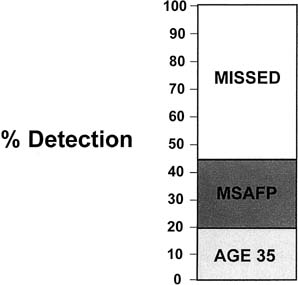
The adoption of wide-scale screening with maternal serum AFP effectively doubled the potential detection of chromosome abnormalities in the population. Even with an aging reproduction population, only 25% to 30% of Down syndrome infants are born to women older than age 35 (Fig. 1). The addition of a well-coordinated maternal serum AFP screening program can detect approximately 30% of the 80% of cases that are born to women younger than age 35. The mechanics of biochemical screening (i.e., with adjustments for gestational age, race, diabetic status, multiple gestation status, and maternal weight and adjustments via a different database or correction factors for maternal race) have been published previously and are not repeated here.27

Fig. 1. Detection by single screening. Of abnormalities, 20% occur in the fetuses of women 35 years old or older. Alpha-fetoprotein screening brings the detection rate to about 45%. MSAFP, maternal serum alpha-fetoprotein.

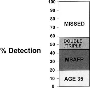
In 1988, Wald and associates28 suggested that a combination of parameters including AFP, β-human chronic gonadotropin (β-HCG), and unconjugated estriol could significantly increase the detection frequency of Down syndrome to approximately 60% of the total (Fig. 2). Multiple studies have corroborated the increased efficacy of multiple marker screening as opposed to AFP alone in detecting chromosome abnormalities, particularly Down syndrome.29,30,31,32,33,34

Fig. 2. Detection by double and triple screening. Concomitant testing of human chorionic gonadotropin and estriol levels improves alpha-fetoprotein screening rate to about 60%. MSAFP, maternal serum alpha-fetoprotein.

Despite overwhelming data and recommendations of national organizations such as the American College of Obstetrics and Gynecology that multiple marker screening be offered, nearly 20% of patients in the United States who have screening still have AFP alone.35 There is essentially universal agreement that among the three common parameters used, AFP, β-HCG, and unconjugated estriol, that if one could choose only one of the above, that β-HCG is the best. There is a virtual tie for second place in efficacy between AFP and unconjugated estriol. Because AFP already is used in North America and much of Western Europe for the detection of neural tube defects, however, the only real remaining question is whether adding unconjugated estriol as a third parameter is cost beneficial.

The debate over the use of unconjugated estriol (i.e., double screening versus triple screening) has become intense and emotional with staunch proponents on both sides. We believe the studies as a whole suggest there is no real effectiveness of adding the third marker. The literature is divided between several studies that say the third marker helps and others that say it does not. Since 1991, when Crossley and associates first proposed the β-HCG-to-AFP ratio be used as a marker, the question of how the data are interpreted has been added into the overall equation of sensitivity and specificity. Our data suggest that the differences may be due to higher variability among unconjugated estriol assays rather than to the other parameters. This variation may explain in part the incongruity among studies.37 Cuckle38 raised the question of whether screening should be offered to all patients or, for example, only to patients age 27 or older and showed that if screening were offered only to women older than age 27, more than 50% of the population would be excluded, and there would be an approximate 9% lower detection of affected pregnancies. Although not reaching the conclusion that this was cost beneficial, Cuckle38 stated that such rationing of services can be considered when resources are scarce.

Evans and colleagues37 investigated many of the “dogmas” of 3 decades of biochemical screening and found that many of these are no longer valid. We believe the wide variance in results reported from around the world is largely due to subtle and sometimes not so subtle differences in laboratory methods.37 The bitter arguments about double versus triple screening are, in part, answered by the fact that there is a much wider variability in estriol measurements than other parameters. Such variation helps explain why labs have such widely diverging experience with estriol. When the methods are standardized, much of the variability disappears and allows for an “apples versus apples” as opposed to “apples versus oranges” comparison. Similarly, much of the reported variation in the literature likewise disappears with standardization, and the diabetic correction factor becomes unnecessary with proper accounting for the fact that diabetic patients are of higher maternal weight.39,40

Many articles in the past several years have looked at the various constituents in the marker regimen, with the most important being touted being use in the second trimester of free β-HCG as opposed to intact β-HCG. Wald and Hackshaw41 reported in 1993 that the use of free β-HCG compared with total β-HCG would increase the detection frequency by about 4% for a given false-positive rate used in conjunction with maternal age, AFP, and unconjugated estriol. Other studies have suggested that, particularly at earlier gestational ages (e.g., 14, and 15 weeks) free β-HCG may have better sensitivity and specificity than the intact molecule.

Another promising marker has been the search for fetal cells in maternal circulation. Studies throughout the 1990s suggested that isolation and analysis of fetal cells may become practical and useful as a screening test.42,43,44 The current state follows essentially 2 decades of various starts and stops that alternatively have looked promising and frustrating since Hertzenberg and coworkers45 first showed detection and enrichment by fluorescent activated cell sorting. Much of the work of the past 2 decades has focused on ways to improve the efficacy of detection methods primarily centered on the need to increase the enrichment of fetal cells from the maternal blood circulation whose prevalence has been estimated to be approximately 1 in 10 million cells.46,47,48

Three types of fetal cells have been sought extensively—trophoblast, lymphocytes, and nucleated fetal red blood cells. The cell type most likely to be successful is thought to be nucleated red blood cells. Bianchi and colleagues46 were the first to use flow sorting to isolate nucleated fetal erythrocytes using an antibody of the transfer interceptor. More recent studies have focused on two general approaches—fluorescent activated cell sorting and magnetic activated cell sorting.43 Trisomic conceptions subsequently confirmed by invasive testing have been found by both methods.46,47,48 Although the results are encouraging, further work needs to be done before the screening test becomes practical. Analysis of progress through the millennium suggested that the magnetic activated cell sorting approach seemed to have a better sensibility than fluorescent activated cell sorting to isolate fetal cells.43 Another approach of looking at freeDNA (i.e., not cellular) is developing as a promising area of study.49 Although the overall sensitivity of fetal cells was not an improvement over current screens, the specificity of fetal cells might be much better. If so, a two-step approach might emerge in which a higher percentage of patients—perhaps 10% using double or triple testing—would be called positive to raise the sensitivity to around 80%. Then these 10% would undergo fetal cell testing to reduce that risk group to 2% to 3% but not lose sensitivity.

Another area of potential applicability of fetal cells in maternal blood is for the isolation of molecular diagnosis of mendelian disorders. Lo and associates50 were able to determine fetal Rh status in women known to be sensitized and married to heterozygous men. Geifman-Holtzman and colleagues51 determined fetal Rh status using polymerase chain reaction fetal nucleated red blood cells sorted from maternal blood. The next several years ultimately will determine how successful fetal cell sorting is as a screening test. It originally was hoped that it could be a diagnostic test and replace the need for invasive testing; however, as of this writing, fetal karyotypes cannot be obtained from cells that are isolated, and only fluorescence in situ hybridization–related results are possible. Although such is good as a screening test for aneuploidy, our experiences show that approximately one third of abnormal karyotypes seen in prenatal diagnosis programs are not ones that would be detected by the standard probes for chromosomes 13, 18, 21, X, and Y.52 Until and unless complete karyotypes can be obtained, fetal cells will not replace invasive testing but may be an important addition to the armamentarium of screening technologies.

Many studies have suggested dimeric inhibin A as an excellent marker that may raise the sensitivity by 3% to 7% for a given screen-positive rate.53 Some have suggested quadruple screening. There are also paradigms that include different parameters at different times combined. Although preliminary data suggest a high sensitivity with improved specificity, hiding results from patients for up to 1 month is ethically problematic in our opinion. No doubt there multiple approaches to screening will emerge, and there will be no one uniform standard approach.

Another two-step approach has been the so called integrated test.54 This is a combination of first-trimester blood and ultrasound. The results are not communicated to the patient, who then waits for second-trimester blood results before a risk assessment is made. Preliminary data suggest a reduced false-positive rate for comparable sensitivity, but the tradeoff is the need for patients to wait 6 weeks for start to finish of the screening process. For patients who do not particularly care about the results, the delay may be fine, but many patients would find such delay intolerable.

Trisomy 18

Although screening generally has focused on trisomy 21, our data and those of others always have shown a varied pattern of anomalies detected by screening.55 A different pattern of analytic levels has been observed in trisomy 18. The values of AFP, β-HCG, and unconjugated estriol appear to be low.56 These low values suggest a different pathophysiology than for Down syndrome. In Down syndrome, the low AFP and unconjugated estriol and high β-HCG can be explained as reflecting inappropriate immaturity or dysmaturity of the fetus (i.e., all values are consistent with a younger gestational age). In trisomy 18, that explanation does not work. We previously showed that there are different patterns of genomically directed intrauterine growth retardation in different aneuploidies, but how this translates into serum markers is unclear. Nevertheless, some reports have shown that an algorithm can be used to identify most trisomy 18 cases, while adding about 0.75% to the population being offered amniocentesis.

Back to Top
FIRST-TRIMESTER BIOCHEMICAL SCREENING

Of all maternal serum markers evaluated, pregnancy associated plasma protein A (PAPP-A) and free β-hCG emerged as the most predictive of all as potential markers for Down syndrome screening in the first trimester.57,58,59,60,61 Canick and Kellner61 reviewed Down syndrome screening in the first trimester (8 to 13 gestational weeks) with maternal serum analytes. PAPP-A and free β-HCG stood out at the most promising markers. They estimated that the Down syndrome screening performance of these analytes in combination with maternal age is a 60% detection rate with a 5% false-positive rate. This is similar to the screening performance of second-trimester double screening, but less efficient than second-trimester triple or quadruple screening. Canick and Kellner61 concluded that first-trimester screening with serum markers alone cannot be recommended except in cases in which second-trimester screening cannot be performed.

Nuchal Translucency

Ultrasound markers of aneuploidy are discussed elsewhere; however, for the sake of completeness and as a preface to combined screening, a brief discussion is presented. Increased skin or soft tissue thickening is a well-described characteristic of Down syndrome, and as reported by Down in 1866, in Down syndrome “[t]he skin…is deficient in elasticity, giving the appearance of being too large of the body.” Nuchal fold or thickening also has been shown in second-trimester Down syndrome fetuses.62 Increased nuchal translucency (NT) thickness in the first trimester has been shown to be an important prognostic marker of fetal aneuploidy and structural anomalies.63 Nicolaides and colleagues63 first found that NT was greater than 3 mm in 86% of the trisomic but in only 4.5% of the chromosomally normal fetuses. A NT greater than 3 mm was associated with a 12-fold increase in maternal age–related risk for fetal aneuploidy.64 Pandya and coworkers65 reported that the risk for fetal trisomy increases with NT and that NT of 3 mm, 4 mm, 5 mm, and greater than 6 mm are associated with a 4-fold, 21-fold, 26-fold, and 41-fold increase in the maternal age–related risks for trisomies 21, 18, and 13. Because the sensitivity of NT in twin gestations is similar to that of singletons, it could be used for individual risk assessment in twins.66

The same group suggested that when performed in experienced hands, NT seems to be the most sensitive method of screening for fetal chromosomal abnormalities, yielding a detection rate of about 85%.67 This optimistic view is not shared by others, however: Roberts and associates68 found poor reproducibility of NT measurement in low-risk populations, highlighting the need for quality control procedures and training of staff to obtain precise measurement of a market that varies by only a few tenths of a millimeter. Haddow and coworkers69 studied 48 Down syndrome pregnancies and 3169 unaffected controls from different centers and reported that NT measurements could be obtained in 61% to 100% of the patients (average 83%). They reported that the Down syndrome detection rate based on NT greater than the 95th percentile was only 31% with a 5% false-positive rate. They concluded that the variability among centers in median NT values and in the ratios of the 95th percentile to the 50th percentile, coupled with the varying ability to obtain the measurement successfully, suggested the performance of NT as a predictor of Down syndrome in leading reporting centers may reflect accurately its long-term performance at individual centers. The National Institute of Child Health and Human Development–supported BUN study (blood and ultrasound nuchal transparency) in the United States reported in 2002 results similar to those found by the London group. Sensitivity of about 80% for a false-positive rate of 5% was reported, with the belief that the theory is now proven. The remaining questions concern how to implement wide-scale utilization.70

Integrated Test (First-Trimester and Second-Trimester Screening)

With the increasing usage of combined first-trimester screening, and owing to the fact that second-trimester screening is not phased out, patients may undergo first-trimester and second-trimester screening, As a result, some patients are given different Down syndrome risk estimates by these tests. To overcome the resulting confusion and to increase the Down syndrome detection rate further, Wald and colleagues54 proposed a new screening method, the “integrated test,” in which measurements obtained during first and second trimesters are integrated to provide a single estimate of a patient’s Down syndrome risk. This strategy employs as first-trimester markers serum PAPP-A and NT and as second-trimester markers various combinations of serum AFP, β-HCG, unconjugated estriol, and inhibin. According to their approach, when a Down syndrome risk of greater than 1:120 is used as the cutoff to define a screen-positive result, a Down syndrome detection rate of 85% is achieved at a 0.9% false-positive rate compared with only 69% at a 5% false-positive rate for the triple screening. This would mean a higher detection rate at a reduction of four fifths in the number of invasive diagnostic procedures and consequent losses of normal fetuses. Alternatively, if the commonly accepted false-positive rate of 5% is maintained, the Down syndrome detection rate is estimated to reach 95%. Despite its theoretical appeal, the practicality and feasibility of the integrated test is still questionable because it would mean forfeiture of the benefits of early detection provided by the combined first-trimester screening. The time gap created between the first-trimester and second-trimester sampling dates is discomforting. The acceptability of this approach is yet to be determined.

Back to Top
PUBLIC POLICY AND ETHICAL ISSUES

The demonstration that multiple marker biochemical screening would detect most fetuses with chromosome abnormalities, such as Down syndrome, has set off policy and ethical debates in North America and Europe. Haddow and colleagues69 have shown that in women older than age 35, nearly 90% of Down syndrome fetuses can be detected, while reducing the number of amniocenteses by perhaps half or more. From purely public health and mathematical perspectives, denying access to women older than age 35 whose biochemical screens do not meet a risk level sufficiently high to warrant expenditure of resources might seem appropriate.38 Such an approach would require a reorientation of philosophy, however, and a removal of patient autonomy over such issues. When autonomy and public dollars come into conflict, however, it is reasonable to expect disagreements over the appropriate usage of these resources.

Czeizel24 summarized the data on what proportion of congenital abnormalities could be detected and prevented. Czeizel believed that approximately 51 of 73 congenital abnormality types (70%) could be evaluated. The birth prevalence of all congenital anomalies could be reduced from about 65 to 26 per 1000; 39 per 1000 or 60% are preventable. Although many congenital abnormalities can be prevented, they do not represent a single pathologic category, and there is no single strategy for their prevention that is appropriate.

Back to Top
SUMMARY

Considerable progress has occurred in the area of biochemical screening. Increasing data have shown the advantages of multiple markers, particularly β-HCG over AFP alone. There continues to be considerable controversy over the best mathematical algorithm and which markers are best (e.g., β-HCG, unconjugated estriol). There seems to be a plateau of detection frequencies at about 65% to 70% with current methodologies. Much further work needs to be done, however, including some new approaches, if there is to be substantial improvement of screening sensitivity. The combination of biochemical with biophysical parameters represents the next level of sophistication in the attempt to identify the highest proportion over the next few years of abnormalities with the fewest false positives. We anticipate a major shift to the first trimester, which would revolutionize genetic counseling, reinvigorate chorionic villus sampling, and eliminate maternal age per se as the major criterion for offering invasive testing.

Back to Top
REFERENCES

1. Evans MI, Chik L, O’Brien JE, et al: MOMs and DADS: Improved specificity and cost effectiveness of biochemical screening for aneuploidy with DADS. Am J Obstet Gynecol 172:1138, 1995

2. Evans MI, Chik L, O’Brien JE, et al: Logistic regression generated probability estimates for trisomy 21 outcomes from serum AFP and BHCG: Simplification with increased specificity. Mat Fetal Med 5:1, 1996

3. Evans MI, Sobecki MA, Krivchenia EL, et al: Parental decisions to terminate/continue following abnormal cytogenetic prenatal diagnosis: “What” is still more important than “when.” Am J Med Genet 61:353, 1996

4. Pryde PG, Odgers AE, Isada NB, et al: Determinants of parental decision to abort or continue for non-aneuploid ultrasound detected abnormalities. Obstet Gynecol 80:52, 1992

5. Brock DJH, Sutcliffe RG: Alpha-fetoprotein in the antenatal diagnosis of anencephaly and spina bifida. Lancet 2:197, 1972

6. Brock DJH, Bolton AE, Monaghan JM: Prenatal diagnosis of anencephaly through maternal serum alpha-fetoprotein measurements. Lancet 2:923, 1973

7. Yen IH, Khoury MJ, Erickson JD, et al: The changing epidemiology of neural tube defects: United States, 1968–1989. Am J Dis Child 146:857, 1992

8. Chan A, Robertson E, Haan EA, et al: Prevalence of neural tube defects in South Australia, 1966–91: Effectiveness and impact of prenatal diagnosis. BMJ 307:703, 1993

9. Steegers-Theunissen Rdgine PM, Smithells RW, Eskes TKAB: Update of new risk factors and prevention of neural-tube defects. Obstet Gynecol Surv 48:287, 1993

10. Van Loon K, Besseghir K, Eshkol A: Neural tube defects after infertility treatment: A review. Fertil Steril 58:875, 1992

11. Shane B, Stokstad ELR: Vitamin B12 folate interrelationships. Ann Rev Nutr 5:115, 1985

12. Shoiania AM: Folic acid and vitamin B12 deficiency in pregnancy and the neonatal period. Clin Perinatol 11:433, 1984

13. Smithells RW, Sheppard S, Schorah CJ: Vitamin deficiencies and neural-tube defects. Arch Dis Child 51:944, 1976

14. Smithells RW, Sheppard S, Schorah CJ, et al: Possible prevention of neural tube defects by periconceptional vitamin supplementation. Lancet 1:339, 1980

15. Smithells RW, Seller MJ, Harris R, et al: Further experience of vitamin supplementation for prevention of neural-tube defect recurrences. Lancet 1:1027, 1983

16. Smithells RW, Sheppard S, Wild L, Schorah CJ: Prevention of neural-tube defect recurrences in Yorkshire: Final report. Lancet 2:498, 1989

17. Steegers-Theunissen RPM, Coers GHJ, Trijbels JMF, et al: Neural-tube defects and derangement of homocysteine metabolism. N Engl J Med 324:199, 1991

18. Yates JRW, Ferguson-Smith MA, Shenkin A, et al: Is disordered folate metabolism the basis for the genetic predisposition to neural-tube defects? Clin Genet 31:279, 1987

19. Wald J, Schorah CJ, Sheldon TA, Smithells RW: Investigation of factors influencing folate status in women who have had a neural tube defect-affected infant. Br J Obstet Gynaecol 100:546, 1993

20. Holmes-Siedle M, Dennis J, Lindenbaum RH, Galliard A: Long term effects of periconceptional multivitamin supplements for prevention of neural tube defects: A seven to 10 year follow up. Arch Dis Child 67:1436, 1992

21. From the Centers for Disease Control and Prevention, Leads from the Morbidity and Mortality Weekly Report, Atlanta, GA:Recommendations for use of folic acid to reduce number of spina bifida cases and otherneural tube defects. JAMA 69:1233, 1993

22. Evans MI, Wapner RJ, O’Brien JE, et al: Impact of folic acid supplementation in the United States: Markedly diminished maternal serum AFPs. ACOG, May 2001

23. Honeim MA, Paulozzi LJ, Mathews TJ, et al: Impact of folic acid fortification of the US food supply on the occurrence of neural tube defects. JAMA 285:3022, 2001

24. Czeizel AE: Primary prevention of neural tube defects and some other major congenital abnormalities: Recommendations for the appropriate use of folic acid during pregnancy. Paediatr Drugs 2:437, 2002

25. Merkatz IR, Nitowsky FM, Macri JN, Johnson WE: An association between low maternal serum alpha-fetoprotein and fetal chromosome abnormalities. Am J Obstet Gynecol 148:886, 1984

26. Nyberg DA, Kramer D, Resta RG, et al: Prenatal monographic findings of trisomy 18: review of 47 cases. J Ultrasound Med 2:103, 1993

27. Evans MI, Dvorin E, O’Brien JE, et al: Alpha-fetoprotein and biochemical screening. In Evans MI (ed): Reproductive Risks and Prenatal Diagnosis. pp 223, 235 Norwalk, CT, Appleton & Lange, 1992

28. Wald NJ, Cuckle HS, Densem JW, et al: Maternal serum screening for Down syndrome in early pregnancy. BMJ 297:883, 1988

29. Cheng EY, Luthy DA, Zebelman AM, et al: A prospective evaluation of a second-trimester screening test for fetal Down syndrome using maternal serum alpha-fetoprotein, hCG, and unconjugated estriol. Obstet Gynecol 81:72, 1993

30. Aitken DA, McCaw G, Crossley JA, et al: First-trimester biochemical screening for fetal chromosome abnormalities and neural tube defects. Prenat Diagn 13:681, 1993

31. Rodriguez L, Sanchez R, Hernandez J, et al: Results of 12 years’ combined maternal serum alpha-fetoprotein screening and ultrasound fetal monitoring for prenatal detection of fetal malformations in Havana City, Cuba. Prenat Diagn 17:301, 1997

32. Wald N, Densem J, Stone R, Cheng R: The use of free 1993, b-hCG in antenatal screening for Down’s syndrome. Br J Obstet Gynaecol 100:550, 1993

33. Goodburn SF, Yates JRW, Raggatt PR, et al: Second-trimester maternal serum screening using alpha-fetoprotein, human chorionic gonadotrophin, and unconjugated oestriol: Experience of a regional programme. Prenat Diagn 14:391, 1994

34. Gardosi J, Mongelli M: Risk assessment adjusted for gestational age in maternal serum screening for Down’s syndrome. BMJ 306:1509, 1993

35. Evans MI, O’Brien JE, Henry GP, et al: Persistence of “single” screen alphafetoprotein tests ordered by practicing physicians. J Matern Fet Neonat Med (in press)

36. Crossley JA, Aitken DA, Connor JM: Prenatal screening for chromosome abnormalities using maternal serum chorionic gonadotrophin, alpha fetoprotein, and age. Prenat Diagn 11:83, 1991

37. Evans MI, O’Brien JE, Dvorun E, et al: Standardization of methods reduces variability: Explanation for the wide historical discrepancy biochemical screening benefits. Am J Obstet Gynecol 184:S111, 2001

38. Cuckle HS: Maternal serum screening policy for Down’s syndrome. Lancet 340:799, 1992

39. Evans MI, Harrison HH, O’Brien JE, et al: Correction for insulin dependent diabetes in alpha fetoprotein testing has outlived its usefulness. Am J Obstet Gynecol (in press)

40. Evans MI, Harrison H, O’Brien JE, et al: Maternal weight correction for alpha fetoprotein: Mathematical truncations revisited. Genetic Testing (in press)

41. Wald NJ, Hackshaw A: Antenatal screening for Down’s syndrome. BMJ 306:1198, 1993

42. Elias S, Simpson JL: Prenatal diagnosis. In Rimoin DL, O’Connor JM, Pyeritz PE (eds): Emery and Rimoin’s Principles and Practice of Medical Genetics. 3rd ed.. New York, Churchill Livingstone, 1997

43. Bianchi DW, Simpson JL, Jackson LG, et al: Fetal gender and aneuploidy detection using fetal cells in maternal blood; analysis of NIFTY I data. Prenatal Diagnosis 22:609, 2002

44. Lewis DE, Schober W, Murrell S, et al: Rare event selection of fetal nucleated erythrocytes in maternal blood by flow cytometry. Cotymetry 23:218, 1996

45. Herzenberg LA, Bianchi DW, Schroder J: Fetal cells in the blood of pregnant women: Detection and enrichment by fluorescence-activated cell sorting. Proc Nat Acad Sci U S A 76:1453, 1979

46. Bianchi DW, Flint AF, Pizzimenti MF, et al: Isolation of fetal DNA from nucleated erythrocytes in maternal blood. Proc Natl Acad Sci U S A 87:3279, 1990

47. Bianchi DW, Klinger KW: Prenatal diagnoisis through the analysis of fetal cells in the maternal circulation. In Milunsky A (ed): Genetic Disorders and the Fetus. p 759, 3rd ed.. Baltimore, Johns Hopkins University Press, 1992

48. Ganshirt-Ahlert D, Borjesson-Stoll R, Burschyk M, et al: Detection of fetal trisomies 21 and 18 from maternal blood using triple gradient and magnetic cell sorting. Am J Reprod Immunol 30:194, 1994

49. Lo YMD, Corbetta N, Chamberlain PF, Sargent JL: Presence of fetal DNA in maternal plasma and serum. Lancet 350:485, 1997

50. Lo YMD, Bowell PJ, Selinger M, et al: Prenatal determination of fetal RhD status by analysis of peripheral blood f rhesus negative mothers. Lancet 341:1147, 1993

51. Geifman-Holtzman O, Bernstein IM, Berry SM, et al: Fetal RhD genotyping in fetal cells flow sorted from maternal blood. Am J Obstet Gynecol 174:818, 1996

52. Evans MI, Henry GP, Miller WA, et al: International collaborative assessment of 146,000 prenatal karyotypes: Expected limitations if only chromosome-specific probes and fluorescent in situ hybridization were used. Hum Reprod 14:1213, 1999

53. Lambert-Messerlian GM, Canick JA, Palomaki GE, Schneyer AL: Second trimester levels of maternal serum inhibin A, total inhibin, alpha inhibin precursor, and active in Down’s syndrome pregnancy. J Med Screen 3:58, 1996

54. Wald NJ, Watt HC, Hackshaw AK: Integrated screening for Down’s syndrome on the basis of tests performed during the first and second trimesters. N Engl J Med 341:461, 1999

55. Drugan A, Dvorin E, Koppitch FC, et al: Counseling for low maternal serum alpha-fetoprotein should emphasize all chromosome anomalies, not just Down syndrome!. Obstet Gynecol 73:271, 1989

56. Palomaki GE, Haddow JE, Knight GJ, et al: Risk-based prenatal screening for trisomy 18 using alpha-fetoprotein, unconjugated estriol, and human chorionic gonadotropin. Prenat Diagn 15:713, 1995

57. Brambati B, Macintosh MC, Teisner B, et al: Low maternal serum levels of pregnancy associated plasma protein A (PAPP-A) in the first trimester in association with abnormal fetal karyotype. Br J Obstet Gynaecol 100:324, 1993

58. Brambati B, Tului L, Bonacchi I, et al: Serum PAPPA-A and free beta-hCG are first trimester screening markers for Down syndrome. Prenat Diagn 14:1043, 1994

59. Forest JC, Masse J, Moutquin MJ: Screening for Down syndrome during first trimester: A prospective study using free beta-human chorionic gonadotropin and pregnancy-associated plasma protein A. Clin Biochem 30:333, 1997

60. Macri JN, Spencer K, Aitken D, et al: First trimester free beta (hCG) screening for Down syndrome. Prenat Diagn 13:557, 1993

61. Canick JA, Kellner LH: First trimester screening for aneuploidy: Serum biochemical markers. Semin Perinatol 23:359, 1999

62. Benacerraf BR, Barss VA, Laboda LA: A sonographic sign for the detection in the second trimester of the fetus with Down’s syndrome. Am J Obstet Gynecol 151:1078, 1985

63. Nicolaides KH, Brizot ML, Snijders RJ: Fetal nuchal translucency: Ultrasound screening for fetal trisomy in the first trimester of pregnancy. Br J Obstet Gynaecol 101:782, 1994

64. Pandya PP, Brizot ML, Kuhn P, et al: First trimester fetal nuchal translucency thickness and risk for trisomies. Obstet Gynecol 84:420, 1994

65. Pandya PP, Snijders RJ, Johnson SP, et al: Screening for fetal trisomies by maternal age and fetal nuchal translucency thickness at 10-14 weeks of gestations. Br J Obstet Gynaecol 102:957, 1995

66. Nicolaides KH, Brizot ML, Snijders RJ: Fetal nuchal translucency: Ultrasound screening for fetal trisomy in the first trimester of pregnancy. Br J Obstet Gynaecol 101:782, 1994

67. Snijders RJ, Johnson S, Seibire NJ, et al: First trimester ultrasound screening for chromosomal defects. Ultrasound Obstet Gynecol 7:216, 1996

68. Roberts LJ, Bewley S, Mackinson AM, Rodeck CH: First trimester fetal nuchal translucency: Problems with screening the general population. Br J Obstet Gynaecol 102:381, 1995

69. Haddow JE, Palomaki GE, Knight GJ, et al: Reducing the need for amniocentesis in women 35 years of age or older with serum markers for screening. N Engl J Med 330:1114, 1994

70. Wapner RJ for: the BUN Study Group: First trimester aneuploid screening. Results of the NICHDMulticenter Study Am J Obstet Gynecol 185:S70, 2001

Back to Top