Chapter 3
Cervical Intraepithelial Neoplasia: History and Detection
L. Stewart Massad and Helen E. Cejtin
Main Menu   Table Of Contents

Search

L. Stewart Massad, MD
Associate Professor of Obstetrics and Gynecology and Radiation Oncology, Cook County Hospital and Rush Medical College, Chicago, Illinois (Vol 4, Chap 3)

Helen E. Cejtin, MD
Attending Physician and Director of Ambulatory Services, Cook County Hospital; Assistant Professor, Chicago Medical School, Chicago, Illinois (Vol 4, Chap 3)

INTRODUCTION
DEFINITION
NATURAL HISTORY
SCREENING
EVALUATING ABNORMAL RESULTS OF SCREENING TESTS
DIAGNOSIS
ACKNOWLEDGMENT
REFERENCES

INTRODUCTION

Hippocrates and Galen described invasive cancers of the cervix, but the existence of asymptomatic neoplasms within the cervical epithelium was not recognized until early in this century, and the preinvasive nature of these lesions has been clarified only in the last decades. Only recently, with the development of techniques that allow molecular biologists to explore genomic changes in dysplastic cells, has the fundamental biology of cervical intraepithelial neoplasia (CIN) begun to emerge.

After defining CIN and briefly reviewing the history of the scientific advances leading to modern practice, this chapter describes the natural history of CIN, strategies for screening, and techniques for diagnosis. Other chapters discuss the epidemiology of cervical cancer, which is identical to that of these precursor lesions; the natural history of CIN; the pathologic characteristics of CIN and cervical cancer; and the biologic makeup of the human papillomavirus (HPV), which is fundamental to understanding CIN. Although a glandular precursor lesion has been identified in the endocervix (adenocarcinoma in situ), this chapter focuses on the common squamous abnormalities.

Back to Top
DEFINITION

Cervical intraepithelial neoplasms are atypical proliferations of immature squamous epithelium that do not penetrate the basement membrane of the epithelium. Mitotic figures, both normal and tripolar or even tetrapolar, are seen above their usual position among the reserve cells at the base of the epithelium. Nuclear abnormalities are characteristic and include a coarse chromatin pattern, abnormal chromatin distribution, pleomorphism, and hyperchromaticity. The nuclear-cytoplasmic ratio is increased. Progressive squamous differentiation is diminished, being restricted to the upper levels of the epithelium; in the most advanced lesions, differentiation is absent, and the full thickness of the epithelium is composed entirely of neoplastic cells.

Table 1 compares the systems for classification of CIN that have been used as understanding of cervical carcinogenesis has changed. The modified Papanicolaou system was developed to distinguish cancer and carcinoma in situ (CIS) from other lesions and now is obsolete. The dysplasia and CIN systems were standard until recently and remain the histologic descriptive terms of choice. They are especially useful for clinicians who elect to treat mild dysplasia or CIN I. The Bethesda system is used most commonly for cytopathologic description and is most appropriate when clinicians plan to treat only high-grade lesions. Multiple descriptors can appear on single reports.

TABLE 1. Comparison of Classification Systems for Precancerous Lesions of the Cervix


Modified Papanicolaou

Dysplasia

CIN

Bethesda

I

Normal

Normal

Normal/Benign

II

Atypia

Atypia

ASCUS/LGSIL

III

Mild dysplasia

CIN I

LGSIL

III

Moderate dysplasia

CIN II

HGSIL

III

Severe dysplasia

CIN III

HGSIL

IV

Carcinoma in situ

CIN III

HGSIL

V

Cancer

Cancer

Cancer


CIN, cervical intraepithelial neoplasia; ASCUS, atypical squamous cells of uncertain significance; LGSIL, low-grade squamous intraepithelial lesion; HGSIL, high-grade squamous intraepithelial lesion.

History

Although cervical CIS was described in the early 1900s,1 the clinical importance of these lesions was not appreciated until useful means for detecting these asymptomatic, invisible lesions were developed. Beforehand, cervical cancer detection relied on inspection and palpation, with biopsy of obvious invasive cancers.2 Schiller developed a technique for iodine staining as a gross means for detecting areas of abnormal epithelium,3 but this test could not distinguish metaplastic from neoplastic areas of the cervix and could not distinguish small areas of invasion present in a field of diffuse nonstaining epithelium. In this era, the nature of intraepithelial lesions was controversial, often described at the margin of invasive lesions but at times noted as a precursor to invasion.3,4 Nevertheless, the description in the 1920s and 1930s of what came to be known as CIN provided the foundation for the development of cytologic study.

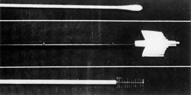
Although cytologic examination of exfoliated cervical epithelial cells was first described by Babes in 1928 in the French literature,5 only with the appearance in 1941 of the findings of Papanicolaou and Traut did this technique enter clinical practice as a means for the early diagnosis of cervical cancer.6 Like any epithelium, the cervical mucosa constantly is being sloughed and regenerated. When examined as thin smears, neoplastic cells from the cervical surface retain characteristic microscopic abnormalities that allow them to be distinguished after staining from normal squames, leukocytes, and glandular cells. Papanicolaou described a technique for aspiration of cells from the posterior vaginal pool, fixation, and cytologic staining that remains the foundation of current screening strategies. Whereas others identified unsuspected neoplasms after abnormal smears and suggested that CIS was a cancer precursor,7-11 Papanicolaou's findings generated significant controversy,12 and his technique required modifications. Over time, vaginal pool aspiration was dropped, to be replaced by spatula collection, as developed by Ayre to facilitate the sampling of cells directly from the ectocervix.13 Augmented with a cotton-tipped applicator for endocervical cell collection,14 this instrument remained the standard collection instrument until the development of endocervical brushes and one-step samplers during the 1980s.

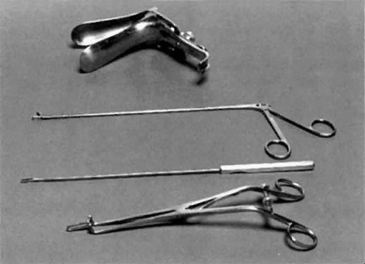
Initially, in the United States, histologic evaluation of abnormal findings on cervical cytologic smears was achieved through blind biopsy of four quadrants of the cervix or through biopsy directed by the Schiller test.15-18 However, although such biopsies were useful when results were positive, the inherent high negative predictive value of this random procedure in identifying lesions invisible to the naked eye soon was recognized.18,19 Cone biopsy became and remains the definitive procedure for histologic evaluation of the cervical transformation zone, punch biopsy being reserved for patients with gross lesions or lesions detected colposcopically.17,18,20-23 This procedure, which involves the excision of the cervical transformation zone, requires anesthesia and hospitalization and carries a significant risk of complications, whereas later studies showed disconcertingly high rates of negative findings in cone biopsies from patients with cytologic tests showing abnormalities of lesser severity than CIS or invasive cancer.18 Alternative methods of diagnosis were sought.

Colpomicroscopy was proposed as an alternative offering magnification as high as 480, but technical factors precluded its wide acceptance.24,25 Hinselmann had described colposcopy in 1925, and the technique had become incorporated into clinical care in other countries, but only in the 1950s was intensive training in colposcopic techniques promoted in the United States.26-30 Initially, a role for colposcopy as a screening modality for CIN was proposed,26,28,29 and it still is used as such in some parts of Europe, but the greater simplicity and wider availability of cytologic screening led to retention of cytologic examination as the standard screening test in the United States, with colposcopy retained as the study of choice for evaluation of women with abnormal cytologic findings. The value of colposcopy for excluding invasive cancer or CIS in women with cytologic results consistent with dysplasia was established,31,32 and studies comparing colposcopy with cone biopsy showed acceptable rates of correlation.33,34 The American Society for Colposcopy and Cervical Pathology, dedicated to promoting skills in colposcopy through research and education, was founded in 1965 and, as a member of the International Federation of Cervical Pathology and Colposcopy, remains a force for the development of strategies for screening, diagnosis, and management of CIN.

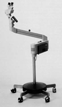
Several technologic enhancements to colposcopy have appeared in the past two decades. Videocolposcopy allows for image recording in real time, with enhanced visualization and opportunities for patient and trainee education. Systems for videocolposcopy include those added to traditional optical colposcopes as well as video-only systems. A comparison study of optical and video systems shows that use of video systems by even experienced colposcopists requires a learning phase but that similar findings result.35 Computerized image analysis and image storage is a recent advance with the potential to minimize subjective analysis of lesions and offer the option of serial follow-up with quantification of changes in lesion size and character.36-38 In addition, computerized colposcopy allows the integration of nonvisible spectra and tissue fluorescence into image analysis—processes with the promise of replacing colposcopy altogether as a diagnostic test.39

An understanding of the natural history of CIN has developed progressively. The preinvasive nature of CIS was the subject of prolonged debate,3,40,41 resolved once the ability of CIS to progress or to regress after biopsy was recognized. After this, the importance of lesser degrees of neoplastic change was investigated. Both the graded severity of lesions of the cervical epithelium and the sometimes prolonged interval between diagnosis of dysplasia and development of invasive cervical cancer came to be understood.42-46 The concept of cervical dysplasia as a continuum of disease was elaborated in the 1950s,47,48 and the term dysplasia was agreed on at the first International Congress of Exfoliated Cytology in 196249 (Fig. 1). Recognizing that pathologic distinctions between severe dysplasia and CIS are difficult to reproduce,50,51 Richart developed the CIN classification system currently used for description of histologic specimens, describing CIN as a continuum of neoplastic change with progressively increasing risk of invasion.52

Fig. 1. Diagram of cervical epithelium showing various terminology used to characterize progressive degrees of cervical neoplasia. (Modified from Richart RM: Can J Med Tech 38:177, 1976; used with permission of the Canadian Society of Laboratory Technologists.)

Once CIS was linked to invasive cancer, principles of management of CIS were derived from those established for cervical cancer, with radical and modified radical hysterectomy recommended for women with CIS.16,22,51,53 This probably was appropriate when no method for thoroughly evaluating the cervical transformation zone existed short of cone biopsy and when cone biopsies were not universally performed or exhaustively sectioned because numerous patients undergoing hysterectomy for CIS were found to have foci of invasive cancer.15,54 Once lesser dysplasias were recognized as cancer precursors, hysterectomy also was recommended for these lesions.55 However, data accumulated to show that intraepithelial neoplasms did not metastasize and that hysterectomy could be avoided when invasion was excluded by meticulous examination of cone biopsy specimens.56-60 Conservative office techniques for treatment of CIN were developed. These include, in order of their incorporation into practice, electrocautery, cryotherapy, carbon dioxide laser ablation and conization, and loop excision.61-67 Recently, studies of natural history suggest that some patients with early CIN may have spontaneous regression of lesions and may require no treatment.68,69

Cytologic and clinical observations led Ayre to the concept that viral infection of “halo cells” represents the first stage of cervical carcinogenesis.70 This concept was validated when Zur Hausen defined the link between HPV and cervical dysplasia in 1977,71 and the concept of high- and low-risk HPV subtypes was recognized in the years after.72-74 Through the 1980s, as HPV testing became more available, substantial epidemiologic and biochemical evidence accumulated supporting the role of HPVs as necessary but not sufficient factors for cervical oncogenesis. A full discussion of these and other areas in HPV research is presented in a separate chapter.

The following ideas and events—that with careful follow-up not all women with CIN require therapy, modification of classification systems, the idiosyncratic interpretations made of classification criteria, a growing appreciation of the malpractice risk inherent in a missed diagnosis of cervical cancer, publicized cases of cervical cancer developing after findings on smears were negative, and reports of cytopathology centers using suboptimal quality controls—all combined in the late 1980s to produce confusion among clinicians about the proper management of women with abnormal findings on cytopathology reports, especially those consistent with early lesions.

One attempted solution to these problems was the development of automated cytologic systems based on image recognition software.75 Adjunctive measures, such as HPV typing, and new instruments were developed to improve the sensitivity of cytologic study as a screening test for cervical cancer, especially for women with borderline abnormalities.76-80

The National Institutes of Health addressed these problems through consensus conferences charged with the development of standards for diagnosis and management of abnormal cytologic findings.81-83 First, the Bethesda system for cervical cytology interpretation was promulgated. Afterward, interim guidelines for management were devised. Recently, criteria for diagnosis have been published to standardize use of the various categories of the Bethesda system. The results of this group have been widely accepted, but not without criticism.84-86

Finally, in the last two decades, molecular biologists have explored the changes that lead cells in the cervical epithelium to become neoplastic. In addition to the inactivation of tumor suppressors from the p53 and retinoblastoma genes by HPV proteins, investigators have identified alterations in the expression of the epidermal growth factor receptor and the ras and myc oncogenes.87-91 Allelic loss follows the induction of tetraploidy and aneuploidy by p53 inactivation.92,93 Rates of apoptosis fall as proapoptotic proteins are down-regulated.94,95 Loss of heterozygosity analyses suggest that other important tumor suppressor genes may be deleted in preneoplastic cervical epithelium,96,97 including alterations in expression of the fragile histidine triad gene on chromosome 3.98,99 Angiogenesis is activated, and matrix interactions are altered as lesions advance.100,101 Despite these advances, however, the precise sequence of genetic alterations after HPV infection that leads to neoplastic transformation in the cervix remains to be elucidated.

The incidence of cytologic abnormalities in the United States has been estimated at approximately 5%,83 implying that CIN develops in hundreds of thousands of women each year. Despite this, the incidence of invasive cervical cancer in 2000 is estimated to be only 12,500.102 Given the magnitude of this public health problem and multitude of poorly understood factors that determine the progression of CIN to invasive cancer, it is understandable that controversy still surrounds the classification, diagnosis, and management of CIN.

Back to Top
NATURAL HISTORY

The sole rationale for the identification of cervical intraepithelial neoplasms is their risk for progression to invasive cancer. These lesions, invisible to the naked eye, cause no symptoms and no morbidity. Considerable attention has been devoted to determining the potential of CIN to progress, and varying recommendations for intervention or observation are based on differing interpretations of these data. Considerable differences exist among estimates of progression and regression rates for CIN, but large series have found that CIN I can progress to invasive cancer and that even CIS sometimes may regress.

Existing studies of the natural history of CIN are limited by several factors, which have been reviewed.103 First, confirmation and definitive classification of CIN requires biopsy. However, biopsy can alter the natural history of CIN, perhaps by inciting a local inflammatory reaction. Some studies have avoided this by diagnosing CIN through cytologic examination, but even when combined with colposcopy, this approach risks both overdiagnosis and underdiagnosis, with consequent overestimation or underestimation of progressive potential. A second limitation to studies of the natural history of CIN is the duration of follow-up. Progression to invasive cancer requires years, especially for early lesions, and many patients in most studies have been lost, leaving their status in doubt. A third factor confounding interpretation of natural history studies is the heterogeneity of some CIN lesions. Colposcopy with directed biopsy may miss significant lesions, and sampling errors may lead to misclassification of some patients, with substantial impact on risk for progression or regression of disease. A fourth confounder is differences in interpretation of lesion grade among pathologists. Finally, patient factors such as age may modify the risk of progression within each grade of CIN. Local immune factors may predispose to regression, and women with impaired immunity, such as those infected with human immunodeficiency virus (HIV) and those with renal allografts, are at increased risk for progression.104,105 When determining the appropriateness of various strategies balancing cervical cancer prevention with the morbidity of treatment, clinicians should keep in mind these limitations and modify general recommendations to fit the needs of individual patients.

The understanding of the invasive potential of CIN began with studies of patients with CIS. McIndoe and colleagues describe a group of 131 women with persistent abnormal cytologic findings after diagnosis of CIS; over time and despite therapy, more than a fifth developed invasive cancer, and the 20-year likelihood of progression to invasion was estimated at almost 40%.106 The lifetime risk of developing invasive cancer among women with untreated CIS is likely to be higher still.

Although most clinicians recommend treatment immediately after diagnosis for women with CIN II, studies of the natural history of this lesion have been performed without therapy. Using cytologic study with microcolposcopy but not biopsy, Richart and Barron estimate the risk of progression of untreated CIN II to CIS at more than 90% after 14 years of follow-up.50 Conversely, a meta-analysis of 15 studies spanning almost 35 years yielded an estimate of progression for CIN II of 20%, with 5% progressing to invasive cancer, 40% regressing, and 40% persisting unchanged.107

Because of its low risk for progression, CIN I has been studied most intensively. Ostor estimates the likelihood of regression at 60%, with only 10% progressing to CIN III and just 1% advancing to invasive cancer.107 Women infected with “high-risk” HPV subtypes may have a greater risk of progression,103 but prospective studies using HPV typing as triage for women with colposcopically demonstrated low-grade lesions have not yet been reported. Epidemiologic data from the large registry in British Columbia suggest that age also determines risk for progression: among women younger than 34 years of age with new lesions, 84% regressed, whereas only 40% of older women did so.108

Back to Top
SCREENING

A useful screening test should identify asymptomatic patients at high risk for a disease of significant morbidity and mortality at a point in the disease course where intervention can alter outcome. Cervical cytologic study does this well. At the same time, an optimal screening test meets these objectives at low cost and with acceptable sensitivity and specificity. Because cervical cytologic study is labor intensive, it is expensive. Nevertheless, it has proven to be competitive with alternative techniques.

Precise definition of the sensitivity and specificity of cervical cytologic study is impossible because the gold standard against which it must be compared is histologic evaluation of the entire cervical transformation zone, and the performance of random cone biopsies on cytologically normal women solely to determine these rates cannot be justified ethically. Still, the false-positive rate of cervical cytologic study has been determined using colposcopy and cone biopsy or cervical loop excision. This rate appears to vary substantially with the grade of CIN identified cytologically, with up to 30% of women with low-grade smears lacking identifiable CIN.109,110 The false-negative rate for cytologic study among patients with cancer can range as high as 50%; because blood and inflammatory cells may obscure neoplastic cells in such patients, this rate appears to be lower for women with CIN.111

The accuracy of cytologic study depends on the prevalence CIN in the population being screened. Epidemiologic risk factors are described in another chapter; populations with a high proportion of women with high-risk factors merit more intensive screening. The incidence of CIN exceeds that of invasive cancer of the cervix. This probably is a result both of the natural history of CIN, as described earlier, and of the identification and eradication of CIN in screened populations before invasion develops. Because CIN is not a reportable disease, its incidence is speculative, based on extrapolation of findings at a few centers; however, if, as noted earlier, 5% of smear results are abnormal, the U.S. incidence must be several hundred thousand.

The false-negative rate falls significantly when serial smears are taken. Using published data and statistical analysis, Eddy estimates that the risk of cervical cancer fell 64% when screening was performed at 10-year intervals, a result that improved to 91% with 3-year screening intervals.112 Increasing the frequency of sampling to annually results in only a small further decrease in risk. A consensus from groups such as the American Cancer Society and the American College of Obstetricians and Gynecologists recommends that cervical cancer screening begin with cytologic evaluation at the onset of sexual activity or age 18 and continue annually until three consecutive normal smears are obtained, after which the frequency of screening can be decreased to 3-year intervals if personal risk factors warrant.113,114 Epidemiologic risk factors that favor annual screening include early initiation of sexual activity, a history of multiple lifetime sexual partners, tobacco use, and immunosuppression. Unfortunately, high-risk women may lack sufficient sophistication to respond correctly to queries about screening.115 Across socioeconomic classes, the reliability of women's recollection of the interval since the last smear and of their history of abnormal findings may be unreliable.116 Without documentation, a lifetime history of normal smear results cannot be assumed.

Cervical cytologic study was adopted without rigorous testing as an important way to detect cervical cancer because of its manifest superiority to techniques of inspection, palpation, and iodine staining with blind biopsy available at its introduction. Despite the absence of clinical trials, cervical cytologic study remains the standard tool for cervical cancer screening. Several reports correlate significant declines in the incidence of cervical cancer with the institution of widespread screening programs.117-120 It has been proposed that cervical cancer could be eradicated if all women complied strictly with screening guidelines.121 Nevertheless, despite widespread incorporation of cytologic screening into clinical practice, cervical cancer remains a significant public health problem.

The reasons for this are many and have been reviewed.121 Unfortunately, many women fail to comply with screening recommendations, and many patients with cervical cancer acknowledge that they have not been screened adequately.116,122 The reasons for this are not clear, but studies suggest that significant reasons for inadequate screening include ignorance of guidelines, dislike of pelvic examination, lack of access to the medical care system, fear of cancer, fear of pain from diagnostic procedures, and mistrust of medical authorities.123 Women who fail to receive adequate screening tend to be older and less educated and belong to minority ethnic groups.123,124 Compliance with screening guidelines has been shown to be improved by education and physician-initiated reminders.125,126

Even when women comply with screening guidelines, inadequate cervical sampling may allow malignant and premalignant lesions to progress untreated. Badly lacerated cervices and endophytic neoplasms may contribute to this problem. Initially, Papanicolaou described vaginal pool aspirates as the source of cellular material for cytologic analysis. However, modern understanding of the location of CIN has led to the development of techniques for sampling the cervical transformation zone. Currently, sampling should be done from both the ectocervix and the endocervix. The Bethesda system classifies smears that lack an endocervical component as “satisfactory but limited” because of the risk that abnormalities at the squamocolumnar junction may have been missed.82 Endocervical cells are commonly absent in true-negative smear results from women with cervical cancer.127 However, for low-risk women, the absence of endocervical cells does not require repeat smear.

A variety of instruments exist to facilitate cytologic sampling of the cervix and endocervix, but none has been shown to be superior in the yield of cervical dysplasia diagnosed (Figs. 2 and 3). An endocervical brush increases the yield of endocervical cells sevenfold more than a moistened cotton-tipped applicator but may induce bleeding that can obscure smears.128,129 Therefore, the ectocervix should be sampled first when an endocervical brush is used. Single-step instruments are available, but little literature is available to determine comparative efficacy.

Fig. 2. Scraping of endocervix and ectocervix. (Herbst AL: Comprehensive Gynecology. St Louis, Mosby-Year Book, 1992.)

Fig. 3. Narrow brushes for endocervical sampling: cotton-tipped swab ( top ), Cervex-Brush (Unimar; middle ); and Cytobrush (Mediscand; bottom ). (Herbst AL: Comprehensive Gynecology. St Louis, Mosby-Year Book, 1992.)

Improper handling of smears also can result in missed lesions. Rapid fixation is essential to proper reading because air-drying introduces artifacts that make diagnosis of dysplasia difficult or impossible. Some suggest that submitting separate endocervical and ectocervical smears increases the sensitivity of cytologic screening, but this has not been shown definitively. Problems in cytologic analysis, including overworked cytotechnicians and inadequate quality control, likely contribute to the underdiagnosis of cervical cancer precursors.121

To minimize human error, computerized screening for cytologic abnormalities has been developed. Systems currently use neural network artificial intelligence technology with iterative improvement.130,131 When used for automated rescreening, these devices review smears classified by cytotechnologists as normal, selecting smears with the most abnormal cells for review. In contrast, when used for primary screening, smears with the least abnormal cells are identified and reported as normal, whereas the remaining slides are rescreened by cytotechnologists. Rescreening improves the sensitivity of cytologic study, although most additional lesions identified are of low grade. Cost is greater than for conventional manual analysis.

An additional strategy for minimizing human error in cytologic study has been the development of liquid-based monolayer technology.132,133 Cells sampled from the cervix are placed in fixative and applied mechanically to slides for review either by cytotechnologists of through an automated screening system. Obscuring of neoplastic cells by blood, leukocytes, and clumping is minimized. Again, the increased sensitivity of monolayer cytology is achieved primarily through the detection of low-grade lesions, whereas cost is increased above that of conventional smears. The liquid fixative can be used for HPV detection to enhance screening.134

A final source of missed opportunities for cervical cancer prevention can occur when communication between physician and cytopathologist is compromised. The Bethesda system for classification of abnormalities was developed to standardize nomenclature to minimize misunderstandings between cytologists and treating physicians. This classification is summarized in Table 2. The initial classification notes whether the smear is satisfactory for interpretation or unsatisfactory. Reasons for unsatisfactory smears include obscuring blood or pus, broken slides, inadequate cellularity, and insufficient patient information. The intermediate category “satisfactory but limited” alerts the clinician to the possibility that a neoplastic lesion cannot be confidently excluded because of defects in the smear.

TABLE 2. The Bethesda System for Reporting Cervical/Vaginal Cytologic Diagnoses

  Format of the Report

  1. A statement on adequacy of the specimen for evaluation
  2. A general categorization, which may be used to assist with clerical triage (optional)
  3. The descriptive diagnosis

  Adequacy of the Specimen
  Satisfactory for evaluation
  Satisfactory for evaluation but limited by … (specify reason)
  Unsatisfactory for evaluation … (specify reason)
  General Categorization (Optional)
  Within normal limits
  Benign cellular changes: see descriptive diagnoses
  Epithelial cell abnormality: see descriptive diagnoses
  Descriptive Diagnoses
  Benign cellular changes

  Infection

  Trichomonas vaginalis
   Fungal organisms morphologically consistent with Candida spp
   Predominance of coccobacilli consistent with shift in vaginal flora
   Bacteria morphologically consistent with Actinomyces spp
   Cellular changes associated with herpes simplex virus
   Other


  Reactive changes

  Reactive cellular changes associated with:

  Inflammation (includes typical repair)
  Atrophy with inflammation (“atrophic vaginitis”)
  Radiation
  Intrauterine contraceptive device


  Other


  Epithelial cell abnormalities

  Squamous cell

  Atypical squamous cells of undetermined significance: qualify*
  Low-grade squamous intraepithelial lesion encompassing:

  HPV†
  Mild dysplasia/CIN I


  High-grade squamous intraepithelial lesion encompassing:

  Moderate and severe dysplasia
  CIS/CIN II and CIN III


  Squamous cell carcinoma


  Glandular cell

  Endometrial cells, cytologically benign, in a postmenopausal woman
  Atypical glandular cells of undetermined significance: qualify*
  Endocervical adenocarcinoma
  Endometrial adenocarcinoma
  Extrauterine adenocarcinoma
  Adenocarcinoma, not otherwise specified


  Other malignant neoplasms: specify
  Hormonal evaluation (applied to vaginal smears only)

  Hormonal pattern compatible with age and history
  Hormonal pattern incompatible with age and history: specify
  Hormonal evaluation not possible because of: specify


CIN, cervical intraepithelial neoplasia; CIS, carcinoma in situ; HPV, human papillomavirus.
*Atypical squamous or glandular cells of undetermined significance should be further qualified as to whether a reactive or a premalignant/malignant process is favored.
†Cellular changes of HPV--previously termed koilocytosis atypia, or condylomatous atypia--are included in the category of low-grade squamous intraepithelial lesion.
(American College of Obstetricians and Gynecologists: Cervical Cytology: Evaluation and Management of Abnormalities. Technical Bulletin No. 183. Washington, DC, ACOG, 1993.)

The Bethesda system also describes whether a smear is normal, shows benign abnormalities, or contains neoplastic or potentially neoplastic cells. Benign abnormalities included in the Bethesda classification system include changes consistent with such infections as candidiasis, trichomoniasis, bacterial vaginosis, and infection with herpes simplex or Actinomyces, as well as reactive changes such as metaplasia and reparative changes that follow radiation, atrophy, inflammation, or intrauterine contraceptive devices.

Epithelial abnormalities are classified under the Bethesda system as glandular or squamous. In addition to adenocarcinoma and the finding of endometrial cells out of cycle or after menopause, glandular abnormalities include “atypical glandular cells of uncertain significance” (AGUS) associated with adenocarcinoma in situ and reactive conditions. Squamous abnormalities include “atypical squamous cells of uncertain significance” (ASCUS), which the cytopathologist should further qualify as favoring neoplastic or reactive conditions. This category should represent no more than 5% of smears from patients of average risk; clinics with many high-risk patients may have a higher proportion of abnormal smears. Some 30% of women with ASCUS smears have dysplasia; the incidence of invasive cancer in this population is low. Cells that show changes consistent with mild dysplasia or HPV infection are classified as consistent with “low-grade squamous intraepithelial lesions” (LGSIL). Most patients with such smears have condylomata or CIN I, whereas fewer than 1% to 2% of women with LSIL smears have invasive cancer, about 5% to 10% may have high-grade dysplasia (CIN II-III), and about 30% have no lesions. Cells that show changes consistent with CIN II-III are classified as “high-grade squamous intraepithelial lesions.” Finally, some smears may be classified as showing frank invasive cancer; this diagnosis is based on the finding of a “tumor diathesis,” blood and inflammatory cells admixed with neoplastic cells. Such a diagnosis requires histologic confirmation before treatment.

Because the sensitivity of cytologic study as a screen for cervical cancer and its precursors is imperfect, alternative techniques for the identification of these abnormalities have been proposed. However, none has yet supplanted cytologic study, and all are currently experimental or limited to certain populations. Most have been tested as adjuncts to cytologic study, improving the sensitivity of Pap smear screening rather than replacing it.

The most widely studied alternative technique for cervical cancer screening is the use of HPV typing to identify women with papillomavirus infections. Women with positive screen results for HPV are at increased risk for CIN, and women infected with HPV types 16, 18, 31, 33, and 35 are more likely to have high-grade CIN (74). However, although the sensitivity of HPV typing appears to be excellent, its specificity is limited.77,79 HPV typing is not indicated for women with smears read as HGSIL or cancer, for whom the prevalence of high-grade CIN justifies immediate colposcopy. The high rate of positivity in women with LGSIL smears negates its use.135 The added expense of HPV testing does not appear to be a cost-effective improvement over serial cytologic study for women with atypical smears,136 although it may have a role in clinical settings where access to colposcopy is restricted.78 The utility of HPV testing may change as new assays are tested, and more definitive recommendations are likely to follow completion of a multicenter trial of HPV testing and other strategies for the management of ASCUS and LGSIL smears.135 The use of HPV testing as a primary screen has shown promise in developing countries but remains experimental.137,138

Colpophotography is a second alternative to cytologic screening, again one most often used as an adjunct rather than a replacement for Pap smears.77,78 It involves the use of special photographic equipment to capture an image of the cervix after staining with acetic acid. Slides then are sent to a diagnostic center, where they are projected for magnified viewing by expert colposcopists who assess the transformation zone for abnormalities. Patients with abnormal or equivocal results can be referred for formal colposcopy.

Colposcopy is used in parts of Europe as a component of the standard gynecologic examination and as part of a screen for cervical cancer and its precursors. However, costly equipment and the need for intensive training make colposcopy prohibitively expensive as an initial screen. It may have a role in populations with a high incidence of CIN, such as women infected with the HIV. HIV-infected women often have multiple risk factors for the development of cervical cancer, and their immunosuppression appears to allow CIN and cancer to develop at an accelerated rate.139,140 In some studies, the prevalence of CIN HIV-infected women exceeds 30%—sufficient frequency to consider screening colposcopy.141 However, the Centers for Disease Control and Prevention recommend that women with HIV be screened cytologically; if initial screening results are normal or if initial smear shows reactive, inflammatory findings, screening should be repeated after 6 months.142 The Bethesda guidelines for management do not apply to this high-risk group, and colposcopy appears to be indicated for all HIV-infected women with other abnormal smears, including ASCUS.143,144

Back to Top
EVALUATING ABNORMAL RESULTS OF SCREENING TESTS

An algorithm that we use for the evaluation and management of abnormal cervical cytologic findings in the Dysplasia Clinic at Cook County Hospital is presented in Figure 4. The approach to each woman must be individualized, and different practitioners have developed varying approaches based on their interpretations of available data.

Fig. 4. Evaluation and management of abnormal cervical cytologic findings.

In 1994, the authors of the Bethesda system and others published interim guidelines for the management of patients with abnormal findings on screening cytologic study. These guidelines have not been prospectively validated, and management decisions should be based on these guidelines, on the local prevalence of CIN, and on individual risk factors.

According to the Bethesda guidelines,83 patients with ASCUS smears should have their smears repeated at 6-month intervals for 2 years. Women with smears suggesting inflammation should be evaluated for cervical and vaginal infections, which should be treated if found. Women with three consecutive normal smears should revert to annual follow-up, according to the guidelines, whereas women with recurrent ASCUS smears should undergo colposcopy to exclude or identify potentially neoplastic lesions. Similarly, the American College of Obstetrics and Gynecology recommends that patients should be notified if a cytology report shows an ASCUS result, emphasizing the need for close follow-up, examination with biopsy of any suspicious cervical or vaginal lesion, and repeat pap in 3 to 6 months, with colposcopy reserved for women with either two or more ASCUS reports or such high-risk factors as HPV or HIV infection, other causes of immunodeficiency, smoking, or multiple sexual partners.145 For postmenopausal women after an ASCUS screen finding, treatment with vaginal estrogen before repeat cytologic study may cause reversion to normal, allowing avoidance of colposcopy.

ASCUS cytologic findings represent 2% to 11% of all smears in the United States.146,147 About 19% to 30% of women with ASCUS smears later prove to have dysplasia,146,148 with 4.8% having high-grade lesions.149 The incidence of dysplasia among women with ASCUS smears declines with age.150,151 A history of treatment for dysplasia also is highly associated with abnormal biopsy findings.151

The Bethesda system was modified in 1991 so that all cytology specimens designated ASCUS include the qualifier “favors reactive” or “favors premalignant/malignant process.” Although this descriptive addendum to ASCUS has increased its specificity, interobserver agreement on this qualifier is suboptimal,149 and its clinical utility is unproven.

Evaluation of a woman with AGUS is difficult, since adenocarcinoma in situ often lies beyond the limits of colposcopic visualization and has no distinctive colposcopic appearance. AGUS smears are associated with a histologic diagnosis of dysplasia or cancer in 37% to 50% of patients, with 20% having high-grade lesions and with endometrial hyperplasia in 3.2%.146,152 These women all should have colposcopy with endocervical curettage (ECC), reserving endometrial biopsy for older, obese, or amenorrheic women or those with atypical endometrial cells. Whether cone biopsy is indicated when no lesion is identified remains uncertain.83

Women with LSIL smears have two options for management according to the Bethesda guidelines. Those who are uncomfortable with the uncertainty of cytologic diagnosis and the small risk of a high-grade lesion should undergo colposcopy. Conversely, because of the propensity for spontaneous regression of low-grade lesions, patients who wish to avoid invasive procedures can be followed at 6-month intervals for up to 2 years. These women should undergo colposcopy if the LSIL smear remains persistent, and they can revert to routine screening intervals after three consecutive normal smears.

About 16% of women with smears containing koilocytes have biopsy-proven dysplasia.153 Interobserver and intraobserver reproducibility in distinguishing koilocytosis and mild dysplasia are poor, and the natural history of the two lesions appears similar.154 Therefore, they are grouped together as LSIL in the Bethesda system and will be so combined in the World Health Organization Histological Classification of Cervical Neoplasms.

Patients with smears showing high-grade squamous intraepithelial lesions or invasive cancer but no visible lesions suspicious for carcinoma should undergo immediate colposcopy. Smears that are unsatisfactory for interpretation should be repeated. The need to repeat smears that are read as “satisfactory but limited” depends on the cause of the limitation, on the patient's risk for cervical cancer as determined by her sexual history and other epidemiologic factors, and on her prior screening history. Absence of endocervical cells should not be the sole indication for repeating a smear. Management decisions should be influenced by individual clinics' rates of loss to follow-up, which in reported series range as high as 30% to 80%,79 compliance being worse in inner-city clinics or in highly mobile populations, such as those treated in college student health clinics.155

Back to Top
DIAGNOSIS

As noted earlier, screening tests for CIN are imperfect, with limited sensitivity and specificity. The definitive diagnosis of CIN requires colposcopy and biopsy, with treatment based on histologic findings.

The colposcopic exam begins with careful visual inspection of the vagina and cervix. A warm speculum moistened with water, not lubricant, is inserted into the vagina so that the entire cervix is visualized. After the cervix is wiped clean of excess mucus and debris, if present, repeat cytologic samples may be obtained, an especially important procedure when a significant delay has occurred since the Pap smear that prompted the colposcopic evaluation.

The cervix next is inspected with the colposcope, a stereoscopic binocular microscope capable of at least 16×× magnification, a center light with green filter, and an adjustable stand. Colposcopes are available with a variety of accessories, including adjustable magnification, real-time video, still cameras, ocular arms that allow simultaneous examination by a student or preceptor, and CO2 lasers (Fig. 5).

The cervix is examined through the colposcope in a clockwise fashion, concentrating on the squamocolumnar junction, the border between squamous epithelium of the exocervix and the columnar epithelium of the endocervix, where most dysplasia appears to originate. If no obvious malignant lesions are noted, 3% to 5% acetic acid then is liberally applied to the cervix. This not only cleans the cervix, it also alters the reflectivity of superficial cells, accentuating atypia and highlighting vasculature. The cervix again is inspected, concentrating on the transformation zone, the area of metaplastic transition between the native squamous epithelium, which is red and smooth, and the columnar epithelium, which has a white, grape-like appearance after the application of acetic acid. Once inspection with white light has been completed, inspection is completed with a green filtered light, which enhances vascular patterns.

Fig. 5. The colposcope. (Courtesy of Carl Zeiss, Inc.)

Colposcopic abnormalities include leukoplakia, acetowhite epithelium, punctation, mosaicism, and atypical vessels. White epithelium occurs from an accumulation of cells with an increased nuclear-cytoplasmic ratio; leukoplakia is white in its native state, whereas acetowhite epithelium appears only after the application of acetic acid. Dull white lesions with rolled peeling edges that are quick to stain may represent higher grade lesions than more transparent, slow-staining lesions with indefinite margins.156 Punctation results from visualization of capillaries that lie perpendicular to the surface epithelium, with coarse punctate patterns associated with higher grade lesions than fine ones. Mosaicism represents capillaries running parallel to and underneath the surface epithelium, with low-grade lesions also having a finer pattern. Atypical vessels are associated with high-grade lesions or invasive cancers and usually show a corkscrew or hairpin configuration, as opposed to the pronounced but normally branching vasculature associated with inflammation or nabothian cysts. In performing colposcopy, keep in mind the association of high-grade lesions with larger transformation zones (63 mm2 or more)157 and take time to evaluate them thoroughly.

Colposcopy can only exclude the presence of an invasive cancer when the cervical transformation zone is adequately seen. An adequate colposcopy is one in which the entire transformation zone is visualized, along with the entire extent of any lesion beginning at the transformation zone; colposcopy is termed inadequate when complete visualization cannot be achieved. During a woman's lifetime, this zone migrates into the endocervix by a process of squamous metaplasia of the columnar epithelium. The three periods of most active metaplasia, which are under the influence of estrogen, progesterone, and vaginal pH, are in the fetus, at adolescence, and at the time of the first pregnancy. The position of the squamocolumnar junction is directly related to age, gravidity, and parity. It is located in the endocervical canal in only 26% of women aged 16 to 20 years, but in 100% of women older than 60 years and in 67% of grand multiparas.158

If the entire transformation zone cannot be visualized because of migration into the endocervical canal, an endocervical speculum may be useful. If this is done, an estimate of the depth of the squamocolumnar junction should be made so that therapy, if required, can be carried to an appropriate level. When the location of the squamocolumnar junction is obscured by mucus or debris, visualization can be improved by manipulation with a cotton-tipped applicator. In postmenopausal women with inadequate colposcopy, 4 to 6 weeks of vaginal estrogen therapy before repeat colposcopy may improve visualization of the squamocolumnar junction. When colposcopy is inadequate after dysplastic cytology, cone biopsy is indicated to exclude invasive cancer.

Although various grading systems for colposcopy have been proposed, colposcopy alone is inadequate to determine the grade of dysplasia, when present, or to reliably exclude small invasive cancers. Rather, colposcopy is used to direct biopsies to the most abnormal areas of the cervix. Greatest accuracy results when biopsy specimens are taken liberally from all areas of colposcopic abnormality. Of 195 cases reported from a public hospital colposcopy clinic, when the colposcopic impression was only a condyloma, 24% of the biopsies revealed high-grade lesions.159

Colposcopically directed biopsies are performed with a Kevorkian-Younge instrument, which can remove a sample 2 to 3 mm wide and several millimeters deep; larger instruments, such as the cup or Tischler forceps, are more appropriate for suspected cancers. The specimen, placed on a paper towel to aid the pathologist in orientation and thus avoid tangential sectioning, then is fixed in formalin or Bouin's solution. Each specimen should be placed in a separate container labeled with the location of the biopsy site. Locations on the cervix are described using the 12 hours of a clock face as reference. The colposcopic appearance of the cervix should be sketched or photographed for comparison with findings a follow-up examination.

ECC has been advocated as an important part of the diagnostic evaluation of women with abnormal results on screening cytologic study. When ECC is performed, a Kevorkian curette is used to scrape twice around the circumference of the endocervix from the internal os to the squamocolumnar junction (Fig. 6). Tissue, mucus, blood, and debris all should be collected into fixative. The purpose of ECC is to identify whether neoplastic cells are present in the endocervical canal beyond the limits of colposcopic visualization. When ECC shows dysplasia, cone biopsy is indicated to exclude invasive cancer in the endocervical canal.

Fig. 6. Instruments used for colposcopy. From the top: Graves speculum, Kevorkian-Younge biopsy forceps, Kevorkian curet, and endocervical speculum.

The need for ECC is controversial. The sensitivity of ECC in identifying endocervical dysplasia is less than that of brush cytologic study (55% versus 92%), although it increases to 83% if abundant tissue is retrieved. However, specificity is higher, at 75% as opposed to 38% for the cytobrush160; because suspicion of endocervical disease requires cone biopsy for confirmation, with attendant morbidity and cost, we prefer the more specific test. Some argue that ECC need not be done in most premenopausal women because of the rarity of endocervical lesions in these patients.161 Further, a study of 28 nonpregnant adolescents younger than 17 years of age evaluated for abnormal cytologic findings in our dysplasia clinic revealed no positive ECC results. However, others conclude that ECC should be done in selected cases,162 and still others argue that it should always be done.163 In one study of 1500 women, 9 of the 11 women with abnormal cytologic findings, adequate colposcopy results, negative biopsy results, and positive ECC findings had dysplasia that would have been missed without ECC.164 A review of 540 patients demonstrated that ECC led to procedures that resulted in diagnosis of a lesion more severe than that identified by colposcopy in 3.8% of women with adequate colposcopy and 21.1% in those with inadequate colposcopy.163 Most persuasively, a study of cervical cancer developing after cryotherapy suggested that omission of ECC was commonly associated with failure.165 Therefore, an ECC is part of the colposcopic evaluation of every nonpregnant women examined in our dysplasia clinic at Cook County Hospital.

Colposcopic biopsies involve moderate discomfort and pain. A variety of methods have been used to decrease pain and anxiety related to the procedure. Topical anesthetic applied to the cervix immediately before biopsy has led to mixed results, leading to a significant reduction of pain in one study using 20% benzocaine166 but not in another using lidocaine.167 For adolescents, watching music videos during colposcopy significantly reduces discomfort.168

If colposcopy of the cervix fails to explain an abnormal cytologic result, the entire vagina should be examined colposcopically after the application of acetic acid. Women with low-grade cytologic abnormalities but normal findings on colposcopy should have careful follow-up because of the 5% to 10% false-negative rate inherent to colposcopy.169,170 In one study of 132 such patients, 42% developed CIN after an average follow-up of 14 months.171 Women with high-grade cytologic findings but normal findings on colposcopy should undergo cone biopsy because many have occult dysplasia.172

Colposcopy alone is insufficient for the specific diagnosis of cervical neoplasia when unsatisfactory in visualizing the squamocolumnar junction, when accompanied by a positive ECC result, when it has failed to identify a lesion consistent with that suggested by cytologic study, or when microinvasion is identified. In these cases, cone biopsy should be performed, and 5% to 20% of all women evaluated for abnormal cytologic findings will need such a procedure.154 Cone biopsy can be done using either a scalpel or a diathermy loop. Comparative trials suggest that in expert hands, loop excision requires less anesthetic, can be performed in clinic, results in less blood loss, and yields an equivalent specimen.173,174 However, when conization is performed using the diathermy loop, care must be taken to excise the entire lesion; in cases of inadequate colposcopy, this may require use of loops up to 2 cm in depth or multiple endocervical excisions under colposcopic guidance. Loop excision does result in coagulation artifact at the margin of excision. Therefore, we prefer cold-knife conization when adenocarcinoma in situ is suspected or when microinvasion is present. In addition, knife conization may be preferable for the multiparous, lacerated cervix because it allows the excision to be tailored to the abnormal anatomy.

Major complications of cone biopsy include the immediate problems of hemorrhage, uterine perforation, and anesthetic risk. Delayed bleeding also may occur. Other late complications may include loss of cervical mucus, infertility, cervical stenosis, and incompetent cervix. Such complications are uncommon. Discrepant results have been reported from studies of the effect of conization on cervical competence and associated premature delivery.175,176

To ensure adequate excision of all lesional tissue and the entire cervical transformation zone, colposcopy or painting with Lugol's solution should be done immediately before cervical conization. The placement of absorbable hemostatic sutures at 3-o'clock and 9-o'clock positions and injection of dilute phenylephrine or vasopressin may facilitate manipulation and minimize blood loss. Some advocate the injection of sterile saline to compress small vessels and facilitate hemostasis.177 Results of ECC performed immediately after excision correlates with prognosis for recurrence.178 To orient the pathologist, the specimen should be marked without injuring the epithelium.

Although the goal of colposcopy in nonpregnant women is the identification of preinvasive neoplasms before ablation, the goal of colposcopy in pregnant patients is the identification or exclusion of invasive cancer. Preinvasive lesions have no effect on pregnancy and should not prompt treatment, but a diagnosis of invasive cancer may require pregnancy termination to save the mother's life. The technique of colposcopy is identical in pregnant and nonpregnant patients, but metaplastic acetowhite epithelium may be more prominent in pregnancy, and the engorged pregnant cervix may present unusually prominent vessels or glands. ECC should never be undertaken during pregnancy because of the risks of rupture of the fetal membranes and perforation of the softened cervix. The safety of a cytobrush in pregnant women has been evaluated without apparent complication but is not advocated by the manufacturer.179

Lurain and Gallup followed 131 women in the second and third trimesters of pregnancy until after delivery who had abnormal cytologic findings but no colposcopic evidence of invasive cancer without biopsy; no cancers were missed.180 Among 401 pregnant women who had colposcopy with biopsy, colposcopic impression was accurate within one degree in 87%; only 3% had a more advanced histologic type. Women older than 30 years of age had a 6% incidence of cancer, whereas among younger women, the incidence was just 1%, suggesting that biopsy is most appropriate for older women or those with findings consistent with CIS.181 Profuse bleeding may follow biopsy during pregnancy, but usually it can be controlled by rapid application of direct pressure and thickened Monsel's solution. If undertaken, biopsy should be limited to a single site. In our dysplasia clinic at Cook County Hospital, colposcopy and cytology are repeated in each trimester if initial evaluation fails to reveal invasive cancer. Dysplasia detected during pregnancy has a greater rate of regression than when detected in a nongravid woman.182 Once evaluation and treatment no longer pose hazards to the fetus, colposcopy with ECC is repeated, usually about 6 weeks after delivery.

Cone biopsy rarely is required during pregnancy. When indicated, it should be performed cautiously after placement of a circumferential hemostatic suture. The biopsy should be as shallow as possible, and some authorities advocate a “wedge” biopsy instead.153 An analysis of 448 cone biopsies performed during pregnancy revealed no increase in pregnancy loss and an estimated blood loss in excess of 500 mL in only 9%.183 Anecdotal reports of loop excision to exclude malignancy exist, but no series large enough to allow estimation of efficacy or morbidity has been reported.

Finally, hysterectomy may rarely be required for the diagnosis of patients with abnormal screening results. Usually, these patients are postmenopausal with cervices that are flush with the vaginal fornices. Often, prior cone biopsy has left insufficient tissue for repeat conization without risk of bladder injury. At times, prior surgery has resulted in agglutination of the cervix, leaving the clinician unable to rely on serial cytologic study to exclude persistent dysplasia or progressive carcinoma high in the canal. Once clinical examination has excluded parametrial involvement by cancer, hysterectomy can be undertaken. The cervix should be serially sectioned, as for a cone biopsy. If deeply invasive carcinoma is found, cure rates equivalent to those after radical hysterectomy can be achieved with radiation or radical reoperation.184, 185

Back to Top
ACKNOWLEDGMENT

The authors thank Dr. David G. Mutch of the Division of Gynecologic Oncology at Washington University School of Medicine, St Louis, MO, for material assistance.

Back to Top
REFERENCES

1. Rubin IC: The pathological diagnosis of incipient carcinoma of the uterus. Am J Obstet Dis Women Child 62: 668, 1910

2. Antoine T: Early diagnosis of cancer of the cervix. Am J Obstet Gynecol 68: 466, 1954

3. Schiller W: Early diagnosis of carcinoma of the cervix. Surg Gynecol Obstet 56: 210, 1933

4. Schiller W: Clinical behavior of early carcinoma of the cervix. Surg Gynecol Obstet 66: 129, 1938

5. Babes A: Diagnostique du cancer uterin par les frottis. Presse Med 36: 451, 1928

6. Papanicolaou GN, Traut HF: The diagnostic value of vaginal smears in carcinoma of the uterus. Am J Obstet Gynecol 42: 193, 1941

7. Meigs JV, Graham RM, Fremont-Smith M et al: The value of the vaginal smear in the diagnosis of uterine cancer. Surg Gynecol Obstet 77: 449, 1943

8. Ayre JE, Bauld WAG, Kearns PJ: Test case to show value of cervical cytology smear in uterine cancer diagnosis. Am J Obstet Gynecol 50: 102, 1945

9. Pund ER, Nieburgs H, Nettles JB et al: Preinvasive carcinoma of the cervix uteri: Seven cases in which it was detected by examination of routine endocervical smears. Arch Pathol 44: 571, 1947

10. Ayre JE: Cervical cytology in diagnosis of early cancer. JAMA 136: 513, 1948

11. Gusberg SB, Graham DA: The development of a vaginal cytology laboratory: Its precision and significance. Am J Obstet Gynecol 59: 1053, 1950

12. Koss LG: Cervical (Pap) smear: New directions. Cancer 71 (Suppl): 1406, 1993

13. Ayre JE: Selective cytology smear for the diagnosis of cancer. Am J Obstet Gynecol 53: 609, 1947

14. Nieburgs HE: A comparative study of different techniques for the diagnosis of cervical carcinoma. Am J Obstet Gynecol 72: 511, 1956

15. Harris JH, Peterson P: Cold-knife conization and residual preinvasive carcinoma of the cervix. Am J Obstet Gynecol 70: 1092, 1956

16. Galvin GA, TeLinde RW: The present-day status of noninvasive cervical carcinoma. Am J Obstet Gynecol 57: 15, 1949

17. Hall EJ, Boyce JG, Nelson JH: Carcinoma in situ of the cervix uteri: A study of 409 cases. Obstet Gynecol 34: 221, 1969

18. Chao S, McCaffrey RM, Todd WD et al: Conization in evaluation and management of cervical neoplasia. Am J Obstet Gynecol 103: 574, 1969

19. Graham RM, Sturgis SH, McGraw J: A comparison of the accuracy in diagnosis of the vaginal smear and the biopsy in carcinoma of the cervix. Am J Obstet Gynecol 55: 303, 1948

20. Ayre JA: Diagnosis of preclinical cancer of the cervix: Cervical cone knife—its use in patients with a positive vaginal smear. JAMA 138: 11, 1948

21. Hulka BS: Punch biopsy and conization as diagnostic procedures after abnormal cervical smears. Obstet Gynecol 36: 54, 1970

22. Carter B, Cuyler WK, Thomas WL et al: The methods of management of carcinoma in situ of the cervix. Am J Obstet Gynecol 64: 833, 1952

23. Boyes DA, Worth AJ, Fidler HK: The results of treatment of 4,389 cases of preclinical cervical squamous carcinoma. J Obstet Gynecol Br Commonw 77: 769, 1970

24. Richart RM: Colpomicroscopic studies of cervical intraepithelial neoplasia. Cancer 19: 395, 1966

25. Fowler WC, Shingleton H: Impact of dysplasia clinic on cervical conization rates. Obstet Gynecol 38: 609, 1971

26. Lang WR, Rakoff AE: Colposcopy and cytology: Comparative values in the diagnosis of cervical atypism and malignancy. Obstet Gynecol 6: 312, 1956

27. Scheffey LC, Bolten KA, Lang WR: Colposcopy: Aid in diagnosis of cervical cancer. Obstet Gynecol 5: 294, 1955

28. Navratil E, Burghardt E, Bajardi F et al: Simultaneous colposcopy and cytology used in screening for carcinoma of the cervix. Am J Obstet Gynecol 65: 1292, 1958

29. Limburg H: Comparison between cytology and colposcopy in the diagnosis of early cervical carcinoma. Am J Obstet Gynecol 75: 1298, 1958

30. Salzer RB: Colposcopy: An aid in the detection of early cancer and precancerous conditions of the cervix. Obstet Gynecol 13: 451, 1959

31. Schmitt A: Colposcopy detection of atypical and cancers lesions of the cervix. Obstet Gynecol 13: 665, 1959

32. Crapanzano JT, Holmquist ND, Mickal A: Evaluation of superficial and intermediate cell dyskaryosis by colposcopy. Am J Obstet Gynecol 94: 405, 1966

33. Ortiz R, Newton M, Langlois PL: Colposcopic biopsy in the diagnosis of carcinoma of the cervix. Obstet Gynecol 34: 303, 1969

34. Krumholz BA, Knapp RC: Colposcopic selection of biopsy sites. Obstet Gynecol 39: 22, 1972

35. Ferris DG, Ho TH, Guijon F et al: A comparison of colposcopy using optical and video colposcopes. J Lower Genital Tract Dis 4: 65, 2000

36. Mikhail MS, Merkatz IR, Romney SL: Clinical usefulness of computerized colposcopy: Image analysis and conservative management of mild dysplasia. Obstet Gynecol 80: 5, 1992

37. Craine BL, Craine ER: Digital imaging colposcopy: Basic concepts and applications. Obstet Gynecol 82: 869, 1993

38. Shafi MI, Dunn JA, Chenoy R et al: Digital imaging colposcopy, image analysis, and quantification of the colposcopic image. Br J Obstet Gynecol 101: 234, 1994

39. Mitchell MF, Cantor SB, Ramanujam N et al: Fluorescence spectroscopy for diagnosis of squamous intraepithelial lesions of the cervix. Obstet Gynecol 93: 462, 1999

40. Hertig AT, Younge PA: A debate: What is cancer in situ of the cervix? Is it the preinvasive form of true carcinoma? Am J Obstet Gynecol 64: 807, 1952

41. McKelvey JL: Carcinoma in situ of the cervix: A general consideration. Am J Obstet Gynecol 64: 816, 1952

42. Lapid LS, Goldberger MA: Exfoliative dyskaryotic cells associated with atypical cervical lesions. Am J Obstet Gynecol 61: 1324, 1951

43. Galvin GA, Jones HJ, TeLinde RW: The significance of basal cell hyperactivity in cervical biopsies. Am J Obstet Gynecol 70: 808, 1956

44. Rawson AJ, Knoblich R: A clinicopathologic stuyd of 56 cases showing atypical epithelial changes of the cervix uteri. Am J Obstet Gynecol 73: 120, 1957

45. Beckham B, Greene RR: Follow-up on cervical epithelial abnormalities. Am J Obstet Gynecol 74: 804, 1957

46. Williamson HO, Hester LL, Pratt-Thomas HR et al: Clinical significance of cervical dyskaryosis. Am J Obstet Gynecol 88: 1029, 1964

47. Peterson O: Spontaneous course of cervical precancerous conditions. Am J Obstet Gynecol 72: 1063, 1956

48. McKay DG, Terjanian B, Poschyachinda D et al: Clinical and pathologic significance of anaplasia (atypical hyperplasia) of the cervix uteri. Obstet Gynecol 13: 2, 1959

49. Bettinger HF, Reagan JW: Proceedings of the international committee on histologic terminology for lesions of the uterine cervix. In Wied GL (ed): Proceedings of the First International Congress on Exfoliated Cytology. Philadelphia: JB Lippincott, 1962

50. Richart RM, Barron BA: A follow-up study of patients with cervical dysplasia. Am J Obstet Gynecol 105: 386, 1969

51. Sedlis A, Cohen A, Sall S: The fate of cervical dysplasia. Am J Obstet Gynecol 107: 1065, 1970

52. Richart RM: Natural history of cervical intraepithelial neoplasia. Clin Obstet Gynecol 10: 748, 1967

53. TeLinde RW, Galvin GA, Jones HW: Therapy of carcinoma in situ: Implications from a study of its life history. Am J Obstet Gynecol 74: 792, 1957

54. Huey TW, Large HL, Kimmelstiel P: Preinvasive carcinoma of the cervix: Correlation of findings in biopsy and hysterectomy specimens. Am J Obstet Gynecol 68: 761, 1954

55. Varga A: The relationship of cervical dysplasia to in situ and invasive carcinoma of the cervix. Am J Obstet Gynecol 95: 759, 1966

56. Krieger JS, McCormack LJ: Graded treatment for in situ carcinoma of the uterine cervix. Am J Obstet Gynecol 101: 171, 1968

57. Peightal TC, Brandes WW, Crawford DB et al: Conservative treatment of carcinoma in situ of the cervix: A clinical and pathological study. Am J Obstet Gynecol 69: 547, 1955

58. Scaramucci JC, Fennell RH, Hepp JA: Carcinoma in situ of the cervix: A report of fifty-one cases, with consideration of minimal therapy. Obstet Gynecol 12: 649, 1958

59. Parker RT, Cuyler WK, Kaufmann LA et al: Intraepithelial (stage 0) cancer of the cervix: A 13 year cumulative study of 485 patients. Am J Obstet Gynecol 80: 693, 1960

60. Krieger JS, McCormack LJ, Bradley VF: Role of conization in the detection and treatment of cervical carcinoma in situ. Am J Obstet Gynecol 86: 120, 1963

61. Richart RM, Sciarra JJ: Treatment of cervical dysplasia by out-patient electrocauterization. Am J Obstet Gynecol 101: 200, 1968

62. Chanen W, Hollyock VE: Colposcopy and electrocoagulation diathermy for cervical dysplasia and carcinoma in situ. Obstet Gynecol 37: 623, 1971

63. Crisp WE, Smith MS, Asadourian LA et al: Cryosurgical treatment of premalignant disease of the uterine cervix. Am J Obstet Gynecol 107: 737, 1969

64. Richart RM, Townsent DE, Crisp W et al: An analysis of “long-term” follow-up results in patients with cervical intraepithelial neoplasia treated by cryotherapy. Am J Obstet Gynecol 137: 823, 1980

65. Baggish MS, Dorsey JH, Adelson M: A ten-year experience treating cervical intraepithelial neoplasia with the CO2 laser. Am J Obstet Gynecol 161: 60, 1989

66. Prendiville W, Cullimore J, Norman S: Large loop excision of the transformation zone (LLETZ): A new method of mangement for women with cervical intraepithelial neoplasia. Br J Obstet Gynaecol 96: 1054, 1989

67. Spitzer M, Chernys AE, Seltzer VL: The use of large-loop excision of the transformation zone in an inner-city population. Obstet Gynecol 82: 731, 1993

68. Nasiell K, Roger V, Nasiell M: Behavior of mild cervical dysplasia during long-term follow-up. Obstet Gynecol 67: 665, 1986

69. Kirby AJ, Spiegelhalter DJ, Day NE et al: Conservative treatment of mild/moderate cervical dyskaryosis: Long-term outcome. Lancet 339: 828, 1992

70. Ayre JE: Role of the halo cell in cervical cancerigenesis: A virus manifestation in premalignancy. Obstet Gynecol 15: 481, 1960

71. zur Hausen H: Human papillomaviruses and their possible role in squamous cell carcinoma. Curr Top Microbiol Immunol 78: 1, 1977

72. Crum CP, Ikenberg H, Richart RM et al: Cervical papillomaviruses segregate within morphologically distinct precancerous lesions. J Virol 54: 675, 1985

73. Ferenczy A, Mitao M, Nagai N et al: Latent papillomavirus and recurring genital warts. N Engl J Med 313: 784, 1985

74. Reid R, Greenberg M, Jenson AB et al: Sexually transmitted papillomaviral infections: I. The anatomic distribution and pathologic grade of neoplastic lesions associated with different viral types. Am J Obstet Gynecol 156: 212, 1987

75. Hutchinson ML, Agarwal P, Denault T et al: A new look at cervical cytology: ThinPrep multicenter trial results. Acta Cytol 36: 499, 1992

76. Borst M, Butterworth CE, Baker V et al: Human papillomavirus screening for women with atypical Papanicolaou smears. J Reprod Med 36: 95, 1991

77. Reid R, Greenberg MD, Lorincz A et al: Should cervical cytologic testing be augmented by cervicography or human papillomavirus deoxyribonucleic acid detection? Am J Obstet Gynecol 164: 1461, 1991

78. Cox JT, Lorincz AT, Schiffman MH et al: Human papillomavirus testing by hybrid capture appears to be useful in triaging women with a cytologic diagnosis of atypical squamous cells of uncertain significance. Am J Obstet Gynecol 172: 946, 1995

79. Spitzer M, Krumholz BA, Chernys AE et al: Comparative utility of repeat Papanicolaou smears, cervicography, and colposcopy in the evaluation of atypical Papanicolaou smears. Obstet Gynecol 69: 731, 1987

80. Massad LS, Lonky NM, Mutch DG et al: Use of Speculoscopy in the evaluation of women with atypical Papanicolaou smears: Improved cost effectiveness by selective colposcopy. J Reprod Med 38: 163, 1993

81. National Cancer Institute Workshop: The 1988 Bethesda system for reporting cervical/vaginal cytological diagnoses. JAMA 262:931, 1989

82. Broder S: The Bethesda system for reporting cervical/vaginal cytologic diagnosis: Report of the 1991 Bethesda Workshop. JAMA 267: 1892, 1992

83. Kurman RJ, Henson DE, Herbst AL et al: Interim guidelines for management of abnormal cervical cytology. JAMA 271: 1866, 1994

84. Herbst AL: The Bethesda system for cervical/vaginal cytologic diagnoses: A note of caution. Obstet Gynecol 76: 449, 1990

85. Syrjanen K, Kataja V, Yliskoski M et al: Natural history of cervical human papilloma lesions does not substantiate the biologic relevance of the Bethesda system. Obstet Gynecol 79: 675, 1992

86. Lonky NM, Navarre GL, Saunders S et al: Low-grade Papanicolaou smears and the Bethesda system: A prospective cytohistopathologic analysis. Obstet Gynecol 85: 716, 1995

87. Hughes RG, Neill WA, Norval M: Papillomavirus and c-myc antigen expression in normal and neoplastic cervical epithelium. J Clin Pathol 42: 46, 1989

88. Riviere A, Wilckens C, Loning T: Expression of c-erb-B2 and c-myc in squamous epithelia and squamous cell carcinomas of the head and neck and the lower female genital tract. J Oral Pathol Med 19: 408, 1990

89. Berchuck A, Rodriguez G, Kamel A et al: Expression of epidermal growth factor receptor and HER-2/Neu in normal and neoplastic cervix, vulva, and vagina. Obstet Gynecol 76: 381, 1990

90. Mittal K, Pearson J, Demopoulos R: Patterns of mRNA for epidermal growth factor receptor and keratin B-2 in normal cervical epithelium and in cervical intraepithelial neoplasia. Gynecol Oncol 38: 224, 1990

91. Pinion SB, Kennedy JH, Miller RW et al: Oncogene expression in cervical intraepithelial neoplasia and invasive cancer of cervix. Lancet 337: 819, 1991

92. Rihet S, Lorenzato M, Clavel C: Oncogenic human papillomaviruses and ploidy in cervical lesions. J Clin Pathol 49: 892, 1996

93. Tarapore P, Fukasawa K: p53 mutation and mitotic infidelity. Cancer Invest 18: 148, 2000

94. Sheets EE, Crum CP, Yeh J: Association between cervical neoplasia and apoptosis as detected by in situ nuclear labeling. Gynecol Oncol 63: 94, 1966

95. ter Harmsel B, Smedts F, Luijpers J et al: BCL-2 immunoreactivity increases with severity of CIN: A study of normal cervical epithelia, CIN and cervical carcinoma. J Pathol 179: 26, 1996

96. Stanley M, Sarkar S: Genetic changes in cervical carcinoma. Papillomavir Rep 5: 141, 1994

97. Rader JS, Gerhard D, O'Sullivan MJ et al: Cervical intraepithelial neoplasia III shows frequent allelic loss in 3p and 6p. Genes Chromosomes Cancer 22: 57, 1998

98. Birrer MJ, Hendricks D, Farley J et al: Abnormal Fhit expression in malignant and premalignant lesions of the cervix. Cancer Res 59: 5270, 1999

99. Nakagawa S, Yoshikawa H, Kimura M et al: A possible involvement of aberrant expression of the FHIT gene in the carcinogenesis of squamous cell carcinoma of the uterine cervix. Br J Cancer 79: 589, 1999

100. Tjalma W, Sonnemans H, Weyler J et al: Angiogenesis in cervical intraepithelial neoplasia and the risk of recurrence. Am J Obstet Gynecol 181: 554, 1999

101. Callagy G, O'Grady A, Butler D: Expression of CD44 in uterine cervical squamous neoplasia: A predictor of microinvasion? Gynecol Oncol 76: 73, 2000

102. Greenlee RT, Murray T, Bolden S et al: Cancer statistics, 2000. CA Cancer J Clin 50: 7, 2000

103. Richart RM, Wright TC: Controversies in the management of low-grade cervical intraepithelial neoplasia. Cancer 71: 1413, 1993

104. Fukuda K, Hachisuga T, Nakamura S et al: Local immune response in persistent cervical dysplasia. Obstet Gynecol 82: 941, 1993

105. Petry KU, Schieffel D, Bode U et al: Cellular immunodeficiency enhances the progression of human papillomavirus-associated cervical lesions. Int J Cancer 57: 836, 1994

106. McIndoe WA, McLean MR, Jones RW et al: The invasive potential of carcinoma in situ of the cervix. Obstet Gynecol 64: 451, 1984

107. Ostor AG: Natural History of cervical intraepithelial neoplasia: A critical review. Int J Gynecol Pathol 12: 186, 1993

108. Oortmarssen GJ, Habbema JDF: Epidemiological evidence for age-dependent regression of pre-invasive cervical cancer. Br J Cancer 64: 559, 1991

109. Alvarez RD, Helm WC, Edwards RP et al: Prospective randomized trial of LLETZ versus laser ablation in patients with cervical intraepithelial neoplasia. Gynecol Oncol 52: 175, 1994

110. Bigrigg MA, Codling BW, Pearson P et al: Colposcopic diagnosis and treatment of cervical dysplasia at a single clinic visit: Experience of low-voltage diathermy loop in 1000 patients. Lancet 336: 229, 1990

111. Van der Graaf Y, Vooijs GP, Gaillaard HLJ et al: Screening errors in cervical cytology screening. Acta Cytol 31: 434, 1987

112. Eddy DM: Screening for cervical cancer. Ann Intern Med 113: 214, 1990

113. Mettlin C, Dodd GD: The American Cancer Society Guidelines for the cancer-related checkup: An update. CA Cancer J Clin 41: 279, 1991

114. American College of Obstetricians and Gynecologists: Cervical cytology: Evaluation and management of abnormalities. Technical Bulletin no. 183, August 1993

115. Massad LS, Meyer P, Hobbs J: Knowledge of cervical cancer screening among women attending urban colposcopy clinics. Cancer Detect Prev 21: 103, 1997

116. Boyce JG, Fruchter RG, Romanzi L et al: The fallacy of the screening interval for cervical smears. Obstet Gynecol 76: 627, 1990

117. Christopherson WM, Lundin FE, Mendez WM et al: Cervical cancer control: A study of morbidity and mortality trends over a 21 year period. Cancer 35: 1357, 1976

118. Miller AB, Lindsay J, Hill GB: Mortality from cancer of the uterus in Canada and its relationship to screening for cancer of the cervix. Int J Cancer 17: 602, 1976

119. Johannesson G, Geirsson G, Day M: The effect of mass screening in Iceland, 1965–1974, on the incidence and mortality of cervical carcinoma. Int J Cancer 21: 418, 1978

120. Adami H-O, Ponten J, Sparen P et al: Survival trend after invasive cervical cancer diagnosis in Sweden before and after cytologic screening: 1960–1984. Cancer 73: 140, 1994

121. Koss LG: The Papanicolaou test for cervical cancer detection: A triumph and a tragedy. JAMA 261: 737, 1991

122. Shy K, Chu J, Mandelson M et al: Papanicolaou smear screening interval and risk for cervical cancer. Obstet Gynecol 74: 838, 1989

123. Peters RK, Bear MB, Thomas D: Barriers to screening for cancer of the cervix. Prev Med 18: 133, 1989

124. Mamon JA, Shediac MC, Crosby CB et al: Inner-city women at risk for cervical cancer: Behavioral and utilization factors related to inadequate screening. Prev Med 19: 363, 1990

125. Lerman C, Hanjani P, Caputo C et al: Telephone counselling improves adherence to colposcopy among lower-income minority women. J Clin Oncol 10: 330, 1992

126. Stewart DE, Lickrish GM, Sierra S et al: The effect of educational brochures on knowledge and emotional distress in women with abnormal Papanicolaou smears. Obstet Gynecol 81: 280, 1993

127. Rylander E: Negative smears in women developing invasive cervical cancer. Acta Obstet Gynecol Scand 56: 115, 1977

128. Koonings PP, Dickinson K, d'Ablaing G et al: A randonmized clinical trial comparing the cytobrush and cotton swab for Papanicolaou smears. Obstet Gynecol 80: 241, 1992

129. Germain M, Heaton R, Erickson D et al: A comparison of the three most common Papanicolaou smear collection techniques. Obstet Gynecol 84: 168, 1994

130. Kok MR, Boon ME: Consequences of neural network technology for cervical screening: Increase in diagnostic consistency and positive scores. Cancer 78: 112, 1996

131. Wilbur DC, Prey MU, Miller WM et al: The AutoPap system for primary screening in cervical cytology: Comparing the results of a prospective, intended-use study with routine manual practice. Acta Cytol 42: 214, 1998

132. Sheets EE, Constantine NM, Dinisco S et al: Colposcopically directed biopsies provide a basis for comparing the accuracy of ThinPrep and Papanicolaou smears. J Gynecol Tech 1: 27, 1995

133. Lee KR, Ashfaq R, Birdsong GG et al: Comparison of conventional Papanicolaou smears and a fluid-based, thinlayer system for cervical cancer screening. Obstet Gynecol 90: 278, 1997

134. Hutchinson ML, Zahniser DJ, Sherman ME et al: Utility of liquid-based cytology for cervical carcinoma screening. Cancer 87: 48, 1999

135. Koutsky L, the ALTS Group: Human papillomavirus testing for triage of women with cytologic evidence of low-grade squamous intraepithelial lesions: Baseline data from a randomized trial. J Natl Cancer Inst 92:397, 2000

136. Bergeron C, Jeannel D, Poveda JD: Human papillomavirus testing in women with mild cytologic atypia. Obstet Gynecol 95: 812, 2000

137. Wright TC, Denny L, Kuhn L et al: HPV DNA testing of self-collected vaginal samples compared with cytologic screening to detect cervical cancer. JAMA 283: 81, 2000

138. Schiffman M, Herrero R, Hildesheim A et al: HPV DNA testing in cervical cancer screening: Results from women in a high-risk province of Costa Rica. JAMA 283: 87, 2000

139. Maiman M, Fruchter RG, Serur E et al: Human immunodeficiency virus infection and cervical neoplasia. Gynecol Oncol 38: 377, 1990

140. Schafer A, Friedmann W, Mielke M et al: The increased frequency of cervical dysplasia-neoplasia in women infected with the human immunodeficiency virus is related to the degree of immunosuppression. Am J Obstet Gynecol 164: 593, 1991

141. Fink MJ, Fruchter RG, Maiman M et al: The adequacy of cytology and colposcopy in diagnosing cervical neoplasia in HIV seropositive women. Gynecol Oncol 55: 133, 1994

142. Centers for Disease Control and Prevention: Cervical cancer screening for women who attend STD clinics or who have a history of STDs. MMWR 42(RR-14):89, 1993

143. Holcomb K, Abulafia O, Matthews RP et al: The signficance of ASCUS cytology in HIV-positive women. Gynecol Oncol 75: 118, 1999

144. Wright TC, Moscarelli RD, Dole P et al: Significance of mild cytologic atypia in women infected with human immunodeficiency virus. Obstet Gynecol 87: 515, 1996

145. American College of Obstetrics and Gynecology Committee on Quality Assessment Criteria Set. No. 6, April 1995

146. Taylor RR, Guerrieri JP, Nash JD et al: Atypical cervical cytology: Colposcopic follow-up using the Bethesda system. J Reprod Med 38: 443, 1993

147. Davis GL, Hernander E, Davis JL et al: Atypical squamous cells in Papanicolaou smears. Obstet Gynecol 69: 43, 1987

148. Jones DED, Creasman WT, Dombroski RA et al: Evaluation of the atypical pap smear. Am J Obstet Gynecol 157: 544, 1987

149. Widra EA, Dookhan D, Jordan A et al: Evaluation of the atypical cytologic smear: Validity of the 1991 Bethesda system. J Reprod Med 39: 682, 1994

150. Kaminiski PF, Stevens CW, Wheelock JB: Squamous atypia on cytology: The influence of age. J Reprod Med 34: 617, 1989

151. Wright TC, Sun XW, Koulos J: Comparison of management algorithms for the evaluation of women with low-grade cytologic abnormalities. Obstet Gynecol 85: 202, 1995

152. Goff BA, Atanasoff P, Brown E et al: Endocervical glandular atypia in Papanicolaou smears. Obstet Gynecol 79: 101, 1992

153. DeSaia PJ, Creasman WT: Preinvasive disease of the cervix. In: Clinical Gynecologic Oncology, p 14. St Louis, Mosby-Year Book, 1993

154. Ismail SM, Colclough AB, Dinnen JS et al: Reporting cervical intra-epithelial neoplasia (CIN): Intra- and interpathologist variation and factors associated with disagreement. Histopathology 16: 371, 1990

155. Richart RM, Wright TC: Controversies in the management of low-grade cervical intraepithelial neoplasia. Cancer 71: 1413, 1993

156. Reid R: Biology and colposcopic features of human papillomavirus-associated cervical disease. Obstet Gynecol Clin North Am 20: 123, 1993

157. Rome RM, Urcuyo R, Nelson JH: Observations on the surface area of the abnormal transformation zone associated with intraepithelial and early invasive squamous cell lesions of the cervix. Am J Obstet Gynecol 129: 565, 1977

158. Ostergard DR: The effect of age, gravidity, and parity on the location of the cervical squamocolumnar junction as determined by colposcopy. Am J Obstet Gynecol 129: 59, 1977

159. Cartwright PS, Dao AH, Reed GW: Human papillomavirus in women: A three-year experience in a county hospital colposcopy clinic. J Reprod Med 37: 167, 1992

160. Andersen W, Frierson H, Barber S et al: Sensitivity and specificity of endocervical curettage and the endocervical brush for the evaluation of the endocervical canal. Am J Obstet Gynecol 159: 702, 1988

161. Swan RW: Evaluation of colposcopic accuracy without endocervical curettage. Obstet Gynecol 53: 680, 1979

162. Urcuyo R, Rome RM, Nelson JH: Some observations on the value of endocervical curettage performed as an integral part of the colposcopic examination of patients with abnormal cervical cytology. Am J Obstet Gynecol 128: 787, 1977

163. Drescher CW, Peters WA, Roberts JA: Contribution of endocervical curettage in evaluating abnormal cervical cytology. Obstet Gynecol 62: 343, 1983

164. Soisson AP, Molina CY, Benson WL: Endocervical curettage in the evaluation of cervical disease in patients with adequate colposcopy. Obstet Gynecol 71: 109, 1988

165. Townsend DE, Richart RM, Marks E et al: Invasive cancer following outpatient evaluation and therapy for cervical disease. Obstet Gynecol 57: 145, 1981

166. Rabin JM, Spitzer M, Dwyer AT et al: Topical anesthesia for gynecologic procedures. Obstet Gynecol 73: 1040, 1989

167. Prefontaine M, Fung-Kee-Fung M, Moher D: Comparison of topical xylocaine with placebo as a local anesthetic in colposcopic biopsies. Can J Surg 34: 163, 1991

168. Rickert VI, Kozlowski KJ, Warren AM et al: Adolescents and colposcopy: The use of different procedures to reduce anxiety. Am J Obstet Gynecol 170: 504, 1994

169. Wetrich DW: An analysis of the factors involved in the colposcopic evaluation of 2194 patients with abnormal papanicolaou smears. Am J Obstet Gynecol 154: 1339, 1986

170. Tovell HMM, Banogan P, Nash AD: Cytology and colposcopy in the diagnosis and management of preclinical carcinoma of the cervix uteri: A learning experience. Am J Obstet Gynecol 124: 24, 1976

171. Byrne P, Jordan J, Williams D et al: Importance of negative results of cervical biopsy directed by colposcopy. Br Med J 296: 172, 1988

172. McCord ML, Stovall TG, Summitt RL et al: Discrepancy of cervical cytology and colposcopic biopsy: Is cervical conization necessary? Obstet Gynecol 77: 715, 1991

173. Oyesanya OA, Amerasinghe C, Manning EAD: A comparison between loop diathermy conization and cold-knife conization for management of cervical dysplasia associated with unsatisfactory colposcopy. Gynecol Oncol 50: 84, 1993

174. Girardi F, Heydarfadai M, Koroschetz R et al: Cold-knife conization versus loop excision: Histopathologic and clinical results of a randomized trial. Gynecol Oncol 55: 368, 1994

175. Buller RE, Jones HW: Pregnancy following cervical conization. Am J Obstet Gynecol 142: 506, 1982

176. Kristensen J, Langhoff-Roos J, Wittrup M et al: Cervical conization and preterm delivery/low birth weight: A systematic review of the literature. Acta Obstet Gynecol Scand 72: 640, 1993

177. Thompson JD, Rock JA: Hysterectomy and cervical intraepithelial neoplasia. In TeLinde's Operative Gynecology, pp 707, 1153. 7th ed. Philadelphia, JB Lippincott, 1992

178. Kobak WH, Roman LD, Felix JC et al: The role of endocervical curettage at cervical conization for high-grade dysplasia. Obstet Gynecol 85: 197, 1995

179. Paraiso MFR, Brady K, Helmchen R et al: Evaluation of the endocervical Cytobrush and Cervex-Brush in pregnant women. Obstet Gynecol 84: 539, 1994

180. Lurain JR, Gallup DG: Management of abnormal Papanicolaou smears in pregnancy. Obstet Gynecol 53: 484, 1979

181. Benedet JL, Selke PA, Nickerson KG: Colposcopic evaluation of abnormal Papanicolaou smears in pregnancy. Am J Obstet Gynecol 157: 932, 1987

182. Kiguchi K, Bibbo M, Hasegawa T et al: Dysplasia during pregnancy: A cytologic follow-up study. J Reprod Med 26: 66, 1981

183. Hannigan EV, Whitehouse HH, Atkinson WD et al: Cone biopsy during pregnancy. Obstet Gynecol 60: 450, 1982

184. Andras EJ, Fletcher GH, Rutledge F: Radiotherapy of carcinoma of the cervix following simple hysterectomy. Am J Obstet Gynecol 115: 647, 1973

185. Chapman JA, Mannel RS, DiSaia PJ et al: Surgical treatment of unexpected invasive cervical cancer found at total hysterectomy. Obstet Gynecol 80: 931, 1992

Back to Top