Chapter 29
Ovarian Tumors of Low Malignant Potential (Borderline Ovarian Tumors)
Antonio J. Amortegui, John T. Comerci and Giuliana Trucco
Main Menu   Table Of Contents

Search

Antonio J. Amortegui, MD
Professor of Pathology, University of Pittsburgh School of Medicine, Magee-Womens Hospital, Pittsburgh, Pennsylvania (Vol 4, Chap 29)

John T. Comerci, Jr, MD
Assistant Professor of Obstetrics and Gynecology University of Pittsburgh School of Medicine Magee-Womens Hospital, Pittsburgh, Pennsylvania (Vol 4, Chap 29)

Giuliana Trucco, MD
Associate Professor of Pathology, University of Pittsburgh School of Medicine, Magee-Womens Hospital, Pittsburgh, Pennsylvania (Vol 4, Chap 29)

INTRODUCTION
CLINICAL FINDINGS
HISTOLOGIC FINDINGS
SEROUS TUMORS OF LOW MALIGNANT POTENTIAL (BORDERLINE SEROUS OVARIAN TUMORS)
IMPLANTS
SEROUS TUMORS OF LOW MALIGNANT POTENTIAL WITH EARLY INVASION OR MICROINVASION (BORDERLINE SEROUS OVARIAN TUMORS WITH EARLY INVASION OR MICROINVASION)
SEROUS PSAMMOCARCINOMA OF THE OVARY
LOW-GRADE SEROUS PERITONEAL CARCINOMA
MUCINOUS CYSTADENOMAS OF LOW MALIGNANT POTENTIAL (BORDERLINE MUCINOUS CYSTADENOMA)
MIXED EPITHELIAL TUMORS OF LOW MALIGNANT POTENTIAL (MIXED BORDERLINE EPITHELIAL TUMORS)
ENDOCERVICAL-TYPE MUCINOUS TUMORS OF LOW MALIGNANT POTENTIAL (BORDERLINE MUCINOUS TUMORS OF MÜLLERIAN OR ENDOCERVICAL TYPE)
INTESTINAL OR ENTERIC-TYPE MUCINOUS OVARIAN TUMORS OF LOW MALIGNANT POTENTIAL (BORDERLINE INTESTINAL OR ENTERIC-TYPE MUCINOUS OVARIAN TUMORS)
PSEUDOMYXOMA PERITONEI
ENDOMETRIOID TUMORS OF LOW MALIGNANT POTENTIAL (BORDERLINE ENDOMETRIOID TUMORS)
CLEAR CELL TUMOR OF LOW MALIGNANT POTENTIAL (BORDERLINE CLEAR CELL TUMOR)
BORDERLINE TRANSITIONAL CELL TUMOR (BRENNER TUMOR OF LOW MALIGNANT)
CYSTADENOFIBROMA WITH ATYPIA (ATYPICALLY PROLIFERATING CYSTADENOFIBROMA)
MANAGEMENT OF OVARIAN TUMORS OF LOW MALIGNANT POTENTIAL EPIDEMIOLOGY
SURGICAL APPROACH
ADJUVANT THERAPY
ACKNOWLEDGMENTS
REFERENCES

INTRODUCTION

Although this chapter is dedicated to ovarian tumors of low malignant potential (LMP; borderline ovarian tumors), we include several other neoplastic entities that are classified as carcinomas but have a clinical behavior similar to that of the tumors of LMP.

This group of ovarian tumors (LMP, borderline) is of special interest because of their favorable behavior even when they are associated with one or more of the following: extraovarian disease, recurrence, or metastasis. They have been designated with a variety of terms that have similar meanings. The terms ovarian tumors of LMP or borderline ovarian tumors are considered the most acceptable designations for these neoplasms. In Europe, the term borderline ovarian tumors is used frequently and has been adopted by the World Health Organization (WHO).1 Russell2 designated these tumors as “atypically proliferating ovarian tumors.” This denomination is misleading because it does not convey the malignant potential of these tumors to the clinician and to the tumor registries, which can result in underreporting. The peculiar behavior of this tumor was first noticed by Taylor,3 who observed that a group of papillary ovarian neoplasms that were classified histologically as malignant, and that were in some cases associated with the presence of peritoneal implants, had a favorable outcome, manifested by a prolonged disease-free survival. The overall survival was 96% at 5 years and 94% at 10 years.4

The histologic criteria used to separate “benign,” “LMP,” or “borderline ovarian tumors,” and “carcinoma” have become more specific. New technology should further contribute to the more accurate separation of these important groups of neoplasms. Still, the biologic behavior of these types of tumors is poorly understood. Each type of epithelial ovarian neoplasm has its corresponding LMP, or borderline ovarian tumor subsets. The LMP ovarian tumors represent from 4% to 19% of all ovarian tumors.4–6 Serous and mucinous types are the most common. A precise diagnosis is important because the prognosis, treatment, and the patient's future reproductive options depend on the type of neoplasm found.

Gynecologists should be aware of their responsibility for the accurate staging of these tumors7 (Table 1). The pathologist should take an adequate number of sections from the primary tumors and other tissues, apply the proper histologic criteria at the time of the frozen section, and later appropriately interpret the permanent sections. In this chapter, we summarize the current concepts and describe our experience with ovarian tumors of LMP at Magee-Women's Hospital (Pittsburgh, PA).

TABLE 1. 1988 Protocol of FIGO Staging of Surface Epithelial---Stromal Tumors, Based on Clinical Examination and Surgical Exploration

  Stage I: Growth limited to the ovaries

  Stage Ia: Growth limited to one ovary; no ascites

  1. No tumor on the external surface; capsule(s) intact
  2. Tumor present on external surface and/or capsule(s) ruptured

  Stage Ib: Growth limited to both ovaries; no ascites
  1. No tumor on the external surface; capsules intact
  2. Tumor present on external surface and/or capsule(s) ruptured

  Stage Ic: Tumor either stage Ia or Ib, but with ascites* present or positive peritoneal washings


  Stage II: Growth involving one or both ovaries with pelvic extension

  Stage IIa: Extension and/or metastases to uterus and/or tubes
  Stage IIb: Extension to other pelvic tissues
  Stage IIc: Tumor either stage IIa or IIb, but with ascites* present or positive peritoneal washings


  Stage III: Growth involving one or both ovaries with intraperitoneal metastases outside the pelvis and/or positive retroperitoneal nodes. Tumor limited to the true pelvis with histologically proved malignant extension to small bowel or omentum
  Stage IV: Growth involving one or both ovaries with distant metastases. If pleural effusion is present, there must be positive cytology to allot a case to stage IV. Parenchymal liver metastases equals stage IV
  Special Category: Unexplored cases that are thought to be ovarian carcinoma


FIGO, International Federation of Gynecology and Obstetricians.
*Ascites is a peritoneal effusion that in the opinion of the surgeon is pathologic or clearly exceeds normal amounts. Final histologic features after surgery are to be considered in the staging, as well as cytology, as far as effusions are concerned.
(Patterson F [ed]: Annual report on gynecologic cancer of FIGO. Vol 20. Stockholm, Stockholm Panorama Press, 1988).

Slides from 39 cases reviewed at Magee-Women's Hospital, Department of Pathology, from 1975 to 1985,8 had the following clinical and histologic characteristics: 36 of the patients were white (92%) and three were black (8%). Ages ranged from 20 to 73 years (mean, 43 years). Most of the patients were clustered between 24 and 33 years of age. Patients with LMP tumors had a bimodal age distribution (Fig. 1).

Fig. 1. Graphic representation of patients' ages with serous and mucinous tumors of low malignant potential. Observe the bimodal distribution of the patients' ages on both types of tumors.

Twenty tumors were diagnosed as serous (51%) and 13 as mucinous (33%); there were 4 cystadenofibromas (10%), one Brenner tumor (3%), and one mixed tumor (3%). Sixteen tumors were present in the right ovary (41%), 15 in the left ovary (38.5%), and 8 were bilateral (21.5%) (Table 2). A total abdominal hysterectomy and bilateral salpingooophorectomy was performed as part of the treatment in 23 of the 39 patients (56%).

TABLE 2. Demographic Data of Patients With Tumors of Low Malignant Potential Studied at Magee-Women's Hospital


Patient Age (yr)

Race

Ovaries

Follow-Up (mo)

LMP Tumor Type

Range

Average

Black

White

Right

Left

Both

Range

Average

Serous

20---73

36.6

1

19

7

8

5

7---139

53.9

Mucinous

26---70

45.2

2

11

8

5

0

11---82

29

Cystadenofibroma

33---65

51.7

1

3

0

1

3

7---57

35

Mixed

35

0

0

1

1

0

0

9

0

Brenner

62

0

0

1

0

1

0

26

0


LMP, low malignant potential.

Peritoneal washings were obtained from 20 patients (51%). Five (25%) were positive for tumor cells, and the rest (75%) were negative. The omentum was examined microscopically in 23 patients (59%), of whom 10 (43%) had peritoneal implants or tumor deposits.

The diameters of the ovarian tumors ranged from 1.5 to 27 cm for both serous and mucinous neoplasms. The endometrioid tumors were smaller, with an average diameter of 2.6 cm. The cystadenofibroma had an average diameter of 5.2 cm. The Brenner tumor and the mixed tumor were 7 cm in diameter (Table 3).

TABLE 3. Pathologic Findings of Patients With Ovarian Tumors of Low Malignant Potential Studied at Magee-Women's Hospital


Size (cm)


Cytologic Findings

Implants


Polarity

Pleomorphism

LMP Tumor Type

Range

Avg

External Surfaces Involved

Pos

Neg

Pos

Neg

Mitosis Mode Per 10 HPF

Up to Moderate

Severe

Up to Moderate

Severe

Serous

1.5---20

10.5

4

4

9

9

6

0---2

17

3

18

2

Mucinous

2.5---27

13.2

0

1

6

2

7

4

11

1

13

0

Cystadeno fibroma-

3.8---8

5.26

1

1

2

0

3

0

4

0

4

0

Mixed

7

0

0

0

1

0

1

0

1

0

1

0

Brenner

7

0

0

0

1

0

1

1

1

0

1

0


Avg, average; HPF, high-power field; LMP, low malignant potential; Neg, negative; Pos, positive.

The International Federation of Gynecologists and Obstetricians (FIGO)9 staging of both LMP tumors and invasive lesions was based on the clinical findings, including laparotomy or laparoscopic exploration, histologic typing and grading of the lesion, and the cytologic findings of the peritoneal washing or ascitic fluid.7,9

Between 50% and 60% of all epithelial ovarian tumors were of serous origin, and one third of these were LMP or borderline tumors. Notice that an incorrect diagnosis of a neoplasm as malignant may subject the patient to unnecessary adjuvant therapy, unnecessary surgery that deprives her the opportunity to bear children, and undue anxiety.

Approximately one third of all epithelial ovarian neoplasms were mucinous. The remaining histologic types were endometrioid, atypically proliferating transitional cell (Brenner tumor), clear cell, squamous, undifferentiated, and unclassified tumors. The percentage distribution of cases in the series at Magee-Women's Hospital8 correlates well with previously reported studies.4,6

Back to Top
CLINICAL FINDINGS

Patients with LMP tumors complain most frequently of an abdominal mass, sometimes accompanied by abdominal discomfort or abdominal cramps. The tumors often are associated with changes in bowel habits, uterine bleeding, increased urinary frequency, dyspareunia, and sometimes with infertility and increased abdominal girth.10 Approximately 30% of patients with these types of tumors are asymptomatic.11 The tumors frequently are discovered during a routine physical examination or during abdominal surgery.

In 20% to 40% of the cases, the LMP ovarian tumors or borderline ovarian tumors are associated with small papillary lesions of the serous type. They usually are located on or under the peritoneal surface of the pelvic organs, omentum, and the retroperitoneal nodes. Of the LMP tumors, 72.8% are diagnosed as stage I, 10% as stage II, 16.7% as stage III, and 0.4% as stage IV.9 The gross characteristics of these neoplasms are not sufficiently distinctive in most cases to distinguish an LMP tumor from a frankly malignant one. The definitive diagnosis of a tumor of LMP depends solely on the histologic examination of multiple representative sections of the ovarian neoplasm and the pathologist's experience with such neoplasms.

Back to Top
HISTOLOGIC FINDINGS

Tumors of LMP are recognized on the bases of the following histologic characteristics:

  • Stratification of the epithelial lining, with up to five layers for serous and three or less for mucinous tumors
  • Papillary formations
  • Exfoliation of cellular clusters (tufting)
  • Presence of interstitial pools of mucus in mucinous neoplasms associated with atypical epithelial cells
  • Increased mitotic activity
  • Absence of stromal invasion (difficult to assess in mucinous tumors)
  • Nuclear atypia including nuclear pleomorphism, hyperchromasia, and occasional prominent nucleoli

Remember that the tumor classification includes a significant component of subjectivity. The interobserver reproducibility among experienced pathologists averages 56%.12 Russell13 recommends that at least two of the histologic characteristics be present to establish the diagnosis of an LMP tumor. Often, not all histologic characteristics are found. As mentioned earlier, the most important step for an accurate diagnosis is extensive tissue sampling and meticulous microscopic examination. One section for every 1 to 2 cm of the tumor's maximum diameter should be taken. The sections should be obtained from solid and papillary areas as well as from areas of nodular thickening, necrosis, or focal hemorrhage.

In the classic report of Taylor,3 patient survival was good, even in patients with tumor implants or metastases. The presence of implants in association with LMP tumors does not necessarily imply malignant metastases. It is not certain whether these implants are real metastases or rather autochronous tumors arising in situ and independent of the primary ovarian neoplasms. The autochronous in situ and independent theory probably is the most likely one. In the series from Magee-Womens Hospital,8 a case in which extraovarian tumor deposits displayed histologic characteristics of a carcinoma was seen, but the neoplasm in the ovary was diagnosed as an LMP tumor. It was not possible to determine whether the malignant-looking extraovarian deposits arose de novo, were implants that became frankly malignant, or were tumor metastasis.

Back to Top
SEROUS TUMORS OF LOW MALIGNANT POTENTIAL (BORDERLINE SEROUS OVARIAN TUMORS)

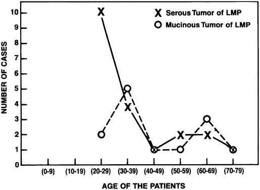
Of all serous tumors, 10% to 15% are LMP tumors.2 In the cases of LMP tumors from Magee-Women's Hospital,8 51% were classified as serous.

Most of the patients in the Magee-Women's Hospital series were between 24 and 33 years of age, which concurs with previous data.7 In general, most of the LMP ovarian tumors occurred in women between 45 and 50 years of age2 (see Table 2). Jensen and Norris,14 however, reported a few cases occurring in patients between 15 and 19 years of age. The mean age varies from 40 years in Michigan to 53 years in Norway.15 The LMP tumors occur at a younger age than do frank carcinomas. The bilaterality 
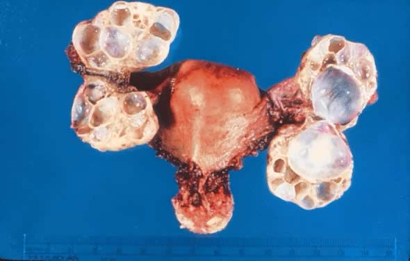
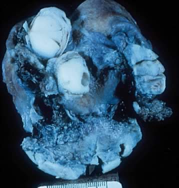
was 25%, which is within the range reported in the literature.16

Gross Examination

Serous tumors of LMP are grossly similar to serous cystadenoma. They are round, ovoid, and multilobulated, and their size varies from a few centimeters to 28 cm in diameter. On inspection, they are reddish, pink, or yellowish blue, sometimes with brown discoloration secondary to intramural or intraluminal hemorrhage. Their surfaces frequently are smooth and sometimes intermingled with irregular indurated areas representing thickened fibrous tissue or dystrophic calcification. Veins present 
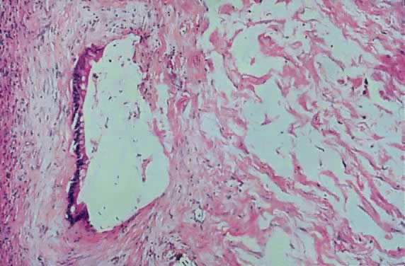
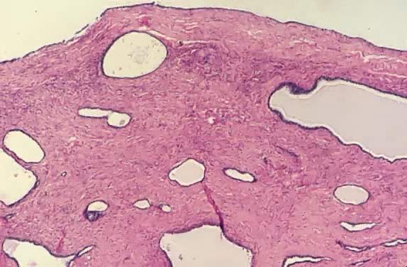
in the cystic wall can be prominent (Fig. 2). Translucency often is observed depending on the cystic contents.

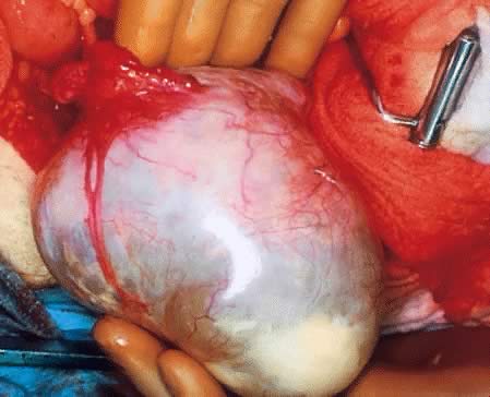
Fig. 2. External surface of a serous cystadenoma of low malignant potential showing a smooth, tense capsule with prominent veins and thickened fibrous areas.(Courtesy of Dr. Richard Stock, Naval Hospital, Portsmouth, VA.)

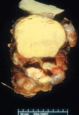
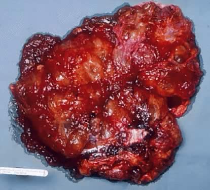
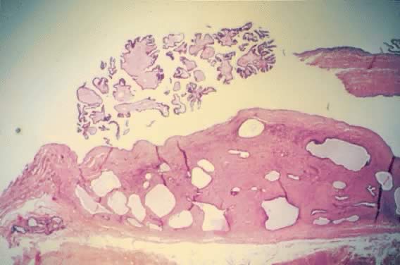
On palpation, the tumors can be fluctuant. Exuberant papillary projections may be observed on the external surface of the cyst. On section, the tumors are cystic (unilocular or multilocular), having secondary or “daughter” cysts of varying sizes and shapes,17 with clear, watery contents (and, therefore, serous cysts). Frequently, the tumor's content is seromucinous, indicating the presence of serous and mucinous lining epithelium,17 which has been confirmed by electron microscopic study.18 Tumors with this histogenic makeup are designated as mixed LMP tumors (described later). Mucus can be secreted by the apical portion of serous tall columnar cells. To designate a tumor as mixed, some investigators require that there be at least a 10% mixture of epithelial components (serous, mucinous, or endometrioid); others consider the required proportion to be at least 40%10 (Fig. 3). Necrosis is rare, although it is more frequent in serous than in mucinous cysts. It is not always possible to distinguish macroscopically a benign tumor from an LMP tumor, or the latter from its malignant counterpart. The presence of papillary projections on the internal or external surface of the cyst, which frequently are tan, suggest a borderline tumor or tumor of LMP.2 The papillary projections in the cyst lumen are more complex and luxuriant 
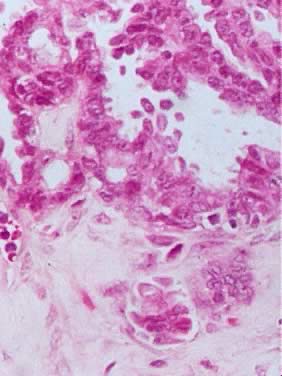
in an LMP tumor than in a benign cystadenoma (Figs. 4 and 5).

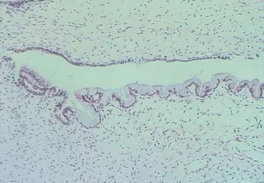
Fig. 3. Cystadenoma exhibiting lining consisting of cuboidal epithelium serous type ( top) and mucus-producing tall, columnar, endocervical-type epithelium with basally arranged nuclei ( bottom ). The mixture of epithelia frequently is found in serous or mucinous cystadenomas of the ovary (hematoxylin and eosin, 520).

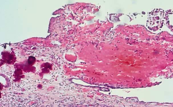
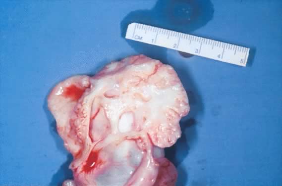
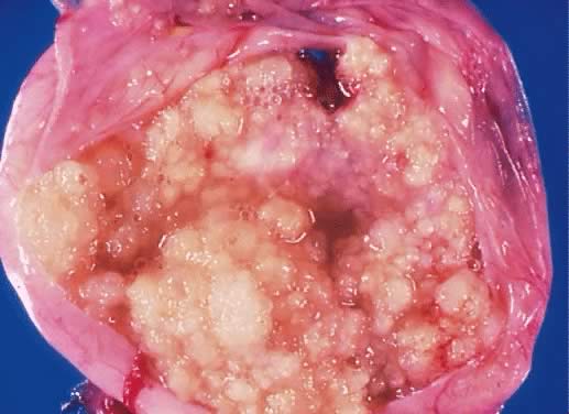
Fig. 4. Serous cystadenoma of low malignant potential demonstrating an exuberant growth of papillary projections arising from the internal surface of the tumor. Bubbly serous fluid is present in the lumen.(Courtesy of Dr. Richard Stock.)

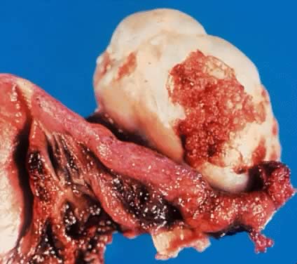
Fig. 5. External surface of an ovary showing a serous cystadenoma of low malignant potential exhibiting papillary growth. The lateral aspect of the uterus and the entire fallopian tube are seen clearly. The papillary growth does not represent invasion.(Courtesy of Dr. Richard Stock.)

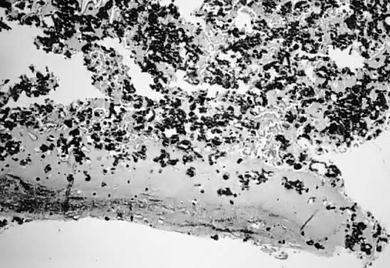
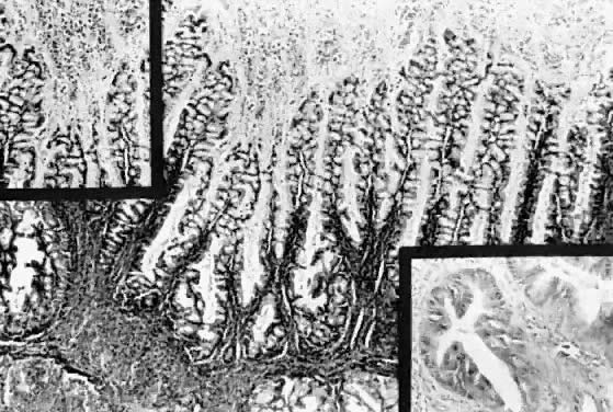
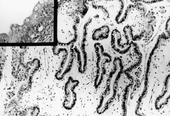
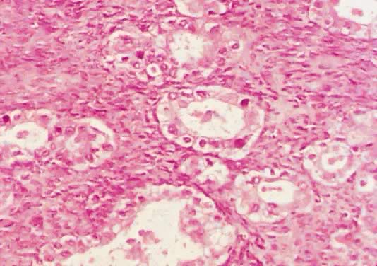
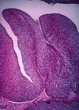
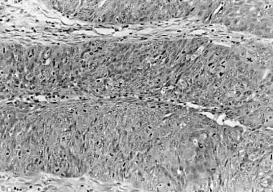
Microscopic Findings

On microscopic examination, the LMP serous tumors are characterized by the presence of simple or complex, broad and fine papillary structures (Fig. 6). They have a distinctive fibrovascular core covered by different types of columnar epithelia with atypical nuclei and tufting. The papillary structures may occur on the external surface in approximately 50% of the stage I tumors (see Fig. 5). Notice that the surface excrescences are the result of tumor growing from the surface epithelium concomitantly with the excrescences in the lumen of the cyst. The excrescences on the external surface do not signify that the tumor has penetrated the wall of the cyst. In few cases, the papillary excrescences represent the only tumor present in the ovary.17,18

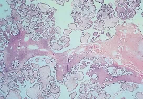
Fig. 6. Photomicrograph of a serous cystadenoma of low malignant potential. The tumor is composed mainly of papillary structures of different sizes resting on stroma made up of fibrous tissue. Tufting is observed in the interpapillary space (100).

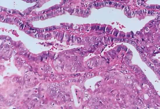
The LMP serous tumor may display all of the types of epithelial cells normally seen in the fallopian tube: ciliated, clear, intermediate, and mucus secreting.19,20 The cytoplasm of the serous cells is clear or eosinophilic. The basally located nucleus is somewhat irregular, hyperchromatic, and slightly pleomorphic. The epithelium, when stratified, should not have more than five layers of cells, which display loss of polarity and atypia. The mitoses are not atypical, although, their activity usually is increased. The frequency of mitosis varies among cases. In our series,8 there were cases in which the mitotic count varied from 0 per 10 high-power fields (HPF) to 9 per 10 HPF (mode, 0 to 2 per 10 HPF).

Psammoma bodies are observed frequently. The border between the abnormal epithelium and the subjacent or surrounding mesenchymal tissue is sharp, regular, and smoothly contoured. The invagination of the epithelium may create diagnostic difficulties; in these cases, observation of their margins reveal smooth rather than jagged edges and the absence of stromal edema, inflammation, or neovascularization. The changes of the epithelium are of the same type throughout. It is unusual to find areas of the tumor exhibiting totally benign epithelium alternating with areas of LMP tumor.21 In some cases, there is penetration of the stroma by glandular-like elements and microcysts with papillae without exhibiting stromal reaction; therefore, it should not be interpreted as an stromal invasion. In some cases, sharply delineated desmoplastic plaques, most frequently on the external surface, are noted. Sometimes these thickened areas are observed on the inner aspect of the wall. These have been designated as autoimplants.22 Micropapillary, cribriform, or solid histologic patterns also can be seen. These histologic patterns have been described only sporadically. Therefore, their behavioral significance are not known. The micropapillary and cribriform types can be associated with poor prognosis, but this has not been corroborated.23 The LMP serous tumor and benign serous cystadenoma usually have cilia. These cilia are absent in the cyst adenocarcinoma.20 Unfortunately, this histologic factor is not of any practical value in 
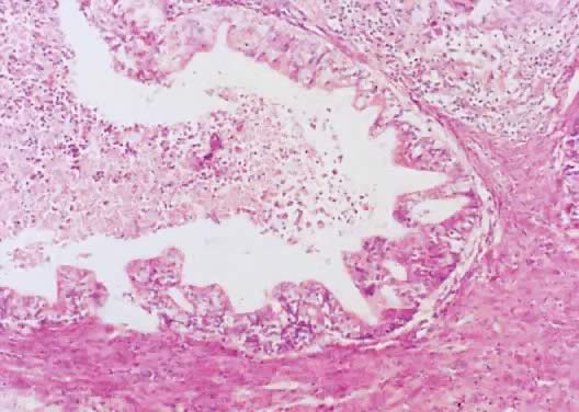
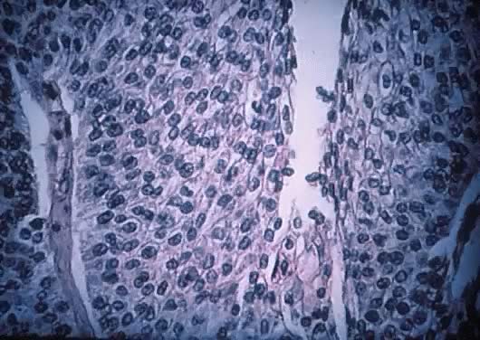
the differential diagnosis.

Epithelial cells devoid of connective tissue detach, fall, and form rosette-like structures that float in the cystic fluid; this phenomenon is designated as tufting (Figs. 7 and 8). Hyalinization of the connective tissue of the papillary structures is seen more often in the LMP serous tumors than in serous cystadenomas.19 Papillae undergoing ischemic changes show edema and frequently slight acute inflammation of the stroma. In more advanced cases, frank necrosis
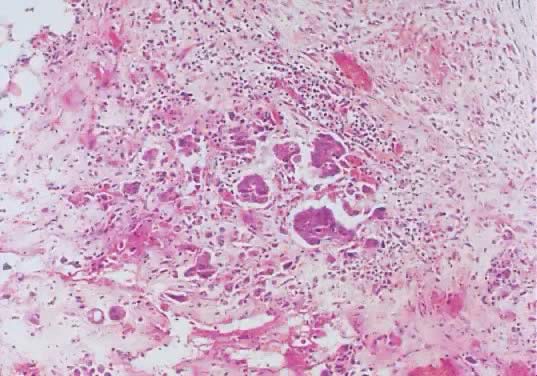
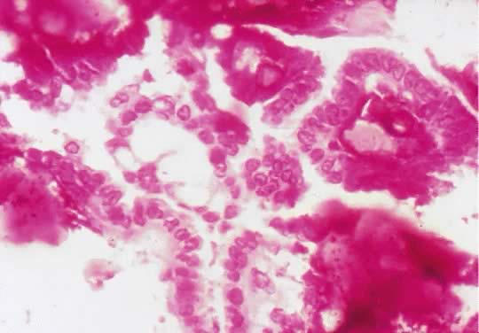
 may be evident.

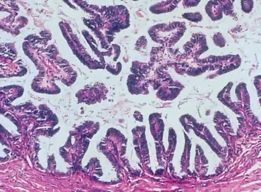
Fig. 7. Inner lining of a serous cystadenoma of low malignant potential showing papillations with epithelial pluristratification, hyperchromasia, and tufting; there is some degree of nuclear atypia (420).

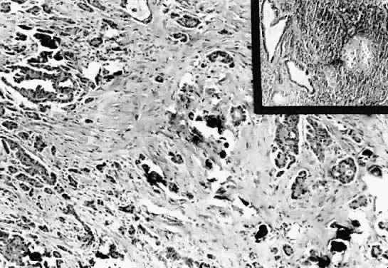
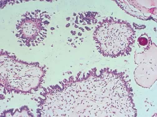
Fig. 8. Serous cystadenoma of low malignant potential demonstrating papillae, stratification of epithelial lining, tufting, stromal edema with hyalinization, and focal acute inflammatory cell infiltrate. On the upper right-hand corner, a psammoma body is clearly seen (hematoxylin and eosin, 420).

Of all the histologic characteristics discussed, the most helpful and important, which can be used to separate the tumor of LMP from a malignant neoplasm, is the stromal invasion by the epithelium. Unfortunately, the determination of stromal invasion can be difficult in some LMP tumors. Another problem is the lack of uniformity among pathologists in determining the existence of invasion. In general, invasive serous carcinoma is typified by the presence of poorly defined nests of cells with marked atypia and irregular borders that infiltrate the stroma. This stroma exhibits a loose, edematous, myxoid, or desmoplastic pattern, sometimes associated with necrosis and inflammatory infiltrate.

Histochemically, the epithelial cells of serous LMP tumors contain material that is periodic acid-Schiff-positive but diastase resistant. The same material also reacts positively with mucicarmine and Alcian blue on its luminal border.17 When viewed with the electron microscope, the cell nuclei are convoluted and the cytoplasm reveals the presence of cilia, which, as mentioned earlier, are absent in serous cystadenocarcinoma.24

Studies of epithelial nuclear deoxyribonucleic acid (DNA) in LMP tumor produce conflicting results. One study demonstrates that these neoplasms exhibit diploid patterns more frequently in early stages (I and II) than in stage III.25

Friedlander and coworkers26 demonstrated that the DNA content of cells derived from LMP serous tumors are diploid in 95% (38 patients) and aneuploid in 5% (2 patients). Of these two, one patient died of progressive disease. Retrospectively, it has been shown than some of the aneuploid cases were carcinomas that were erroneously diagnosed as LMP tumors. In a study27 of 44 primary ovarian serous tumors of LMP and 16 implants, about half of the primary tumors had a diploid pattern, and the other half, an aneuploid pattern. In 30% of the cases, the ploidy of the implants differed from that of the primary neoplasms. Of the 44 patients, 4 died: of these 4 patients, 1 had a diploid DNA pattern and the other 3, an aneuploid pattern. The implants in the four patients were aneuploid. Therefore, if the implants are invasive and aneuploid, the prognosis is unfavorable. Overall, however, there is little utility to flow cytometric examination in LMP tumors.

In a series investigated by Kabawat and associates,28 most of the serous malignant tumors, the LMP serous tumors, and 11% of the benign serous cystadenomas expressed the OC-125 monoclonal murine antigen on their surface. Its presence does not correlate with the morphologic pattern and, therefore, probably is not significant for the determination of tumor differentiation.

Another frequent finding in the LMP serous tumors is the presence of calcium deposits, some of which have a concentric laminated structure and are known as psammoma bodies (see the section of the serous psammocarcinoma of the ovary later). They are more abundant in the peritoneal implants within the fibrous tissue than in the ovarian tumor itself. In our review of LMP tumors, psammoma bodies were present in 60% of the cases. The psammoma bodies are discussed in more detail later.

Eichhorn and coworkers29 report 40 cases of serous tumors diagnosed as borderline with cribriform pattern, micropapillary pattern, or both. Of the 40 cases, 26 tumors (65%) were of the micropapillary type, 11 (27.5%) were cribriform, and 3 (7.5%) had both patterns. The micropapillary pattern was found in patients 21 to 76 years of age (mean, 38 years). The patients with cribriform pattern were 34 to 79 years (mean, 60 years). Those with both patterns also had a mean age of 38 years.

The criteria30,31 for the diagnosis of these two subsets of serous tumors of LMP are as follows:

  1. Tumors with micropapillary pattern characterized by small, uniform papillae with cores containing little or no stroma emanating in a nonhierarchical fashion from large fibrotic, edematous, or myxoid stalk or directly from the cyst wall; or a cribriform pattern of the cells lining the stalk or cyst walls; or both
  2. Involvement by either or both patterns of at least one confluent area (5 mm) in greatest dimension on at least one slide
  3. No more than grade 2 nuclear atypia (with grade 3 atypia being defined as the presence of “markedly enlarge pleomorphic and vesicular” nuclei with “prominent eosinophilic nucleoli”)

MICROPAPILLARY PATTERN.

The criteria just outlined for micropapillary LMP serous tumors was modified by Eichhorn and coworkers,29 who require that the papillae are at least “five times as long as wide.” Microinvasion was defined as the presence in the stroma of single cells or small nests of cells with little or no stromal response occupying one or more foci with an area of less than 10 mm2. To establish the presence of invasive implants, invasion of the underlining tissue with tumor replacing or destroying it should be demonstrated.

The micropapillary tumors were between 2.5 and 19 cm in diameter; 69% were bilateral, and 50%, intracystic. Only 15% were exophytic, and 35% were both intracystic and exophytic. Four percent of the tumors were microinvasive. The micropapillary component of the epithelial area was 10% to 25% in seven cases. In another seven cases, the micropapillary area was between 25% and 50%. In 12 cases, the micropapillary area was over 50%. In all cases, the mitotic rate was not more than 1 per 10 HPF.

CRIBRIFORM PATTERN.

The size of the tumors with cribriform pattern was similar to that of the micropapillary tumors, and the bilaterality was only 36%. These tumors were intracystic in 55% of the cases—more frequent than the micropapillary tumors. Of 11 tumors, 1 (9%) was exophytic, 4 (36%) were both intracystic and exophytic, and 1 showed microinvasion. In 3 of 11 cases, the cribriform pattern represented 10% to 25% of the epithelial surface. In two cases, the cribriform pattern occupied between 25% and 50% of the epithelial area. In two cases, the cribriform architecture occupied more than 50% of the epithelial surface. Mitoses were no more than 1 of 10 HPF.

The micropapillary and cribriform subtypes of serous tumors of LMP were more frequently associated with invasive implants. In the series by Eichhorn and coworkers,29 12% of serous LMP tumors were micropapillary and cribriform; of those, 65% were pure micropapillary, 27.5% were cribriform, and 7.5% were mixed. Both micropapillary and cribriform types of serous LMP tumors were treated almost 10 years earlier than those with pure serous LMP tumors. The patients with micropapillary-type tumors presented for treatment more frequently in a more advanced stage, the ovarian surface was involved more frequently, and they were bilateral more often. In the cases reported by Burks and colleagues,30 67% were stage II or III, whereas the typical serous LMP tumors were stage II or III in 32% of the cases. The micropapillary tumors were bilateral twice as frequently as were the typical serous LMP tumors. They also exhibited surface components more often than the typical LMP tumors. As mentioned earlier, micropapillary tumors had invasive implants more frequently than the typical serous LMP tumors. Patients with invasive implants died twice as frequently as those without invasive implants. Siedman and Kurman31 state that “a micropapillary pattern even in the absence of destructive infiltrative growth is a manifestation of an invasive tumor.” Therefore, it has been suggested that this group of tumors be designated as micropapillary carcinomas.24,30,32 In the cases reported by Eichhorn and coworkers,29 none of the noninvasive implants were micropapillary. These authors also state that some implants of overt ovarian serous carcinoma are not invasive, but cells with obviously malignant features present in a noninvasive implant are sufficient to make the diagnosis of carcinoma. These controversial findings should be resolved by studies of larger series of the subtypes in question. It appears that patients with micropapillary or cribriform types of LMP serous tumors had 5- and 10-year survival rates similar to those of typical LMP serous tumors and different from those observed with serous carcinomas. The survival rates vary considerably among studies. In the annual report of FIGO,33 the 5-year patient survival rate for LMP ovarian serous tumors for stages I to III was between 90% and 95%. Similar figures have been reported in subsequent series.

Back to Top
IMPLANTS

Implants are defined as the presence of extraovarian tumor deposits on or below the peritoneal surface. They are present in 16% to 48% of the cases of LMP tumors at the time of the initial diagnosis.21 In the Magee-Womens Hospital series of LMP tumors,8 implants were found in 45% of the cases. As stated earlier, it is uncertain whether these tumor deposits are true metastases or independent lesions growing de novo from mesothelial cells in the surface or subserosal mesenchymal tissue. There is evidence that some implants persist without progressing, that some continue growing without being lethal to the patient, that others may recur after their removal, and that some may behave like a malignant tumor. Furthermore, some implants may disappear after the removal of the ovarian lesion. There also is evidence that tumor implants with histologic characteristics of cystadenomas,6 LMP tumors, or frankly malignant serous carcinomas have been found in the absence of an ovarian serous carcinoma or with a small ovarian serous neoplasm.34,35 This phenomenon supports the theory of the multicentric autochronous metaplastic transformation of the peritoneal surface, where this metaplastic process results in the formation of a neoplasm, possibly by a field affect.4,36 These lesions correspond to what is called extraovarian müllerian peritoneal neoplasms. Using monoclonality studies and X chromosome inactivation analysis, it is possible to demonstrate the multifocal origin of the ovarian and peritoneal neoplastic growths.36

When implant-like or exophytic lesions are present only on the ovarian surface, the condition is considered a separate entity. Itskoviz and associates37 labeled this condition “surface papillomatosis of borderline malignancy” or borderline ovarian surface papillomatosis of LMP (Figs. 9 and 10). There is also sufficient evidence that other tumor deposits probably are the result of the implantation of neoplastic cells on the peritoneal surface. This assumption supports the presence of multiple implants and their frequent association with ovarian exophytic tumors.38

Fig. 9. Photomicrograph of a serous tumor of low malignant potential showing a focus of microinvasion represented by small nests of tumor cells penetrating the tumor stroma near the epithelial-stromal interphase. The nests are surrounded by a small clear space that should be filled with serous fluid. There is no stromal necrosis or inflammation around the tumor nests (400).

Fig. 10. Serous surface ovarian papilloma of low malignant potential with dystrophic calcifications. Secondary papillae lined with atypical cuboidal cells are noted at the upper right-hand corner (hematoxylin and eosin, 420).

Implants are more common in patients with all types of serous tumors but often are seen in LMP ovarian tumors with exophytic lesions, particularly if both ovaries are involved. Exophytic tumors of the ovary are found in 94% of patients with peritoneal implants, whereas only 27% of the patients without peritoneal implants have exophytic ovarian lesions. Of patients with exophytic ovarian lesions, 62% have peritoneal implants compared with only 4% of patients without ovarian exophytic lesions. Based on these data, diagnosing LMP serous tumors with exophytic lesions provides significant information related to the presence of extraovarian deposits.

According to Segal and Hart,39 the presence of exophytic lesions on the ovary predicts the existence of implants with a sensitivity of 94% and a specificity of 73%.

Implants are classified into three categories:

  Benign implants: Tumor deposits formed by glandular and tubular structures lined with benign-appearing epithelium without the presence of endometrial stromal cells surrounding the glandular structures have been designated by some authors as endosalpingiosis (because they have the type of epithelium present in the fallopian tubes), müllerian inclusions, and gland inclusions. The presence of psammoma bodies is not rare in this type of lesion.38 Benign implants are found in 22% of patients with LMP serous tumors. According to Gershenson and Silva38, this type of implant should be staged and treated as stage I lesion.
  Noninvasive implants: These implants are defined as tumor deposits with histologic characteristics similar to those found in LMP tumors but without invasion of the surrounding stroma and often with a subserosal location. In these cases, it is believed that they are formed from invaginations of mesothelial cells.17 Occasionally, they are intracystic. This type of implant is found in 37% of patients with LMP serous tumors.38
  Invasive implants: Tumor deposits similar to noninvasive implants, but with invasion of the desmoplastic stroma by individual tumor cells are defined as invasive. The margins of the invasive implants are poorly demarcated. The invasive tumor resembles a well-differentiated invasive serous adenocarcinoma. The desmoplastic stroma displays loose fibrous connective tissue with an inflammatory response (Fig. 11). This type of implant is found in 13% of patients with LMP serous tumors. Implants from LMP serous tumors are found (from the highest to the lowest frequency) in the pelvic peritoneum, omentum, uterus, fallopian tubes, nonpelvic colon, appendix, abdominal peritoneum, colon (except pelvic), small intestine, periaortic lymph nodes, liver capsule, and diaphragm.38 Several authors4,16 indicate that patients with invasive implants have a less favorable prognosis. More than one type of implant may be found in the same patient. No definition of an invasive implant is universally accepted.

Fig. 11. Omental invasive implants associated with a serous ovarian tumor of low malignant potential. The malignant histologic characteristics of the epithelium, its stromal invasion by single and papillary nests of cells, the inflammatory response, and the fibroblastic proliferation are evident (hematoxylin and eosin, 420).

Spread of disease to the lymph nodes was not found in any of our cases of LMP tumors reported in the Magee-Women's Hospital series.8 This also is considered a rare occurrence by others.10,17 Rare cases of distant metastases to the brain, lung, bone, and liver have been reported.40 In our series8 of LMP tumors, one patient showed radiologic evidence of metastasis to the liver parenchyma with gross involvement of the liver capsule. In general, the histologic type of the implants remains unchanged with the time. As mentioned earlier, the biologic behavior of the tumors is best predicted by the histologic features of the implants.4,26,36 Michael and Roth35 suggest that the prognosis depends more on the stage of the tumor than on the histologic type of the implants. Gershenson and coworkers41 conclude that the only factor that influenced the time of recurrence, progression, and survival was the presence of macroscopic residual disease at the time of the initial surgery. Evidence in the literature suggests that the presence of implants (especially invasive implants), cellular atypia, and residual gross disease after the initial surgery yield a significantly higher recurrence rate and a lower survival.41 Unfortunately, there is no consensus on the definition of the criteria for the classification of the implants; therefore, there is a lack of correlation among authors in assessing disease persistence, progression, and patient survival.

Some authors42–44 divide the implants into “superficially invasive” and “deeply invasive.” Others41 further subdivide the implants into early invasive and frankly invasive. The early invasive implants are defined as those in which only few individual cells are observed in the stroma. Frankly invasive implants are defined as those in which numerous single cells or groups of cells are observe in the stroma. These definitions do not mention stromal necrosis, desmoplasia, or inflammatory infiltrate. The persistence or progression of the tumor and patient survival do not change with either classification. Bell and associates44,45 use criteria for invasion based on the regularity or irregularity of the epithelial clusters invading the subperitoneal tissue, the presence of mitoses, and the nuclear atypia. More studies are needed to standardize the criteria to obtain more reproducible and comparable results.

Back to Top
SEROUS TUMORS OF LOW MALIGNANT POTENTIAL WITH EARLY INVASION OR MICROINVASION (BORDERLINE SEROUS OVARIAN TUMORS WITH EARLY INVASION OR MICROINVASION)

The concept of microinvasion in an LMP tumor of the ovary has been recognized for approximately 30 years.46–49 Histologically, microinvasion is characterized by the presence in the tumor stroma of one or more discrete foci of tumor with LMP features, and the tumor should be formed by cuboidal or columnar cells that appear singly or in groups. These cells have an abundant eosinophilic cytoplasm, round nuclei, and sometimes prominent nucleoli. Occasional hobnail-type cells are identified. The groups of invasive cells are small, and the foci of invasion range from one to eight groups. The cells with eosinophilic cytoplasm can be confused with decidual cells, especially in patients that are pregnant at the time of diagnosis. These types of cells were present in all tumors with microinvasion described by Bell and Scully.49 The eosinophilic cells constitute from less than 10% to more than 90% of the neoplastic cells of the tumors. The cells are single or form nests that must not exceed an area of 10 mm2.50 Occasionally, these nests of tumor cells are associated with psammoma bodies. The groups of cells representing microinvasion generally are surrounded by a clear zone that separates the infiltrating cells from the stroma. This zone or space can be filled with fluid probably secreted by the tumor cells. The invading cells do not involve the ovarian stroma. In some cases, these cells exhibit a cribriform pattern. Typically, there is no stromal reaction to the invading cells; however, when present, it is represented by a slight increase in the number of immature fibroblasts. In the study by Bell and Scully,49 lymphatic invasion was present in 10% of the patients, and all patients were younger than 37 years of age.

The definition of microinvasion, as with the definition of implants, varies among authors. The prognosis of the patients with tumors with microinvasion in the series by Katzenstein and associates,46 Tavassoli,48 and Bell and Scully49 was similar to that of the LMP tumors, that is to say, the prognosis was excellent in each of the studies despite the fact that some patients were treated conservatively with limited surgery. Therefore, in cases of young women with LMP tumors with early invasion who are concerned about their fertility, surgery can spare the uterus and the contralateral ovary without significantly increasing the risk.

Back to Top
SEROUS PSAMMOCARCINOMA OF THE OVARY

Although the histologic characteristics of psammocarcinoma has been known for years,1,51 only recently has this type of ovarian and peritoneal neoplasm been characterized as a specific entity.52 This tumor previously was considered to be an invasive papillary serous adenocarcinoma of the ovary, most of the time stage III composed of cells with moderate atypia and with psammoma bodies involving at least 75% of the tumor nests or papillae. Psammocarcinoma is considered to be a low-grade serous carcinoma that involves the ovaries, the ovaries and peritoneum, the omentum, mesentery, and gastrocolic ligament. This type of tumor can form large cystic masses accompanied by implants in many intraabdominal or pelvic organs.

Psammoma bodies are concentrically laminated calcifications designated as calcospherules. Chemically, they are crystals of hydroxyapatite. They are formed as a result of dystrophic calcification that takes place in epithelial cells and stromal histiocytes that undergo degeneration. These calcific bodies are not specific for psammocarcinoma; they are commonly found in all serous ovarian neoplasias including adenocarcinomas, where they are more abundant than in LMP tumors. Other malignant and benign neoplastic processes, including thyroid carcinomas and meningiomas, may display psammoma bodies.44,52 Several investigators51,53 report on the importance of the presence of psammoma bodies as a prognostic indicator in ovarian carcinoma, but their results have been contradictory.

Gilks and colleagues52 report on 11 cases in which the histologic characteristics of the tumor indicated invasive serous carcinomas of the ovaries and peritoneum, whereas their behavior was comparable with that of serous tumors of LMP.

Clinically, the patients had the same signs and symptoms as those with full-blown serous adenocarcinoma or serous LMP tumors, including some that were asymptomatic. The patients' mean age was similar to that of patients with serous carcinoma, (i.e., they were older than patients with LMP tumors).52 In some cases, disease may be suspected by the presence of psammoma bodies in the Papanicolaou smear. This event has occurred more than once at our institution.

The histologic criteria for the diagnosis are as follows (Figs. 13 to 15):

Fig. 13. Serous psammocarcinoma of the ovary. The section shows numerous psammoma bodies in the stroma, cyst lumen, and papillae (hematoxylin and eosin, 85).

Fig. 14. Photomicrograph of a psammocarcinoma to illustrate psammoma bodies in the papillary component of the tumor. Atypical cuboidal epithelial cells with prominent nucleoli line the papillae (200).

Fig. 15. Serous psammocarcinoma. The epithelial component diffusely infiltrates the desmoplastic stroma. Few psammoma bodies are present (hematoxylin and eosin, 212). Inset. Section of a periaortic lymph node involved by tumor. Observe the atypical glandular epithelium. The glands in the center exhibit atypical papillary structures (hematoxylin and eosin, 212).

  1. Destructive invasion of the stroma, vascular invasion, or, in extraovarian tumors, invasion of intraperitoneal viscera
  2. Mild to moderate cytologic atypia
  3. Occasional nests of solid areas of epithelial proliferation no more than 15 cells in diameter
  4. At least 75% of papillae and nests associated with or completely replaced by psammoma body formation

Kelley and associates54 reported on a case of psammocarcinoma occurring in an 18-year-old nulligravid woman. Her symptoms were abdominal pain and a pelvic mass whose size was comparable with that of a fetus of 15 weeks' gestation (see Fig. 12). The sonogram revealed a tumor with cystic and solid areas containing papillary excrescences and the presence of ascites. The preoperative CA-125 levels were 25 U/mL. At laparotomy, tumor was found in both ovaries accompanied by exophytic lesions. Implants were found on the serosa of the uterus, cul-de-sac, appendix, and the bladder flap.The right and left periaortic lymph nodes were palpable and positive for neoplastic disease (see Fig. 15). The tumor from the adnexa involved the sigmoid colon. The tumor was FIGO9 stage IIIC. Morphologically, all described histologic criteria were present (see Figs. 12 to 15). The DNA content of the tumor was aneuploid. The patient was treated with total abdominal hysterectomy, bilateral salpingo-oophorectomy, omentectomy, and sigmoid colectomy. Adjuvant chemotherapy was administered consisting of nine courses of carboplatin and cyclophosphamide. Ten years later, the patient is alive and disease free, and her CA-125 levels are normal. This case clearly demonstrates that this type of tumor can occur in the young. The gynecologist and the pathologist should be familiar with this entity, which probably often is misdiagnosed as a papillary cystadenocarcinoma; such misdiagnosis can lead to a graver prognosis for the patient and an increased possibility of overtreatment.

Fig. 12. Gross view of an ovarian psammocarcinoma. Multiple tumor nodules are noted. The largest one is yellowish. The cut surface has a gritty and solid consistency.

Poggi and coworkers55 report on a case of psammocarcinoma with an aggressive behavior manifested after the original diagnosis. This is contrary to what was suggested by Gilks and colleague52 in their original publication.

Back to Top
LOW-GRADE SEROUS PERITONEAL CARCINOMA

Although described by Swerdlow56 in 1959, only a few cases of low-grade serous peritoneal carcinoma have been reported. Before the criteria for the diagnosis of this lesion were established, these tumors probably were diagnosed either as well-differentiated extraovarian serous adenocarcinoma, serous cystadenoma of low malignant potential (serous borderline tumor), or psammocarcinoma. This type of tumor shares several of the histologic characteristics of the lesions mentioned earlier. The patient's average age is 58 years (youngest, 42; oldest, 73 years), 57% of the patients are postmenopausal, 38% were premenopausal, and 7% are intramenopausal.

The symptoms are vaginal bleeding, pelvic mass, abdominal pain, gastrointestinal disturbance, and ascites. In most cases, the neoplastic process was confined to the abdominal cavity; in a few (1 of 3) cases, the tumors are limited to the pelvis (ovaries, fallopian tubes, uterus, and peritoneal surface), mimicking fibrous adhesions or a case of pelvic inflammatory disease. In about half of the cases, the tumor is found while performing abdominal or pelvic surgery for other reasons. More than half of cases involve the omentum, which on palpation appears indurated and nodular, sometimes forming plaques. The neoplastic process may involve the ovarian surface or any other area of the peritoneal lining.

To establish a precise histologic diagnosis, the tumor should be generously and carefully sampled.

Microscopically, the tumor is characterized by the presence of nodularities, most of the time less than 1 cm in diameter. The neoplastic process is represented by solid epithelial papillary masses, generally between 20 and 30 cells across. Cuboidal or columnar cells devoid of cilia form the epithelial component of the tumor. These cell exhibit a moderate amount of eosinophilic cytoplasm, and the nuclei are round or ovoid, frequently with a macronucleoli. The mitoses are between 1 and 6 per 10 HPF; the cytoplasmic borders are not well defined. Cellular atypia is moderate. The epithelial component of the tumor often is associated with abundant psammoma bodies, which should not involve more than 75% of epithelial elements (in few cases, this component is more than 75%, but the epithelial masses are larger than 15 cells across; therefore, it is not a psammocarcinoma). Epithelial nests and psammoma bodies are seen, especially on the surface of the ovaries or fallopian tubes, in approximately 50% of the cases, either unilaterally or bilaterally. The tumor can show glandular, cribriform, or slit-like patterns. One of the criteria for the diagnosis of this tumor is the presence of stromal invasion, which probably is seen in all omental tumors. Invasion is less frequently observed in the other organs involved. Occasionally, lymphovascular invasion is identified by lymph node metastases. The metastasis in the lymph nodes maintains the histologic characteristics observed in the primary tumor.

The criteria for diagnosis of extraovarian low-grade serous peritoneal carcinoma reported by Weir and coworkers57 is extensive and detailed:

  1. Ovaries are normally sized or are involved by a benign process; most of the tumor is peritoneal, and, according to the surgeon or pathologist, the extent of tumor involvement at one or more extraovarian sites is greater than on the ovarian surface.
  2. Tumor is mostly serous and comparable with ovarian papillary serous carcinoma, any grade.
  3. On microscopic examination of the ovaries, one of the following is evidenced: no tumor, surface tumor without cortical invasion, surface tumor with cortical stromal involvement of less than 5 5 mm2, or a tumor smaller than 5 5 mm2 with ovarian stroma surface tumor.
  4. Patients who have had an oophorectomy before the diagnosis of serous carcinoma must have a pathology report documenting the absence of carcinoma in ovaries and a review of all slides of ovaries (or an attempt to review if the oophorectomy occurred more than 5 years before the diagnosis of peritoneal serous carcinoma).

The low-grade peritoneal serous carcinomacan be associated with multiple benign or malignant processes that frequently involve the ovaries and fallopian tubes (cystadenomas, cystadenofibromas, endosalpingiosis, Brenner tumor or transitional tumor, mucinous cystadenomas, and well-differentiated endometrial endometrioid adenocarcinomas). The follow-up information on the cases so far reported is too short to draw meaningful conclusions related to this tumor's behavior.

Back to Top
MUCINOUS CYSTADENOMAS OF LOW MALIGNANT POTENTIAL (BORDERLINE MUCINOUS CYSTADENOMA)

Mucinous ovarian neoplasms of all types constitute 12% to 15% of all ovarian tumors.9,50 Nearly 75% of mucinous tumors are benign and are FIGO9 stage I at the time of diagnosis; 6% to 10% are mucinous cystadenocarcinomas, and 10% are mucinous tumors of LMP.21,50,51 From the Magee-Women's Hospital series from 1975 to 1986,8 13 of 39 (33%) tumors were classified as LMP mucinous cystadenomas. This proportion is higher than the 15% reported by others.4,15 The mucinous tumors are consider as epithelial-stromal tumors. In these tumors, it has been possible to observe transition between mucinous, serous, or endometrioid elements, which are cells clearly of epithelial origin. Nevertheless, evidence shows that some mucinous tumors can be of germ cell origin.50 In few cases, mucinous glands are observed in teratomas of the ovary, and some mucinous tumors coexist with teratomas58 more often than any other epithelial tumor. The process of neometaplasia offers an explanation for the presence of gastrointestinal-type epithelium in mucinous ovarian tumors. In some instances, it is possible to observe the transition between the epithelium of an endometrioid cyst to endocervical-type epithelium and to a mucinous cyst tumor. Also, rarely, Brenner tumors or transitional cells tumors have been found in the wall of mucinous cysts; conversely, Brenner tumors or transitional cell tumors contain mucinous cyst. These phenomena suggest that urothelium of the Brenner tumor can be a source of mucinous neoplasms.50

LMP mucinous tumors occur in women with a mean age of 51 years, and they are bilateral in 10% to 15% of the patients.10,19,49 There is a significant difference between the bilaterality of LMP mucinous tumors of intestinal or enteric epithelial type (6%) and that of LMP mucinous tumors of endocervical or müllerian type (40%).59–61

On gross examination, the mucinous tumors are large. They frequently are larger than serous tumors. The large size does not necessarily indicate malignancy, even when they reach a diameter of 50 cm or more and weigh 50 to 150 kg.19,62 On average, the mucinous tumors measure 15 to 30 cm in diameter. The cysts usually are multiloculated and exhibit many infoldings of the inner epithelial lining, which contributes to the formation of a frequent, complex papillary architecture.19,59 These frequent infolding patterns usually make the diagnosis of invasion cumbersome and frustrating.

The gross examination in most cases does not permit differentiation between LMP mucinous tumors and its malignant counterparts (Figs. 16 and 17). As these figures show, the external appearance of the cyst is clear; sometimes blue, brown, or reddish coloration is observed, which is secondary to subperitoneal intramural or intracystic hemorrhage. Papillations seldom are found on the external surface of the tumor. The wall of the tumors have focal thick or nodular areas. Often, the ovary itself is part of the cyst wall. Some of the tumors exhibit a fine honeycomb pattern.

Fig. 16. Uterus with bilateral ovarian mucinous cystadenomas of low malignant potential showing multioculations and thick cyst walls.(Courtesy of Dr. Richard Stock.)

Fig. 17. Gross appearance of a mucinous cystadenoma of low malignant potential. The external surface of the tumor is irregular and multilobulated. The lobules are of different size and shape. The tumor is reddish brown. The lobules were fluctuant on palpation. Some areas of the tumor are firm. On cut section, multiple mucus-filled cysts are seen. Numerous small tan papillary structures are present in the inner surface of some of the cysts.

Microscopically, the cell cytoplasm of LMP mucinous tumors is more basophilic than that of the epithelium of the mucinous cystadenoma. Acute and chronic inflammatory infiltrate is found in a few cases. In general, the junction between the atypical epithelium lining the papillary structures of the cyst wall of the tumor with the underlining stroma is sharp and clear. Spread of the tumor outside of the ovary occurs in less than 15% of cases, and when it occurs, it is in the form of pseudomyxoma peritonei. Microinvasion, as defined for LMP serous tumors, is rare in LMP mucinous tumors.63

Back to Top
MIXED EPITHELIAL TUMORS OF LOW MALIGNANT POTENTIAL (MIXED BORDERLINE EPITHELIAL TUMORS)

Although mixed epithelial tumors of LMP (mixed borderline tumors) have long been known, they have not been well characterized until the publication of Rutgers and Scully.60 This group of tumors was defined by the WHO1 as a tumor in which “a second and/or a second and third type of epithelial cell accounts for at least 10% of the neoplastic lining.”

The ages of patients with mixed tumors range between 17 and 60 years (mean, 35 years). Ninety-one percent of the cases occur among premenopausal women. The most common presenting signs and symptoms are abdominal mass, abdominal pain, dysfunctional uterine bleeding, and infertility. Many patients are asymptomatic. All of the cases reported by Rutgers and Scully61 were FIGO9 stage I at the time of diagnosis. Of 36 cases, 28 were unilateral (78%); the remaining 22% were bilateral, with a prevalence similar to that reported for LMP serous tumor bilaterality. In 14% of the cases, the ovarian surface was involved by tumor. They can be unilocular or multilocular.

Tumor size ranges from 2 to 16 cm (average, 8.5 cm). Microscopically, the tumors have a papillary architecture, which is similar to that of the serous tumors. The papillae, as well as the inner cyst wall, are lined by various types of atypical epithelia, all derived from the müllerian system: mucinous, cervical, enteric, ciliated, and squamous in different proportions. Tufting also is present. The most prevalent component (10% to 60%) is the mucinous epithelium of endocervical type. Of the 27 cases described by Rutgers and Scully,60 only 1 was intestinal-type epithelium.

Acute inflammatory infiltrate is present in the epithelium, in the stroma, and in the mucus of the cyst lumen in more than 80% of the cases. There is not a clear explanation for the cause of the inflammatory reaction. One important component of the tumor is the presence of glandular formations similar to the type observed in microglandular hyperplasia of the endocervix. Of the five cases exhibiting this histologic glandular pattern, one patient was pregnant, one was in the postpartum period, and one was taking oral contraceptives. The other two patients did not have a history of hormonal therapy.61,64

Another interesting observation regarding the LMP mixed epithelial tumors is their association with pelvic or ovarian endometriosis in 53% of the cases. In five of the cases, a gradual transition between normal glandular epithelium, mild atypia, and severe glandular atypia was demonstrated. Pure müllerian LMP tumors are associated with endometriosis in 30% of the cases, whereas endometriosis is present only in 4% of all LMP tumors. In a few cases, the tumor is positive for the Grimelius stain, which confirms the presence of epithelium of intestinal type. Generally, the atypical part of a mixed LMP tumor is present in the intestinal-type epithelium.65

Mucinous tumors of the ovary can occur simultaneously with mucinous tumors of the gastrointestinal tract, especially with tumors of the vermiform appendix, in which case they usually are associated with pseudomyxoma peritonei. They also can coexist with mucinous tumors of the cervix, particularly with minimal deviation adenocarcinoma. At Magee-Womens Hospital, we treated a patient (personal experience) with bilateral ovarian tumors that were diagnosed as atypical mucinous cystadenomas and mucinous LMP tumor. Almost a year later, a minimal deviation adenocarcinoma of the cervix was found. The histologic features of the ovarian and cervical lesions were identical. Therefore, the ovarian tumors could have been metastatic lesions from the minimal deviation adenocarcinoma of the cervix.

Mucinous tumors of the ovary frequently are associated with secretion of steroid hormones, probably produced in the tumor stroma. Some cases of Zollinger-Ellison syndrome are caused by gastrin produced by the cells of neuroendocrine type found in the tumor epithelial lining. Also, elevation of the serum levels of inhibin are found in LMP mucinous tumors and in carcinoma.

Of the patients who were treated conservatively (unilateral salpingo-oophorectomy), 20% experience recurrence. Of those who were treated with hysterectomy and bilateral salpingo-oophorectomy, the recurrence rate was 5%.

Back to Top
ENDOCERVICAL-TYPE MUCINOUS TUMORS OF LOW MALIGNANT POTENTIAL (BORDERLINE MUCINOUS TUMORS OF MÜLLERIAN OR ENDOCERVICAL TYPE)

The müllerian- or endocervical-type mucinous tumors of LMP represent 15% of all the LMP mucinous tumors.61,64 In these studies, the average patient age is 34 years. Some patients are asymptomatic, and others complain of abdominal mass, abdominal pain, vaginal bleeding, and infertility. Seventy-seven percent of the patients are FIGO9 stage I at the time of diagnosis. Forty percent of tumors are bilateral. The diameters of the tumors ranged from 3 to 24 cm (average, 8 cm), which is significantly smaller than the 19-cm average diameter of the LMP mucinous tumors of intestinal type. Fifty-seven percent of the tumors are unilocular, and a few had papillae on the external surface. The cyst contents usually are thick, yellow, brown, or greenish mucus.

Microscopically, the tumors exhibited fine, elongated branching papillae supported by delicate fibrovascular cores (Figs. 18 and 19). In some cases, the stroma is broad and edematous. The papillae are similar to those of the serous tumors. The lining epithelium is tall columnar with a basally located nucleus. These tumors are lined mostly with endocervical-type epithelium. The cell cytoplasm is basophilic. The cells could be stratified up to 20 layers, usually present at the tip of the papillae. These peculiar epithelial “nests” are observed in most of the tumors of this type, invalidating the criterion of three layers, which generally is accepted as a dividing line between LMP mucinous tumors and cystadenocarcinoma (see the section on intestinal-type mucinous ovarian tumors of LMP later). Tufting is present, similar to that seen with the serous tumors. Epithelium forming a filigree pattern is a common finding (see Fig. 19), and occasionally a few ciliated cells are observed.

Fig. 18. Mucinous cystadenoma of low malignant potential, endocervical or müllerian type. Observe the marked papillation similar to that seen in serous tumors. Tufting and inflammatory exudate in the cyst lumen are seen (hematoxylin and eosin, 212). Inset, upper left. Papillation and inflammatory exudate. Inset, lower right. Epithelial atypia of gland-like spaces (420).

Fig. 19. Mucinous cystadenoma of low malignant potential, endocervical type, exhibiting a filigree pattern, pluristratification with atypia, stroma with mucinous material and inflammatory cell infiltration at the upper right-hand corner, and cellular debris with inflammatory cell exudate in the lumen (hematoxylin and eosin, 520).

Nuclear atypia is present, varying from mild to severe. Polymorphonuclear leukocytes often are found in the stroma, epithelium, and in the cyst lumen, intermingling with plasmalymphocytic infiltrates. Occasionally, multinucleated cells of foreign body type are seen in the tumor, presumably in response to mucus infiltration of the intramural stroma. The surface of the ovary is involved by tumor in 13% of the cases of endocervical type, compared with 6% of tumors of the intestinal epithelial type.

Implants are found in 18.5% of the endocervical-type LMP mucinous tumor. Microscopically, they are represented by glands immersed in a fibrous stroma with desmoplastic reaction. The implants are of the noninvasive type. The glandular epithelium is slightly atypical and occasionally is stratified. No mitoses are observed in these cases. The same types of epithelial cells are identified in the affected lymph nodes. According to Rutgers and Scully,61 no patients with LMP mucinous ovarian tumors of müllerian type have recurrences of tumor outside of the ovary.

The mucicarmine stain was positive in the cytoplasm of most cases of endocervical epithelial-type tumor, confirming the presence of intracytoplasmic mucin.

Ovarian müllerian-type tumors are associated with endometriosis in 30% of cases.60 In approximately 22% of LMP tumors of intestinal type, a transition from endometriosis to atypical glandular hyperplasia to an LMP tumor and to carcinoma is observed. Intestinal-type epithelium has been identified in cases in which there is an association with cystic teratomas.22

Back to Top
INTESTINAL OR ENTERIC-TYPE MUCINOUS OVARIAN TUMORS OF LOW MALIGNANT POTENTIAL (BORDERLINE INTESTINAL OR ENTERIC-TYPE MUCINOUS OVARIAN TUMORS)

The intestinal or enteric-type mucinous ovarian tumors of LMP constitute more than 85% of the LMP mucinous tumors.

Patients with LMP intestinal mucinous tumors are, according to Rutgers and Scully,61 significantly older (45% of their patients were postmenopausal) than those with the müllerian-type tumors (average age, 52 years). Eighty-three percent of the tumors were FIGO9 stage IA at the time of diagnosis, and 17%, stage III. The enteric type of mucinous tumors has a higher staging because of the presence of pseudomyxoma peritonei (discussed later) and lymph node metastasis or inclusions. As mentioned earlier, the average tumor diameter is 19 cm. Seventy-two percent of the tumors are multiloculated; the rest are unilocular (see Fig. 16). Only 23% of the multilocular tumors reported by Rutgers and Scully61 had papillary excrescences in their lumen.

Microscopically, all cases of LMP mucinous tumors of intestinal type have goblet cells, Paneth cells, and Grimelius-positive neuroendocrine-type cells19,61,64,65 lining the papillae or cyst walls (Fig. 20). This indicates the presence of intestinal-type epithelium. Other types of epithelia (e.g., squamous, serous, and endometrioid) can be found but should be less than 10% of the total epithelial mass. Some of the papillary structures are long and thin, and others are small with minimal branching. Epithelial bridging in the glands was seen, often rendering a filigree pattern similar to that observed in the malignant tumors. The number of layers of atypical epithelial cells in the mucinous tumors does not exceed three layers (see Fig. 20, inset). Mitoses and tufting are present but not as often as in serous tumors.13 The epithelial-stromal junction is sharp and clear. Complex glandular and cystic patterns resulting from the outpouching of the inner lining of the tumors often are present and created artifacts, resulting in a more difficult assessment of the stromal invasion than with the assessment of LMP serous tumors.

Fig. 20. Papillations and tufting in a mucinous cystadenoma of low malignant potential, intestinal type. Spaces are lined with a mixture of goblet cells and tall, columnar intestinal-type epithelium. The stroma is edematous and exhibits fine capillaries. Areas with metaplastic changes are present on the left side and upper right-hand corner (hematoxylin and eosin, 212). Inset. Higher magnification illustrating goblet cells (hematoxylin and eosin, 420).

Spread of the tumor outside of the ovaries occurs in less than 15% of the cases, and when it occurs, it usually takes the form of pseudomyxoma peritonei (discussed later). True peritoneal or omental implants are rare.

Because accurate establishment of stromal invasion, and therefore, the presence of carcinoma often is difficult, Hart and Norris66 suggest different criteria for the diagnosis of LMP mucinous tumors and carcinoma. Neoplasms diagnosed as LMP mucinous tumors not using the stromal invasion criterion and classified as stage I have survival rates of 98% and 96% at 5 and 10 years, respectively. These survival rates also are obtained when stromal invasion is used as the dividing line between LMP tumors and frank malignant neoplasms. Sumithran and associates67 demonstrated that tumors that had more than four layers of epithelial stratification usually followed a malignant course. Their findings support the aforementioned criteria of Hart and Norris.66 Those who use the criteria based on stromal invasion should look for irregular borders between the epithelium and the stroma, irregular gland arrangement, focal chronic inflammatory infiltration, and the presence of a loose immature stroma, or necrosis, or both. Russell13 emphasizes that diagnosing carcinoma without stromal invasion ignores the most important histologic hallmark of malignancy. Colgan and Norris17 determine the existence of invasion by the presence of islets of tumor within connective tissue greater than 2 mm in diameter or more than half-power field.

Counting the number of layers present on the papillae or wall of the cyst may be a difficult task for both mucinous and serous tumors in some cases. Therefore, it should be done only on histologic preparations that are clearly perpendicular to the plane of section of the epithelium.34 For more accurate diagnosis, it is ideal to consider as many histologic parameters as possible, including stromal necrosis.15,17,65,68

Nuclear DNA measurement has been used in LMP tumors to identify individual neoplasms with aggressive potential. It is known that aneuploidy is an adverse prognostic factor in ovarian serous carcinomas, independent of clinical stage.

Baak and coworkers69 used quantitative microscopic examination to differentiate between LMP mucinous tumors and mucinous carcinomas. The overall agreement between the computer assessment quantitation and light microscopic examination was more than 92%. Unfortunately, this technique is not practical and probably is not cost effective in determining the potential of LMP tumor progression. Some of the reports with meaningful values from both morphometric study and DNA flow cytometric study have problems related to accurate histologic descriptions, tumor staging, lack of description of types of implants (invasive, noninvasive), extent of tumor, sampling, interlaboratory and intralaboratory reproducibility, variations in methodology, and variation in criteria for diploidy and tetraploidy. Therefore, the data available from flow cytometric study of LMP tumors show contradictory results.26,32 When the technique becomes well standardized, flow cytometric study may be of diagnostic value. More studies are needed before a definitive conclusion can be reached. These types of tests, however, can be highly desirable in tumors such as those of LMP, where the separation of these tumors from cystadenocarcinomas with light microscopic examination can be difficult and sometimes impossible.

Vimentin can help in the differential diagnosis of serous LMP tumors. In serous carcinoma and endometrioid carcinoma, this immunostain often gives positive results, whereas results are negative in mucinous tumors. The cytoplasm of mucinous carcinomas frequently is positive for carcinoembryonic antigen. Most of the problems of differential diagnosis stem from the possibility that the tumor in question can be a metastatic mucinous adenocarcinoma, especially when the tumor is bilateral and when there is history of a mucinous adenocarcinoma in other parts of the body.

Mural Nodules

Nodules in the wall of mucinous tumors (benign, LMP, and malignant) have been reported. They are rare in other types of tumors. Histologically, they are different from the mucinous tumors. They can be of three types:

  Sarcoma-like mural nodules: These nodules are represented by tumor-like masses that protrude in the cyst inner lining. They usually are firm, yellow-tan with a brown discoloration, and frequently are associated with hemorrhage. They are found in benign cystadenomas, both types of LMP mucinous tumors (endocervical and intestinal), and in mucinous carcinomas. They can be single or multiple. In general, they are round and well circumscribed. Histologically, they are formed by different types of cells, some simulating osteoclast; some exhibit bizarre nuclei and atypical mitotic figures that can be up to 10 per 10 HPFs. Inflammatory infiltrate, hemorrhage, and necrosis are common. Etiologically, they are considered to be reactive. Their presence does not affect the prognosis of the tumor with which they are associated.16
  Sarcomatous nodules: These lesions are grossly similar to the previous one and occur in cystadenomas and carcinomas. They have not been described in mucinous tumors of LMP.
  Nodules of anaplastic carcinoma: Tumor nodules formed by anaplastic carcinoma have been reported.70,71 These tumors also are poorly circumscribed and may show vascular invasion. The tumor stroma is formed by epithelial cells that occasionally have a sarcomatoid appearance. They do not show inflammatory infiltrate or giant cells.16 These lesions must be differentiated properly from the sarcomatous-like nodule because the nodule of anaplastic carcinoma can be associated with metastases and death.

Back to Top
PSEUDOMYXOMA PERITONEI

Pseudomyxoma peritonei is a poorly understood and controversial condition characterized by the accumulation of large amounts of mucous material within the peritoneal cavity with involvement of the omentum. This unusual form of neoplasia often is associated with rupture, spillage, leakage, or metastases from a primary appendiceal tumor, a tumor of other areas of the gastrointestinal tract, especially the colon, or from the ovaries. The tumors of the appendix and ovaries can be synchronous, which makes the precise determination of the origin difficult. Most of the time, the pseudomyxoma peritonei is associated with well-differentiated malignant tumors, but it can be secondary to benign mucinous neoplasms. It is argued that the tumors of the ovaries associated with pseudomyxoma peritonei probably are secondary to a tumor in the appendix. In many cases, the ovaries are completely free of tumor; therefore, the primary tumor must have been in the gastrointestinal tract. It also is known that the simple rupture of a mucinous lesion is not sufficient to cause pseudomyxoma peritonei.64,72 Some investigators73–75 suggest metaplasia of the mesothelium as a possible cause of pseudomyxoma peritonei. Therefore, in this case, pseudomyxoma peritonei should be consider as a third site of an independent primary form of neoplasia. This idea is entertained based on the existence of other forms of ovarian type of tumors originating on the peritoneal surface only. On the opposite side of this theory are those that believe that this form of neoplastic process does not occur on the peritoneal surface because mucinous metaplasia of the peritoneum is rare. Also, when a mucinous tumor of the abdomen does not involve the ovary or the appendix, it can develop in the retroperitoneum. Clinically, the fact that mucinous tumors of the ovary associated with pseudomyxoma peritonei are more frequent on the right side suggests an appendiceal origin. By histochemical technique, Ronett and colleagues76 provided evidence that pseudomyxoma peritonei and the mucous cyst present concomitantly in the appendix and ovaries are of appendiceal origin. Other investigators77 using molecular genetic studies failed to demonstrate the association and common origin of the appendiceal and ovarian tumor.

It has been suggested that the presence of free mucin in the abdominal cavity should be designated “mucinous ascites.” The presence of mucin with evidence of organization (inflammatory cells, fibroblast, capillaries, and adherence to surfaces) but without neoplastic cells should be designated pseudomyxoma peritonei. The collection of intraabdominal mucus with fibrous adhesions, inflammation, and histologically malignant cells should be designated as mucinous metastatic carcinoma.

Pseudomyxoma peritonei is found in approximately 2 of 10,000 laparotomies and is three to four times more common in women than in men.78–80 Most of the patients with pseudomyxoma peritonei have a prolonged clinical course that can lead to progressive bowel dysfunction, intercurrent infection, inanition, and death. In some individuals, pseudomyxoma peritonei does not recur after cytoreduction therapy.

Based on two studies,79,80 the most probable origin of pseudomyxoma peritonei is believed to be a benign or malignant tumor of the appendix. These authors based their assumption on anatomic location, careful gross examination of the organs, determination of bilaterality of the ovarian tumors, and meticulous histologic evaluation and comparison of the types of mucus-secreting epithelia.

According to Ronnett and coworkers,80 to arrive at the most likely site of origin of the tumor causing pseudomyxoma peritonei, the pathologist should observe the following:

  1. Is the tumor confined to the ovarian or superficial ovarian cortical stroma, or both?
  2. Is the ovarian tumor bilateral and histologically consistent with colon or appendiceal morphologic types, including mixed glandular and signet ring cell neoplasms resembling goblet cell carcinoid, carcinoid-adenocarcinoma, or crypt cell carcinoma of the appendix?
  3. Is the unilateral ovarian adenocarcinoma consistent with colonic or appendiceal morphologic type with a history of colonic or appendiceal adenocarcinoma?

According to Grodinsky and Rubnitz,81 in most cases, the primary site of origin can be established, and in most cases, the primary site is an appendiceal adenoma, followed by an appendiceal or colonic carcinoma. In many cases, the gross inspection and even the microscopic examination fail to reveal the presence of a mucinous appendiceal neoplasm or the site of its rupture.

Other possibilities for the origin of pseudomyxoma peritonei are the ovaries and the rest of the peritoneal surface by mucinous metaplasia of the mesothelium, as mentioned earlier. However, mucinous metaplasia of the mesothelium is rare, if it exists. If it does exist, the metaplasia would be the result of irritation produced by the ascitic fluid on the surface of the mesothelium. In cases of colonic carcinoma as the source of pseudomyxoma peritonei, transmural neoplastic invasion by the primary tumor can be documented. Animal experiments with artificially produced appendiceal mucoceles that do not have a neoplastic process indicate that the presence of the mucous alone, as mentioned earlier, is not sufficient to generate pseudomyxoma peritonei.81,82 The mucus material present in the pseudomyxoma peritonei is either cellular or acellular. The acellular mucous collection probably should be designated “mucinous ascites”; these cases behave in a more favorable fashion than pseudomyxoma with cells.83 Acellular pseudomyxomas peritonei probably results in some cases from inadequate sampling. Ronnett and colleagues80 found that pseudomyxoma peritonei behaves in a way that is related to the type and differentiation of the tumor of origin. Consequently, if the cells in the mucus are rare or are of benign appearance, the tumor of origin is an appendiceal adenoma. In cases in which the tumor of origin is an adenocarcinoma, the mucus exhibits cells with marked atypia or malignant characteristics. The patients, in these cases, are diagnosed as having intraabdominal carcinomatosis with parenchymal organ and lymph node metastases.

The pseudomyxoma peritonei does not characteristically involve the bowel surface peritoneum in contrast to what happens with carcinoma. The peritoneal implants are noninvasive and are found in the omentum, undersurface of diaphragm, pelvis, right retrohepatic space, left abdominal gutter, and ligament of Treitz. Morbidity and mortality are intimately associated with the presence of pseudomyxoma peritonei in the abdominal cavity. Pseudomyxoma peritonei may produce bowel obstruction. Metastasis of pseudomyxoma peritonei outside of the abdominal cavity does not occur. In pseudomyxoma peritonei, the mucus should be accompanied by noninvasive implants, the presence of an appendiceal adenoma without significant atypia, and mitotic activity. The cells in the mucus should have the same histologic characteristics as the adenoma. This lesion can be treated with surgery or intraperitoneal chemotherapy with good results.84

The stroma of the ovaries can be infiltrated by the mucous of pseudomyxoma peritonei, in which case it is designated as pseudomyxoma ovarii.58 Epithelial cells with atypia or malignant characteristics are present in the mucus pools (Fig. 21). As just described, these cases are secondary to appendiceal or colonic neoplasia with secondary ovarian involvement, not to a primary mucinous tumor of the ovary whether benign, LMP, or malignant.

Fig. 21. Stroma of an ovarian mucinous cystadenoma of low malignant potential showing pools of mucus and atypical mucus-producing columnar epithelial cells of intestinal type in the stroma. Goblet cells are seen easily. The mucus pools are mostly acellular, although three hyperchromatic atypical nuclei are seen at the upper right-hand corner (hematoxylin and eosin, 420).

Back to Top
ENDOMETRIOID TUMORS OF LOW MALIGNANT POTENTIAL (BORDERLINE ENDOMETRIOID TUMORS)

The existing lack of uniformity for the histologic criteria to diagnose LMP tumors of the ovary is even more prevalent when dealing with endometrioid tumors.

The name “endometrioid neoplasm” is derived from their histologic similarities to atypical adenomatous hyperplasia (atypical complex hyperplasia) of the endometrium or to a well-differentiated endometrial adenocarcinoma. The designation suggested few years ago, “atypically proliferating tumors,” may generate confusion because these tumors have been designated and classified by several authors85,86 as “proliferating endometrioid tumors” independent of the designation given by Russell2 in 1994. The term “proliferating endometrioid” was suggested to designate a benign neoplastic endometrioid process with mild atypical changes of the epithelium forming the glands, not yet sufficient to diagnose it as an endometrioid tumor of LMP or borderline endometrioid tumor. No cases of metastasis or death have been reported as result of an endometrial tumor with mild atypical changes.87–89

Microscopically, two types of endometrioid tumors of the ovary are described. One is similar to a cystadenofibroma, and it is characterized by endometrial-like glands in a dense, fibrous stroma (Fig. 22). In general, these types of ovarian tumors are rare; no large series has been evaluated, and, therefore, their biologic evolution has not been assessed completely.85–87 Two percent to 10% of all ovarian tumors of LMP are endometrioid9,29,88,89 and represent about 20% of all the endometrioid neoplasias.58 These tumors may be associated with synchronous or metachronous hyperplasia of the endometrium.88,89

Fig. 22. Photomicrograph of an endometrioid tumor of low malignant potential. The tumor is composed of endometrial-like glands simulating atypical complex hyperplasia. The glands are arranged in groups separated by dense fibrous stroma (100).

The patients usually are between the fifth and sixth decades in age. The tumors are small and predominantly solid. They are tan or yellow-white with small cystic areas, which gives the lesion a honeycombed appearance. The size ranges from 1 cm (the smallest to qualify as endometrioid LMP tumor) to 10 cm in diameter, and they can be unilateral or bilateral. As with other LMP tumors, LMP endometrioid tumors usually are asymptomatic,13 but as with other ovarian tumors, the patient may complain of pelvic mass, pelvic pain, and vaginal bleeding.

The other form of LMP endometrioid tumor is found less frequently and is composed of solid areas in which hemorrhage or necrosis is observed. These tumors simulate endometriosis.

In summary, the WHO50 criteria for the diagnosis of LMP endometrioid tumors are as follows:

  • Atypia, mild to severe lining glands or cysts
  • Back-to-back arrangement of the glands
  • Absence of stromal invasion

Bell and Scully90 labeled endometrioid tumors “atypical endometrioid adenofibromas or proliferating endometrioid tumors.” They are characterized by crowded, complex endometrial glands with epithelial stratification and mild to moderate atypia (Fig. 23; see Fig. 22). Bridging of the glands can form a cribriform pattern. Tufting is noted in the cystic or glandular spaces. Smooth muscle and areas of luteinization can be observed. There are lesions with low-grade malignant characteristics.

Fig. 23. Photomicrograph of an endometrioid tumor of low malignant potential. Glands similar to those observed in atypical complex, and atypical simple hyperplasia are observed. The glands are irregular. The epithelium form, in some areas, solid masses of atypical cells showing glands within glands. The stroma is formed by dense fibrous tissue. No stromal necrosis is observed (200).

The group of LMP endometrioid tumors simulating endometriosis is characterized by the presence of epithelial stratification and marked atypia. The adenomatous elements have less stroma and more exuberant back-to-back glands within cystic spaces (see Figs. 22 and 23). This marked epithelial proliferation may mimic an endometrioid adenocarcinoma.91 In accordance with the definition of Snyder and coworkers,92 the endometrioid tumors are classified according to the degree of glandular atypia and the presence of nest of epithelium that are smaller or larger than 5 mm without intervening stroma. Following the parameters of this classification, if the atypia is mild to moderate and the nests of epithelium are less than 5 mm, the tumor should be classified as benign, and they could be designated as atypical proliferating endometrioid tumors. Conversely, if the atypia is severe, the nests are larger than 5 mm, the mitotic activity is increased up to 10 per 10 HPF in the most active epithelial areas, and the nuclei exhibit hyperchromasia and pleomorphism, the tumor should be designated as LMP endometrioid tumor. In all of these lesions, there is no evidence of stromal invasion by the atypical epithelium. Bell and Scully90 consider endometrioid papillae that protrudes into the lumen of cystic space lesion as well-differentiated adenocarcinomas, even with absence of destructive invasion. The papillae in these cases should be thicker and broader than those seen in papillary endometrioid carcinoma. This process mimics well the atypical complex endometrial hyperplasia or a well-differentiated adenocarcinoma (see Figs. 22 and 23). Squamous metaplasia was present in 20% to 80% of the cases of LMP endometrioid tumors; rarely, mucinous or serous metaplasia was seen.33,92 Morule metaplasia was present in close to 77% of the cases and of those, 90% showed central necrosis and it was possible to observe focal areas of microinvasion. In two of the cases described by Snyder and coworkers,92 there were epithelial cells with atypical koilocytic appearance. They did not mention whether they tried to confirm the presence of human papillomavirus DNA in this lesion. In a 1988 study by Snyder and colleagues,92 52% of the cases of LMP endometrioid tumors and 15% to 25% of the endometrioid adenocarcinomas showed an association with preexisting endometriosis. In some cases, the gradual transition between a normal endometriotic gland to a mildly, moderately atypical epithelium can be observed. Some investigators consider the possibility that endometrioid tumors also can arise from metaplasia of the surface coelomic epithelium.17,20,93 They consider this possibility because there are relatively few cases of tumors arising from endometriosis. It is believed that LMP endometrioid tumors rarely metastasized or recurred. Therefore, in a young patient, a unilateral salpingo-oophorectomy should be curative.

Because of the lack of uniformity in criteria to establish the diagnosis of LMP endometrioid tumors, determining the diagnosis of the tumor should be done as accurately as possible, whatever method is used. It is probably safe to use destructive invasion as the most important parameter to distinguish LMP endometrioid tumors from carcinoma. According to other investigators,90 the existence of overt malignancy can be established by confirming the presence of glands or islands of epithelium with marked atypia. Remember that the LMP endometrioid tumors, as other LMP tumors, have a microinvasive status characterized by the presence of nests or cords of cells with a malignant appearance invading the stroma in a disorganized fashion. Scully and associates50 arbitrarily defined microinvasion as one or more foci of tumor cells of 10 mm2 or less in area. One or more areas larger than 10 mm2 is considered, therefore, frank invasion. Nevertheless, microinvasive LMP endometrioid tumors behave similarly to neoplasms without microinvasion.

Back to Top
CLEAR CELL TUMOR OF LOW MALIGNANT POTENTIAL (BORDERLINE CLEAR CELL TUMOR)

According to Czernobilsky,86 clear cell tumors of LMP represent a subset of the endometrioid adenofibromatous tumors of the ovary. This type of tumor is rare and represents only 1% of all LMP clear cell tumors.19,50 These neoplasms are found in the ovary, endometrium, and vagina and are the tumors more often associated with pelvic endometriosis and hyperplasia of paraendocrine tissue.50 Most of the tumors are unilateral. Their size varies between a few centimeters and 30 cm in diameter.

Dystrophic calcifications comparable with those seen in the mucinous or Brenner tumors have been observed in LMP clear cell tumors.

Grossly, these tumors are solid but exhibit small cystic spaces that may give a honeycomb appearance to the tumor. The spaces are filled with serous, mucinous, or seromucinous fluid similar to that in cystadenofibromas.58,93 In some cases, cystic spaces with polypoid masses, which are yellowish brown, are observed. Clear, mucoid, or bloody fluid may be present. The external surface can be shaggy because of fibrous adhesions. The clear cell tumors currently are accepted to be of müllerian and not mesonephric origin.93–95 This assumption is based on the following: frequent association of clear cell tumors with endometriosis, occurrence in the same ovary, the coexistence with endometrioid adenocarcinoma, the fact that this type of neoplasia occurs also in the endometrium, and the fact that it can be found in vaginal adenosis, which is of müllerian origin.50

Microscopically, these neoplasms have a fibrous stroma. Within the stroma, there are glands and tubules arranged in a disorganized fashion, contrary to what is seen in the adenofibromas (Fig. 24). The stroma is definitively more abundant than the epithelial component. The infolding tubular and glandular spaces characteristic of these tumors are lined by atypical clear cells that may form papillary structures (see Fig. 24). The clear cells may be arranged in single or multiple rows. The nuclear atypia is of varying degrees, sometimes with prominent nucleoli and epithelial tufting. Mitotic figures are seen occasionally. The epithelial cells of some of these tumors are of the hobnail type, and they are characterized by scanty cytoplasm with the nucleus situated on one edge of the cell, simulating the changes of the Arias-Stella phenomenon. The cytoplasm of the cells exhibiting a clear cell pattern is abundant, granular, and eosinophilic. Frequently, squamous, ciliated, and other types of metaplasia and endometriosis are seen. This peculiarity emphasizes the müllerian origin of the tumor.

Fig. 24. Photomicrograph of a clear cell tumor of LMP showing tubular structures lined by low columnar epithelium. The cytoplasm in most epithelial cells is clear. Some nuclei are hyperchromatic. The intertubular stroma is dense (200).

Microscopically, the glandular lumen and tubular structures may show mucinous contents, and the cytoplasm may display diastase-digestible periodic acid-Schiff-positive material, confirming the presence of glycogen.95

As is emphasized in the description of other LMP tumors, invasion of the stroma by the epithelial components of the tumor is the cardinal feature that separates LMP clear cell tumor from clear cell carcinoma.34 Roth and associates95 report the simultaneous existence of clear cell adenofibromatous tumor and clear cell carcinoma. It is difficult to affirm categorically that this type of lesion can coexist as two separate entities; it could be either an LMP clear cell tumor that has become a clear cell carcinoma or a tumor that started as a carcinoma de novo with areas so well-differentiated so as to be confused with LMP clear cell tumor. We have treated a patient with ovarian tumors with areas of LMP and areas of well-differentiated clear cell carcinoma (personal experience).

In the cases reported by Roth and associates,95 some patients with clear cell carcinoma, as expected, died of their malignant disease; others survived up to 16 years. In the 39 tumors that we studied,8 there was not a single case of LMP clear cell tumor. The coexistence of a clear cell tumor of LMP and malignant lesions, as mention earlier, emphasizes the importance of generous sampling (one section for every 1 to 2 cm of the tumor diameter) so that a malignant tumor is not missed. The most important microscopic feature, as with other LMP tumors, is the destructive invasion of the stroma by the tumor cells.

Back to Top
BORDERLINE TRANSITIONAL CELL TUMOR (BRENNER TUMOR OF LOW MALIGNANT)

Borderline transitional cell tumor is an unusual neoplasm recognized as originating from mesothelium that has undergone metaplasia, resulting in an urothelium-like epithelium.96,97 This assumption is based on the following: (1) the frequent presence of mucinous or serous epithelium in the tumors and their frequent association with mucinous cysts; (2) the observation of communication or transition between the tumor epithelium and the surface ovarian epithelium that has undergone transitional cell metaplasia; and (3) the close similarities of immunohistochemical reactions and ultrastructural characteristics between the nests of Brenner tumor and the Walthard nests, which are of mesenchymal origin.50 Also, the transitional cell or Brenner tumor may have a germ cell origin. This possible origin has been suggested because of the occasional association of the transitional cell tumor (Brenner tumor) with a dermoid cyst and, more rarely, with the struma ovarii or carcinoid tumor.

Transitional cell tumor of LMP, or borderline transitional cell tumor (Brenner tumor of LMP), represents 1.5% to 2.5% of all ovarian neoplasms.17 In our review at Magee-Womens Hospital,8 only one case of transitional cell tumor of LMP (Brenner tumor of LMP) was found (2.6%). The patients range in age from 45 to 80 years. In most series, the average age is about 50 years for transitional cell tumor of LMP or (Brenner tumor of LMP) and 60 for the frankly malignant tumor.98 The tumor size varies from 1 to 40 cm in diameter.13,95,99 In most cases (93%), these tumors are unilateral and occasionally are associated with teratomas (5% to 6%), and in 25% of cases, they coexist with mucinous elements. Two percent of mucinous cystadenomas are known to have a transitional cell or Brenner tumor component.18,58 They usually are well circumscribed, firm, nodular masses that may have a smooth surface.

Transitional cell tumor of LMP or Brenner tumors of LMP are predominantly cystic and unilocular (Fig. 25). On cross-section, these tumors show a white-yellow surface and spaces that are totally or partially filled with friable or granular papillae growing from the inner lining of the cyst wall. The cyst lumen contains clear watery fluid.

Fig. 25. Gross appearance an ovarian transitional cell tumor of low malignant potential (Brenner tumor of LMP). Notice irregular cystic spaces and two round-oval masses approximately 1.5 cm in diameter. These masses are solid. In the lower mass, a small cyst space is noted. The inner lining of the cystic spaces is shaggy.

In the 1973, histologic classification of ovarian tumors by the WHO7 criteria for transitional cell tumors of LMP (Brenner cell tumor of LMP) were defined, including cases described by Roth in 197197 and later by Hallgrimson and Scully.100

Roth and collaborators99,101 in 1985 classified the transitional cell tumor (Brenner tumor) into the following categories: metaplastic, proliferating, LMP, and malignant. This classification was based on the comparison of the histologic changes present in the transitional epithelium of the urinary tract with those of the transitional cell tumors of the ovary (Brenner tumors). The classic benign transitional cell tumor (Brenner tumor) exhibits cell morphologic features similar to those in the von Brunn nests. These nests are observed in the inner surface of the urinary bladder, may be single or multiple, and are associated with chronic cystitis.58,95

The metaplastic type of transitional cell tumor, or metaplastic Brenner tumor, is equated with changes seen in cystitis glandularis.

Proliferating transitional cell tumor, or proliferating Brenner tumor, is equated with the changes observed in papillary transitional cell carcinoma grade 1 to 2 of the urogenital tract.6,34 No stromal invasion is seen in this type of tumor. The ovarian transitional cell tumor of LMP (Brenner cell tumor of LMP) is compared with the papillary transitional cell carcinoma grade 3 of the urinary tract or with a cervical squamous cell carcinoma in situ of the cervix (Figs. 26 and 27). The well-differentiated malignant transitional cell tumor of the ovary (well-differentiated malignant Brenner tumor) is compared with the invasive transitional cell tumor of the urogenital tract grade 1 to 2. The poorly differentiated transitional cell tumor of the ovary (poorly differentiated malignant Brenner tumor) is compared or equated with the invasive transitional cell tumor of the urogenital tract grade 334,101 (Fig. 28) For some,99 the Brenner tumor forms part of a continuous spectrum characterized by a gradual increase of epithelial abnormalities. None of the tumor subsets (metaplastic proliferating and LMP) have become malignant. Therefore, their behavior and prognosis is similar in the three types.

Fig. 26. Papillations showing a thin, central fibrovascular core present in a proliferating Brenner tumor. The epithelial lining consists of 10 to 20 layers of transitional cell-type epithelium. The cellular atypia and pattern resemble those of transitional cell carcinoma (grades 1 to 2) of the urinary tract (hematoxylin and eosin, 400).(Courtesy of Dr. Laurence Roth.)

Fig. 27. Transitional cell-like epithelium of proliferating Brenner tumor showing atypical nuclear changes similar to those of transitional cell carcinoma of the urinary bladder (grades 1 to 2) (hematoxylin and eosin, 700).(Courtesy of Dr. Laurence Roth.)

Fig. 28. Section illustrating Brenner tumor of low malignant potential with histologic characteristics simulating a transitional carcinoma grade 3. Observe the number of layers, loss of polarity of nuclei, hyperchromasia, and abundant and abnormal mitosis (hematoxylin and eosin, 420).

We agree with Fox,21 that it would appear preferable to retain the single diagnostic entity of transitional cell tumor of LMP or Brenner tumor of LMP rather than subdividing it in the three subsets described earlier. The transitional cell tumor of LMP, or Brenner cell tumor of LMP, sometimes is associated with endocrine manifestations of androgenic or, less frequently, estrogenic types, probably secondary to steroid hormone secretion by the tumor stroma.50 The real biologic behavior of these neoplasm is not well known because of their rarity.

Back to Top
CYSTADENOFIBROMA WITH ATYPIA (ATYPICALLY PROLIFERATING CYSTADENOFIBROMA)

Cystadenofibroma with atypia is known to be formed by tissue of epithelial and mesenchymal origin. The epithelial component usually is serous, but mucinous, clear cell, and endometrioid types may be seen.85 Occasionally, these tumors exhibit atypia but without evidence of stromal invasion.

Czernobilsky has suggested the term proliferating adenofibroma for this type of tumor.102 The degree of atypical change is similar to that present in serous ovarian tumors of LMP. Nevertheless, these tumors have a more favorable prognosis than the serous tumors of LMP. This peculiarity may result from the presence of a dense stroma and a thick capsule, which act as a barrier to the progression of the epithelial neoplasm (Figs. 29 and 30), and the lack of papillary structure so typical of the other tumors of LMP, resulting in less surface area for aggressive epithelial growth.93,102 On gross inspection, these tumors are a mixture of solid and cystic areas, but the solid parts outnumber the cystic components (Fig. 31). Occasionally, the cystic parts may be visible only under the microscope. The solid areas exhibit a thick fibrous stroma and are the most characteristic elements of the tumors (see Fig. 31). Sometimes, the epithelial elements are more prominent than the stroma; in this case, this tumors are called adenofibromas. Often, these tumors exhibit squamous metaplasia.103

Fig. 29. Cystadenofibroma with atypia. The stroma is composed of dense, fibrous tissue and dilated cyst-like glands lined with cuboidal epithelial cells. Papillary areas at the top demonstrate dense, fibrous stroma lined with atypical epithelial cells (hematoxylin and eosin, 170).

Fig. 30. Close-up of cystadenofibroma with atypia showing dense, fibrous stroma and dilated cystic glands lined with cuboidal epithelial cells showing nuclear hyperchromasia (hematoxylin and eosin, 420).

Fig. 31. Gross appearance of a cystic adenofibroma of LMP. The tumor involves part of the ovary. It is slightly lobulated and made up of cystic and solid structures. The tumor is firm and hard on cut section. It is yellowish gray. The cystic spaces show a smooth inner lining.

Most of the cystadenofibromas of LMP are found in the ovary,4,15,26,102 but they also can be present in the fallopian tube and other areas of the pelvis, always in the neighborhood of the ovary.91,102–105

Most cystadenofibromas are serous, but endometrioid or mucinous types occasionally are found.93 Cystadenofibromas represent at least 1% of all ovarian tumors and approximately 46% of all serous tumors.93,106 In general, patients with adenofibromas with atypia are older than those with cystadenofibromas without atypia; however, the tumor occurs at younger ages.

Back to Top
MANAGEMENT OF OVARIAN TUMORS OF LOW MALIGNANT POTENTIAL EPIDEMIOLOGY

In regard to management, age of presentation figures prominently in the control of tumors of LMP. For both serous and mucinous tumors of LMP, mean age at diagnosis in institutional series approaches 40 years of age—two decades earlier than the mean age at diagnosis for invasive epithelial ovarian cancer.8,107 A meta-analysis of 12 case-control studies by Harris and associates108 found a mean age of 44 ± 14.6 years for women with tumors of LMP compared with a mean age of 52.9 ± 11.7 years for women with invasive ovarian carcinoma. These data are consistent with data from FIGO, in which 8.7% and 31.8% of invasive ovarian carcinomas and ovarian tumors of LMP, respectively, occurred in women younger than age 40.34

The Collaborative Ovarian Cancer Group found protective factors against the development of tumors of LMP to be pregnancy (odds ratio, 0.54), breast-feeding (odds ratio, 0.86), and the use of oral contraceptives (odds ratio, 0.80).108 A history of infertility increased the risk of tumors of LMP (odds ratio, 1.9).

Goldberg and Runowicz109 and Shushan and colleagues110 suggest an association between exposure to fertility drugs and the development of ovarian cancer.109,110 A recent case-control study by Mosgaard and colleagues,111 which reviewed data from the Danish Cancer Registry, found an odds ratio of 1.5 for borderline ovarian cancer among infertile treated women compared with untreated women. The Collaborative Ovarian Cancer Group found that the use of fertility drugs increased the risk of developing a tumor of LMP (odds ratio, 4.0).108

The designations “tumors of LMP” and “borderline ovarian carcinomas” imply a progression from normal ovary to benign ovarian tumors to tumors of LMP to invasive ovarian cancer. A pathophysiologic progression, however, is unlikely. Harlow and associates112 did not find that tumors of LMP occurred more frequently in women whose pedigree was consistent with an ovarian cancer syndrome. Furthermore, tumors of LMP rarely have been reported in ovarian cancer families, and the risk of malignant transformation in individual tumors appears to be less than 1%, which is comparable with the incidence of ovarian cancer in the general population.113

Back to Top
SURGICAL APPROACH

Preoperative evaluation for any women with a complex pelvic mass should include a complete history and physical examination. Preoperative blood work should include epithelial cell tumor markers, including CA-125, CA19-9, and carcinoembryonic antigen, as well as germ cell markers in younger premenopausal women, including quantitative β-human chorionic gonadotropin, α-fetoprotein, and lactate dehydrogenase. CA-125 has been shown to correlate well with tumor mass, response to chemotherapy, and survival in invasive ovarian cancer. However, CA-125 levels in borderline tumors are not well characterized. Rice and associates114 found an elevated CA-125 level only in patients with advanced serous borderline tumors. All stage I patients had normal CA-125 levels.

Preoperatively, radiologic confirmation of findings on physical examination should be performed according to Bourne and coworkers,115 Kurjak and colleagues,116 and Fleischer and associates.117 Transvaginal color Doppler ultrasound has a proven use in differentiating between benign and malignant ovarian neoplasms.115 A study by Emoto and colleagues118 preoperatively evaluated 12 patients with borderline tumors and compared them with 100 patients with benign ovarian neoplasms and 31 patients with malignant ovarian tumors. Both the mean resistance index (RI) and pulsatility index (PI) were significantly lower in borderline and malignant ovarian tumors (RI 0.39; PI 0.58) than those of benign tumors (RI 0.61; PI 1.05).

Because borderline ovarian tumors arise in younger women, for whom the index of suspicion for cancer is low, surgical interventions for these neoplasms are likely to be performed by community physicians without gynecologic oncology specialization. Current guidelines for the surgical staging of borderline ovarian tumors are identical to those for frankly invasive ovarian tumors.119 Recommended biopsy sites include pelvic peritoneum (cul-de-sac, pelvic wall, and bladder peritoneum), abdominal peritoneum (paracolic gutters and diaphragmatic surfaces), omentum, intestinal serosa and mesentery, and retroperitoneal lymph nodes (pelvic and para-aortic). Patients with mucinous borderline tumors also should undergo and appendectomy. Most patients (80% to 92%) with ovarian tumors of LMP present with stage I disease. Stage II represents 2% to 14%; stage III, 6% to 14%;and stage IV, 1%.120 However, only 24% to 39% of patients with LMP tumors have complete surgical staging, and about 16% to 30% of patients with presumed stage I localized ovarian cancer and up to 75% of those with presumed stage II are upstaged after reoperation and staging procedures.120

Hopkins and Morely121 report the experience at the University of Michigan from 1970 to 1987, during which time 15 patients with borderline tumors underwent restaging surgery within 3 months of initial diagnosis and after referral. Further disease was found in seven patients (47%). In another study involving 29 patients with borderline ovarian tumors who had presumed stage I or II disease based on initial surgical findings, 7 patients (24%) were upstaged to stage III at the time of restaging surgery.122 The most common sites of metastases were the pelvic lymph nodes (27%) and omentum (13%). Snider and associates89 found that 4 of 13 patients with ovarian serous borderline tumors (31%) were upstaged after referral to their institution. However, none of 12 patients with mucinous borderline tumors were upstaged, suggesting a lower propensity for intraperitoneal spread for mucinous tumors. This propensity has been observed in other studies.114,123

A large study by Lin and coworkers124 examined 255 patients referred to the University of Texas M.D. Anderson Cancer Center for second-opinion consultation for the diagnosis of LMP between 1990 and 1996. Most patients (58%) underwent hysterectomy with salpingo-oophorectomy, which was followed in frequency by unilateral salpingo-oophorectomy (17%), bilateral salpingo-oophorectomy (17%), and ovarian cystectomy or biopsy (8%). Most primary surgical procedures (78%) were performed by an obstetrician-gynecologist, followed in frequency by a gynecologic oncologist (10%), general surgeon (6%), and obstetrician-gynecologist plus general surgeon (5%). Most surgeries (70%) were performed by obstetrician-gynecologists in a community hospital setting. Of the 193 cases for which the frozen-section diagnosis was known, 117 (61%) were diagnosed correctly as borderline ovarian tumors, 52 (27%) as invasive tumors, and 24 (12%) as benign ovarian tumors. Pathologists at teaching hospitals were significantly more accurate (86%) than those at community hospitals (58%) in identifying borderline ovarian tumors on frozen-section analysis. In order of frequency, the omentum was the most frequent site for biopsy, followed by the pelvic peritoneum, abdominal peritoneum, pelvic lymph nodes, and para-aortic lymph nodes. Overall, 66% had no staging performed. Overall, 95% of cases performed by gynecologic oncologists had at least some staging biopsies compared with 82% by obstetrician-gynecologist plus general surgeons, 65% by obstetrician-gynecologists alone, and 42% by general surgeons alone. To determine whether staging biopsies yielded a high enough frequency of positive results to justify staging, the frequency of sites positive for tumor implants (noninvasive and invasive) was examined. Overall, the biopsy specimens were positive for noninvasive implants in 29% of cases and for invasive implants in 8% of cases, for a total positivity rate of 37%. Of the patients who underwent some type of staging biopsy, 80 of 169 (47%) were upstaged as a result of sampling biopsies, with 70 of 169 (41%) having extrapelvic implants.

It is unclear why the rate of staging biopsies among obstetrician-gynecologists in the study by Lin and coworkers124 is low. Possible explanations include a view that ovarian serous borderline tumors behave in a benign fashion regardless of stage, a view that postoperative treatment is unnecessary or ineffective for patients with peritoneal implants, or the fact that training with respect to the biologic behavior of borderline ovarian tumors has been ineffective.

Diversity exists in the surgical management of LMP tumors among gynecologic oncologists. Menzin and colleagues,125 in a survey of 274 members of the Society of Gynecologic Oncologists, found that 96% perform peritoneal washings, 97% sample the omentum, and 92% submit random peritoneal biopsies. Eighty-eight percent perform lymph node sampling: para-aortic biopsies by 89% and pelvic biopsies by 97%. Of this latter group, 91% sample the external iliac chain, 82% submit hypogastric nodal tissue, and 70% remove obturator lymph nodes. In this study, the staging procedure appeared to vary. Features that seem technically less challenging, such as obtaining peritoneal washings and performing an omentectomy, were mostly included, whereas lymph node sampling was performed by 88% of respondents. Also, notice that the extent of pelvic nodal sampling appeared inversely proportional to the surgical complexity associated with the anatomic site (e.g., external iliac, hypogastric, and obturator). Leake and associates126 report that women with localized intraperitoneal disease and lymph node metastases had a higher recurrence rate than those with localized intraperitoneal disease alone.

Does the information gained from comprehensive surgical staging have any impact on clinical management and outcome? The answer to this question cannot be addressed without acknowledging the debate over the biologic makeup of ovarian serous tumors. The prevailing view is that borderline tumors are low-grade malignant neoplasms that behave in an indolent fashion, and when confined to the ovary, have an extremely low probability of recurrence and death. Conversely, advanced-stage serous borderline tumors are associated with a higher probability of recurrence and death. However, some believe that serous borderline tumors, regardless of stage, are benign. Regardless of which side of the debate is taken, no study has demonstrated a beneficial effect of postoperative therapy for patients who have ovarian serous borderline tumors and peritoneal implants.11,38,127–129 Although some peritoneal implants respond to chemotherapy, a survival benefit has not been shown.38,130

Does surgical staging provide useful prognostic information? A literature review reveals that tumor recurrence in women with serous borderline tumors has been reported in 45% of patients with invasive implants and in 11% of those with noninvasive peritoneal implants.15,27,31,35,43,44,131,132 At the time of these reports, 37% of patients with invasive peritoneal implants and 6% of those with noninvasive peritoneal implants were dead of tumor. Even if it is believed that postoperative treatment for patients with advanced stage borderline ovarian tumors is not indicated, surgical staging data provide women and their families with important prognostic information.

What are the management options for a patient referred after surgery for an ovarian borderline tumor seemingly confined to the ovary, but without surgical staging or appendectomy? First, the clinician should define the extent of abdominal exploration performed at the initial surgery by reviewing the operative note. This indicates whether the diaphragmatic surfaces were visualized; whether the liver, gallbladder, spleen, stomach, and large and small bowel were palpated; and whether the uterus and contralateral adnexa were evaluated. Discussion of the operative findings with the surgeon who performed the initial operation also may be helpful. The size and location of the scars on the patient's abdomen also may shed light on the extent of visualization possible at initial surgery.

Management options for the incompletely staged patient include (1) observation; (2) surgical staging with preservation of the contralateral ovary, tube, and uterus; and (3) surgical staging with total hysterectomy and bilateral salpingo-oophorectomy. Issues that may influence the choice among the three options outlined include the histologic cell type of the tumor, the patient's interest in future fertility, and her options for hormone replacement therapy. Patients with serous borderline tumors are more likely to be upstaged at a second laparotomy because of their greater propensity for upper abdominal spread. Of 468 patients with serous borderline tumors in 10 retrospective studies, 65% had stage I disease and 20% had stage III disease compared with 236 patients in 8 studies where the percentages of stage I and III tumors were 89% and 9%, respectively.113 It may be reasonable to offer a staging operation to a patient with a serous borderline tumor grossly confined to the ovary, particularly if the first surgery failed to provide a thorough evaluation of the upper abdomen and pelvis. Restaging of mucinous tumors is less likely to yield new information in that they are less likely to be bilateral and have upper abdominal spread. If the physician believes that restaging is necessary, operative laparoscopy should be considered as an alternative to laparotomy. When the patient has had a thorough evaluation, however, with no abnormalities appreciated, tumor grossly confined to one ovary, and a desire to preserve fertility, observation alone is a reasonable option after performing computed tomography and checking serum tumor markers.

A recent Gynecologic Oncology Group report suggests that retention of reproductive tissue, including uterus, tubes, and contralateral ovary, is safe.133 Gotlieb and associates134 reviewed 82 patients with confirmed borderline tumors diagnosed over a 25-year period with a special focus on fertility-associated issues. Thirty-nine patients underwent conservative management. Of these, only three patients had a contralateral recurrence (8%). Two of the three again opted for conservative management and were without evidence of disease at last follow-up. Eleven patients had received or were scheduled to receive ovulation induction. Twenty-two pregnancies were achieved in 15 patients after conservative treatment. No influence of the disease or its treatment on the pregnancy (or vice versa) was observed during a mean follow-up of 69 months. In a report by Lim-Tan and colleagues,135 35 patients with serous borderline tumors underwent unilateral ovarian cystectomy, bilateral cystectomy, or unilateral cystectomy with contralateral oophorectomy or salpingo-oophorectomy. All but two of the patients had stage I disease. Tumor persisted or recurred only in the ovary that had been subjected to cystectomy in 2 (6%) of 33 patients with stage I tumors, in both the ipsilateral and contralateral ovaries in 1 patient (3%), and in the contralateral ovary only in 1 patient (3%). At the time of the report, all patients were alive and free of disease. If bilateral borderline tumors are present, then bilateral salpingo-oophorectomy is appropriate therapy for older patients. For young patients, portions of one or both ovaries may be preserved with ovarian cystectomies, if feasible. In young patients for whom bilateral adnexectomy cannot be avoided, uterine preservation should be considered for use with in vitro fertilization using a donor egg.

In addition to fertility issues, the patient's need for estrogen replacement therapy also must be considered. Bilateral oophorectomy may place a young patient in need of estrogen replacement therapy for many years. Complication rates for such regimens have been estimated at 50%.136 In addition, a personal or family history of breast cancer may preclude estrogen replacement therapy. Bilateral oophorectomy in a patient unwilling or unable to take estrogen replacement therapy may substantially increase her risk of osteoporosis, heart disease, and sexual dysfunction.

Back to Top
ADJUVANT THERAPY

The decision regarding adjuvant therapy for serous borderline tumors is complicated by our understanding of their behavior, which has been impeded by the failure in the literature to clearly report the mechanisms leading to patient deaths. In studies of borderline tumors, fatalities are reported as “deaths from disease.” Because borderline tumors are regarded as a subset of carcinoma, the implication is that these deaths result from intraabdominal carcinomatosis. However, most deaths result from benign complications of the disease itself, such as bowel obstruction from fibrous adhesions and complications of chemotherapy and radiation therapy. Few deaths result from progression of a borderline tumor to true frankly invasive carcinoma.137

Another area of confusion is that according to the WHO guidelines, borderline tumors with extraovarian lesions should be classified as borderline based on the primary tumor, regardless of the nature of the extraovarian disease. As noted previously, the nature of the implant rather than the characteristics of the primary tumor is a significant prognostic indicator. It appears that the classification of an ovarian tumor as borderline based on the appearance of the primary neoplasm, regardless of the appearance of the extraovarian implants, no longer is justified.

The prognosis for women with stage I borderline tumors is excellent. For those with serous borderline tumors, cure approaches 100%. A literature review, including a report of a study from the Gynecologic Oncology Group,133 found that only 7 of 988 (0.7%) patients were dead of tumor. The seven deaths from tumor occurred at 0.75, 4,4, 7.5, 9.5, 13, and 16 years, respectively, after diagnosis. In none of the cases of death reported in the literature was the extent of histologic sampling documented. In this Gynecologic Oncology Group study, none of the 146 patients with stage I serous borderline tumor had relapsed, although the median follow-up time was only 42.4 months. Essentially, all mucinous borderline tumors are stage I. Those that are thought to be metastatic usually are associated with the condition of pseudomyxoma peritonei. For stage I mucinous borderline tumors, the cure rate ranges from 90% to 100%.

Advanced-stage serous borderline tumors are those associated with peritoneal implants. In several series examined by Gershenson138 for 220 patients with invasive peritoneal implants, the relapse rate was 45% and the death rate was 37%. For patients with noninvasive implants, the relapse rate was 11% and the death rate was 6%. Currently, there is no established role for chemotherapy or abdominal radiation in patients with advanced-stage borderline tumors.113,139–141 The rarity of recurrent disease raises the possibility that treatment-related complications may exceed the theoretical benefits. A variety of analyses have been studied to identify a subgroup of patients requiring early treatment. Immunohistochemical study, computer-assisted microscopic analysis, oncogene expression, and flow cytometric study yield conflicting results. These technologies do not provide clinically useful prognostic information.113,140,141 Currently, only advanced-stage borderline tumors with invasive implants are believed by some to warrant the use of chemotherapy in a setting where all visible disease has been removed.31

Few studies examine the impact of adjuvant therapy on patients with serous borderline tumors with invasive implants. A study by Gershenson and coworkers142 examines 39 patients with invasive peritoneal implants. Of 31 patients who received postoperative chemotherapy, 22 received platinum-based chemotherapy, and 9 received single-agent melphalan chemotherapy (1 in conjunction with radiotherapy). Platinum-based chemotherapy included cisplatin plus cyclophosphamide in 13 patients; carboplatin plus cyclophosphamide in 3 patients; a combination of cisplatin and doxorubicin plus cyclophosphamide in 2 patients; single-agent cisplatin in 1 patient; single-agent carboplatin in 1 patient; cisplatin plus thiotepa in 1 patient; and cisplatin plus paclitaxel in 1 patient. Eighteen of the 39 patients (46%) underwent second-look surgery after completion of initial postoperative chemotherapy. For 11 of these 18 patients, residual disease at the completion of initial surgery was microscopic. Of the remaining 7 patients, 1 (14%) had a complete surgicopathologic response, 3 (43%) had a partial surgicopathologic response, and 3(43%) had no response to chemotherapy. The only factor found to influence time to recurrence or progression and survival was macroscopic residual disease at initial surgery. Patients who had no macroscopic residual tumor had a significantly better progression-free survival and overall survival than patients with any macroscopic residual tumor. There was no difference in time to recurrence or in survival time when patients who received any type of postoperative therapy were compared with those who received no postoperative therapy. The authors comment that possibly there is a beneficial influence of postoperative treatment that is not demonstrated by these studies because of the relatively few patients and variable follow-up times in reported series. They recommend that until definitive information is obtained, patients with serous borderline tumors and invasive peritoneal implants be treated with six cycles of platinum-based chemotherapy.

Back to Top
ACKNOWLEDGMENTS

We thank the coauthors of the 1989 version of this chapter, Drs Elio Madan and Rajnikant M. Amin for their collaboration, and Kelly Bogda, Ammar Hindi, and John McCaulley for technical assistance.

Back to Top
REFERENCES

1. Serov SF, Scully RE, Sobin LH: International Histologic Classification of Tumors, p 53. Histological Typing of Ovarian Tumors, Publication No. 9, Geneva, World Health Organization, 1973

2. Russell P: Surface epithelial-stromal tumors of the ovary. In Kuerman RJ (ed): Blaustein's Pathology of the Female Genital Tract, p 712. New York, Springer-Verlag, 1994

3. Taylor HC: Malignant and semi-malignant tumors of the ovary. Surg Gynecol Obstet 48: 702, 1929

4. Levi F, LaVecchia C, Randimbison L et al: Borderline ovarian tumors in Vaud, Switzerland: Incidence, survival and second neoplasms. Br J of Cancer 79: 4, 1999

5. Fulcheri E, Lapertosa G, Baracchini P et al: Mucinous ovarian cystomas of intestinal type: Malignant transformation in relation to their histochemical pattern. Eur J Gynecol Oncol 8: 460, 1987

6. Scully RE: Tumors of the ovaries and maldeveloped gonad. In Atlas of Tumor Pathology, series 2, fascicle 16, pp 16–72. Washington DC, Armed Forces Institute of Pathology, 1978

7. Piver MS: Importance of proper staging of ovarian carcinoma. Clin Obstet Gynecol 10: 223, 1983

8. Amortegui AJ, Madan E, Amin RM: Ovarian tumors of low malignant potential (borderline ovarian tumors). In Sciarra JJ, Buchsbaum HJ (eds): Gynecology and Obstetrics. Philadelphia, Lippincott-Raven, 1991

9. Pattersson F (ed): Annual Report on Gynecologic Cancer of FIGO. Vol 20. Stockholm, Stockholm Panorama Press, 1988

10. Bostwick DG, Tazelaar HD, Ballon SC et al: Ovarian epithelial tumors of borderline malignancy: A clinical and pathological study of 109 cases. Cancer 58: 2052, 1986

11. Chambers JT: Borderline ovarian tumors: A review of treatment. Yale J Biol 62: 351, 1989

12. Stalberg H, Abeler V, Blom GP et al: Observer variation in histologic classification of malignant and borderline ovarian tumors. Hum Pathol 19: 1030, 1988

13. Russell P: The pathological assessment of ovarian neoplasms: II. The proliferative “epithelial” tumors. Pathology 11: 251, 1979

14. Jensen RD, Norris HJ: Epithelial tumors of the ovary: Occurrence in children and adolescents less than 20 years of age. Arch Pathol 94: 29, 1972

15. Kliman L, Rome RM, Fortune DW: Low malignant potential tumors of the ovary: A study of 76 cases. Obstet Gynecol 68: 338, 1986

16. Michael H, Roth LM, Kotylo PK: Recent developments in the pathology of ovarian epithelial tumors of low malignant potential and related neoplasms. Pathol Annu 28 (pt2):1, 1993

17. Colgan TJ, Norris HJ: Ovarian epithelial tumors of low malignant potential: A Review. Int J Gynecol Pathol 1: 367, 1983

18. Fenoglio C, Ferenczy A, Richart RM: Mucinous tumors of the ovary: Ultrastructural studies of mucinous cystadenoma with histogenetic considerations. Cancer 36: 1709, 1975

19. Hart WR: Pathology of malignant and borderline (low malignant potential) epithelial tumors of ovary. In Copleson M (ed): Gynecologic Oncology, p 863. 2nd ed, New York, Churchill Livingstone, 1992,

20. Klemi PJ, Nevalainen TJ: Ultrastructural and histochemical observations on serous ovarian cystadenomas. Acta Pathol Microbiol Scand A 86: 303, 1978

21. Fox H: The concept of borderline malignancy in ovarian tumors: A reappraisal. Curr Top Pathol 78: 111, 1989

22. Bell DA: Ovarian serous cystadenoma of borderline malignancy with stromal microinvasion ASCP check sample. Anat Pathol 215: 1, 1992

23. Silva EG, Kurman RJ, Russell P et al: Symposium: Ovarian tumor of borderline malignancy. Int J Gynecol Pathol 15: 281, 1996

24. Gondos B: Electron microscopic study of papillary serous tumors of the ovary. Cancer 27: 1455, 1971

25. Fu YS, Ro J, Reagan JW et al: Nuclear deoxyribonucleic acid heterogeneity of ovarian borderline malignant serous tumors. Obstet Gynecol 67: 478, 1986

26. Friedlander ML, Russell P, Taylor IW et al: Flow cytometric analysis of cellular DNA content as an adjunct to the diagnosis of ovarian tumors of borderline malignancy. Pathology 16: 301, 1984

27. De Nictolis M, Montironi R, Tommasoni S et al: Serous borderline tumors of the ovary: A clinicopathologic, immunohistochemical, and quantitative study of 44 cases. Cancer 70: 152, 1992

28. Kabawat SE, Bast RC, Welch WR et al: Immunopathologic characterization of a monoclonal antibody that recognizes common surface antigens of human ovarian tumors of serous, endometrioid and clear cell types. Am J Clin Pathol 79: 98, 1983

29. Eichhorn JH, Bell DA, Young RH et al: Ovarian serous borderline tumors with micropapillary and cribriform patterns: A study of 40 cases and comparison with 44 cases without these patterns. Am J Surg Pathol 23: 397, 1999

30. Burks RT, Sherman ME, Kurman RJ: Micropapillary serous carcinoma of the ovary: A distinctive low-grade carcinoma related to serous borderline tumors. Am J Surg Pathol 20: 319, 1996

31. Siedman JD, Kurman RJ: Subclassification of serous borderline tumors of the ovary into benign and malignant types: A clinicopathological study of 65 advanced stage cases. Am J Surg Pathol 20: 1331, 1996

32. Seidman JD, Kurman RJ: Borderline ovarian tumors: Author's reply. Am J Surg Pathol 22: 904, 1998

33. Pettersson FG: Annual Report of the Results of Treatment in Gynecological Cancer. Stockholm, Stockholm International Federation of Gynecology, 1991

34. Ulbright TM, Roth LM: Common epithelial tumors of the ovary proliferating and of low malignant potential. Semin Diagn Pathol 2: 2, 1985

35. Michael H, Roth LM: Invasive and non-invasive implants in ovarian serous tumors of low malignant potential. Cancer 57: 1240, 1986

36. Lu KH, Bell DA, Welch WR et al: Evidence for the multifocal origin for bilateral and advanced human serous borderline ovarian tumors. Cancer Res 58: 2328, 1998

37. Itskovitz J, Kerner H, Brendes JM: Ovarian surface papillomatosis of borderline malignancy. J Reprod Med 22: 144, 1979

38. Gershenson DM, Silva EG: Serous ovarian tumors of low malignant potential with peritoneal implants. Cancer 65: 578, 1990

39. Segal GH, Hart WR: Ovarian serous tumors of low malignant potential (serous borderline tumors): The relationship of exophytic surface tumor to peritoneal implants. Am J Surg Pathol 16: 577, 1992

40. Bergman F: Carcinoma of the ovary: A clinicopathologic study of 86 autopsy cases with special reference to the mode of spread. Acta Obstet Gynecol Scand 45: 211, 1966

41. Gershenson DM, Silva EG, Levy L et al: Ovarian serous borderline tumors with invasive peritoneal implants. Cancer 82: 1096, 1998

42. Norris HJ: The identification and prognosis of borderline epithelial tumors. In Dellenbach-Helweg G (ed): Ovarial Tumoren, p 167. Berlin, Spring-Verlag, 1982

43. McCaughey WTE, Kirk ME, Lester W et al: Peritoneal epithelial lesions associated with proliferative serous tumors of the ovary. Histopathology 8: 195, 1984

44. Bell DA, Weinstock MA, Scully RE: Peritoneal implants of ovarian serous borderline tumors: Histologic features and prognosis. Cancer 62: 2212, 1988

45. Bell DA: Ovarian surface epithelial-stromal tumors. Hum Pathol 22: 750, 1991

46. Katzenstein A-L, Mazur MT, Morgan TE et al: Proliferative serous tumors of the ovary: Histologic features and prognosis. Am J Surg Pathol 2: 339, 1978

47. Russell P: The pathological assessment of ovarian neoplasms: III. The malignant “epithelial” tumours. Pathology 11: 493, 1979

48. Tavassoli FA: Serous tumor of low malignant potential with early stromal invasion (serous LMP with microinvasion). Mod Pathol 1: 407, 1988

49. Bell DA, Scully RE: Ovarian serous borderline tumors with stromal microinvasion: A report of 21 cases. Hum Pathol 21: 397, 1990

50. Scully RE, Young RH, Clement PB: Tumors of the ovary, maldeveloped gonades, fallopian tube, and broad ligament. In Rosai J, Sobin LH (eds): Atlas of Tumor Pathology. Washington, DC, Armed Forces Institute of Pathology, 1998

51. Aure JC, Hoeg K, Kolstad P: Clinical and histological studies of ovarian carcinoma: Long-term follow-up of 990 cases. Obstet Gynecol 37: 1, 1971

52. Gilks CB, Bell DA, Scully RE: Serous psammocarcinoma of the ovary and peritoneum. Int J Gynecol Pathol 9: 110, 1990

53. Sorbe B, Frankendal B: Prognostic importance of psammoma bodies in adenocarcinomas of the ovaries. Gynecol Oncol 14: 6, 1982

54. Kelley JL, Capelle SC, Kanbour-Shakir A: Serous psammocarcinoma of the ovary in an adolescent female. Gynecol Oncol 59: 9, 1995

55. Poggi SH, Bristow RE, Nieberg RK et al: Psammocarcinoma with an aggressive course. Obstet Gynecol 92: 659, 1998

56. Swerdlow M: Mesothelioma of the pelvic peritoneum resembling papillary cystadenocarcinoma of the ovary. Am J Obstet Gynecol 77:197

57. Weir MM, Bell DA, Young RH: Grade 1 peritoneal serous carcinoma: A report of 14 cases and the comparison with 7 peritoneal serous psammocarcinoma and 19 peritoneal serous borderline tumors. Am J Surg 22: 849, 1998

58. Russell P: The pathological assessment of ovarian neoplasms: I. Introduction to the common “epithelial” tumors and analysis of benign “epithelial” tumors. Pathology 11: 5, 1979

59. Hart WR: Ovarian epithelial tumors of borderline malignancy (carcinoma of low malignant potential). Hum Pathol 8: 541, 1977

60. Rutgers JL, Scully RE: Ovarian mixed epithelial papillary cystadenomas of borderline malignancy of mullerian type: A clinicopathologic analysis. Cancer 61: 546, 1988

61. Rutgers JL, Scully RE: Ovarian mullerian mucinous papillary cystadenomas of borderline malignancy: A clinicopathologic analysis. Cancer 61: 340, 1988

62. O'Hanlan KA: Resection of a 303. 2 pound ovarian tumor. Obstet Gynecol 54: 365, 1994

63. Nayar R, Siriaunkgul S, Robbinson KM et al: Microinvasion in low malignant potential tumors of the ovary. Hum Pathol 27: 521, 1996

64. Rutgers JL, Baergen RN: Mucin histochemistry of ovarian borderline tumors of mucinous and mixed epithelial types. Mod Pathol 7: 825, 1994

65. Szymanska K, Szamborski J, Miechowiecka N et al: Malignant transformation of mucinous ovarian cystadenomas of intestinal type. Histopathol 7: 497, 1983

66. Hart WR, Norris HJ: Borderline and malignant mucinous tumors of the ovary: Histologic criteria and clinical behavior. Cancer 31: 1031, 1973

67. Sumithran E, Susil BJ, Looi L-M: The prognostic significance of grading in borderline mucinous tumors of the ovary. Hum Pathol 19: 15, 1988

68. Czernobilski B, Roth LM: Common epithelial tumors of the ovary. In Roth LM, Czernobilski B (eds): Tumors and Tumors-Like Conditions of the Ovary: Contemporary Issues in Surgical Pathology. New York, Churchill Livingstone, 1985

69. Baak JP, Fox H, Buckley CH: The prognostic value of morphometry in ovarian epithelial tumors of borderline malignancy. Int J Gynecol Pathol 4: 186, 1985

70. Bruijn JA, Smit VTHBM, Que D-Q et al: Immunohistology of sarcomatous mural nodules in an ovarian mucinous cystadenocarcinoma. Int J Gynecol Pathol 6:287, 1987

71. Chan YF, Ho HC, Yau SM et al: Case report: Ovarian mucinous tumor with mural nodules of anaplastic carcinoma. Gynecol Oncol 35: 112, 1989

72. Jones DH: Pseudomyxoma peritonei. Br J Clin Pract 19: 675, 1965

73. Khan MA, Demopuolus RI: Mucinous ovarian tumors with pseudomyxoma peritonei: A clinicopathological study. Int J Gynecol Pathol 11: 15, 1992

74. Sandembergh HA, Woodruff JD: Histogenesis of pseudomyxoma peritonei: Review of nine cases. Obstet Gynecol 49: 339, 1977

75. Seidman JD, Elsayed AM, Sobin LH et al: Association of mucinous tumors of the ovary and appendix: A clinicopathologic study of 25 cases. Am J Surg Pathol 17: 22, 1993

76. Ronnett BM, Schmookler BM, Dieer-West M et al: Immunohistochemical evidence supporting the appendiceal origin of pseudomyxomaperitoney in women. Int J Gynecol Pathol 16: 1, 1997

77. Chuaqui RF, Zhuang Z, Emmert-Buck MR et al: Genetic analysis of synchronous nucinbous tumors of the ovary and appendix. Hum Pathol 27: 165, 1996

78. Aho AJ, Heinonen R, Lauren P: Benign and malignant mucocele of the appendix. Acta Chir Scand 139: 392, 1973

79. Prayson RA, Hart WR, Petras RE: A clinicopathologic study of 19 cases with emphasis on site of origin and nature of associated ovarian tumors. Am J Surg Pathol 18: 591, 1994

80. Ronnett BM, Kurman RJ, Zahn CM et al: Pseudomyxoma peritonei in women: A clinicopathologic analysis of 30 cases with emphasis on site of origin, prognosis and relationship to ovarian mucinous tumors of low malignant potential. Hum Pathol 26: 509, 1995

81. Grodinsky PH, Rubnitz AS: Mucocele of the appendix and pseudomyxoma peritonei: A clinical review and experimental study with case report. Surg Gynecol Obstet 73: 345, 1941

82. Cheng K: An experimental study of mucocele of appendix and pseudomyxoma peritonei. J Pathol Bacteriol 61: 217, 1949

83. Young RH, Gilsk CB, Scully RE: Mucinous tumors of the appendix associated with mucinous tumors of the ovary and pseudomyxoma peritonei: A clinicopathological analysis of 22 cases supporting an origin in the appendix. Am J Surg Pathol 15: 415, 1991

84. Sugarbaker PH, Zhu MB, Baanez-Sese G et al: Peritoneal carcinomatosis from appendiceal cancer: Results in 69 patients treated by cytoreductive surgery and intraperitoneal chemotherapy. Dis Colon Rectum 36: 323, 1993

85. Roth LM, Czernobilsky B, Langley FA: Ovarian endometrioid adenofibromatous and cystadenofibromatous tumors: Benign, proliferating and malignant. Cancer 48: 1838, 1981

86. Czernobilsky B: Endometrioid neoplasia of the ovary: A reappraisal. Int J Gynecol Pathol 1: 203, 1982

87. Russell P, Merkur H: Proliferating ovarian “epithelial” tumors: A clinicopathological analysis of 144 cases. Aust NZ J Obstet Gynecol 19: 45, 1979

88. Richardson GS, Scully RE, Nikrui N et al: Common epithelial cancer of the ovary. N Engl J Med 312 (Pt 2):474, 1985

89. Snider DD, Stuart GCE, Nation JG et al: Evaluation of surgical staging in stage I low malignant potential ovarian tumors. Gynecol Oncol 40: 129, 1991

90. Bell DA, Scully RE: Atypical and borderline endometrioid adenofibromas of the ovary: A report of 27 cases. Am J Surg Pathol 9: 205, 1985

91. Creasman WT, Park R, Norris HJ: Stage I borderline ovarian tumors. Obstet Gynecol 59: 93, 1982

92. Snyder RR, Norris HJ, Tavassoli FA: Endometrioid proliferative and low malignant potential tumors of the ovary: A clinicopathologic study of 46 cases. Am J Surg Pathol 12: 661, 1988

93. Kao GF, Norris HJ: Unusual cystadenofibromas: Endometrioid, mucinous, and clear cell types. Obstet Gynecol 54: 729, 1979

94. Scully RE, Barlow JF: “Mesonephroma” of ovary: Tumor of mullerian nature related to the endometrioid carcinoma. Cancer 20: 1405, 1967

95. Roth LM, Langley FA, Fox H et al: Ovarian clear cell adenofibromatous tumors, benign, of low malignant potential and associated with invasive clear cell carcinoma. Cancer 53: 1156, 1984

96. Arey LB: The origin and form of the Brenner tumor. Am J Obstet Gynecol 81: 743, 1961

97. Roth LM, Stermberg WH: The proliferating Brenner tumor. Lab Med 2: 42, 1971

98. Trebekeck CE, Friedlander ML, Russell P et al: Brenner tumors of the ovary: A study of the histology, immunohistochemistry and cellular DNA content on benign, borderline, and malignant ovarian tumors. Pathology 19: 241, 1987

99. Roth LM, Dellenbach-Helweg G, Czernobilsky B: Ovarian Brenner tumors I: Metaplastic, proliferating and of low malignant potential. Cancer 56: 582, 1985

100. Hallgrimsson J, Scully RE: Borderline and malignant Brenner tumors of the ovary. Acta Pathol Microbiol Immunol Scand (A) 8 (Suppl 233): 56, 1972

101. Roth LM, Czernobilsky B: Ovarian Brenner tumors: II. Malignant. Cancer 56: 592, 1985

102. Czernobilsky B: Cystadenofibroma, adenofibroma, and malignant adenofibroma of the ovary. Pathol Annu 12(Pt 1):20 1977

103. Malloy JJ, Dockerty MB, Welch JS et al: Papillary ovarian tumors: I. Benign tumors and serous and mucinous cystadenocarcinomas. Am J Obstet Gynecol 93: 867, 1965

104. Wolfe SA, Secinges DL: Varied anatomical types of ovarian adenofibromas: A proposed classification. Am J Obstet Gynecol 99: 121, 1967

105. Motlik K: Ovarian adenofibroma: Ovarian neoplasms, morphology and classification. Acta Univ Carol (Med Monogr) 41: 227, 1970

106. Czernobilsky B, Borenstein R, Lancet M: Cystadenofibroma of the ovary. Cancer 34: 1971, 1971

107. Trimble EL, Trimble CL: Epithelial ovarian tumors of low malignant potential. In Markman M, Hoskins WJ (eds): Cancer of the Ovary. New York, Raven Press, 1993

108. Harris R, Whittemore, AS, Iitnyre J, and the Collaborative Ovarian Cancer Group: Characteristics relating to ovarian cancer risk: Collaborative analysis of 12 U.S. case-control studies: III. Epithelial tumors of low malignant potential in white women. Am J Epidemiol 136:1204, 1992

109. Goldberg GL, Runowicz CD: Ovarian carcinoma of low malignant potential, infertility, and induction of ovulation: Is there a link? Am J Obstet Gynecol 166: 853, 1992

110. Shushan A, Paltiel O, Iscovich J et al: Human menopausal gonadotropin and the risk of epithelial ovarian cancer. Fertil Steril 65: 13, 1996

111. Mosgaard BJ, Lidegaard O, Kjaer S et al: Ovarian stimulation and borderline ovarian tumors: A case-control study. Fertil Steril 70: 1049, 1998

112. Harlow BL, Weiss NS, Lofton S: Epidemiology of borderline ovarian tumors. J Natl Cancer Inst 78: 71, 1987

113. Trimble CL, Trimble EL: Management of epithelial ovarian tumors of low malignant potential. Gynecol Oncol 55: 852, 1994

114. Rice LW, Berkowitz RS, Mark SD et al: Epithelial ovarian tumors of borderline malignancy. Gynecol Oncol 39:195 1990

115. Bourne T, Campbell S, Steer C et al: Transvaginal color flow imaging: A possible new screening technique for ovarian cancer. Br Med J 299: 1367, 1989

116. Kurjak A, Jukovic D, Alfirevic Z et al: Transvaginal color Doppler imaging. J Clin Ultrasound 18: 227, 1990

117. Fleischer AC, Rodgers WH, Rao BK et al: Assessment of ovarian tumor vascularity with transvaginal color Doppler sonography. J Ultrasound Med 10: 563, 1991

118. Emoto M, Udo T, Obama H et al: The blood flow characteristics in borderline ovarian tumors based on both color Doppler ultrasound and histopathologic analysis. Gynecol Oncol 70: 351, 1998

119. International Federation of Gynecology and Obstetrics: Annual report and results of treatment in gynecologic cancer. Int J Gynaecol Obstet 28:189, 1989

120. lchalal U, Dgani R, Piura B et al: Current concepts in management of epithelial ovarian tumors of low malignant potential. Obstet Gynecol Surv 50: 62, 1994

121. Hopkins MP, Morley GW: The second-look operation and surgical reexploration in ovarian tumor of low malignant potential. Obstet Gynecol 74: 375, 1989

122. Yazigi R, Sandstad J, Munoz AK: Primary staging in ovarian tumors of low malignant potential. Gynecol Oncol 31: 402, 1988

123. Casey AC, Bell DA, Lage JM et al: Epithelial ovarian tumors of borderline malignancy: Long term follow-up. Gynecol Oncol 500: 316, 1993

124. Lin PS, Gershenson DM, Bevers MW et al: The current status of surgical staging of ovarian serous borderline tumors. Cancer 85: 905, 1999

125. Menzin, AW, Gal D, Lovecchio JL: Contemporary surgical management of borderline ovarian tumors: A survey of the Society of Gynecologic Oncologists. Gynecol Oncol 78:7, 2000

126. Leake JF, Currie JL, Rosenshein NB et al: Long term follow-up of serous ovarian tumors of low malignant potential. Gynecol Oncol 47: 150, 1992

127. O'Quinn AG, Hannigan EV: Epithelial ovarian neoplasms of low malignant potential. Gynecol Oncol 21: 177, 1985

128. Nation JG, Krepart GV: Ovarian carcinoma of low malignant potential: Staging and treatment. Am J Obstet Gynecol 154: 290, 1986

129. Fort MG, Pierce VK, Saigo PE et al: Evidence for the efficacy of adjuvant therapy in epithelial ovarian tumors of low malignant potential. Gynecol Oncol 32: 269, 1989

130. Barakat RR, Benjamin I, Lewis JL et al: Platinum-based chemotherapy for advanced stage serous ovarian carcinoma of low malignant potential. Gynecol Oncol 59: 390, 1995

131. Russell P: Borderline epithelial tumors of the ovary: A conceptual dilemma. Clin Obstet Gynecol 11: 259, 1984

132. Manchul LA, Simm J, Levin W et al: Borderline epithelial ovarian tumors: A review of 81 cases with assessment of the impact of treatment. Int J Radiat Oncol Biol Phys 22: 867, 1992

133. Barnhill DR, Kurman RJ, Brady MF: Preliminary analysis of the behavior of stage I ovarian serous tumors of low malignant potential: A Gynecologic Oncology Group study. J Clin Oncol 13: 2752, 1995

134. Gotlieb WH, Fikker S, Davidson B et al: Borderline tumors of the ovary: Fertility treatment, conservative management, and pregnancy outcome. Cancer 82: 141, 1997

135. Lim-Tan SK, Cjigas HE, Scully RE: Ovarian cystectomy for serous borderline tumors: A follow-up study of 35 cases. Obstet Gynecol 72: 775, 1988

136. Missile DR Jr: Menopause. In Herbst AL, Mishell DR Jr, Stenchever MA et al (eds): Comprehensive Gynecology, p 1245. 2nd ed. St Louis, Mosby-Year Book, 1991

137. Kurman RJ, Trimble CL: The behavior of serous tumors of low malignant potential: Are they ever malignant? In J Gynecol Pathol 12: 120, 1993

138. Gershenson D: Contemporary treatment of borderline ovarian tumors. Cancer Invest 17: 206, 1999

139. Kaern J, Trope CG, Abeler VM: A retrospective study of 370 borderline tumors of the ovary treated at the Norwegian Radium Hospital from 1970 to 1982: A review of clinicopathologic features and treatment modalities. Cancer 71: 1810, 1993

140. Trope C, Kaern J: Management of borderline tumors of the ovary: State of the art. Semin Oncol 25:372 1998

141. Gershenson D, Silva E, Tortolero-Luna G et al: Serous borderline tumors of ovary with noninvasive peritoneal implants. Cancer 83: 2157, 1998

142. Gershenson DM, Silva EG, Levy L et al: Ovarian serous borderline tumors with invasive implants. Cancer 82: 1096, 1998

Back to Top