Chapter 30
Benign, Proliferative Noninvasive (Borderline), and Invasive Epithelial Tumors of the Ovary
Jeffrey D. Seidman and Robert J. Kurman
Main Menu   Table Of Contents

Search

jeffery D. Seidman, MD
Director of Gynecological Pathology, Department of Pathology, Washington Hospital Center, Washington, DC (Vol 4, Chap 30)

Robert J. Kurman, MD
richard Telinde Distinguished Professor of Gynecologic Pathology, The John Hopkins University School of Medicine, Baltimore, Maryland (Vol 4, Chap 30)

 
INTRODUCTION
SEROUS TUMORS
MUCINOUS TUMORS
ENDOMETRIOID TUMORS
CLEAR CELL TUMORS
TRANSITIONAL CELL (BRENNER) TUMORS
ACKNOWLEDGMENT
REFERENCES

INTRODUCTION

The approximate distribution of ovarian epithelial tumors is shown in Table 1. Ovarian epithelial tumors comprise approximately half of all ovarian tumors, and account for approximately 40% of benign tumors and 86% of malignant tumors.

Table 1. Approximate Distribution of Ovarian Epithelial Tumors (% of All Ovarian Epithelial Tumors)


Atypical


Proliferative


Benign

(Borderline)

Carcinoma

Total

Serous

30.7

5.5

16.5

52.7

Mucinous

23.7

3.8

3.6

31.1

Endometrioid

*

0.4

5.7

6.1

Clear cell

*

0.2

2.4

2.6

Transitional

3.1

0.1

0†

3.2

Undifferentiated

2.1

2.1

Mixed

0.5

0.1

1.8

2.4

Total‡

57.5

9.9

32.6

100


*Less than 0.5%.
†This percentage is inaccurate because older studies did not include this category.
‡The totals do not exactly equal 100% because of rounding and lack of comparable data in some studies.
(Seidman JD, Russell P, Kurman RJ: Surface epithelial tumors of the ovary. In Kurman RJ [ed]:Blaustein's Pathology of the Female Genital Tract, pp 791–904, 6th ed. New York, Springer-Verlag, 2002)
Back to Top
SEROUS TUMORS

The classification of serous neoplasms of the ovary presented in this chapter, unlike the World Health Organization (WHO) classification,1 categorizes the proliferative noninvasive epithelial ovarian neoplasms as atypical proliferative tumors rather than borderline or of low malignant potential. The borderline category of ovarian epithelial tumors was introduced in the early 1970s to describe a group of tumors that did not display overtly malignant features but that occasionally appeared to behave in a malignant fashion.2,3 Their behavior appeared to be intermediate between benign cystadenomas and frank serous carcinomas. The International Federation of Gynecology and Obstetrics (FIGO) committee charged with the development of the classification, which subsequently was adopted by the WHO, stated that this “intermediate” group may be composed of different types of tumors but that the differences could not be discerned.3 The classification was therefore viewed as provisional, but with its continued use over the past 3 decades, the borderline category has become firmly entrenched and is now regarded as a specific entity. Although it has been recognized for many years that the category includes a heterogeneous group of tumors, it is only recent studies that have clearly documented the biologic spectrum encompassed by the borderline category. Noninvasive serous tumors with a papillary architecture in which papillae display a hierarchical branching pattern are termed atypical proliferative serous tumors (APSTs), whereas those displaying a more complex papillary architecture characterized by delicate micropapillae are classified as micropapillary serous carcinomas (MPSCs). The intermediate behavior of tumors in the borderline group results from the inclusion of benign and malignant neoplasms, thereby creating the illusion of intermediate behavior. With the subdivision of the borderline group into benign and malignant tumors, the need for a borderline category disappears. Investigators who prefer to retain the borderline category cite recurrences and deaths in a minority of patients.4 They also maintain that, in at least some, peritoneal implants reflect spread from the ovarian tumors, that microinvasion indicates transformation to bona fide carcinoma, and that some associated lymph node lesions represent metastases.

The literature shows that assessment of the prognosis of APST and MPSC requires evaluation of the peritoneal implants that often accompany serous ovarian tumors. 5 Studies over the past 20 years6 have shown that classifying implants as invasive or noninvasive plays a critical role in determining prognosis. Patients whose tumors are associated with noninvasive implants have a 10-year survival that is close to 100%.6–8 In contrast, patients whose tumors are associated with invasive implants have a mortality rate of 34% after more than 7 years of follow-up.6 The difference in survival for patients with invasive versus noninvasive implants is highly significant and supports the subclassification of implants for clinical management.

Benign Serous Tumors

Benign serous tumors include cystadenomas, adenofibromas, cystadenofibromas, and surface papillomas. These tumors are common, accounting for approximately 25% of all benign ovarian neoplasms and 58% of all ovarian serous tumors. In patients, the peak incidence is in the 4th and 5th decades, and the median age is 41 years. The symptoms and signs are nonspecific and most commonly include pelvic pain, discomfort, or an asymptomatic pelvic mass discovered on routine examination. Bilaterality rates are in the range of 12% to 23%.

Cystadenomas are composed of cysts filled with clear, watery (serous) fluid or thin mucoid material. Occasionally, they contain thick mucus-like material more typical of mucinous neoplasms. The external surfaces of the cysts are smooth and glistening, often with a prominent vascular pattern. Occasionally, small papillary excrescences are found on the external surface of the cyst. The tumors may be unilocular or multilocular and vary in size up to 30 cm, with a median of 9 cm. The lining of the cyst is either entirely flat or may have a varying number of coarse papillary projections. Such papillary excrescences rarely cover the entire inner surface of the cyst. Cystadenofibromas are solid neoplasms composed of tough, rubbery tissue with interspersed glandular spaces.

Normal-sized ovaries often have small papillary projections with a fibrotic stromal component resembling a microscopic adenofibroma or cystadenofibroma arising from the surface; furthermore, simple germinal or cortical inclusions may become cystically dilated. Therefore, it has been suggested that serous neoplasms be diagnosed only if the lesion is greater than 1 cm in diameter.9 This is obviously arbitrary and therefore is unlikely to distinguish true examples of neoplastic growth from simple serous cysts or nonneoplastic hyperplasias of the ovarian cortex.

There is a broad spectrum of epithelial proliferation in benign serous tumors that is manifested by variation in the prominence and complexity of the papillae, from a simple, single layer and blunt papillae to focal epithelial stratification and detachment of cell clusters approaching the degree of proliferation seen in APSTs. Identification of these features in 10% of the histologic material is the boundary between a cystadenoma and an APST. Cystadenomas are generally lined by a single layer of flattened-to-cuboidal cells with uniform basal nuclei. In addition, the epithelial cells can be pseudostratified and tubal in type, with the characteristic elongated (secretory cell) or rounded (ciliated cell) nuclei. In large cysts, the epithelium often becomes attenuated because of the pressure exerted by the cyst contents. Mitoses and atypia are generally absent. Psammoma bodies are present in the stroma in 15% of cystadenomas.

The stroma of benign serous tumors can resemble normal ovarian stroma but is generally more fibrous. When the stroma is highly cellular and fibrous, the tumor can be designated as an adenofibroma.

A variety of benign cysts may occur in and around the ovary and broad ligament and may simulate serous cystadenomas, both grossly and microscopically. These include functional ovarian cysts, endometriotic cysts, hydatid cysts of Morgagni, mesonephric cysts, and mesothelial (peritoneal) cysts. Any of these cysts, if large, may adhere to or compress the ovary, thus suggesting an ovarian origin. Because serous cystadenomas are benign, unilateral salpingo-oophorectomy or ovarian cystectomy is adequate treatment. Recurrence is extremely rare and reflects either incomplete resection or a new primary tumor.

Atypical Proliferative (Borderline) Serous Tumors

CLINICAL FEATURES AND OPERATIVE FINDINGS.

Atypical proliferative serous tumors comprise approximately 10% of ovarian serous neoplasms and 56% of ovarian tumors classified as borderline. The clinical features of patients with APSTs are similar to those for serous cystadenomas, except that the mean patient age is slightly older for APSTs compared to cystadenomas.

Nearly 40% of atypical proliferative serous tumors are bilateral. Exophytic papillae reflecting ovarian surface involvement are common and are more often found in patients who also have peritoneal implants.10

The APSTs are often associated with serous-type lesions involving the peritoneum. These include endosalpingiosis (benign glandular inclusions), found in 40% of patients,11 and noninvasive implants, found in approximately 30% of patients.6 The latter may appear as granular lesions or fibrous plaques.

PATHOLOGY.

Atypical proliferative serous tumors have gross features similar to cystadenomas but tend to have finer, more friable, and more exuberant papillary projections. Papillae are nearly always present on internal surfaces of the cyst and are present on the external surfaces in up to 70% of cases. The adenofibromatous variant of APST is uncommon and grossly resembles its nonatypical counterpart.

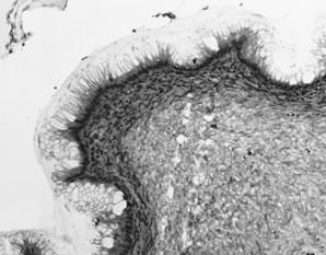
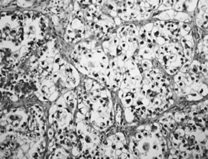
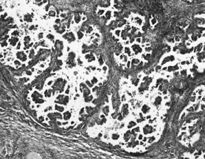
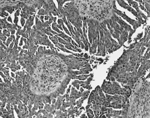
APSTs display extensive epithelial stratification, tufting and detachment of cell clusters, in addition to hierarchical branching with successively smaller papillae emanating from the larger, more centrally located papillae (Fig. 1). The two most easily quantifiable proliferative changes in these tumors are epithelial stratification and the extent of tufting or budding with detachment of cells from the surface. It has been recommended that a serous neoplasm should display stratification and budding in at least 10% of the available material to qualify as an APST.

Fig. 1. Atypical proliferative serous tumor. Hierarchical branching pattern with detachment of cell clusters and without invasion characterizes this tumor, which is also referred to as serous borderline tumor.

The cells in APSTs show features of epithelial and mesothelial differentiation. Ciliated cells resembling those in the fallopian tube are present in approximately one third of tumors. In addition, cells with abundant eosinophilic cytoplasm and rounded nuclei resembling mesothelial cells are also present, particularly on the tips of the papillae. The nuclei of APSTs resemble those in cystadenomas but tend to display slightly more atypia. Nuclei are basally located and tend to be ovoid or rounded. The chromatin is usually fine, but nucleoli are sometimes prominent. Mitoses are not common and rarely exceed four per 10 high-power fields. Psammoma bodies are present in up to half of APSTs.

The most common type of microinvasion in APSTs is characterized by isolated cells with abundant eosinophilic cytoplasm that appear to be budding from the epithelium into the superficial stromal cores of the papillae.6,12 When carefully searched for, 10% of APSTs contain microinvasion; however, in studies not specifically searching for microinvasion, it has been noted in only 1.3% of cases.6 The survival of 94 patients with microinvasion followed for a mean of 7.4 years was 100%.6 Thus, it is likely that microinvasion is frequently overlooked with no adverse affect on outcome. Although more data are needed, from the standpoint of prognosis and patient treatment, microinvasion at present appears to have no clinical relevance.

The peritoneum in patients with APSTs often contains serous epithelial proliferations displaying a range of proliferative changes: benign glands designated endosalpingiosis, papillary epithelial proliferations, sometimes with stromal desmoplasia, designated, noninvasive implants, and invasive carcinoma, also designated invasive implants. Some patients may have both invasive and noninvasive implants.

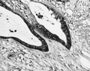
Endosalpingiosis, or benign glandular inclusions, may involve the peritoneal surfaces in patients with or without benign or malignant serous ovarian tumors. It is found in 40% of patients with APSTs. These glands typically are lined by simple columnar epithelium, often displaying tubal-type differentiation. The epithelium may display minor degrees of cytologic atypia and form simple papillary structures; psammoma bodies are sometimes present and may persist after degeneration of the associated epithelial structures. Mitotic figures are absent.

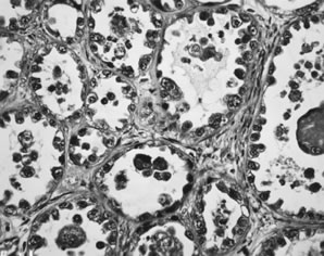
Among women with APSTs, 31% have peritoneal epithelial lesions that display a degree of proliferation beyond that usually seen in endosalpingiosis, but that lack features of invasion. These have been designated noninvasive implants and have two morphologic forms: epithelial and desmoplastic. Mixtures of both types are not uncommon.

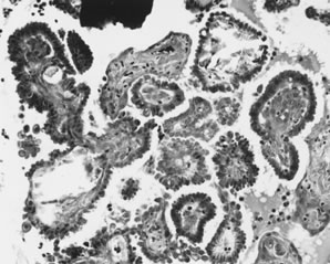
Epithelial implants are papillary and resemble the ovarian APST5,8 (Fig. 2). Mild atypia is often present, and occasionally atypia may be marked. Mitoses are usually absent. Calcification, usually in the form of psammoma bodies, is common and may be extensive.

Fig. 2. Noninvasive peritoneal implant. A papillary proliferation resembling the ovarian atypical proliferative serous tumor is present on the peritoneal surface and does not invade underlying tissue.

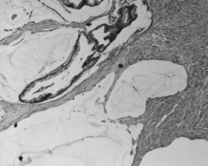
Desmoplastic implants are densely fibrotic lesions that entrap glandlike structures. The characteristic architectural feature of the desmoplastic noninvasive implant is a plaquelike thickening overlying peritoneal surfaces that appears tacked on. They may extend into the septae that separate omental lobules and create a low-power appearance, which is suggestive of invasion. A chronic inflammatory response is usually present, and in 20% of cases, an acute inflammatory exudate overlies the implant. Mitotic figures are usually absent, and psammoma bodies are present in more than 90% of cases.5,8

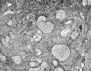
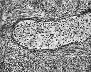
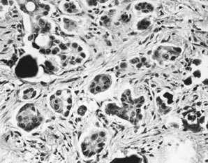
Invasive implants have been reported in association with advanced stage APSTs in approximately 6% of cases. In contrast, they are found in association with nearly half of advanced stage MPSCs (see Micropapillary serous carcinoma below). The characteristic architectural feature of an invasive implant is a haphazard infiltrative growth pattern (Fig. 3). A confluent or cribriform glandular pattern may be present. An exophytic micropapillary pattern, as described below (see Micropapillary serous carcinoma below), also qualifies for the designation of invasive implant (Fig. 4),8 but more often, micropapillae are nonbranched, embedded in fibrous stroma, and surrounded by a clear space or cleft. Sometimes, micropapillae are present within glands and may fuse with one another to create a weblike appearance. Invasive implants often display only mild cytologic atypia, but occasionally atypia is moderate and rarely marked. Mitotic figures are occasionally present. The distinction of desmoplastic implants from invasive implants (carcinoma) may be very difficult at times, but is important since it is this feature that is the best predictor of outcome for tumors with extraovarian disease.

Fig. 3. Invasive peritoneal implant. A haphazard infiltrative growth pattern of solid nests and glands with moderate cytologic atypia characterizes this type of implant.

Fig. 4. Peritoneal implant with micropapillary features. Elongated, fine micropapillae with fusion are present. These features in a peritoneal implant are interpreted as invasive by some investigators.

Lymph node involvement has been reported in approximately 63 cases of APSTs.6 Excluded from consideration is endosalpingiosis involving lymph nodes, which are nonneoplastic glandular inclusions of müllerian type that occur in pelvic lymph nodes of 5% to 14% of unselected women and in 45% to 65% of women with borderline ovarian tumors. The most common type of metastatic lymph node lesion that has been associated with APSTs is characterized by individual cells and clusters of cells with abundant eosinophilic cytoplasm in the sinuses, predominantly subcapsular sinuses. The nature of these cells is unclear, but recently it has been suggested that they may be mesothelial in origin. Similar cells are nearly always present on the surface of primary APSTs and are often present in the stroma of noninvasive desmoplastic implants. It is plausible that these cells exfoliate and are filtered from the peritoneal fluid by regional lymph nodes—so-called deportation.

Identification of lymph node metastases of APSTs has been cited as evidence of malignant potential for APSTs (borderline tumors). However, this conclusion is not supported by the data since the survival rate of 43 reported patients with lymph node metastases is 98% after a mean follow-up of 6.5 years.6

Ovarian epithelial tumors are often heterogeneous, and carcinomas may have benign-appearing areas resembling a cystadenoma or an APST. It should therefore come as no surprise that approximately 20% to 30% of ovarian epithelial tumors diagnosed as atypical proliferative (borderline) at the time of frozen section examination prove to be carcinomas on further sampling. Because 15% of unilateral tumors are associated with extraovarian disease, it is important that the surgeon perform a thorough exploration when the frozen section is diagnosed as an APST, but formal staging is not necessary for a unilateral ovarian tumor unless suspicious peritoneal lesions are found. In contrast, 56% of bilateral tumors are associated with extraovarian disease, and therefore staging in this setting is advisable.10

BEHAVIOR.

The disease-specific survival rate of patients with APSTs confined to the ovaries after a mean of approximately 6.7 years, based on more than 2000 reported cases, exceeds 99.5%.6 As indicated above, survival of 94 reported patients who had tumors that showed microinvasion is 100%, and survival of 43 reported patients with lymph node involvement was 98%.6 In six prospective, randomized trials including approximately 373 patients with serous borderline tumors followed for a mean of 6.7 years, the survival was 100%.6 The behavior of APSTs with extraovarian disease is based on the type of implants that are present. The survival rate of patients with APSTs with noninvasive implants is 95% to 100% after a mean follow-up of 7.4 years.6–8 Recurrences and deaths reported in the literature are poorly documented in the majority of cases.6 When carefully documented, most deaths are either treatment related or are the result of complications from adhesions and bowel obstruction rather than carcinoma. Finally, in a literature review of more than 18,000 borderline tumors, we were unable to identify a single well-documented case of an APST with noninvasive peritoneal implants of which primary ovarian tumor had been adequately sampled to exclude invasion (one section per centimeter of maximum tumor diameter) that had progressed to documented invasive carcinoma,6 although it is conceivable that a noninvasive peritoneal implant can undergo alignant transformation on rare occasions. Among 27 cases that reportedly progressed to invasive carcinoma, none were documented to have been adequately sampled for pathologic examination.6

In general, the noninvasive serous tumors associated with invasive implants are MPSCs, not APSTs. Based on a literature review of 467 noninvasive serous tumors, which included both invasive and noninvasive implants, the survival rate for patients with invasive implants was 66% after a mean follow-up of 7.4 years compared to 95% for patients with noninvasive implants. The difference was highly significant (p < .0001).6 The finding of invasive implants in association with an APST is very unusual and is probably because of inadequate sampling and reflects foci of occult invasion in the primary tumor.

Malignant Serous Tumors

MICROPAPILLARY SEROUS CARCINOMA.

In 1979, Russell and Merkur13 described a noninvasive serous tumor, the morphology of which closely approached the degree of proliferation displayed by low-grade carcinomas but lacked invasion and, therefore, fulfilled the WHO criteria for serous borderline tumor. They designated these tumors as “grade IV proliferating serous tumors.” This pattern was also recognized by others around the same time.14 But despite numerous studies of ovarian borderline tumors or “tumors of low malignant potential” in the ensuing 2 decades, little data on this variant were published until 1996, when two series appeared, 8,15 followed by a third in 1999.7 At this time, some investigators observed that these tumors, despite no apparent evidence of invasion, behaved like low-grade carcinomas in contrast to the other tumors in this group, and therefore proposed that they be designated “micropapillary serous carcinoma”8,15; other investigators preferred the term serous borderline tumor with a micropapillary pattern.7

Because the published data on MPSC are limited to only a few large studies, much of the data describing the clinical presentation, behavior, and treatment of this tumor are preliminary. MPSCs that do not display destructive infiltrative growth are considered noninvasive for purposes of this discussion, although it is conceivable that the complex exophytic micropapillary pattern is a form of invasion.

CLINICAL FEATURES AND OPERATIVE FINDINGS.

The mean age of patients with the noninvasive form of MPSC is 42 years. The most common presentation is an asymptomatic pelvic mass, but abdominal pain, fullness, and distention are common symptoms in advanced stage cases. Sixty-five percent of tumors are bilateral. Forty-seven percent of patients are stage I and the remainder are stages II and III. The mean tumor size is 8.2 cm. Surface involvement is present in 54% of cases.

PATHOLOGY.

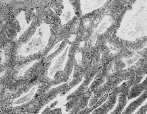
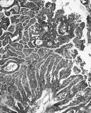
Because these tumors are very well differentiated, they tend to have a papillary and cystic gross appearance like APSTs and little, if any, necrosis in contrast to many typical serous carcinomas, which often have solid areas and extensive necrosis. Noninvasive MPSC is a proliferative serous neoplasm that displays a high degree of epithelial proliferation and complexity but is morphologically intraepithelial because diagnostic features of invasion are not present. Some investigators believe that the association of MPSC with invasive carcinoma and a high mortality rate warrants classification of this variant as a well-differentiated papillary serous carcinoma, thus the designation MPSC. In either case, this variant displays a characteristic pattern of papillary branching (Fig. 5). The distal papillary branches are thin and delicate with minimal fibrovascular support and emanate abruptly from thick, more centrally located papillae without intervening branches of successive intermediate sizes, unlike the hierarchical branching pattern of APSTs. The papillae often fuse to form a cribriform pattern. Because micropapillary and cribriform areas may be focally present in APSTs, a 5-mm-in-diameter area of a confluent micropapillary pattern is required for the designation of MPSC; anything less than this, in the absence of other features of invasion, should be classified as an APST. Extensive sampling of the ovarian tumor may be necessary to resolve difficult cases. MPSC displays no apparent invasion of the stromal cores of the papillae. High-grade malignant nuclear features, even in the presence of the typical micropapillary patterns, warrant a diagnosis of ordinary papillary serous carcinoma. Invasion is recognized by a haphazard infiltrative growth composed of solid nests or complex glandlike structures displaying micropapillae or a confluent pattern. The nests, glands, and glandlike structures are often surrounded by a clear space or cleft. Psammoma bodies may be numerous in either the MPSC or invasive areas. The distinguishing morphologic features of serous neoplasms, including the invasive and noninvasive variants of MPSC, are shown in Table 2.

Fig. 5. Micropapillary serous carcinoma. Elongated, fine micropapillae with fusion and bridging at the surfaces of the larger thick papillae characterize this tumor. Morphologic features of destructive invasion are not present.

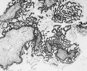
Table 2. Distinguishing Morphologic Features of Serous Ovarian Neoplasms


Stratification and

Micropapillary

Stromal

Diagnosis

Atypia

Detachment

Pattern

Invasion

Serous cystadenoma

Absent, or

Absent, or present

Absent

Absent


  present in <10%

  in <10%


APST

Present in >10%

Present in >10%

May be present,

Absent


  <5 mm of


  confluence


MPSC, noninvasive

Present

Usually present

Present, >5mm

Absent


 of confluence


MPSC, invasive

Present

Usually present

Present

Present

Serous carcinoma

Present

May be present

May be present

Present


APST, atypical proliferative serous tumor; MPSC, micropapillary serous carcinomas.
(Seidman JD, Russell P, Kurman RJ: Surface epithelial tumors of the ovary. In Kurman RJ [ed]:Blaustein's Pathology of the Female Genital Tract, pp 791–904, 6th ed. New York, Springer-Verlag, 2002)

The peritoneal implants associated with MPSC are frequently invasive (i.e., carcinoma). Among 39 reported cases of advanced stage noninvasive MPSC, 19 (49%) of the implants were invasive7,8,15 in comparison to 6% of the implants associated with APSTs.8

BEHAVIOR.

Despite the apparent absence of destructive infiltrative growth in the noninvasive variant of MPSC, the limited available data indicate that it behaves as a low-grade serous carcinoma. Stage I MPSC appears to be cured by adnexectomy alone, although only a small number of stage I tumors have been reported. Based on relatively limited data from three studies, the 5- and 10-year survival rates for patients with advanced stage MPSC are approximately 84% and 58%, respectively.7,8,15 Documented recurrences of invasive carcinoma have been reported in 34% of advanced stage MPSCs. One invasive recurrence from an inadequately sampled primary ovarian MPSC that was reportedly stage I but for which slides of the omentum were not reviewed by the authors has been reported. Insufficient data are available on the invasive variant of MPSCs to comment on their behavior.

Invasive Serous Carcinoma

CLINICAL AND OPERATIVE FINDINGS.

Serous carcinoma is the most common type of ovarian cancer and accounts for approximately 50% of malignant ovarian neoplasms. The peak age group is 45 to 65 years, and the mean age is 57 years.16 Inasmuch as 70% to 84% of patients present in advanced stage (FIGO stage II or higher) with tumor disseminated throughout the abdominal and pelvic cavities, common presenting symptoms are abdominal pain and distention due to ascites or bulky abdominal tumor. Gastrointestinal symptoms are also common. Other symptoms may include urinary frequency, dysuria, and vaginal bleeding. Stage I tumors usually present as an asymptomatic mass on a routine pelvic examination.

Two thirds of cases involve both ovaries. For stage I cases, approximately 40% are bilateral. Nearly all advanced stage ovarian carcinomas spread along peritoneal surfaces, including the pelvic peritoneum (stage II), the surfaces of the bowel, and other abdominal organs (stage III). Both pelvic and abdominal spread can be by direct extension or metastasis. For example, direct extension to the rectosigmoid, broad ligament or uterus can occur by contiguous growth, or exfoliation of malignant cells can result in seeding of the peritoneal surfaces of the bowel or pelvic peritoneum. Tumor rupture before or during surgery may occur and warrants assignment of a stage I or II tumor to stage IC or IIC, respectively. Assessment of the character of the external surface is an important component of staging for tumors that are confined to the ovaries. This is best done by the surgeon but should also be evaluated by the pathologist. Grossly exophytic papillary tumor on the surface of an ovarian neoplasm that is confined to the ovaries warrants a stage of IC. Intracystic neoplasms without gross or microscopic tumor on the external surfaces do not qualify for surface involvement unless tumor cells invade through the full thickness of the cyst wall and are thus exposed to the peritoneal cavity. Dense adhesions to pelvic structures or bowel are reassigned to FIGO stage II or III.

Patients with stage III disease usually have omental involvement which, in advanced cases, is manifested by a solid tumorous mass (omental cake). Pelvic and para-aortic lymph node metastases are found frequently, with a direct relationship to the extent of disease. Rarely, inguinal lymph nodes contain metastatic tumor. Patients with tumors that otherwise appear to be stage I have lymph node metastases in 4% to 14% of cases; the corresponding figures for stages II, III, and IV are 36%, 41% to 68%, and 88%, respectively. Liver metastases usually manifest as studding of the peritoneal surface of the liver. Parenchymal liver metastases are rarely present, and very rarely, splenic metastases are found, necessitating splenectomy.

In 8% to 12.5% of women with advanced stage papillary serous carcinoma, the ovaries are small and display predominantly surface involvement; occasionally, the ovaries are completely uninvolved by tumor. These findings warrant a diagnosis of primary peritoneal serous carcinoma.

PATHOLOGY.

Serous carcinomas range from microscopic (referred to as early de novo carcinoma) to approximately 20 cm in diameter. They are typically multilocular and cystic, with soft, friable papillae filling the cyst cavities and containing serous, turbid, or bloody fluid. The external surfaces may be smooth or bosselated and sometimes display surface papillae. Tumors are often solid, pink to gray with less-obvious papillae, and may be soft or firm depending on the character of the tumor stroma. Hemorrhage and necrosis are often present. Omental metastases are characterized by firm nodules of variable size with white or gray cut surfaces, which may coalesce into an omental cake. If the omentum is grossly normal, microscopic omental metastases are found in 22% of cases.

Well-differentiated serous carcinomas are discussed above (see micropapillary serous carcinomas above). Typical serous carcinomas display complex papillary and solid patterns and marked nuclear atypia (Fig. 6). A frequently encountered and quite characteristic pattern is a lacelike or labyrinthine pattern. This pattern may be focal but often predominates and is characterized by extensive bridging and coalescence of papillae, resulting in slitlike spaces between the papillae. Areas of solid growth are common. A glandular pattern is not necessarily diagnostic of endometrioid differentiation, and the distinction of a high-grade endometrioid carcinoma from a serous carcinoma may be difficult. The cells may be small and uniform with grade 2 nuclear atypia, but more often are large, pleomorphic, and display obviously malignant cytologic features.

Fig. 6. Serous carcinoma. Markedly complex and stratified papillary proliferation with moderate-to-severe nuclear atypia.

Serous carcinomas that display extensive solid areas are usually composed of uniform sheets of cells with high-grade nuclear atypia and may display isolated bizarre mononuclear giant cells or syncytial-like aggregates. Mitoses, including abnormal mitoses, are usually numerous and necrosis is often pronounced. Areas with grade 2 nuclei often merge with areas of grade 3 nuclei. Nuclear grade is subjective and not very reproducible; we therefore designate these tumors as “high grade.” Usually there are focal areas with papillary architecture that permit the diagnosis of a serous carcinoma as opposed to an undifferentiated carcinoma. Psammoma bodies are present in 25% of cases. A completely solid carcinoma without any evidence of glands or papillae warrants a designation of undifferentiated carcinoma.

When faced with a neoplasm in the ovary, particularly when bilateral, the physician should always consider the possibility of metastatic tumor to the ovary. Although metastatic carcinomas to the ovary most often mimic mucinous or endometrioid ovarian carcinomas, metastases may display a variety of patterns.

Serous carcinomas may invade the fallopian tubes, and on occasion, the distinction of an ovarian from a primary tubal carcinoma may be difficult. Meticulous dissection of the fallopian tube, which is nearly always dilated and filled with tumor when primary, can be of value. Microscopically, the presence of carcinoma in situ in uninvolved tubal epithelium is also helpful.

Grossly normal-sized ovaries with microscopic surface involvement or no involvement by serous carcinoma are classified as peritoneal carcinomas. From a practical standpoint, the distinction is not critical since the behavior and treatment of serous carcinomas of the ovary and peritoneum are similar.

BEHAVIOR.

Stage I patients who have been carefully staged have a 5-year survival rate that exceeds 90%.17 Survival for stages III and IV is generally very poor. Some of the older literature overestimates the survival rate for stage III patients because of the inclusion of borderline tumors. Stage II tumors are uncommon (8% of serous carcinomas) and represent an intermediate group, which, depending on other factors including completeness of surgical removal and substage, can have widely varying survival and cure rates. Early de novo carcinoma, despite its small size, is associated with recurrence and death in approximately a third of cases, usually with widespread peritoneal disease.18

Back to Top
MUCINOUS TUMORS

In the early 1970s, FIGO and WHO introduced a new classification in which tumors of surface epithelial origin were not only classified according to cell type but also were subdivided into benign, frankly malignant, or “of borderline malignancy” or “low malignant potential.”2,3 This tripartite categorization, with the category of borderline malignancy at its core, was conceived to reconcile the disparate behavior and histologic appearance of a certain group of serous tumors, but the terminology was extended to include mucinous tumors as well as the other cell types.19 In the ensuing years, it was found that the long-term survival for mucinous borderline tumors (MBTs) confined to the ovary was close to 100%, whereas survival for advanced stage MBTs was 40% to 50%.20 Thus, the only MBTs that displayed malignant behavior were those that were advanced stage. A recent review showed that approximately 85% of all reported advanced stage MBTs was associated with pseudomyxoma peritonei (PMP).20 Furthermore, when analyzed carefully, virtually all of these can be shown to be secondary tumors from a mucinous cystadenoma of the appendix that ruptured and thus discharged mucinous epithelium into the peritoneal cavity.21,22 This epithelium is capable of implanting on peritoneal surfaces and producing copious amounts of mucin. Bona fide primary ovarian MBTs that have ruptured before or at the time of surgery have never been reported to have recurred or resulted in the development of PMP. Thus, most investigators believe that PMP is a condition associated with certain gastrointestinal neoplasms, almost always mucinous cystadenomas of the appendix, but not with primary ovarian mucinous tumors.

Other studies have shown that metastatic carcinomas from the upper gastrointestinal tract, notably the pancreas and biliary tract, and occasionally the cervix can display a deceptive appearance in the ovary such that they are easily mistaken for ovarian MBTs. Furthermore, the ovarian tumors in these cases are typically much larger than the primary gastrointestinal tumor, thus heightening the resemblance to a primary ovarian tumor. Ovarian MBTs are a heterogeneous group of neoplasms. The majority are benign proliferative tumors confined to the ovary. The minority that are advanced stage are either secondary tumors from a ruptured mucinous adenoma of the appendix associated with PMP or are metastatic carcinomas from a primary site in the upper gastrointestinal tract or cervix that masquerade as advanced stage MBTs. When diagnosed properly, ovarian MBTs are benign tumors that rarely, if ever, involve extraovarian sites. Thus, the need for a category of borderline mucinous tumors no longer exists and accordingly in this chapter, the term atypical proliferative replaces borderline or low malignant potential. Cystadenomas with increased proliferative activity and varying degrees of cytologic atypia are classified as atypical proliferative mucinous tumors (APMTs). Mucinous tumors with marked cytologic atypia represent “APMT with intraepithelial carcinoma.” APMTs with clear-cut foci of invasion measuring less than 5 mm in diameter are designated “APMT with microinvasion.”

Benign Mucinous Tumors

Mucinous cystadenomas and adenofibromas comprise 41% of all benign ovarian epithelial neoplasms, and 76% of ovarian mucinous neoplasms. Benign mucinous neoplasms occur most often in the 3rd to 6th decades with a mean age of approximately 50 years. Small tumors are often found incidentally, whereas large tumors present as an obvious pelvic or abdominal mass.

Bilaterality is uncommon, occurring in 2% to 5% of cases. Mucinous cystadenomas are typically large multiloculated cystic tumors that measure up to 50 cm in diameter. Locules are usually small and multiple but are variable in number; rarely, mucinous adenomas are large simple cysts with no loculations. These are the largest ovarian tumors on record; mucinous cystadenomas measuring 25 to 30 cm in diameter are not unusual, and tumors weighing more than 100 kg have been reported. The outer cyst wall is usually thick with a smooth opaque surface. The cysts contain thick tenacious gelatinous material that is usually pale yellow but may be turbid, brown, and more watery when associated with areas of infarction. Fibrous stroma is usually inconspicuous, but when it is abundant, these tumors are termed mucinous adenofibromas.

Microscopically, mucinous cystadenomas are lined by a single layer of uniform, tall columnar cells with clear or basophilic cytoplasm and small basal hyperchromatic nuclei (Fig. 7). Goblet cells are almost always present and indicate gastrointestinal-type differentiation. Rarely, endocervical-type mucinous epithelium predominates and is usually associated with a papillary architecture. Not infrequently, the cyst is lined by nondescript, tall columnar epithelium more typical of gastric antral-type epithelium. The intestinal-type tumors are usually glandular and cystic. Simple mucinous cysts have thick, collagenous, acellular outer walls, sometimes with dystrophic calcification. More complex cystadenomas have multiple locules that may be of similar or widely varying size, some of which display a peripheral arrangement of small acini, or daughter cysts, which may create a pseudoinvasive pattern. The complexity of glandular and papillary arrangements may be quite florid, and the presence or absence of invasion is often difficult to assess. Accumulation of mucin within the cysts leads to attenuation of the epithelium, which may lead to leakage of mucin into the stroma; the latter can also be caused by infarction. The mucin in the stroma may elicit no stromal reaction, an acute or chronic inflammatory response with numerous muciphages, or a foreign body-type reaction. Extensive dissection of mucin through the tumor stroma is designated “pseudomyxoma ovarii”. The stroma of mucinous cystadenomas is fibrocollagenous and the cellularity is variable but rarely approaches the high cellularity of the stroma of serous tumors.

Fig. 7. Mucinous cystadenoma. A single layer of benign mucinous epithelium displaying pale basophilic cytoplasm overlies a cellular stroma resembling ovarian stroma.

Atypical Proliferative (Borderline) Mucinous Tumors

CLINICAL AND OPERATIVE FINDINGS.

These tumors comprise about 38% of borderline tumors and 12% of mucinous ovarian neoplasms. The clinical features associated with APMTs are similar to those for mucinous cystadenomas. The age distribution of patients with these tumors peaks in the 5th and 6th decades.

APMTs are bilateral in up to 6% of cases. Like mucinous cystadenomas, APMTs are often very large neoplasms and may be adherent to other pelvic or abdominal organs. However, these tumors are noninvasive and therefore adherence to adjacent organs is not an ominous feature.

PATHOLOGY.

The characteristic gross features of APMT are identical to those of mucinous cystadenomas. This is not surprising since APMTs arise in pre-existing mucinous cystadenomas (see below).

Atypical Proliferative Mucinous Tumors, Gastrointestinal Type

Atypical proliferative mucinous tumor of gastrointestinal type is characterized by cytologic atypia and epithelial stratification of mucinous epithelium that is otherwise similar to that of gastrointestinal-type mucinous cystadenomas (Fig. 8). At least 10% of the neoplasm should display these features to be included in the APMT category. Stromal invasion is absent. The epithelial stratification is often manifested by intraluminal papillary tufts of varying size that lack fibrovascular support. A villoglandular architecture is occasionally present. Although the older literature suggests that epithelial stratification greater than three cell layers conveys a worse prognosis,19 these data have not been substantiated, and furthermore, assessment of the degree and extent of epithelial stratification may not be reproducible. The glands are often crowded and may be back to back. Foci of a true cribriform pattern characterized by absence of fibrovascular support between adjacent glands may occur, but must be confined within cysts to qualify as an APMT. Nests or masses of epithelium displaying a cribriform pattern within the stroma reflect invasion and depending on the size of these foci qualify as microinvasive (less than 5 mm) or frank carcinoma (see below). Cytologic atypia and mitotic activity are variable. Marked cytologic atypia qualifies for the diagnosis of intraepithelial carcinoma (see below) and should prompt further sampling for areas of invasion.20,23–25

Fig. 8. Atypical proliferative mucinous tumor. Stratification and cytologic atypia of mucinous epithelium characterize this tumor. Invasion is not present.

The term atypical proliferative mucinous tumor with intraepithelial carcinoma is relatively new and has been used to describe a subset of atypical mucinous neoplasms displaying extensive epithelial stratification (greater than three cell layers) or marked cytologic atypia, which is a characteristic feature of invasive carcinoma in many sites; but in these cases, unequivocal stromal invasion cannot be shown.20 We reserve the diagnosis of APMT with intraepithelial carcinoma only for those tumors displaying areas of high-grade nuclear atypia. APMT with intraepithelial carcinoma comprises approximately one quarter of all mucinous neoplasms in the atypical proliferative and invasive carcinoma groups combined. The survival of patients with intraepithelial carcinoma exceeds 95% after salpingo-oophorectomy and for all practical purposes is the same as that of APMTs.20,23–25

To qualify as APMT with microinvasion, no single invasive area should exceed 5 mm in diameter. Multiple foci of invasion are permitted.20 Some authors have proposed a 3-mm criterion as the maximum size of the invasive area to qualify for this diagnosis. In most cases, microinvasion measures less than 2 mm. Stromal invasion in mucinous neoplasms is described below (see mucinous carcinomas). Limited data on microinvasion in APMTs indicate a survival of 100% based on 42 patients followed for a mean of 6.0 years.

The most important differential diagnostic consideration when evaluating a mucinous neoplasm in the ovary is whether the tumor is primary in the ovary or metastatic. If primary, an APMT must be distinguished from invasive carcinoma (see mucinous carcinoma below). Features suggestive of metastatic ovarian involvement include bilaterality, and if bilateral, size less than 10 cm, ovarian surface involvement, extensive pseudomyxoma ovarii, and certain patterns of haphazard, nodular infiltrative growth.

Atypical Proliferative Mucinous Tumors, Endocervical-Like Type

Fifteen percent of ovarian APMTs are of the müllerian, endocervical-like type, also referred to as seromucinous to reflect the combination of serous and mucinous features.26 The mean patient age is 34 years. Seventy-seven percent are stage I. Forty percent are bilateral. In the largest reported series,26 all patients who had stage II and III disease (23% of all cases) did not have PMP, suggesting that the peritoneal “implants” associated with this type of APMT are more akin to those seen with APSTs. This is also consistent with the papillary architecture of these tumors, a feature that closely resembles the architecture of serous tumors and is dissimilar to the architecture of intestinal-type mucinous neoplasms. The mean tumor size is 8 cm. The microscopic architectural features as noted above are similar to APSTs, with a hierarchical branching pattern, stratification, tufting, and detachment of cell clusters. A prominent acute inflammatory infiltrate is nearly always present at least focally. The epithelium is tall, columnar, and mucinous, resembling endocervical epithelium. Cells with cilia can be found in a third of cases, reflecting serous-type differentiation. Atypia is usually minimal. Cells with slightly larger and rounded nuclei containing densely eosinophilic cytoplasm, particularly at the tips of the papillae, are present in nearly all cases. A small component (less than 10%) of other müllerian epithelial cell-type differentiation (e.g., endometrioid) is common. There is a strong association with endometriosis, which is present in 30% of patients; in two thirds of these patients, the neoplasm has been observed to arise directly from the atypical epithelial component of an endometriotic cyst.

BEHAVIOR.

Atypical proliferative mucinous tumors are benign, with survival rates of 99% based on literature reviews, including approximately 900 patients.20 Rare reports of aggressive behavior most likely reflect inadequate sampling with failure to detect occult invasion, or the ovarian tumor may be a metastasis from an occult primary source elsewhere. Although the literature suggests that the survival of patients with advanced stage MBTs is only 54%, it is now clear that bona fide advanced stage MBT rarely, if ever, occurs. Eighty-five percent of women who have been reported to have advanced stage MBTs present with the syndrome of pseudomyxoma peritonei, a condition that is not of ovarian origin20 (see below). For patients with APMT, endocervical-like type, after a short mean follow-up of 3.7 years, all reported patients were alive and well, but two who underwent unilateral salpingo-oophorectomy had contralateral APMTs develop, which most likely reflect new primary tumors.

Mucinous Tumors Associated With Pseudomyxoma Peritonei

Historically, the term pseudomyxoma peritonei (PMP) referred to the gross appearance of mucinous ascites associated with a mucinous tumor of the appendix, colon, or ovary. The literature on PMP before the 1990s is difficult to evaluate for several reasons. First, PMP has never been a specific histopathologic diagnosis but, rather, an operative/gross description. Second, much of the literature on this entity lacks descriptive data on the histology of the lesion. Third, PMP in women often involves the ovaries, and until recently, it had not been clear whether the primary site was in the appendix, ovary, or both in such women. Some investigators have also proposed that the primary site may be the colon. Another problem plaguing the diagnosis of PMP is the subclassification, by some authors, of cases into those with and without epithelial cells. These problems have recently been addressed. Some investigators have proposed that the clinical presentation and gross appearance of PMP encompass two distinct clinicopathologic entities, both of gastrointestinal origin: peritoneal mucinous carcinomatosis (PMCA), a highly aggressive tumor associated with a high early mortality rate; and disseminated peritoneal adenomucinosis (DPAM), an indolent condition nearly always derived from the appendix.27 In the latter condition, the mucinous epithelium released from the appendiceal neoplasm implants on peritoneal surfaces and is capable of producing copious amounts of mucin that persistently reaccumulate in the form of mucinous ascites. The mucinous epithelium has little capacity to invade tissue, but the mucin that accumulates in the peritoneal cavity is an irritant that induces fibrosis and dense adhesions. Patients with this entity usually have prolonged survival, but eventually many die of bowel obstruction. Although most cases of PMP can be reliably classified into DPAM or PMCA, approximately 10% display discordant or intermediate histologic features and, on average, have a prognosis intermediate between the two major groups.27

The development of bona fide invasive carcinoma in patients with DPAM is extremely rare. Conversely, mucinous neoplasms of the appendix that implant on the peritoneum as PMP are classified as carcinomas by some expert gastrointestinal pathologists, even if they display no significant cytologic atypia.28 We favor the view that these tumors are not true carcinomas. This is a very low-grade, clinically indolent neoplastic process that needs to be distinguished from the clinically aggressive PMCA rather than lumped together under the rubric PMP. The term PMP should be restricted to the description of the clinical entity and should not be used as a pathologic term.

Only 39% of patients with PMP in the largest series reported are women, and of these, 44% have ovarian involvement by a mucinous neoplasm.27 It is now generally accepted that ovarian involvement in women with PMP is secondary; PMP is nearly always of gastrointestinal origin, usually from a mucinous adenoma of the vermiform appendix.21,22 Mucinous ovarian tumors that are associated with PMP are typically classified as borderline because tumor is present throughout the peritoneal cavity. As noted above, these tumors are not ovarian in origin but are derived from appendiceal mucinous cystadenomas. Finally, it should be noted that the majority of the mucinous ovarian tumors associated with PMP show gross and microscopic features that are distinctive from those of primary ovarian MBTs (see below). We prefer to refer to the ovarian tumors in these cases descriptively as “ovarian involvement by DPAM from an appendiceal mucinous neoplasm.” They are identical to the appendiceal mucinous neoplasms from which they are derived.

CLINICAL AND OPERATIVE FINDINGS.

The mean patient age is 44 years with two thirds of patients presenting in the 4th and 5th decades. Presenting clinical symptoms are often dominated by the effects of mucinous ascites, including abdominal distention and abdominal pain.

Mucinous ovarian tumors associated with PMP are bilateral in 80% of cases with a mean diameter of 7 cm. When unilateral, there is a right-sided predominance. These findings contrast with primary ovarian APMTs, which are almost invariably unilateral, large (mean diameter greater than 15 cm), stage I, and equally distributed in the right and left ovaries. Mucinous ascites or mucoid nodules scattered throughout the peritoneal surfaces is characteristic.

Ovaries involved by DPAM are often cystic and usually display mucoid surfaces, surface nodules, or implants. Metastatic mucinous carcinomas (PMCA) in the ovaries appear similar but are more often solid. In 75% of patients with PMP, there is gross or microscopic evidence of rupture of the appendiceal tumor. Gastrointestinal perforations can be very small, can heal, and can be overlooked because of inadequate sampling by the pathologist; these are the best explanations for most of the remaining 25% of apparently unruptured appendices. It is also possible that there can be chronic leakage of mucus because of a small perforation that repeatedly heals and reruptures. In contrast, rupture of bona fide stage I primary ovarian MBTs is not associated with recurrence or development of PMP.

PATHOLOGY.

Microscopically, the majority of tumors associated with PMP display mucoid nodules or implants involving the ovarian surface; in 20% of cases, the involvement is confined to the surface. Surface and superficial or deep cortical involvement is seen in more than 50% of cases, and approximately 25% are confined to the ovarian stroma. Pseudomyxoma ovarii, an appearance characterized by dissection of acellular lakes of mucin through the ovarian stroma, is present in two thirds of cases and is usually multifocal or extensive.

The ovarian tumors associated with mucinous tumors that are disseminated throughout the peritoneum are of two types. Tumors composed of pools of mucin with scanty simple or focally proliferative mucinous epithelium displaying minimal cytologic atypia and rare mitotic figures have been designated DPAM along with the abdominal disease (Fig. 9). Additional morphologic features that are characteristically seen in these secondary ovarian lesions include strips of mucinous epithelium floating in mucin pools, often with a hypersecretory or hypermucinous appearance. These are nearly always associated with appendiceal adenomas lacking significant atypia. The ovarian mucinous proliferation closely resembles the peritoneal mucinous lesions as well as the appendiceal adenoma.

Fig. 9. Disseminated peritoneal adenomucinosis. In this form of pseudomyxoma peritonei, large pools of acellular mucin dissect through tissues. Occasional mucinous glands, seen at upper left, display minimal cytologic atypia.

In the past, ovarian mucinous tumors characterized by epithelial proliferation and moderate-to-marked atypia or signet ring cells, significant mitotic activity, and obvious stromal invasion were often included in the category of PMP because of the abundant extracellular mucin production (Fig. 10). However, because they contain overtly histologically malignant epithelium, are associated with gastrointestinal carcinomas, and have a poor prognosis, they are not diagnosed as PMP, but rather as metastatic mucinous carcinomas. They are the ovarian manifestation of PMCA and are virtually always metastatic.27 Because these tumors display obvious cytologically malignant features, they are rarely confused with ovarian APMTs.

Fig. 10. Peritoneal mucinous carcinomatosis. In this form of pseudomyxoma peritonei, angulated glands with moderate-to-severe nuclear atypia invade tissues.

Immunohistochemical and molecular biologic studies have provided confirmatory evidence that the bland-appearing mucinous tumors associated with PMP involving the ovaries are secondarily derived from the associated appendiceal mucinous tumor.29,30

BEHAVIOR.

The behavior of mucinous tumors that are disseminated throughout the peritoneal cavity depends on tumor morphology. Cases designated DPAM, which are nearly always derived from appendiceal neoplasms, constitute the classic description of PMP found in the older literature, in which the behavior is characterized as slowly progressive, with reaccumulation of mucinous ascites and compatible with prolonged survival if treated symptomatically by periodic evacuation of the ascites. The 5- and 10-year survival rates are 75% and 68%, respectively.27 In contrast, those cases designated PMCA are metastatic mucin-secreting carcinomas from the appendix or colon and are associated with an aggressive clinical course, with more than 90% of patients dead within 3 years. It is clear, therefore, that the term pseudomyxoma peritonei, although useful as an operative and gross description, lacks sufficient specificity for use as a histologic diagnosis.

Mucinous Carcinoma

CLINICAL AND OPERATIVE FINDINGS.

After exclusion of metastatic tumors to the ovaries, primary ovarian mucinous carcinomas are uncommon and, in fact, are much less common than metastatic mucinous carcinomas.20 Because of the limitations of the published data, clinical and pathologic features including proportion of ovarian neoplasms, bilaterality rates, and stage distribution are variable and may be unreliable. Mucinous carcinomas appear to comprise fewer than 5% of ovarian epithelial neoplasms, 12% of ovarian mucinous neoplasms, and 11% of ovarian carcinomas. However, these are probably overestimates because some of the studies from which these data are derived were published before the recognition of the variety of metastatic mucinous carcinoma patterns in the ovaries.

Mucinous carcinomas occur in women in the 4th to 7th decades. The mean age is 54 years. The clinical presentation is similar to that of benign mucinous neoplasms, inasmuch as the majority of cases are stage I. Therefore, these patients do not generally present with signs and symptoms of abdominal carcinomatosis as do the majority of women with serous ovarian carcinomas. Rather, because these tumors can be quite large, the clinical presentation is generally that of a large pelvic or abdominal mass and abdominal distention.

Nearly 60% of patients with mucinous carcinoma have FIGO stage I disease. The majority of stage I cases are unilateral. If microinvasive tumors are excluded from the carcinoma category, this figure drops to approximately 50%. The proportion of stage I cases varies widely among studies, a finding that reflects variation in criteria as well as the difficulty of distinguishing intraepithelial carcinoma from invasive carcinoma.20,23–25 Nearly all intraepithelial carcinomas have been stage I. Ascites may occur, but the gelatinous, mucinous ascites characteristic of PMP does not occur in association with primary ovarian mucinous tumors (see PMP above).

PATHOLOGY.

Because mucinous carcinomas arise from atypical proliferative mucinous neoplasms, their gross appearance is similar. Mucinous carcinomas are typically large, cystic, and multiloculated. Solid areas and intracystic nodules are more common in carcinomas than in cystadenomas and APMTs. Rarely, the tumor is predominantly solid.

Invasive mucinous carcinomas resemble APMTs but display unequivocal stromal invasion measuring more than 5 mm.20 Stromal invasion in primary ovarian mucinous neoplasms is characterized by three different patterns: (1) a confluent glandular growth with crowded glands that merge together without intervening stroma, and back-to-back cribriform glands with well-defined, sharply etched rounded spaces; (2) clusters of single cells with abundant eosinophilic cytoplasm surrounded by clear spaces infiltrating the stroma; and (3) glands of varying sizes infiltrating the stroma in a haphazard pattern. A nodular pattern with glands and nests of tumor in a nodular arrangement surrounded by normal ovarian stroma is strongly suggestive of metastatic disease.

Cytologic atypia is usually marked and characterized by an increased nuclear/cytoplasm ratio, irregularly clumped chromatin, and prominent nucleoli. The cytoplasm is scant and eosinophilic with irregular borders and occasional mucin droplets. Stratification is usually prominent but exceeds three cell layers in fewer than 50% of cases. Necrosis is often present. Mitotic figures are usually prominent, with more than five per 10 high-power fields in the majority of cases. Although there are little data on grading, most mucinous carcinomas are well differentiated.

Before making a diagnosis of mucinous carcinoma of the ovary, it is imperative to exclude metastatic mucinous carcinoma. Mucinous adenocarcinomas of the pancreas, biliary tract, colon, appendix, and cervix can mimic ovarian mucinous tumors.

The distinction of APMT from mucinous carcinoma can be difficult. Stromal invasion must be identified to diagnose mucinous carcinoma; the various patterns of stromal invasion are discussed above. If cytologically malignant features are present but unequivocal stromal invasion cannot be confidently identified, a diagnosis of APMT with intraepithelial carcinoma is warranted.

BEHAVIOR.

The overall prognosis of patients with mucinous carcinoma is better than that for serous carcinoma because a high proportion of patients present in stage I. Nonetheless, despite the differing clinical and pathologic features of ovarian serous and mucinous carcinomas, their behavior, when stratified by stage, is similar. Recurrence and mortality in stage I mucinous carcinoma occur in 8% to 12% of cases, which is similar to well-staged stage I serous carcinomas.16 Advanced stage mucinous carcinoma (FIGO III and IV) is uniformly fatal, as is serous carcinoma of advanced stage. Limited data are available on FIGO stage II tumors, as only 10% of patients are in stage II. Similarly, little data are available on high-grade stage I mucinous carcinomas. Occult advanced stage mucinous carcinoma is rare compared to serous carcinoma.

Back to Top
ENDOMETRIOID TUMORS

The majority of endometrioid ovarian neoplasms are carcinomas. Endometrioid adenofibromas and atypical proliferative endometrioid tumors (APETs) are less common. Recent refinements in the criteria for these tumors suggest that many tumors diagnosed previously as well-differentiated endometrioid carcinomas would now be classified as APETs. Thus, APET may comprise up to 5% of ovarian epithelial neoplasms.

Benign Endometrioid Tumors

Endometrioid adenofibromas are usually unilateral. The median age of women with these tumors is 57 years. The mean diameter is approximately 10 cm. Seventeen percent are bilateral. The external surface is smooth and the cut surface is densely fibrous, often with intermixed cystic areas creating a honecomb appearance similar to a serous cystadenofibroma. The cysts contain clear or yellowish fluid. Microscopically, the dominant pattern is that of an adenofibroma or cystadenofibroma. The epithelial elements are arranged in branching tubular glands and cysts and usually resemble proliferative endometrium. The epithelium lining the glands is tall and columnar with oval nuclei containing coarse chromatin and small nucleoli; the cytoplasm is basophilic to amphophilic. Sometimes, the nuclei resemble those of atrophic or inactive endometrium, with uniform, elongated dark nuclei and scanty cytoplasm. Mitoses are variable but usually rare. Secretory changes and focal squamous differentiation may be present. The stroma is usually densely fibrotic; focal areas may resemble ovarian cortex. Occasional endometrioid adenofibromas are associated with endometriosis. These tumors are benign, although rarely may recur.

Atypical Proliferative Endometrioid Tumors

There is a spectrum of epithelial proliferation, glandular crowding, and cytologic atypia in benign endometrioid neoplasms ranging from mild atypia, mild glandular crowding, and epithelial stratification slightly beyond that seen in typical endometrioid adenofibromas to confluent epithelial proliferation lacking stromal support in areas up to 5 mm in diameter resembling atypical hyperplasia and well-differentiated adenocarcinoma of the endometrium. A variety of terms have been used for these tumors, including proliferating or proliferative endometrioid tumor, atypical endometrioid adenofibroma, endometrioid tumor of borderline or low malignant potential, and atypical proliferative endometrioid tumor.31,32 The criteria for these diagnoses have differed among these series. Because the behavior of these tumors has been benign, we prefer to combine these groups and refer to them as APET.

Among the three largest series, there were 96 patients with a mean patient age of 50 years. Five patients (5%) had bilateral tumors, and all but one were confined to the ovaries; one had a colonic “implant”. The mean tumor size was approximately 8 cm. The characteristic gross appearance is solid and cystic; the cyst fluid is usually hemorrhagic, brown or green. A few patients have had endometriosis and some have also had endometrial hyperplasia.

The two characteristic microscopic architectural appearances of APET are adenofibromatous and glandular/papillary.31,32 The glandular/papillary proliferation can show varying degrees of glandular complexity and crowding. An underlying adenofibroma is often present. When the glandular proliferation becomes confluent, this is considered evidence of invasion by some experts but could also reasonably be considered examples of APET with intraepithelial carcinoma. A confluent epithelial proliferation that exceeds 5 mm in diameter warrants a diagnosis of carcinoma according to one group.32 The glands show crowding and mild or moderate cytologic atypia and epithelial stratification; tufting and bridging may be present. Severe cytologic atypia warrants a diagnosis of intraepithelial carcinoma. Squamous metaplasia is present in up to half of cases. The stroma is usually cellular with periglandular cuffing. Necrosis is common and is often confined to gland lumens or cysts. The single case with a colonic implant most likely represents an independent lesion arising in endometriosis. All reported cases have had a benign behavior after a mean follow-up period of approximately 5 years. In summary, microinvasion, intraepithelial carcinoma, and confluence of glandular growth, when limited in extent, are atypical morphologic features and appear to be biologically benign, but only a few cases have been reported and clinical follow-up data are limited.

Endometrioid Adenocarcinoma

Endometrioid carcinomas comprise 5.7% of ovarian surface epithelial neoplasms, 17.5% of ovarian carcinomas, and 93% of endometrioid ovarian neoplasms.

CLINICAL AND OPERATIVE FINDINGS.

These tumors are most common in the 5th and 6th decades, and the mean patient age is 56 years. The most common symptoms are abdominal distention and pelvic or abdominal pain. Abnormal vaginal bleeding is also frequent. This is, in part, related to the association of endometrioid ovarian carcinoma with endometrial hyperplasia and carcinoma (see below). Most patients have an adnexal mass on pelvic examination.

Tumor size ranges from 12 to 20 cm with a mean of approximately 15 cm. The stage distribution differs significantly from serous carcinoma. A high proportion (43%) of endometrioid carcinomas are diagnosed in stage I. Approximately 13% of early stage (FIGO I-II) cases are bilateral. Endometriosis, which may be extraovarian, in the ipsilateral or contralateral ovary or within the tumor itself, is found in 15% to 20% of patients.

PATHOLOGY.

Endometrioid carcinomas have a smooth outer surface. On cut section, they are solid and cystic, with the cysts containing friable soft masses and bloody fluid. Cysts occasionally contain mucus or greenish fluid. Less often, the tumor is solid with extensive hemorrhage and necrosis. Tumors arising in endometriosis may display gross findings of an endometriotic cyst containing chocolate-colored fluid, with a solid nodule in the wall reflecting the focus of malignant transformation.

Destructive infiltrative growth is definitional of ovarian carcinoma. Some authors also interpret a confluent glandular epithelial proliferation exceeding 5 mm as a pattern of invasion, which corresponds to a pattern of stromal invasion for tumors in the uterine corpus.32

Well-differentiated endometrioid adenocarcinoma accounts for the majority of cases and is characterized by a confluent or cribriform proliferation of glands lined by tall, stratified columnar epithelium with sharp luminal margins (Fig. 11). A villoglandular growth pattern also occurs. Mitotic figures are often seen. Squamous differentiation is present in up to 50% of cases. Degeneration of squamous cells may induce a foreign body-type giant cell reaction in the stroma. Focal secretory changes are seen in up to a third of cases. This may be because of endogenous or exogenous progestin effect or may occur in the absence of hormonal stimulation. Areas of APET are commonly associated with endometrioid carcinoma.32 Moderately and poorly differentiated endometrioid carcinomas show solid growth and complex glandular and microglandular patterns. Nuclear pleomorphism and mitotic activity are marked and necrosis and hemorrhage are often prominent.33

Fig. 11. Endometrioid carcinoma. Crowded, back-to-back glands lined by tall columnar epithelium resembling endometrial epithelium invade the stroma.

Approximately one third of ovarian carcinomas classified as endometrioid based on the predominant epithelial component are mixed with other epithelial types. A clear cell component is present in 20%, and a papillary serous component is seen in 10% of cases.

In addition to pure endometrioid carcinoma, which is the most common, several of the variants of endometrioid carcinoma of the uterine corpus also occur in the ovary. These include endometrioid carcinoma with squamous differentiation and secretory and ciliated variants. Other variant microscopic patterns of endometrioid ovarian carcinoma have been described. One variant has been designated “sertoliform endometrioid carcinoma” or “endometrioid carcinoma resembling sex cord-stromal tumor.”

The sertoliform or sex cordlike variant of endometrioid carcinoma creates the most problems in the differential diagnosis. This has important therapeutic and prognostic implications since sex cord-stromal tumors are usually low grade and require no therapy if confined to the ovaries, whereas patients with carcinomas usually require chemotherapy and have a worse prognosis. The age of the patient is helpful since the mean age of patients with Sertoli-Leydig cell tumors is 25 years, while women with endometrioid carcinoma are usually perimenopausal or postmenopausal. In addition, hormonal manifestations such as virilization are often associated with sex cord-stromal tumors and generally not with endometrioid carcinomas. In most cases of sertoliform endometrioid carcinoma, extensive sampling of the tumor discloses areas of typical endometrioid carcinoma.

Another common problem in the differential diagnosis of endometrioid carcinoma is metastatic adenocarcinoma, particularly metastatic adenocarcinoma from the colon. Colonic metastases to the ovaries are often cystic and grossly mimic primary ovarian neoplasms. Useful features in the distinction are a characteristic garland pattern characterized by cysts lined by cytologically malignant tall, columnar cells with bridging and cribriforming in addition to bilaterality and surface involvement.34,35 Although extensive dirty necrosis has also been cited as a feature of metastatic colonic carcinoma, this type of necrosis, characterized by abundant basophilic karyorrhectic nuclear debris intermixed with eosinophilic necrotic material, often in a geographic or zonal distribution, is often seen in primary ovarian carcinomas and therefore is of limited value.36 The immunoperoxidase panel of CK7/20 can be of value in the distinction of primary ovarian versus metastatic colonic carcinoma.

BEHAVIOR.

It has often been stated that patients with endometrioid carcinomas of the ovary have a better prognosis than do those patients with typical serous carcinomas of the ovary. However, this is largely, if not completely, because of the high proportion of patients presenting in stage I. When stratified by stage, essentially all subtypes of ovarian carcinoma have a similar prognosis. The apparent favorable prognosis may also be due, in part, to a high proportion of grade I cases, although data on the influence of grade on prognosis are less clear. Treatment for endometrioid carcinoma is generally the same as that for other ovarian carcinomas. However, progestational agents, antiestrogens, tamoxifen, and other hormonal therapies have been used with limited success in previously treated endometrioid carcinomas; 10% to 15% response rates have been reported. There may be a correlation between the presence of steroid hormone receptors in tumor tissue and response rates, but data are limited. Hormonal therapy may be an option for treatment of recurrence in patients who did not respond or cannot tolerate chemotherapy or surgery.

Endometrioid Carcinoma Associated With Uterine Carcinoma

Approximately 14% of women with endometrioid ovarian carcinoma also have endometrial cancer of the uterine corpus. Endometrial hyperplasia is also commonly present. Because both tumors are often well-differentiated endometrioid adenocarcinomas, they usually resemble each other, and excluding the possibility that the ovarian tumor is metastatic can be a problem. Usually this can be determined based on a careful evaluation of the clinicopathologic features. If the uterine endometrial tumor is low grade with no or only inner half myometrial invasion, its metastatic potential is very low and the ovarian tumor can confidently be regarded as independent. If the endometrial tumor is high grade or deeply myoinvasive or both, the features of the ovarian tumors come into play. Bilaterality and a multinodular pattern, as well as other patterns characteristic of metastatic disease, indicate metastatic tumor. Close association of the ovarian tumors with either an underlying adenofibroma or endometriosis can provide evidence that the ovarian tumor is independent.

Follow-up of patients considered to have independent primary tumors also supports their independence, because most of these patients survive without recurrence, a finding compatible with stage I endometrial and ovarian carcinomas but not with a stage III endometrial cancer. Molecular studies can be of value in difficult cases.

Endometrioid Carcinoma Associated With Endometriosis

On average, 15% to 20% of endometrioid carcinomas of the ovary are associated with endometriosis, which may occur within the tumor, the ipsilateral or contralateral ovary, or elsewhere. The frequency can be as high as 42%. In only a small minority of cases, however, can direct continuity from endometriosis to atypical hyperplasia to carcinoma be shown. This is most commonly seen in the lining of an endometriotic cyst, which may contain a thickening of the cyst wall or a solid nodule protruding into the cyst. The mean age of women with endometrioid carcinoma associated with endometriosis is 5 to 10 years younger than when unassociated with endometriosis. Tumors associated with endometriosis, and particularly those arising in an endometriotic cyst, are usually well differentiated and stage I, and therefore the prognosis is excellent. On rare occasions, a limited atypical epithelial proliferation in an endometriotic cyst raises the differential diagnosis of atypical hyperplasia similar to the type seen in the uterine corpus versus well-differentiated endometrioid adenocarcinoma. In such cases, criteria for this distinction have been modified from those used in the uterine corpus.

Back to Top
CLEAR CELL TUMORS

The majority of clear cell neoplasms of the ovaries are carcinomas. Clear cell adenofibromas and atypical proliferative clear cell neoplasms are rare.

Clear Cell Adenofibromas

Among approximately 12 reported cases of benign clear cell tumors, the mean age is 45 years. One case was bilateral. The median diameter was 12 cm. The tumors display a smooth, lobulated external surface, and the cut surfaces have a fine honeycomb appearance with minute cysts embedded in firm, rubbery stroma. The cyst fluid is clear. Microscopically, the tumor is characterized by tubular glands lined by one or two layers of peglike or hobnail cells that bulge into the lumen. The cytoplasm is either scanty, often in the hobnail cells, or abundant, clear, granular, or eosinophilic in the large polyhedral cells. Nuclear atypia and mitotic activity are minimal. The stroma is compact and fibrocollagenous. The cytoplasm usually contains glycogen. The clinical behavior is benign.

Atypical Proliferative Clear Cell Tumors

Among approximately 30 patients with APCCT (clear cell adenofibroma of borderline malignancy or low malignant potential) in the literature, the mean age is 60 to 70 years. The mean tumor diameter is approximately 15 cm. The gross appearance is similar to that of the clear cell adenofibroma, but in addition there are softer and fleshier areas. Microscopically, the architecture is similar to the clear cell adenofibroma. The tumor has greater epithelial proliferation and atypia than the adenofibroma and lacks stromal invasion. The cell types lining the glands and cystic spaces are similar to those in benign tumors but display significant nuclear atypia with coarse chromatin clumping, prominent nucleoli, and mitotic activity up to three per 10 high-power field. The epithelium often displays stratification and budding; true papillary structures are uncommon. Small solid nests of clear cells may be present and can raise the possibility of stromal invasion.37

Peritoneal implants, as occur in the more common atypical proliferative serous tumor, have not been described with APCCTs. Among the limited reported cases, there is one alleged recurrence and no tumor deaths. Accordingly, we prefer the term atypical proliferative clear cell tumor for these neoplasms.

Clear Cell Carcinomas

CLINICAL AND OPERATIVE FINDINGS.

Clear cell carcinomas comprise 2.4% of ovarian epithelial neoplasms and 7.4% of ovarian carcinomas. The mean age of patients with clear cell carcinoma is 57 years. Symptoms usually relate to a pelvic or abdominal mass. Interestingly, clear cell carcinoma is the most common epithelial ovarian neoplasm to be associated with paraneoplastic hypercalcemia.

Because the relationship with endometriosis is strongest for clear cell carcinoma among all types of ovarian carcinoma, endometriotic implants are commonly present in close proximity to the tumor or elsewhere in the pelvis or abdomen. Approximately half of patients present in FIGO stage I and 15% in stage II.16 Four percent of stage I cases are bilateral.

PATHOLOGY.

Tumors range up to 30 cm in diameter with a mean of approximately 15 cm. Although they may be solid and fibrous with a honeycomb-cut surface resembling benign and atypical proliferative clear cell tumors, more commonly the cut surfaces show a thick-walled unilocular cyst with multiple yellow-beige fleshy nodules protruding into the lumen, or a multiloculated cystic mass with cysts containing watery or mucinous fluid. Tumors arising in endometriosis may display features of an endometriotic cyst, which typically contains chocolate-brown fluid and a thickened, polypoid or nodular area in the wall reflecting the area of malignant transformation.

Clear cell carcinomas display several different patterns that often occur together.38 The most common patterns are solid and tubulopapillary. The solid pattern is characterized by sheets of polyhedral cells with abundant clear cytoplasm separated by delicate fibrovascular septae or dense hyalinized fibrotic stroma (Fig. 12). The tubulopapillary pattern is characterized by papillae with varying degrees of complexity. The fibrovascular cores of the papillae are often hyalinized. Complex tubules and cysts may be intermixed with the papillae, creating a tubulocystic pattern. The cell borders are usually prominent and the nuclei vary from small and rounded or angular to large and pleomorphic with large, prominent nucleoli. The cytoplasm is filled with glycogen. In the tubulopapillary pattern, the cells are often columnar with a hobnail appearance with the nucleus protruding from the papillae, gland or cyst, into the lumen (Fig. 13). Occasionally, the epithelium lining the glands and cysts is flattened. Mitotic activity is often prominent. Necrosis, hemorrhage, and stromal lymphocytic infiltrates are variable. Luteinized stromal cells and microcalcifications are occasionally present.

Fig. 12. Clear cell carcinoma. Sheets of cells with clear cytoplasm and mild-to-moderate nuclear atypia are characteristic.

Fig. 13. Clear cell carcinoma. Glands lined by epithelium displaying severe nuclear atypia and a hobnail pattern characterized by nuclei protruding into the lumena of the glands are present. In this pattern, clear cytoplasm is less conspicuous.

On average, 30% to 35% of ovarian clear cell carcinomas are associated with endometriosis, either in the involved ovary or elsewhere in the pelvis or abdomen. These figures vary widely and several studies have observed 50% or greater associated with endometriosis. When carefully sought, the majority of ovarian clear cell carcinomas are associated with endometriosis and one third of cases can be convincingly shown to have arisen within endometriosis, usually an endometriotic cyst.

BEHAVIOR.

There are conflicting data on the behavior of clear cell carcinoma of the ovary. In some studies, the prognosis appears similar to that for other ovarian carcinomas, but in others, the prognosis is said to be worse. In two series in which analyses were stratified by stage, clear cell carcinoma had a significantly worse prognosis than did serous carcinoma only in stage III, based on a total of 81 stage III patients with clear cell carcinoma in these studies.16,39 In summary, when controlled for stage, survival of women with clear cell carcinoma may be slightly lower than that of patients with serous carcinoma, but in most studies, a rigorous comparison to a control group of nonclear cell carcinomas, stratified by stage, has not been made. It is possible that survival analyses are confounded by the possibility that clear cell carcinomas tend to be of higher histologic grade more often than other epithelial cell types. However, there are insufficient reproducible data correlating grade with prognosis of ovarian carcinomas at present to confirm this. The treatment for clear cell carcinoma is similar to that of other epithelial cell types of ovarian carcinoma.

Back to Top
TRANSITIONAL CELL (BRENNER) TUMORS

The preferred term by WHO for this group of tumors is transitional cell; however, the term Brenner tumor, based on one of the earliest descriptions of the tumor by Brenner, remains firmly entrenched in the literature and remains an approved term for some variants in this tumor group. Transitional cell tumors comprise 3.2% of ovarian epithelial neoplasms. Nearly all of these are benign Brenner tumors; atypical proliferative and malignant forms are very uncommon. The transitional epithelial cell type, characterized by a relatively uniform population of stratified cells with ovoid nuclei displaying nuclear grooves, is named because of its resemblance to urothelium.

Benign Transitional Cell Tumors

Benign transitional cell tumors comprise approximately 3.1% of ovarian epithelial neoplasms. The mean patient age is 50 years. They are common incidental findings and are often microscopic in size. Therefore, the presenting symptoms are often unrelated to the ovarian tumor. Most tumors are 2 cm or smaller, but occasionally they are large and may exceed 10 cm. They are well-circumscribed, firm, and rubbery, with a smooth or slightly bosselated serosal surface. The cut surfaces are typically solid and fibrous, usually gray, white, or yellow, and may be whorled or lobulated; occasionally, a cystic component is present. Calcification is sometimes seen. Small tumors can usually be seen arising in the ovarian cortex.

Microscopically, the characteristic feature is sharply demarcated epithelial nests in a dense fibrous stroma (Figs. 14 and 15). The epithelial cells are relatively uniform in size with prominent cell borders and pale to eosinophilic cytoplasm. The nuclei are oval, often with small nucleoli, and longitudinal grooves are usually present. Atypia and mitotic activity are generally not present. The nests often become cystic and contain eosinophilic debris or mucin. Occasionally, metaplastic endocervical-like mucinous epithelium lines the cystic nests; some investigators have termed these tumors metaplastic Brenner tumor. The metaplastic mucinous component may occasionally form the dominant part of the tumor and may account for the association of Brenner tumors with mucinous cystadenomas. The stroma varies from closely resembling ovarian cortical stroma to densely fibrous. Hyalinized areas are common, and dystrophic calcification is present in 50% of cases, often in the hyalinized areas. The stroma is occasionally luteinized. The clinical behavior is benign.

Fig. 14. Benign transitional cell (Brenner) tumor. Rounded nests of transitional-type epithelium are embedded in a fibrotic stroma resembling ovarian stroma.

Fig. 15. Benign transitional cell (Brenner) tumor. Ovoid nuclei with longitudinal grooves characterize the transitional cell type.

Atypical Proliferative Transitional Cell Tumors

These uncommon neoplasms, also termed proliferating or borderline transitional cell or Brenner tumor, or transitional cell or Brenner tumor “of low malignant potential,” present in patients whose mean age is 59 years. They are always unilateral and confined to the ovary. The tumors are larger than their benign counterparts and are usually cystic and measure 10 to 28 cm with a mean diameter of 18 cm. Friable papillary or polypoid masses project into the cyst lumens, and there is usually a benign Brenner component with a more solid and fibrous cut surface. Microscopically, the intracystic papillary component is composed of transitional-type epithelium resembling low-grade noninvasive papillary transitional cell neoplasms of the urinary tract. Mucinous metaplasia may be present in the epithelium lining the papillae. Underneath the papillae and within the wall there may be solid sheets of transitional epithelium with little intervening stroma. Nearly all cases display areas of benign transitional cell neoplasm, and occasionally, the proliferating component can be seen arising directly from the benign component. The cytologic features are similar to those in benign transitional cell tumors, but occasionally significant atypia and mitotic activity are present. Some authors have separately classified “proliferative” and “low malignant potential” transitional cell tumors, the former characterized by a resemblance to grade 1–2 urothelial papillary transitional cell carcinoma (TCC) and the latter displaying high-grade nuclear atypia resembling grade 3 urothelial papillary TCC or in situ squamous cell carcinoma. In all cases, stromal invasion is absent. Among more than 50 reported cases of atypical proliferative transitional cell tumors, there has been one local recurrence and no convincing evidence of malignant behavior.40 Accordingly, we prefer the term atypical proliferative transitional cell (Brenner) tumor.

Malignant Transitional Cell Tumors

Malignant transitional cell tumors are difficult to define clinicopathologically for several reasons. First, published data are very limited. Second, large databases including the FIGO annual report and the Surveillance, Epidemiology, and End Results (SEER) database do not separately classify ovarian TCCs; in addition, malignant Brenner tumor is erroneously classified by SEER in a miscellaneous category for noncarcinomas. Third, papillary serous carcinomas not infrequently have focal areas that display features resembling TCC. Currently, many experts believe that most tumors classified as ovarian TCCs are serous carcinomas with a pseudopapillary pattern and that genuine TCC of the ovary is very rare.

Approximately 10% to 15% of advanced stage ovarian carcinomas contain a transitional cell component; some authors have found that a transitional cell pattern predominates in 22% of advanced stage ovarian carcinomas. Some investigators have defined two clinicopathologic types: malignant Brenner tumor, in which a benign or atypical proliferative Brenner component is identified, and TCC, in which no benign or atypical proliferative Brenner component is identified.41

CLINICAL AND OPERATIVE FINDINGS.

The clinical presentation is nonspecific and is similar to those of the more common cell types of ovarian cancer; thus, pelvic or abdominal pain and a mass are common. Malignant Brenner tumors occur at a mean age of 63 years while TCCs present at a median age of approximately 55 years.

The size of malignant Brenner tumors ranges up to 25 cm with a mean diameter of 14 cm. Among stage I tumors, 16% are bilateral. The stage distribution is as follows: stage I, 64%; stage II, 12%; stage III, 18%; stage IV, 6%. In contrast, 69% of TCCs present in advanced stage. The median tumor size is 13 cm.

PATHOLOGY.

The gross appearance is typically solid and cystic. The cysts may exhibit polypoid, friable mural nodules, and the cyst fluid is watery or mucinous. Hemorrhage and necrosis may be prominent, and half have foci of gritty calcification. In the malignant Brenner tumor, the benign Brenner component may be identifiable as a solid fibrous nodule within a cyst wall. Serous carcinomas that have a minor transitional cell component have the gross features of papillary serous carcinoma (see above, malignant serous tumors).

The histologic patterns of TCC are often mixed with other types of carcinoma, most often serous. Approximately 10% of ovarian carcinomas containing the TCC pattern are said to be pure. More than 50% of the tumor should display the patterns of TCC for a diagnosis of TCC. A diagnosis of malignant Brenner tumor is warranted when a benign or atypical proliferative Brenner component is identified.

The characteristic microscopic feature is thick, blunt, and often elongated papillary folds with fibrovascular cores, lined by transitional-type epithelium resembling urothelium. The papillae often appear to arise from a cyst wall with a similar lining of stratified and atypical transitional cells. Foci of squamous or glandular differentiation are common. Stromal invasion is present and characterized by haphazard infiltrative growth of epithelium at the base of the papillae into the cyst wall or extensive areas of solid epithelial proliferation with scant or no fibrovascular support. A less-common architectural pattern is characterized by solid areas resembling benign Brenner tumor, but the epithelial nests have a disorderly growth pattern that appears invasive and is sometimes associated with stromal desmoplasia. Cytologic atypia is usually prominent and corresponds to grade 2 or 3 papillary TCC of the urinary tract. High-grade pleomorphic nuclear features and bizarre giant cells may occur, further supporting the opinion that these tumors are transitional-like variants of papillary serous carcinoma. Mitotic activity is prominent. Stromal inflammation, necrosis, and dystrophic calcification are often present. Benign or atypical proliferative Brenner components are required for the diagnosis of a malignant Brenner tumor and are absent in TCCs (see above). In addition, malignant Brenner tumors, like their benign counterparts, often have prominent stromal calcification in contrast to TCCs, in which calcification is uncommon.

TCCs must be distinguished from metastatic TCC from the urinary tract. This is usually done clinically; urinary tract TCCs that metastasize to the ovaries generally do so late in their evolution and therefore are generally large, deeply invasive, and thus clinically evident.

The presence or absence of a benign or atypical proliferative Brenner component distinguishes a TCC from a malignant Brenner tumor as discussed above. Poorly differentiated papillary serous carcinomas may have transitional-like areas in up to 22% of cases. The appearance of thick, blunt papillae of transitional cell differentiation contrasts with the papillae of serous carcinoma, which are generally thinner.

BEHAVIOR.

Limited data indicate that ovarian TCC is more aggressive than malignant Brenner tumor. This is suggested by the tendency for TCC to present in more advanced stage, with most malignant Brenner tumors presenting in stage I.41 However, it is not clear that the behavior is different when stratified by stage. The apparent behavioral difference may arguably be due to obliteration of a recognizable benign Brenner component by the more rapidly growing, clinically aggressive tumors, rendering their classification as TCC only because a benign component can no longer be identified. Alternatively, TCC may simply reflect a variant of papillary serous carcinoma; this is supported by many similar clinical and pathologic features between these two types of ovarian carcinoma, and an immunophenotype that resembles papillary serous carcinoma of the ovary rather than TCC of the urinary tract.42

Limited data suggest that advanced stage TCCs are more chemosensitive to platinum-containing chemotherapeutic regimens in comparison to advanced stage serous carcinomas; however, other investigators have not confirmed these findings.

Rare types of ovarian epithelial tumors including psammocarcinoma, mucinous tumors with mural nodules, small cell carcinoma, squamous cell carcinoma, and carcinosarcoma are discussed in more specialized texts.

Back to Top
ACKNOWLEDGMENT

The authors thank Peter Russell, M.D., for his contributions to this chapter.

Back to Top
REFERENCES

1. Scully RE: Histological typing of ovarian tumors (ed 2). World Health Organization International Histological Classification of Tumors. New York, Springer-Verlag, 1999

2. Serov SF, Scully RE, Sobin LH: Histologic typing of ovarian tumors. In International Histological Classification and Staging of Tumors (no. 9). Geneva, Switzerland, World Health Organization, 1973

3. International Federation of Gynecology and Obstetrics: Classification and staging of malignant tumors in the female pelvis: Acta Obstet Gynecol Scand 50:1, 1971

4. Kempson RL, Hendrickson MR: Ovarian serous borderline tumors: The citadel defended (editorial). Hum Pathol 31: 525, 2000

5. Bell DA, Weinstock MA, Scully RE: Peritoneal implants of ovarian serous borderline tumors: Histologic features and prognosis. Cancer 62: 2212, 1988

6. Seidman JD, Kurman RJ: Ovarian serous borderline tumors: A critical review of the literature with emphasis on prognostic indicators. Hum Pathol 31: 539, 2000

7. Eichorn JH, Bell DA, Young RH et al: Ovarian serous borderline tumors with micropapillary and cribriform patterns: A study of 40 cases and comparison with 44 cases without these patterns. Am J Surg Pathol 23: 397, 1999

8. Seidman JD, Kurman RJ: Subclassification of serous borderline tumors of the ovary into benign and malignant types: A clinicopathologic study of 65 advanced stage cases. Am J Surg Pathol 20: 1331, 1996

9. Scully RE, Young RH, Clement PB: Tumors of the ovary, maldeveloped gonads, fallopian tube and broad ligament. In Atlas of tumor pathology, 3rd series, fascicle 23. Washington, DC, Armed Forces Institute of Pathology, 1998

10. Segal GH, Hart WR: Ovarian serous tumors of low malignant potential (serous borderline tumors): The relationship of exophytic surface tumor to peritoneal “implants.” Am J Surg Pathol 16: 577, 1992

11. Copeland LJ, Silva EG, Gershenson DM et al: The significance of mullerian inclusions found at second-look laparotomy in patients with epithelial ovarian neoplasms. Obstet Gynecol 71: 763, 1988

12. Bell DA, Scully RE: Ovarian serous borderline tumors with stromal microinvasion: a report of 21 cases. Hum Pathol 21: 397, 1990

13. Russell P, Merkur H: Proliferating ovarian “epithelial” tumours: A clinico-pathological analysis of 144 cases. Aust NZJ Obstet Gynaecol 19: 45, 1979

14. Katzenstein AA, Mazur MT, Morgan TE et al: Proliferative serous tumors of the ovary: Histologic features and prognosis. Am J Surg Pathol 2: 339, 1978

15. Burks RT, Sherman ME, Kurman RJ: Micropapillary serous carcinoma of the ovary: a distinctive low grade carcinoma related to serous borderline tumors. Am J Surg Pathol 20: 1319, 1996

16. Pecorelli S (ed): FIGO annual report on the results of treatment in gynaecological cancer, Vol 23. J Epidemiol Biostat 3: 1, 1998

17. Dembo AJ, Davy M, Stenwig AE et al: Prognostic factors in patients with stage I epithelial ovarian cancer. Obstet Gynecol 75: 263, 1990

18. Bell DA, Scully RE: Early de novo ovarian carcinoma: A study of fourteen cases. Cancer 73: 1859, 1994

19. Hart WR, Norris HJ: Borderline and malignant mucinous tumors of the ovary: Histologic criteria and clinical behavior. Cancer 31: 1031, 1973

20. Riopel MA, Ronnett BM, Kurman RJ: Evaluation of diagnostic criteria and behavior of ovarian intestinal-type mucinous tumors: Atypical proliferative (borderline) tumors, and intraepithelial, microinvasive, invasive, and metastatic carcinomas. Am J Surg Pathol 23: 617, 1999

21. Prayson RA, Hart WR, Petras RE: Pseudomyxoma peritonei: A clinicopathologic study of 19 cases with emphasis on site of origin and nature of associated ovarian tumors. Am J Surg Pathol 18: 591, 1994

22. Ronnett BM, Kurman RJ, Zahn CM et al: Pseudomyxoma peritonei in women: A clinicopathologic analysis of 30 cases with emphasis on site of origin, prognosis, and relationship to ovarian mucinous tumors of low malignant potential. Hum Pathol 26: 509, 1995

23. Hoerl HD, Hart WR: Primary ovarian mucinous cystadenocarcinomas: A clinicopathologic study of 49 cases with long-term follow-up. Am J Surg Pathol 22: 1449, 1998

24. Chaitin BA, Gershenson DM, Evans HL: Mucinous tumors of the ovary: A clinicopathologic study of 70 cases. Cancer 55: 1958, 1985

25. de Nictolis M, Montironi R, Tommasoni S et al: Benign, borderline, and well-differentiated malignant intestinal mucinous tumors of the ovary: A clinicopathologic, histochemical, immunohistochemical, and nuclear quantitiative study of 57 cases. Int J Gynecol Pathol 13: 10, 1994

26. Rutgers JL, Scully RE: Ovarian mullerian mucinous papillary cystadenomas of borderline malignancy: A clinicopathologic analysis. Cancer 61: 340, 1988

27. Ronnett BM, Zahn CM, Kurman RJ et al.: Disseminated peritoneal adenomucinosis and peritoneal mucinous carcinomatosis: A clinicopathologic analysis of 109 cases with emphasis on distinguishing pathologic features, site of origin, prognosis, and relationship to “pseudomyxoma peritonei.” Am J Surg Pathol 19: 1390, 1995

28. Carr NJ, McCarthy WF, Sobin LH: Epithelial noncarcinoid tumors and tumor-like lesions of the appendix: A clinicopathologic study of 184 patients with a multivariate analysis of prognostic factors. Cancer 75: 757, 1995

29. Cuatrecasas M, Matias-Guiu X, Prat J: Synchronous mucinous tumors of the appendix and the ovary associated with pseudomyxoma peritonei: A clinicopathologic study of six cases with comparative analysis of c-Ki-ras mutations. Am J Surg Pathol 20: 739, 1996

30. Szych C, Staebler A, Connolly DC et al.: Molecular genetic evidence supporting the clonality and appendiceal origin of pseudomyxoma peritonei in women. Am J Pathol 154: 1849, 1999

31. Bell DA, Scully RE: Atypical and borderline endometrioid adenofibromas of the ovary: A report of 27 cases. Am J Surg Pathol 9: 205, 1985

32. Bell KA, Kurman RJ: A clinicopathologic analysis of atypical proliferative (borderline) tumors and well differentiated endometrioid adenocarcinomas of the ovary. Am J Surg Pathol 24: 1465, 2000

33. Czernobilsky B, Silverman BB, Mikuta JJ: Endometrioid carcinoma of the ovary: A clinicopathologic study of 75 cases. Cancer 26: 1141, 1970

34. Daya D, Nazerali L, Frank GL: Metastatic ovarian carcinoma of large intestinal origin simulating primary ovarian carcinoma: A clinicopathologic study of 25 cases. Am J Clin Pathol 97: 751, 1992

35. Lash RH, Hart WR: Intestinal adenocarcinoma metastatic to the ovaries: A clinicopathologic evaluation of 22 cases. Am J Surg Pathol 11: 114, 1987

36. DeCostanzo DC, Elias JM, Chumas JC: Necrosis in 84 ovarian carcinomas: a morphologic study of primary versus metastatic colonic carcinoma with a selective immunohistochemical analysis of cytokeratin subtypes and carcinoembryonic antigen. Int J Gynecol Pathol 16: 245, 1997

37. Bell DA, Scully RE: Benign and borderline clear cell adenofibromas of the ovary. Cancer 56: 2922, 1985

38. Czernobilsky B, Silverman B, Enterline HT: Clear cell carcinoma of the ovary: A clinicopathologic analysis of pure and mixed forms and comparison with endometrioid carcinoma. Cancer 25: 762, 1970

39. Kennedy AW, Markman M, Biscotti CV et al: Survival probability in ovarian clear cell adenocarcinoma. Gynecol Oncol 74: 108, 1999

40. Roth LM, Gersell DJ, Ulbright TM: Ovarian Brenner tumors and transitional cell carcinoma: Recent developments. Int J Gynecol Pathol 12: 128, 1993

41. Austin RM, Norris HJ: Malignant Brenner tumors and transitional cell carcinoma of the ovary. Int J Gynecol Pathol 6: 29, 1987

42. Ordonez NG: Transitional cell carcinomas of the ovary and bladder are immunophenotypically different. Histopathology 36: 433, 2000

Back to Top