Chapter 35
Disorders of the Thyroid Gland
Jorge H. Mestman
Main Menu   Table Of Contents

Search

Jorge H. Mestman, MD
Professor of Clinical Medicine, and Obstetrics and Gynecology, University of Southern California, School of Medicine, Los Angeles, California (Vol 3, Chap 23; Vol 5, Chaps 34, 35)

INTRODUCTION
HYPOTHYROIDISM
HYPERTHYROIDISM
REFERENCES

INTRODUCTION

Second to diabetes mellitus, thyroid disorders are the most common endocrine problems found in daily practice. With the advent of more precise diagnostic tests, diagnosis of thyroid disease can be made before the classic symptoms become evident.

Back to Top
HYPOTHYROIDISM

Hypothyroidism is defined as the constellation of symptoms and signs resulting from insufficient synthesis of thyroid hormone. The clinical spectrum of hypothyroidism varies from the subclinical form, in which clinical symptoms and signs are absent, to the severe form of myxedema. The term myxedema, often used as a synonym for hypothyroidism, has another connotation: the presence of mucinous edema that can be recognized clinically. Because many patients with hypothyroidism do not show such marked clinical changes, the term myxedema should be applied only to those with severe disease and specific tissue alterations.

Hypothyroidism may occur as a result of intrinsic disorders of the thyroid gland (primary hypothyroidism), lack of normal stimulation of the thyroid gland due to hypothalamic or pituitary disease (secondary hypothyroidism), or peripheral resistance to thyroid hormone, a rare but intriguing syndrome.1,2 More than 95% of cases of hypothyroidism are due to primary thyroid disease (Table 1). The diagnosis in most cases is readily available with the determination of serum thyroid-stimulating hormone (TSH) in the presence of low free thyroxine (FT4). Serum TSH is elevated in cases of primary hypothyroidism and within normal limits in patients with secondary hypothyroidism. Other causes of low FT4 in the presence of normal TSH (“euthyroid sick syndrome”) will be discussed briefly below.

TABLE 1. Etiology of Hypothyroidism

  1. Primary Hypothyroidism
    1. Destruction or loss of thyroid tissue
      1. Primary idiopathic hypothyroidism or primary thyroid atrophy
      2. Post-131I ablation
      3. Postsurgical ablation
      4. Postexternal radiation
      5. Congenital sporadic thyroid aplasia or dysplasia

    2. Thyroid hormone biosynthetic defects
      1. Iodine deficiency or excess
      2. Use of antithyroid drugs
      3. Inherited enzymatic defects

    3. Goiter
      1. Chronic autoimmune (Hashimoto's) thyroiditis
      2. Endemic goiter
      3. Infiltrative disease


  2. Secondary Hypothyroidism
    1. Pituitary disease
    2. Hypothalamic disease

  3. Generalized thyroid hormone resistance
  4. Transient hypothyroidism
    1. Subacute thyroiditis
    2. Silent thyroiditis
    3. Postpartum thyroid dysfunction
    4. Drugs (lithium, iodine, amiodarone)
    5. Autoimmune chronic thyroiditis

Classification

PRIMARY HYPOTHYROIDISM.

Primary hypothyroidism may result from the destruction of thyroid tissue, biosynthetic defects in T4, or intrinsic disease of the thyroid gland with formation of goiter.3

SECONDARY HYPOTHYROIDISM. An impairment in TSH secretion produces decreased production of thyroid hormone. TSH deficiency is usually accompanied by hyposecretion of other pituitary hormones, such as adrenocorticotropic hormone (ACTH), follicle-stimulating hormone (FSH), luteinizing hormone (LH), and growth hormone (GH). Secondary hypothyroidism includes (1) pituitary hypothyroidism (e.g., pituitary tumor, postpartum pituitary necrosis); and (2) hypothalamic hypothyroidism (e.g., hypothalamic tumors, head injury, granulomatous disease, isolated TSH-releasing-hormone deficiency).

GENERALIZED THYROID HORMONE RESISTANCE.

A rare disorder characterized by reduced target tissue responsiveness to circulating free thyroid hormone (not discussed in this chapter).1,2

TRANSIENT HYPOTHYROIDISM SYNDROME.

Although most hypothyroid patients require thyroid replacement therapy for life, in the past few years the occurrence of transient hypothyroidism has been recognized. In this situation, the patient may experience a period of hypothyroidism in the course of an episode of subacute thyroiditis, painless thyroiditis, postpartum thyroid dysfunction, or following the discontinuation of thyroid therapy in euthyroid patients. Transient hypothyroidism also has been reported in some patients undergoing iodine or lithium therapy. There may be few or no clinical symptoms of hypothyroidism; the diagnosis can be confirmed with thyroid tests. In most of these patients, thyroid levels will return to normal spontaneously, and thyroid medication is not indicated unless the patient is symptomatic; however, patients with transient hypothyroidism should be followed closely because the hypothyroidism could recur and become permanent, such as in cases of postpartum thyroid dysfunction.4 Recently, resolution of hypothyroidism has been reported in a group of patients with chronic thyroiditis, as a result of the disappearance of TSH-receptor-blocking antibodies.5 This mechanism would also explain several reports of spontaneous hyperthyroidism in patients with longstanding hypothyroidism.6

Primary Hypothyroidism

The prevalence of hypothyroidism in women has been reported to be between 2% and 6% of the population in the United States and England. It is three to four times more common in women than in men, and the incidence increases with age.7 In the Framingham study, in persons aged 60 years or more, hypothyroidism was found in 5.9% of women and 2.4% of men.8,9

CLINICAL MANIFESTATIONS.

Chronic thyroid failure develops over a period of many months or years, and many of the initial manifestations (e.g., fatigue, constipation, dry skin, weight gain), are not specific for hypothyroidism, but are frequent complaints in the general population. Mental and physical fatigue are among the first symptoms of most patients with hypothyroidism; this may go unnoticed for some time or may be attributed to other reasons, such as aging.10 With progression of the disease, cold intolerance, slow speech, hoarseness, periorbital edema, and puffiness of the hands and feet become common complaints.

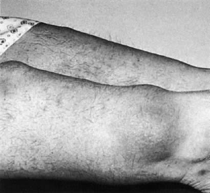
As is the case with hyperthyroidism, thyroid failure involves most organ systems. The degree of involvement depends on several factors, the most important perhaps being chronicity of the condition. Changes in the central nervous system are manifested by a decrease in mental activity, lethargy, and loss of mental concentration. Sleepiness at the end of the day, hearing loss, and mental depression may be prominent manifestations. Paresthesias of the fingers as a consequence of carpal tunnel syndrome may be the first and only manifestation in some hypothyroid patients. The relaxation phase of the deep tendon reflexes is delayed, and this has become a characteristic sign of the disease, although it is present only in patients with advanced symptoms. Hypothyroid patients may present with arthralgia and stiffness of the small joints of the hands, and muscle cramps are not infrequent complaints. On physical examination, the skin may be rough, scaly and thickened and cold to the touch. Hair becomes coarse and brittle, and loss of axillary, pubic, and scalp hair is not unusual in severe cases. Features of periorbital edema may be evident when previous pictures of the patient covering a span of several years are compared.

Bradycardia is noticed in some patients, and the electrocardiogram (ECG) may show a low voltage with nonspecific ST abnormalities. Occasionally a patient may suffer from cardiomegaly due to pericardial effusion or cardiomyopathy.

Gynecologic and obstetric abnormalities are not infrequent in hypothyroid women. Menorrhagia is commonly reported, although the true incidence is unknown. Anovulation is said to be common, although pregnancy with normal fetal outcome is not an uncommon event. If hypothyroidism is not promptly corrected, however, there is a higher risk of pregnancy-induced hypertension and premature delivery.11,12 Most women on thyroid therapy need an increase in dosage during pregnancy.13,14 TSH determinations are advisable in these women when pregnancy is diagnosed. It is strongly advisable to perform thyroid studies in patients with galactorrhea-amenorrhea because primary hypothyroidism is one of the causes of the syndrome.15

LABORATORY DIAGNOSIS.

A decrease in serum FT4 or its estimated FT4 index (FT4I) is characteristic of almost all forms of hypothyroidism, regardless of the cause. Serum TSH concentration is elevated in patients with primary hypothyroidism, and this measurement is the most sensitive test for the diagnosis of primary thyroid failure.16 Serum TSH levels may be elevated in patients with a normal FT4I, and this condition has been called “decreased thyroid reserve,” or subclinical hypothyroidism. It may represent the first indication of primary hypothyroidism in the majority of cases (see below).

A low serum FT4I in the presence of normal serum TSH concentrations is diagnostic of secondary hypothyroidism in ambulatory patients. In hospitalized patients with intercurrent illness, euthyroid sick syndrome must be ruled out.17

Other biochemical abnormalities seen in hypothyroid patients include elevations in serum cholesterol and triglycerides, elevations in serum creatinine phosphokinase, and nonspecific electrocardiographic abnormalities. It is helpful to obtain serum thyroid antibody concentrations to confirm the cause of hypothyroidism; they are elevated in most patients with chronic thyroiditis. Radioisotope studies are not indicated in evaluating hypothyroid patients.

SUBCLINICAL HYPOTHYROIDISM.

The syndrome of subclinical hypothyroidism is characterized by an asymptomatic state associated with normal free thyroid hormone concentrations in the presence of mild elevations in serum TSH levels (up to 20 to 25 mU/L).18 The most common causesare Hashimoto's thyroiditis and post-thyroid ablation (e.g., 131I therapy, surgery). Recognition of this entity is important because in a significant number of such patients, frank hypothyroidism can develop.19 The incidence is higher in women than in men, estimated at 15% to 20% of women older than 50 years.7 In more than 50% of these women, increased thyroid antibody titers are detected. Most patients are asymptomatic, but some present with complaints of fatigue, dry skin, and cold intolerance. Goiter is detected in approximately 30% to 60% of patients.

Subclinical hypothyroidism also may be diagnosed in patients taking insufficient thyroid replacement therapy for the treatment of overt hypothyroidism.20 Medications such as lithium, iodine, and those containing iodine (e.g., amiodarone) may cause mild thyroid dysfunction, particularly in patients with chronic autoimmune thyroid disease.

Some studies have shown subtle abnormalities of organ dysfunction in patients with subclinical hypothyroidism.21,22 Elevations in cholesterol and low-density lipoprotein cholesterol levels and decreases in high-density lipoprotein levels have been reported in some studies, correction being achieved after thyroid replacement therapy. Changes in left ventricular function have been reported as well. In one study,21 left ventricular ejection fraction improved with thyroid replacement therapy. In one double-blind randomized study, patients with subclinical hypothyroidism had fewer symptoms while receiving T4 therapy.23 Because most women are asymptomatic, treatment is controversial; however, because of the rate of progression to overt hypothyroidism in patients with positive antibodies (80% in a 4-year follow-up study19), it is advisable to start thyroid therapy in patients with subclinical hypothyroidism and positive thyroid antibodies.

SCREENING. Although screening for primary hypothyroidism cannot be recommended for every patient, there are patients that are at high risk for developing overt hypothyroidism. Currently, determinations of serum TSH is cost-effective for evaluating patients with the following high risk factors for development of hypothyroidism:

  Autoimmune thyroiditis
  Goiter
  Previous treatment for hyperthyroidism (thyrotoxicosis, see below)
  Previous high-dose neck radiation therapy
  Current lithium or amiodarone therapy
  Type I diabetes mellitus
  Postpartum, particularly in combination with a goiter
  Other autoimmune diseases (e.g., pernicious anemia, idiopathic thrombocytopenia).

Other risk factors include sleep apnea and hyperlipidemia in adults and failure to grow properly in children. Because the incidence of subclinical hypothyroidism is increased after the age of 50, particularly in women, a case can be made for screening all postmenopausal women (Fig. 1).

Fig. 1. Evaluation of patients with suspected primary hypothyroidism (screening)

TREATMENT.

Before treatment is contemplated in hypothyroid patients, a proper diagnosis between primary and secondary hypothyroidism is imperative. In patients with secondary hypothyroidism, a complete evaluation of the hypothalamic-pituitary axis should be made to rule out involvement of other hormones. Isolated TSH deficiency is uncommon.

One of the most gratifying experiences in medicine for the patient as well as the physician is the treatment of hypothyroidism, because all symptoms due to thyroid deficiency resolve with appropriate therapy. Several preparations are available, but regardless of the type of thyroid medication to be used, several principles should be kept in mind before therapy is started:

  1. Most patients will need lifelong replacement therapy.
  2. Therapy should be started with a low dosage, especially in the elderly and those with heart problems, and gradually increased to the full replacement dosage.
  3. The goal of any treatment is to achieve euthyroidism, from both the clinical and the biochemical point of view.

Because the hormonal content in synthetic levothyroxine is more reliably standardized than that of desiccated thyroid hormone, synthetic hormone preparations have replaced desiccated thyroid as the mainstay of thyroid therapy. With very few exceptions, there is no justification nowadays to treat hypothyroid patients with preparations other than levothyroxine. Thyroxine (T4) is deiodinated to triiodothyronine (T3) in extrathyroid tissues in humans; therefore T4 more closely simulates the physiology of normal subjects. It is well absorbed and tolerated and is given in a single daily dose.24 Its half-life is 6 days in normal persons and 8 to 11 days in hypothyroid patients. Some medications (e.g., ferrous sulfate, cholestyramine, sucrosulfate, aluminum hydroxide) have been reported to decrease thyroid absorption in the gastrointestinal tract if taken concomitant with thyroid therapy. Malabsorption and hypertrophic gastritis also may produce a decrease in T4 absorption by the gastrointestinal tract.

Normal serum TSH and FT4I concentrations are the goal of levothyroxine therapy. Studies in the last decade have shown that the amount of levothyroxine needed to produce normal tests is less than that previously recommended. Increased bioviability of levothyroxine preparations and more sensitive TSH measurements are responsible for this adjustment in the dosage. The recommended replacement dose is approximately 120 μg/day ± 40 or approximately 1.7 μg/kg body weight/day.25 The physician should be aware of the potential differences in the levothyroxine content among several generic thyroid preparations available today, although one study has indicated that most, but not all, of the generic brands contain the stated amount of levothyroxine.26

In young and healthy patients, therapy can be initiated with 0.05 to 0.075 mg levothyroxine daily; this amount can be increased every 2 to 6 weeks until the proper dose is achieved. For most patients, by the end of 6 to 8 weeks the full replacement dose is achieved. The serum TSH level is determined 4 to 6 weeks after therapy is initiated, and the dose is adjusted accordingly. Thereafter, patients should be seen at yearly intervals, at which time the dose may be adjusted depending on the thyroid tests results. It should also be kept in mind that as patients get older, they require smaller doses of levothyroxine.27

In older patients and those with heart disease, a more cautious approach is recommended; the initial dose should be 0.025 mg levothyroxine daily, and this dose should be increased every 4 to 8 weeks until full therapeutic effect or normalization of thyroid tests is obtained.28,29

MYXEDEMA COMA.

This is a rare clinical situation that constitutes a life-threatening emergency. Myxedema coma can occur in untreated patients with longstanding myxedema who are under stressful situations, particularly infection. Occasionally, the use of narcotics and analgesics can precipitate myxedema coma in patients with severe hypothyroidism. Clinically, patients present with progressive stupor and coma, seizures, hypotension, hypoventilation, and hypothermia. Respiratory acidosis and hyponatremia are also characteristic findings. Because the mortality is high, aggressive treatment is indicated: An initial dose of 0.3 mg IV levothyroxine is given, followed by an IV dosage of 0.05 to 0.1 mg/day, which is continued until the patient is able to take medication by mouth. Concomitant with levothyroxine administration, 100 mg hydrocortisone should be administered every 8 hours because many of these patients may present with relative adrenal insufficiency, or the hypothyroidism may be due to pituitary or hypothalamic disease. Assisted ventilation may be required, and fluid replacement should be given cautiously.30 A potential cause of hypothyroid coma is the discontinuation of thyroid therapy in hypothyroid patients on thyroid therapy who are admitted to the hospital for severe intercurrent disease. Lack of thyroid hormone for 2 weeks or longer can produce severe hypothyroidism.

PATIENTS ON LONG-TERM THYROID THERAPY.

Thyroid hormone preparations are among the most commonly prescribed drugs. In 1994, approximately 35 million prescriptions were written for thyroid tablets, about 92% for synthetic levothyroxine, and 8% for desiccated thyroid. Thyroid therapy is used for the treatment of hypothyroidism; it is also used for other conditions in euthyroid persons, such as nodular goiters, and empirically for the treatment of obesity, chronic fatigue, hypercholesterolemia, and infertility.28,29 It has been reported in the last few years that doses of thyroid hormones suppressing serum TSH, may produce subtle symptoms of hyperthyroidism such as nocturnal tachycardia, shortened systolic interval time, increased sodium excretion by the kidney, decreased total serum cholesterol, and alterations in liver enzymes.31,32 All the above abnormalities are reversed by normalizing thyroid tests.

One of the most significant clinical findings is the potentially deleterious effect of excessive thyroid therapy on bone metabolism. Hyperthyroidism due to intrinsic thyroid disease or excessive thyroid dosage affects bone resorption and bone formation.33,34 Patients with a history of hyperthyroidism are at an increased risk for bone fractures and tend to have fractures at an earlier age than persons who had never been hyperthyroid. Prompt treatment of thyrotoxicosis reverses bone mineral density. Most studies have shown significant decreases in bone mineral density in patients on thyroid therapy and low or suppressed serum TSH levels. Most of these studies, however, were cross-sectional and therefore confounding factors, such as race, obesity, menopausal status, drug therapy (e.g., calcium, estrogens, diuretics), and previous history of thyrotoxicosis, were not always evaluated. Therefore, although still a controversial issue and until prospective studies become available, it is recommended that women on thyroid therapy have serum TSH maintained within normal limits, thyroid function tests being assessed on a yearly basis.35 The exception are women who require suppressive thyroid therapy (serum TSH < 0.01 mU/L) after ablation therapy for treatment of thyroid carcinoma. In this situation, bone mass should be monitored at regular intervals. Recent evidence suggests that estrogen therapy36 or biphosphonates37 prevent the osteopenia induced by thyroid hormone therapy.

Finally, there are patients on thyroid medication for whom the original indication for thyroid therapy was not medically sound; in such situations and with the patients' consent, the physician may discontinue thyroid therapy for 4 to 6 weeks and repeat thyroid function tests. In cases of true primary hypothyroidism, serum TSH will be high and serum FT4 in the hypothyroid range at the end of this 4- to 6-week period; in euthyroid persons thyroid tests will return to normal limits.38 In some patients, however, it may take many weeks for the TSH to become elevated after being withdrawn from thyroid therapy; therefore it is prudent to repeat the serum TSH test in a few months before a final diagnosis of euthyroidism is confirmed.39

CHRONIC THYROIDITIS.

Hashimoto's thyroiditis (chronic or autoimmune) is one of the causes of goiter or hypothyroidism, or both. The diagnosis is confirmed by the clinical findings and by high titers of thyroid autoantibodies in serum. The disease is seen much more commonly in female patients than in males (4:1). The clinical picture is characterized by a goiter that is firm in consistency, has a tendency to be lobulated, and affects both lobes of the thyroid gland. The histologic changes consist of a combination of diffuse lymphocytic infiltrations, obliteration of thyroid follicles, and fibrosis. The natural course of the goiter is to remain unchanged or to enlarge gradually over many years if left untreated. Most patients develop hypothyroidism over several years. The use of iodine in patients with chronic thyroiditis may trigger the development of hypothyroidism or hyperthyroidism.40,41 Patients and their families have shown an increased incidence of other autoimmune diseases, such as pernicious anemia, insulin-dependent diabetes mellitus, vitiligo, Sjögren's syndrome, rheumatoid arthritis, chronic active hepatitis, and adrenal insufficiency.

Patients with chronic thyroiditis may develop transient thyroid dysfunction during pregnancy and in the postpartum period. They may have mild symptoms of hyperthyroidism 4 to 6 weeks postpartum, with enlargement of the thyroid gland and subsequent hypothyroidism at 3 to 5 months postpartum. These patients will return to the euthyroid state 5 to 10 months after delivery without undergoing specific treatment. It has been proposed that these changes are due to changes in the immune response during pregnancy.3,42

Treatment of chronic thyroiditis with thyroid hormone is used in euthyroid patients to decrease the size of the goiter, although this practice is controversial. In longstanding goiter that is firm on palpation, therapy is usually not effective in reducing its size because the gland is fibrotic. Otherwise, patients are followed yearly to detect any tendency toward hypothyroidism. These patients should be advised to have their thyroid tests checked during pregnancy and in the postpartum period because these are the times when they are at significant risk for development of thyroid dysfunction. Surgery is justified only in the unusual case of local pressure symptoms (e.g., dysphagia, cough, respiratory distress).

Subacute Thyroiditis.

Subacute thyroiditis is an acute, self-limiting viral infection affecting the thyroid gland. On pathologic examination, the gland is enlarged, with an inflammatory reaction involving the capsule. Polymorphs, lymphocytes, and foreign-body giant cells are recognized. Women are affected twice as often as men. The disease is preceded by a protracted phase of low-grade fever, malaise, and muscle aches for 2 to 3 weeks, followed by neck pain that often radiates to the angle of the jaw and to the ear on the involved side. Some patients may complain of dysphagia. The thyroid gland is tender to palpation and enlarged, and sometimes one of the lobes is as hard as stone. In a few days it returns to normal and the opposite side becomes enlarged and tender. Mild signs of hyperthyroidism may develop in some patients. The course of the disease varies from a few weeks to several months, with eventual spontaneous recovery. Patients may experience a period of hypothyroidism before final recovery.43

Diagnostic findings include elevation in serum FT4I and FT3I concentrations and, in most cases, suppression of TSH; there might be a slight elevation in the thyroid antibody titer. Diagnosis is confirmed by an elevation in the erythrocyte sedimentation rate and a low or depressed 24-hour radioiodine 131I thyroid uptake. In the early phase of the disease, treatment consists of analgesics. In patients in whom analgesic medication is ineffective, the use of corticosteroid (e.g., 30 to 40 mg prednisone daily in divided doses) is very effective in controlling symptoms, particularly neck pain. Prednisone dosage may be reduced after a few days and discontinued by 3 to 6 weeks in most cases. In some patients, however, symptoms recur when the amount of glucocorticoids is decreased and has to be increased again to control the pain. Sometimes symptoms may persist for several months. If a patient has significant hyperthyroid symptoms, 10 to 40 mg propranolol three times per day is effective in controlling the symptoms.

A similar clinical picture without thyroid pain has been described. This has been termed “painless thyroiditis,” or silent thyroiditis; its relationship to the classic subacute thyroiditis is not clear. Biopsies in patients with painless thyroiditis have shown lymphocytic infiltrations, rather than the typical histology of subacute thyroiditis. Clinically, more than 90% of patients with silent thyroiditis have manifestations of hyperthyroidism, with elevations in serum T4 and T3 concentrations and suppressed TSH. The 24-hour 131I thyroid uptake is low. The most common symptoms are palpitations, nervousness, weakness, and heat intolerance. Eye signs typical of Graves' disease are absent, although approximately one third have lid lag or lid retraction. The thyroid gland is enlarged and nontender.44 Recovery from hyperthyroidism usually takes 3 to 5 months. Many of these patients go through a period of transient hypothyroidism lasting a few weeks. The course is similar to patients with postpartum thyroid dysfunction.

Multinodular Goiter.

Multinodular goiter can be defined as an enlargement of the thyroid gland characterized by excessive growth and structural or functional transformation of one or several areas within the normal thyroid tissue. The pathogenesis of nodular transformation is not known, although chronic TSH stimulation in thyroid glands with abnormal follicular growth pattern plays an important role, as does iodine deficiency in endemic areas. However, the etiology of sporadic nodular goiter is unclear.45 The prevalence of nodular goiter is 4% to 8% in the adult US population and is more prevalent in women than in men (4:1).

The natural history of nontoxic goiter indicates that a diffuse enlargement of the thyroid may begin in adolescence with a gradual appearance of nodularities. These nodules grow slowly, over several decades, without producing significant clinical difficulties. They are seen more often in women and the elderly. Some goiters will remain quiet and stable, whereas others will keep growing steadily or may regress.46 In some patients, thyroid antibodies may be elevated, indicating that some of these goiters are related to chronic lymphocytic thyroiditis (Hashimoto's thyroiditis).

Most commonly, the diagnosis of nodular goiter is made during a routine physical examination. The patient may be unaware of the goiter. Very seldom, compression or local symptoms are the presenting complaint; occasionally, rapid growth of the nodule or sudden, acute pain indicating hemorrhage into a nodule causes the patient to seek help from the physician.

For the thyroid gland to be readily palpable, there must be a diffuse enlargement about two times the normal size.47 Inspection of the neck is an important part of the thyroid examination because nodules and asymmetry of the thyroid gland can be demonstrated easily in this way, particularly as the patient swallows gulps of water. The consistency of the nodules varies widely, from soft to hard. Thyroid tests are usually normal; however, if the sensitive TSH test is suppressed in the presence of normal FT4 and FT3 values, the possibility of an autonomous thyroid nodule should be entertained. 131I thyroid scanning is helpful in defining the presence of an autonomous or “hot” nodule within a multinodular goiter. Although no therapy is indicated, such patients should be followed closely for the development of hyperthyroidism.

Two complications in the natural history of multinodular goiter deserve discussion: (1) the development of thyrotoxicosis; and (2) the possible occurrence of malignancy in one of the nodules. Thyrotoxicosis in patients with multinodular goiters may develop insidiously, in which case it is difficult to recognize. Exposure to iodides (saturated solution of potassium iodide) therapy, iodine contrast materials, or amiodarone therapy may produce thyrotoxicosis in some patients. Therefore, patients with known nodular goiters should be followed, and thyroid tests should be performed at regular intervals because it may not always be possible to recognize this complication clinically. Ultrasound may be indicated to assess the size of the gland.

There is no evidence in the literature that nodular goiter predisposes the patient to the development of thyroid carcinoma. Carcinoma should be suspected when the thyroid scan shows hypofunctioning nodules, or if there has been a recent increase in the size of one of the nodules; needle aspiration biopsy should be used in such cases.

In some cases, levothyroxine therapy has proved effective in suppressing further goiter growth, although it is unusual to obtain complete regression. It should be kept in mind, however, that giving thyroid therapy to patients with multinodular goiter can lead to thyrotoxicosis in patients with autonomous nodules, in which case the serum TSH is low or suppressed. Therefore, thyroid therapy for multinodular goiter should be used only in patients with normal serum TSH values and only to decrease goiter size.

The indication for surgical therapy in multinodular goiter varies from center to center, but nearly everyone agrees that surgery should be performed only by highly trained surgeons and with facilities for intraoperative examination of frozen section. Surgery is indicated for a goiter producing symptoms, for a suspicion of malignancy, or for cosmetic reasons.48 In euthyroid patients who have scintigraphic evidence of one or several autonomous nodules, no therapy is indicated. These patients should be followed closely, however, because they are at increased risk for development of hyperthyroidism.

Solitary Nodule of the Thyroid Gland. As in the case of multinodular goiters, the finding of a single thyroid nodule is usually incidental during a routine physical examination. It has been estimated that the incidence of clinically detected thyroid nodules in the general population is approximately 4%, women having a fourfold higher incidence than that for men. For the nodule to be palpable, it should be more than 1 cm in diameter. Very seldom does the patient have local symptoms, such as dysphagia, hoarseness, and occasionally acute neck pain, due to bleeding into the nodule.

The discovery of a thyroid nodule always raises the possibility of malignancy. Several factors should be considered when evaluating such nodules. Young persons, particularly those less than 20 years old, have a higher incidence of malignancy. Benign nodules are much less common in men than in women, so nodules found in men, particularly those older than 50, are suggestive of malignancy. Family history is sometimes relevant: It has been reported that 6% of patients with thyroid cancer have a family history of malignant thyroid neoplasm. A nodule detected in a patient with a history of irradiation to the head or neck during infancy or childhood is suggestive of malignancy.49 Rapid nodule growth is an indication for more aggressive investigation.

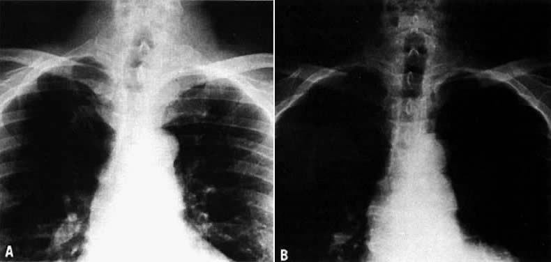
On physical examination, an asymptomatic neck mass is a presenting sign in more than 90% of patients. Cervical lymph nodes, cord paralysis, pain, or dysphagia may indicate thyroid carcinoma. Deviation of the trachea demonstrated on an x-ray is not always an indication of malignancy. A cystic lesion can produce a deviation of the trachea that resolves after aspiration of the fluid (Fig. 2). More than 90% of patients with thyroid nodules are clinically and chemically euthyroid. A little fewer than 10% may have an autonomous or hyperfunctioning nodule evident on a scintigraphic scan, in which case the patient may show symptoms of hyperthyroidism.50

Fig. 2. Single cold nodule of the left lobe of the thyroid gland. Routine chest x-ray demonstrates deviation of the trachea. A. Fine-needle aspiration obtained 7 mL of clear fluid. The cytology was benign. B. Repeat chest x-ray approximately 6 months later demonstrates complete resolution of the indentation of the left side of the trachea. The diagnosis was cystic adenomatoid nodule.

The evaluation of a single thyroid nodule is controversial, and several approaches have been recommended.51,52 As an initial step, some authorities suggest the determination of thyroid tests with thyroid antibodies,51 because the presence of thyroid dysfunction in the presence of a single thyroid nodule is almost always an indication of a benign process, although exceptions occur particularly in “cold” nodules in the presence of Graves' disease.53 The next step suggested is a thyroid scan to determine whether the nodule is hypofunctioning (cold) or hyperfunctioning (warm, hot). Hyperfunctioning nodules rarely harbor cancer. Fine-needle aspiration biopsy of the nodule is indicated for hypofunctioning nodules. Experienced cytopathologists give a correct interpretation in approximately 80% of specimens, and the incidence of false-positive or false-negative results is between 5% and 6%. As suggested by Ingbar,54 “the reading should be performed by a cytopathologist especially interested and experienced in interpretation of the resulting specimens.” Improvement in the sensitivity and specificity of the cytological interpretation of a thyroid nodule has resulted in a higher rate of excision of cancerous nodules and a decline in surgical intervention for noncancerous nodules. In most series reported, 70% of the nodules are benign; approximately 4% to 10% are malignant, 10% indeterminate or suspicious, and 10% insufficient for analysis because of an inadequate biopsy.49

A different approach to the evaluation of thyroid nodules is preferred in some centers, fine-needle aspiration biopsy being the first test used in the presence of a single or predominant thyroid nodule.52,55 If the diagnosis is malignancy, surgical excision is indicated; the type of surgery (lobectomy vs subtotal or total thyroidectomy) depends on the experience and criteria of the surgeon, and a full discussion is beyond the scope of this review.56 After surgery, thyroid suppression with levothyroxine is recommended; surgery is followed by 131I therapy in the presence of either residual tumor detected by thyroid scan or persistent elevation of serum thyroglobulin, or both.57,58 Long-term prognosis for thyroid cancer patients depends on the type of tumor, presence of local or distant metastasis, and other factors.59,60 My own approach to the evaluation of single thyroid nodules is presented in Figure 3.

Fig. 3. Evaluation of patients with single thyroid nodules (*fine-needle aspiration biopsy)

For lesions that are benign on cytologic examination, long-term thyroid suppression therapy has been classically recommended; however, recent observations appear to indicate that thyroid suppression therapy offers no benefit over placebo therapy in reducing the size of the nodule,61 but this issue is still controversial.62 It is my own practice to use thyroid suppression therapy for 3 to 6 months in benign nodules and to continue it if there has been a significant reduction in the size of the nodule. To avoid the potential complications of osteoporosis and subclinical hyperthyroidism, however, the serum TSH should be kept between 0.02 and 0.4 mU/L. After surgery for a benign thyroid nodule, thyroid therapy is indicated for patients in whom postsurgical hypothyroidism develops or for those who had received radiation therapy during childhood for benign conditions. In the latter group of patients, the recurrence rate of thyroid nodules has been reported to be 4.5 times higher in patients not treated with thyroxine therapy.63

Most malignant tumors of the thyroid are papillary or follicular carcinoma, although mixed lesions are not unusual. Undifferentiated carcinoma occurs in the elderly, and the prognosis is poor. Medullary thyroid carcinoma accounts for approximately 2% of thyroid cancer; it may occur alone or as part of the syndrome of multiple endocrine neoplasia in association with pheochromocytomas, parathyroid adenomas, and mucosal neuromas. In patients suspected of having this type of tumor, it is useful to measure serum calcitonin levels because they are characteristically elevated.64

Back to Top

HYPERTHYROIDISM
Hyperthyroidism, or thyrotoxicosis*, is defined as the constellation of physical and biochemical findings as a consequence of chronic exposure of the tissues to excessive concentrations of thyroid hormones. The clinical manifestations of the disease vary depending on the patient's age, duration of the disease, complications, abnormalities in other systems, and the cause of the syndrome. The most common cause of hyperthyroidism is Graves' disease. Other causes are listed in Table 2. Therefore, in addition to making the diagnosis of thyrotoxicosis, it is important to identify the cause, not only because different causes have different natural histories, but also because they require different forms of therapy.
 
*Thyrotoxicosis is a biochemical and physiological complex that results when the tissues are presented with excessive quantities of the thyroid hormones. I prefer the general term of thyrotoxicosis rather than hyperthyroidism to describe this syndrome, since the disorder need not originate in the thyroid gland. The term hyperthyroidism is best reserved for disorders in which thyrotoxicosis results from overproduction of hormones by the thyroid itself (e.g., Graves' disease, which is the most interesting and most important among them);54 however, in most papers and textbooks, the term hyperthyroidism is generally used.

TABLE 2. Etiology of Hyperthyroidism

  Most Common

  1. Graves' disease
  2. Toxic adenoma
  3. Multinodular goiter

  Less Common
  1. Subacute thyroiditis
  2. Thyrotoxicosis factitia
  3. Iodine-induced thyrotoxicosis
  4. Chronic autoimmune thyroiditis
  5. Silent (painless) thyroiditis

  Unusual Cases
  1. Trophoblastic hyperthyroidism
  2. Struma ovarii
  3. Thyroid carcinoma
  4. Thyrotropin-stimulating hormone hypersecretion
  5. Hyperthyroxinemia in hyperemesis gravidarum

Graves' Disease

Graves' disease—also known as Basedow's disease, exophthalmic goiter, or diffuse toxic goiter—is of autoimmune etiology and characterized by diffuse goiter, thyrotoxicosis, infiltrative ophthalmopathy, pretibial myxedema, and acropachy. Only a minority of patients, however, have a combination of all of these manifestations; hyperthyroidism and goiter with some type of eye involvement is the characteristic presentation. It has been known for some time that infiltrative ophthalmopathy and, rarely, infiltrative dermopathy may be present without hyperthyroidism (euthyroid Graves' disease). It occurs in predisposed families and could be associated with other autoimmune disorders, as is seen in chronic thyroiditis.

Adams and associates65 identified a substance in the serum of some patients with Graves' disease that was able to stimulate the thyroid gland of the guinea pig; because of its prolonged effect, this substance was named long-acting thyroid-stimulating (LATS) hormone. This observation and those of others seemed to indicate that LATS was a thyroid-stimulating antibody occurring in the blood of some patients with Graves' disease and possibly causing hyperthyroidism. It is now recognized that this substance is a TSH-receptor antibody. Several commercially available techniques are used for its determination, but the clinical indications are few (Table 3).66

TABLE 3. Clinical Use of TSH-receptor Antibody (TRHAb)

  1. Pregnancy
    1. Prediction of neonatal hyperthyroidism
    2. Diagnosis of fetal hyperthyroidism

  2. Euthyroid Graves' ophthalmopathy
  3. Euthyroid Graves' dermopathy

The most common clinical manifestations include nervousness, increased sweating, hypersensitivity to heat, palpitations, fatigue, weight loss, shortness of breath, muscle weakness, and eye complaints. In some patients, edema of the lower extremities, frequent bowel movements without diarrhea, anorexia, and eye complaints may be the initial manifestations. In approximately 2% of patients, there may be weight gain. Although these signs and symptoms may develop over a period of months, in some cases the disease is of rapid onset and the clinical picture develops within a few weeks. In older persons, the disease may have few presenting symptoms, such as severe weakness, wasting of muscles, and atrial fibrillation (masked hyperthyroidism).

On physical examination, the thyroid gland tends to be bilaterally enlarged two to four times the normal size, firm, and painless. In approximately 5% of patients, mainly in the elderly, there is no palpable goiter. In large glands, a thrill may be felt and bruit may be heard. Retraction of the eyelids leads to widening of the palpebral fissure, and this may produce the lid lag typical of hyperthyroidism, regardless of the cause. On close observation, a tremor of the lids can be seen. Changes in the nails are seen commonly, separation of the distal margin of the nail from the nail bed (onycholysis).

The skin is warm, moist, and smooth. Tremor of the hands and tongue is sometimes easily detected. Vitiligo is present in 10% of patients with Graves' disease. Tachycardia is the most frequent cardiovascular manifestation and presents as palpitations. Tachycardia during sleep (pulse rate greater than 90 beats/min) serves to distinguish tachycardia of thyrotoxic origin from that caused by anxiety. Cardiac arrhythmias (particularly in the form of atrial fibrillation) are common in the elderly. Edema of the lower extremities may be seen in patients with thyrotoxicosis unrelated to congestive heart failure; it resolves after the hyperthyroidism is treated.

In women, menstrual function may be disturbed. Oligohypomenorrhea, anovulation, and sometimes amenorrhea are not uncommon. However, pregnancy in hyperthyroid women, particularly when the disease is not severe, is not an uncommon event. Changes in menstrual function could be related to alterations in estrogen metabolism reported in patients with thyrotoxicosis.

The ophthalmopathy of Graves' disease is a disorder of unknown etiology and variable clinical presentation: It may precede, accompany, or follow hyperthyroidism, or it may be seen in the absence of recognizable thyroid disease. The clinical presentation varies from minimal disease to severe infiltrative disease. The most common symptoms include mild pain or eye discomfort, lacrimation, photophobia, and blurring of vision. Patients may complain of diplopia, or it may be discovered for the first time on careful physical examination. The most common signs are periorbital edema, lid edema, lid lag, conjunctival edema and injection, and exophthalmos. Exophthalmos is probably the most common manifestation and is usually bilateral, but often slightly asymmetric. Unilateral exophthalmos may be present; in a series of more than 200 cases of unilateral exophthalmos, Graves' ophthalmopathy was the cause in 28%.67 When unilateral exophthalmos occurs, the differential diagnosis includes orbital tumors, lymphomas, and Sjögren's syndrome. The use of noninvasive techniques, mainly computerized tomography and ultrasonography, has replaced invasive techniques used in the past in the differential diagnosis of exophthalmos.68

Pretibial myxedema, or infiltrative dermopathy, is an uncommon manifestation of Graves' disease that occurs in 5% of patients. The lesion is usually localized in the pretibial area; it appears as a violaceous induction of the skin or, at times, as localized nodular areas (Fig. 4). Thyroid acropachy describes an unusual presentation of clubbing of the digits and osteoarthropathy.

Fig. 4. Pretibial myxedema (nodular form)

In some patients, spontaneous remissions occur; however, in most patients the disease must be treated because of the severity of the symptoms and potential systemic complications.

The diagnosis is confirmed by a suppressed serum TSH and an elevation in FT4I or FT3I. Sometimes, the FT4I is within normal limits despite clinical symptoms of thyrotoxicosis, but there is an elevated serum T3 concentration (the term T3 toxicosis has been applied to this syndrome). Once the diagnosis is made, the second step is to determine the cause of hyperthyroidism. When the clinical diagnosis of Graves' disease is obvious (e.g., diffuse goiter, infiltrative ophthalmopathy, and infiltrative thermopathy), no other tests are indicated. In other situations, in which the cause of hyperthyroidism is not readily apparent, 131I thyroid uptake is a very important tool.

Treatment

There are three principal forms of treatment for hyperthyroidism due to Graves' disease. Antithyroid drug therapy, 131I therapy, and surgery are all very effective in controlling the symptoms. The treatment options are discussed with the patient; however, indications for choosing any one of them can depend on the physician's experience and preference.69,70,71

ANTITHYROID DRUGS.Thionamides.

Thionamides have been available since 1943. The most commonly used drugs in the United States are propylthiouracil and methimazole. They block thyroid hormone synthesis by different mechanisms. In addition, propylthiouracil acts at the periphery as a potent inhibitor of type I 5-deiodinase enzyme, which converts T4 to T3. Initial divided doses of 100 to 150 mg propylthiouracil every 8 hours, or 10 to 20 mg methimazole twice a day, are expected to produce euthyroidism in 4 to 8 weeks.72 After the euthyroid state is achieved, the amount of medication is decreased and adjusted according to the clinical picture and the determination of serum T4 and T3 concentrations. Treatment with this regimen for 6 to 24 months may produce remission in approximately 20% to 60% of patients.72 Long-term combination of thyroxine and antithyroid medication is used by some clinicians.73

Combined therapy, however, should not be administered to pregnant patients.74 Recently, it was reported that relapse from hyperthyroidism may be significantly reduced after initial treatment with methimazole for 6 months, followed by methimazole plus 100 μg of levothyroxine for 1 year, and levothyroxine alone for another 3 years.75 More studies are needed before these data can be confirmed.76

At present, there are no adequate tests to predict which patients will have a prolonged remission. It appears that the remission rate is higher in patients with small goiters and in those with mild symptoms and short-term duration of the disease. Side effects due to antithyroid therapy occurs in about 5% of patients. The most common side effects are pruritus and mild skin rashes, which are treated with antihistamines and switching to a different thionamide. The most severe complication is agranulocytosis (neutrophilic granulocytosis 250/mm3) which occurs in fewer than 0.2% of patients treated. Because this complication occurs suddenly, frequent routine blood counts are not useful in predicting an idiosyncratic response.70 The patient should be aware of this possible complication and report it to the physician if both fever and sore throat develop.72

On the prescription, the physician should write that the patient should stop taking the antithyroid medication and notify the physician immediately if sore throat and fever develop. If the diagnosis of agranulocytosis is confirmed, the patient should be hospitalized and treated with antibiotics. Recently, treatment with granulocytosis-stimulating factors has shown to shorten the period of recovery.77

Beta-Adrenergic Antagonist Drugs.

Beta-adrenergic antagonist drugs have been used in the treatment of symptoms of hyperthyroidism for many years with good results. After a few days of treatment, 20 to 80 mg propranolol three to four times a day is very effective in controlling the symptoms of the disease (e.g., palpitations, tremor, anxiety). Propranolol does not, however, play a role in the long-term treatment of thyrotoxicosis. It should be used in conjunction with antithyroid drugs until symptoms improve and the antithyroid drug becomes effective, which takes 7 to 14 days. Other beta-blocker agents, such as nadolol (80 mg/day) or atenolol (50 to 100 mg/day) are as effective as propranolol and are given once a day.69,70

RADIOIODINE THERAPY.

131I therapy for thyrotoxicosis has been used for more than 40 years and is effective in patients with Graves' disease. There is no evidence that this form of therapy increases the incidence of leukemia or thyroid cancer.78 The possible genetic damage has not been fully evaluated. The gonadal dose from 131I is approximately 0.4 rad/mCi, resulting in an increase in the birth defect rates from 4% to 4.025%. Radiotherapy is contraindicated in pregnancy and, given after 8 to 10 weeks of gestation, may produce congenital hypothyroidism. Women of reproductive age should receive the treatment in the first 10 days of the onset of menses or after a negative pregnancy test. The usual dose of 131I is 5 to 15 mCi, and the most common complication is permanent hypothyroidism. In patients with mild symptoms of hyperthyroidism, 131I may be given as the only treatment; beta-blockers can be used for controlling the symptoms for a few weeks before and after treatment. In more symptomatic patients and in the elderly, it is advisable to control hyperthyroidism with antithyroid drugs before radiotherapy is given. The antithyroid drug can be discontinued 3 or 4 days before radiotherapy, although this is not always necessary.70 There are several reports of aggravation of ophthalmopathy with the use of 131I therapy in patients with Graves' disease. It is recommended that radiotherapy be postponed until the ophthalmopathy is stable or that concurrent glucocorticoid therapy be given at the time of 131I administration.79,80,81

SURGERY.

Nowadays there are few indications for subtotal thyroidectomy in the management of patients with Graves' disease.82 For patients refusing 131I therapy, or those with large goiters or a hypofunctioning nodule, surgery is indicated. It also has been used in the first half of pregnancy to avoid the use of antithyroid drugs or in the rare pregnant patient who is allergic to both antithyroid medications.83 Permanent hypothyroidism is a potential complication, and patients should be followed up with thyroid tests on a regular basis.84 Relapses of hyperthyroidism can occur up to 30 years after the original surgery, hence the need for long-term follow-up in these patients.85

Other Causes of Hyperthyroidism

TOXIC ADENOMA.

In toxic adenoma, thyrotoxicosis is caused by overproduction of thyroid hormones due to an autonomous single thyroid nodule. On physical examination, a single nodule is felt. The clinical symptoms are generally mild in most patients, and signs of Graves' disease (exophthalmopathy and dermopathy) are characteristically absent. The diagnosis is confirmed by a thyroid scan showing a warm or hyperfunctioning nodule and suppression of the rest of the thyroid gland. Occasionally, a hyperfunctioning nodule may be present on scan in a patient with normal thyroid tests. It is possible for some of these euthyroid nodules to progress to toxic adenomas. Most of them, however, remain euthyroid or may regress over many years.86 Radiotherapy is the therapy of choice in the majority of these patients.87

TOXIC MULTINODULAR GOITER. The occurrence of thyrotoxicosis in patients with multinodular goiter is seen mainly in patients older than 50 years who have had a longstanding goiter. Iodine therapy has been reported to trigger this development.40 It may also occur in patients who have changed their diet from low to high iodine intake. Surgery or multiple-dose 131I therapy is the treatment of choice.

SUBACUTE THYROIDITIS AND SILENT THYROIDITIS.

Patients with both subacute and silent thyroiditis may present with symptoms of hypermetabolism. The clinical history and physical examination and a low 131I thyroid uptake are helpful in the diagnosis. Beta-blockers are used to treat symptoms.43

IODINE-INDUCED THYROTOXICOSIS (JOD-BASEDOW).

Patients developing hyperthyroidism after administration of iodine have been reported in the literature.88,89 The majority of patients have an underlying thyroid disease, mainly multinodular goiters or endemic goiter,40 or a previous history of Graves' disease. Iodine-induced thyrotoxicosis occasionally occurs in patients who initially had normal thyroid glands. For example, this syndrome should be considered in patients developing symptoms of thyrotoxicosis after receiving iodine therapy for the treatment of a respiratory condition90 or iodine-containing drugs for diagnostic purposes (e.g., cholecystogram, pyelogram). Amiodarone, an iodine-containing antiarrhythmic drug, should be added to the list.91 The symptoms can be mild to severe; both serum FT4 and FT3 are elevated, and characteristically the 24-hour 131I uptake is suppressed. Treatment may be difficult because patients do not respond promptly to antithyroid therapy, and the course of the disease may be prolonged. The use of propranolol may be effective in controlling the symptoms.

THYROTOXICOSIS FACTITIA.

In thyrotoxicosis factitia, the patient may chronically ingest an excess of thyroid hormone, commonly with the hope of losing weight. In many circumstances, these patients have personality disorders and deny the use of exogenous thyroid medication. The diagnosis sometimes is difficult to confirm, but thyroid tests in the hyperthyroid range, together with a suppressed 24-hour 131I thyroid uptake are suggestive of the condition. Inasmuch as many of these patients take T3 (Cytomel), measurement of serum T4 alone could be misleading because the value will be low in the presence of thyrotoxic symptoms; however, serum T3 concentrations or FT3I will be elevated. Determination of serum thyroglobulin levels is helpful in the differential diagnosis because suppressed values are characteristic of patients with the factitious form of the disease.92

THYROID-STIMULATING HORMONE-DEPENDENT HYPERTHYROIDISM.

TSH-dependent hyperthyroidism (excess TSH secretion) is a rare form of thyrotoxicosis in which serum TSH concentrations are elevated; in approximately 60% of patients a pituitary tumor is present. Abnormalities in TSH dynamics have been suggested as the cause of this syndrome: There is an increase in the α-subunit of TSH in patients with a demonstrable pituitary tumor, whereas there is suppression of the α-subunit and elevation of the β-subunit in patients without a pituitary tumor.93

THYROID CARCINOMA.

Hyperthyroidism due to thyroid carcinoma may be secondary to a hot autonomous thyroid nodule (rare) or to metastatic follicular carcinoma. No more than 30 such cases have been reported in the literature.94

MOLAR PREGNANCY AND CHORIOCARCINOMA. Hyperthyroidism may occur in patients with hydatidiform moles, as first recognized by Tisné and associates95 in 1955. In most patients, clinical signs of hypermetabolism are absent in the presence of abnormally high circulating thyroid hormones. The thyroid gland is generally not enlarged or is only slightly increased in size. Severe hyperthyroidism complicated by life-threatening situations has been reported.96 Excess levels of human chorionic gonadotropin, with its TSH-like activity, appear to be the cause of the syndrome.97

Choriocarcinoma can produce hyperthyroidism by a similar mechanism. As in the case of molar pregnancy, the patient may be clinically euthyroid despite having high levels of circulating thyroid hormones.

The thyrotoxicosis resolves quickly after evacuation of the mole. It can be controlled just before surgery with iodine therapy, propranolol, or antithyroid drugs. In cases of choriocarcinoma, it is advisable to treat the hyperthyroidism while chemotherapy is used.

STRUMA OVARII.

Struma ovarii is a rare condition that should be suspected in hyperthyroid women with a palpable ovarian lesion or ascites in the presence of a low 131I uptake in the neck area.98

THYROID STORM.

Thyroid crisis or storm is a medical emergency with a high mortality rate. It often occurs in patients with history of thyrotoxicosis, or as the first manifestation of hyperthyroidism. It is usually triggered by infection, acute illness, or trauma, or after subtotal thyroidectomy or other operations. Patients develop marked hyperthermia, severe hypermetabolic symptoms, and changes in the central nervous system.

Treatment consists of IV fluids, alcohol sponge baths to reduce the temperature, oxygen, and specific antithyroid therapy. Propylthiouracil is the drug of choice because, in addition to suppressing the synthesis of new thyroid hormones in the gland, it decreases T3 generation from T4 in the circulation. Because IV preparations are not available, 200 mg propylthiouracil should be administered orally every 4 hours. This is followed within 1 hour by the slow IV administration of 1 g sodium iodide over a 24-hour period or the oral administration of 5 drops of saturated solution of potassium iodide every 8 hours. The radiographic contrast agent ipodate sodium or iopanoic acid (1 g/day by mouth) also can be used. Treatment with both drugs is continued until a satisfactory response has occurred, and iodides are gradually withdrawn during a 2- to 3-week period. The use of 40 to 80 mg propranolol orally every 6 hours or 1 mg/min up to a maximum of 10 mg IV, decreases the heart rate and relieves the catecholamine-mediated effects of thyroid storm. For many years, 100 mg IV hydrocortisone every 8 hours, or equivalent amounts of other glucocorticoids, has been used empirically in the treatment of thyroid storm. There is now good evidence indicating that the beneficial effects of corticosteroids are derived from their suppression of the peripheral conversion of T3 from T4; this effect occurs shortly after the administration of the drug.99

After the treatment of thyroid storm, definite therapy for thyrotoxicosis in the form of subtotal thyroidectomy or 131I is imperative.

TRANSIENT THYROTOXICOSIS OF HYPEREMESIS GRAVIDARUM (PREGNANCY-INDUCED THYROTOXICOSIS).

Thyroid tests in women with severe hyperemesis gravidarum may be in the hyperthyroid range, and in many situations the differential diagnosis from hyperthyroidism is difficult. Patients suffer from severe nausea and vomiting, frequently requiring hospitalization, with a mean weight loss of 5 kg. Sensitive serum TSH is usually suppressed. Because severe nausea and vomiting could be an early manifestation of hyperthyroidism, the physician is presented with a difficult task. Tachycardia is common, but other symptoms of thyrotoxicosis are usually absent. In most cases, the symptoms abate after a few days of hospitalization, and the thyroid tests return to normal before 20 weeks' gestation. The detection of TSH-receptor antibodies could be helpful as a marker of Graves' disease. If the thyroid tests do not normalize spontaneously in a short period of time, or if the patient has other signs and symptoms of thyrotoxicosis, treatment with antithyroid drugs should be instituted.100,101

POSTPARTUM THYROID DYSFUNCTION.

Women with autoimmune thyroid disease (e.g., Graves' disease, chronic lymphocytic thyroiditis) frequently experience symptoms of hyperthyroidism 4 to 8 weeks postpartum.3 This has been reported to occur in 3% to 6% of all women after delivery.42,102 In most cases the symptoms are mild, and spontaneous recovery is the rule. Postpartum thyroid dysfunction usually recurs in future pregnancies. The clinical course is similar to that for patients with subacute thyroiditis, the episode of hyperthyroidism being followed by a hypothyroid phase and finally complete recovery. Patients with a previous history of hyperthyroidism due to Graves' disease may suffer recurrence of thyrotoxicosis after delivery. The differential diagnosis of postpartum thyroiditis may be difficult at times. However, recurrence of hyperthyroidism due to Graves' disease is accompanied by an elevation in 131I thyroid uptake, rather than a low uptake characteristic of postpartum thyroiditis.3

Back to Top

REFERENCES

1. Refetoff S, Weiss RE, Usala SJ: The syndromes of resistance to thyroid hormone. Endocr Rev 14: 348, 1994

2. Beck-Peccoz P, Chatterjee KK: The variable clinical phenotype in thyroid hormone resistance syndrome. Thyroid 4: 225, 1994

3. Larsen PR, Ingbar SH: The thyroid gland. In Wilson JD, Fosteer DW (eds): Williams Textbook of Endocrinology, 8th ed, p 358. Philadelphia, WB Saunders, 1992

4. Mestman JH, Goodwin TM, Montoro MM: Thyroid disorders of pregnancy. Endocrinol Metab Clin North Am 24: 41, 1995

5. Takasu N, Yamada T, Takasu M et al: Disappearance of thyrotropin-blocking antibodies and spontaneous recovery from hypothyroidism in autoimmune thyroiditis. N Engl J Med 326: 513, 1992

6. Takasu N, Yamada T, Sato A et al: Graves' disease following hypothyroidism due to Hashimoto's disease: Studies of eight cases. Clin Endocrinol 33: 687, 1990

7. Bilous RW, Tunbridge WMG: The epidemiology of hypothyroidism—an update. In Lazarus JH, Hall R (eds): Bailliere's Clinical Endocrinology and Metabolism, p 531. London, Bailliere Tindall, 1988

8. Sawin CT, London MH: “Natural” desiccated thyroid: A “health-food” thyroid preparation. Arch Intern Med 149: 2117, 1989

9. Bahemuka M, Hodkinson H: Screening for hypothyroidism in elderly patients. Br Med J 2: 601, 1975

10. Robuschi G, Safran M, Braverman LE et al: Hypothyroidism in the elderly. Endocr Rev 8: 142, 1987

11. Davis LE, Leveno KJ, Cunningham FG: Hypothyroidism complicating pregnancy. Obstet Gynecol 72: 108, 1988

12. Leung AS, Millar LK, Koonings PP et al: Perinatal outcome in hypothyroid pregnancies. Obstet Gynecol 81: 349, 1993

13. Mandel SJ, Larsen PR, Seely EW et al: Increased need for thyroxine during pregnancy in women with primary hypothyroidism. N Engl J Med 323: 91, 1990

14. Kaplan MM: Monitoring thyroxine therapy treatment during pregnancy. Thyroid 2: 147, 1992

15. Onishi T, Miyai K, Aono T et al: Primary hypothyroidism and galactorrhea. Am J Med 63: 373, 1977

16. Hershman JM, Pittman JA: Utility of the radioimmunoassay of serum thyrotropin in man. Ann Intern Med 74: 481, 1971

17. Wartofsky L, Burman KD: Alterations in thyroid function in patients with systemic illness: The “euthyroid sick syndrome.” Endocr Rev 3: 164, 1982

18. Cooper DS: Subclinical hypothyroidism. In Mazzaferri EL (ed): Advances in Endocrinology and Metabolism, Vol 2, pp 77–88. St. Louis, Mosby Year Book, 1991

19. Rosenthal MJ, Hunt WC, Garry PJ et al: Thyroid failure in the elderly. JAMA 258: 209, 1987

20. Sawin CT, Geller A, Hershman JM et al: The aging thyroid. JAMA 261: 2653, 1989

21. Forfar JC, Wathen CG, Todd WTA: Left ventricular performance in subclinical hypothyroidism. Q J Med 57: 857, 1985

22. Staub JJ, Althaus BU, Engler H et al: Spectrum of subclinical and overt hypothyroidism: Effect on thyrotropin, prolactin, and thyroid reserve, and metabolic impact on peripheral target tissues. Am J Med 92: 631, 1992

23. Cooper DS, Halpern R, Wood LC et al: L-thyroxine therapy in subclinical hypothyroidism. Ann Intern Med 101: 18, 1984

24. Choe W, Hays MT: Absorption of oral thyroxine. Endocrinologist 5: 222, 1995

25. Fish LH, Schwartz HL, Cavanaugh J et al: Replacement dose, metabolism, and bioavailability of levothyroxine in the treatment of hypothyroidism. N Engl J Med 316: 764, 1987

26. Hennessey JV, Evaul JE, Tseng YC et al: L-thyroxine dosage: A reevaluation of therapy with contemporary preparations. Ann Intern Med 105: 11, 1986

27. Sawin CT, Herman T, Molitch ME et al: Aging and the thyroid. Am J Med 75: 206, 1983

28. Roti E, Minelli R, Gardini E et al: The use and misuse of thyroid hormone. Endocr Rev 14: 401, 1993

29. Toft AD: Thyroxine therapy. New Engl J Med 331: 174, 1994

30. Nicoloff JT, LoPresti JS: Myxedema coma. Endocrinol Metab Clin North Am 22: 279, 1993

31. Biondi B, Fazio S, Carella C et al: Cardiac effects of long term thyrotropin suppressive therapy with levothyroxine. J Clin Endocrinol Metab 77: 334, 1993

32. Ladenson PW: Editorial: Thyrotoxicosis and the heart: Something old and something new. J Clin Endocrinol Metab 77: 332, 1993

33. Harvey RD, McHardy KC, Reid IW et al: Measurement of bone collagen degradation in hyperthyroidism and during thyroxine replacement therapy using pyridinium cross-links as specific urinary markers. J Clin Endocrinol Metab 72: 1189, 1991

34. Ross DS, Neer RM, Ridgway EC et al: Subclinical hyperthyroidism and reduced bone density as a possible result of prolonged suppression of the pituitary-thyroid axis with L-thyroxine. Am J Med 82: 1167, 1987

35. Baran DT: Thyroid hormone and bone mass: The clinician's dilemma. Thyroid 4: 143, 1994

36. Schneider DL, Barrett-Connor EL, Morton DJ: Thyroid hormone use and bone mineral density in elderly women: Effects of estrogen. JAMA 271: 1245, 1994

37. Rosen HN, Moses AC, Gundberg C et al: Therapy with parenteral pamidronate prevents thyroid hormone-induced bone turnover in humans. J Clin Endocrinol Metab 77: 664, 1993

38. Krugman LG, Hershman JM, Chopra IJ et al: Patterns of recovery of the hypothalamic-pituitary-thyroid axis in patients taken off chronic thyroid therapy. J Clin Endocrinol Metab 41: 70, 1975

39. Molitch ME, Spare SV, Arnold GL et al: Pituitary thyroid function after cessation of prolonged thyroid suppression. N Engl J Med 295: 231, 1976

40. Martin FIR, Tress BW, Colman PG et al: Iodine-induced hyperthyroidism due to nonionic contrast radiography in the elderly. Am J Med 95: 78, 1993

41. Mizukami Y, Michigishi T, Nonomura A: Iodine-induced hypothyroidism: A clinical and histological study of 28 patients. J Clin Endocrinol Metab 76: 466, 1993

42. Amino N, Mori H, Iwatani Y et al: High prevalence of transient postpartum thyrotoxicosis and hypothyroidism. N Engl J Med 306: 849, 1982

43. Greene JN: Subacute thyroiditis. Am J Med 51: 97, 1971

44. Nikolai TF, Brosseau J, Ketrick M et al: Lymphocytic thyroiditis with spontaneously resolving hyperthyroidism (silent thyroiditis). Arch Intern Med 140: 478, 1980

45. Studer H, Gerber H: Clinical manifestations and management of nontoxic diffuse and nodular goiter. In Braverman LE, Utiger RD (eds): Werner and Ingbar's The Thyroid, 6th ed, p 114. Philadelphia, JB Lippincott, 1991

46. Rallison ML, Dobyns BM, Meikle AW et al: Natural history of thyroid abnormalities: Prevalence, incidence, and regression of thyroid diseases in adolescents and young adults. Am J Med 91: 363, 1991

47. Siminoski K: Does this patient have a goiter? JAMA 273: 813, 1995

48. Edis AJ: Prevention and management of complications associated with thyroid and parathyroid surgery. Surg Clin North Am 59: 83, 1979

49. Caruso D, Mazzaferri EL: Fine needle aspiration biopsy in the management of thyroid nodules. Endocrinologist 3: 194, 1991

50. Hamburger JI: Evolution of toxicity in solitary nontoxic autonomously functioning thyroid nodules. J Clin Endocrinol Metab 50: 1089, 1980

51. Ridgway EC: Clinician's evaluation of a solitary thyroid nodule. J Clin Endocrinol Metab 74: 231, 1992

52. Mazzaferri EL: Management of a solitary thyroid nodule. N Engl J Med 328: 553, 1993

53. Belfiore A, Garofalo MR, Giuffrida D et al: Increased aggressiveness of thyroid cancer in patients with Graves' disease. J Clin Endocrinol Metab 70: 830, 1990

54. Ingbar SH: Thyroid gland. In Wilson JD, Foster DW (eds): Williams Textbook of Endocrinology, 7th ed, pp 802. Philadelphia, WB Saunders, 1985

55. Gharib H: Fine needle aspiration biopsy of thyroid nodules: Advantages, limitations, and effect. Mayo Clin Proc 69: 44, 1994

56. Clark OH: Total thyroidectomy: The thyroidectomy: The treatment of choice for patients with differentiated thyroid cancer. Ann Surg 196: 361, 1982

57. Ozata M, Suzuki S, Miyamoto T et al: Serum thyroglobulin in the follow up of patients with treated differentiated thyroid cancer. J Clin Endocrinol Metabol 79: 98, 1994

58. Spencer CA, Wang CC: Thyroglobulin measurement: Techniques, clinical benefits and pitfalls. Endocr Metab Clin North Am 24: 841, 1995

59. Robbins K, Merino MJ, Boice J et al: Thyroid cancer: A lethal endocrine neoplasm. Ann Intern Med 115: 133, 1991

60. Samaan NA, Schultz PN, Hickey RC et al: The results of various modalities of treatment of well differentiated thyroid carcinoma: A retrospective review of 1599 patients. J Clin Endocrinol Metab 75: 714, 1992

61. Gharib H, James EM, Charboneau JW et al: Suppressive therapy with levothyroxine for solitary thyroid nodules: A double blind controlled clinical study. N Engl J Med 317: 70, 1987

62. La Rosa GL, Lupo L, Giuffrida D et al: Levothyroxine and potassium iodide are both effective in treating benign solitary solid cold nodules of the thyroid. Ann Intern Med 122: 1, 1995

63. Fogelfeld K, Wiviott MVT, Shore-Freedman E et al: Recurrence of thyroid nodules after surgical removal in patients irradiated in childhood for benign conditions. N Engl J Med 320: 835, 1989

64. Gharib H, McConahey WM, Tiegs RD et al: Medullary thyroid carcinoma: Clinicopathologic features and long-term follow up of 65 patients treated during 1946 through 1970. May Clin Proc 67: 934, 1992

65. Adams DD, Purvis HD, Sirett NE: Response of hypophysectomized mice to injections of human serum containing long acting thyroid stimulator. Endocrinology 68: 154, 1961

66. McKenzie JM, Zakarija M: The clinical use of thyrotropin receptor antibody measurements. J Clin Endocrinol Metab 69: 1093, 1989

67. Dallow RL: Evaluation of unilateral exophthalmos with ultrasonography: Analysis of 258 consecutive cases. Laryngoscope 85: 1905, 1975

68. Fells P: Thyroid-associated eye disease: Clinical management. Lancet 338: 29, 1991

69. Franklyn JA: The management of hyperthyroidism. New Engl J Med 330: 1731, 1994

70. Klein I, Becker DV, Levey GS: Treatment of hyperthyroid disease. Ann Intern Med 121: 281, 1994

71. Singer PA, Cooper DS, Levy EG et al: Treatment guidelines for patients with hyperthyroidism and hypothyroidism. JAMA 273: 808, 1995

72. Cooper DS: Antithyroid drugs. New Engl J Med 311: 1353, 1984

73. Origazzi J: Management of Graves' hyperthyroidism. Clin Endocrinol Metab 16: 365, 1987

74. Mestman JH, Manning PR, Hodgman J: Hyperthyroidism and pregnancy. Arch Intern Med 134: 434, 1981

75. Hashizume K, Ichikawa K, Sakurai A et al: Administration of thyroxine in treated Graves' disease: Effects on the level of antibodies of thyroid stimulating hormone receptors and on the risk of recurrence of hyperthyroidism. N Engl J Med 324: 947, 1991

76. Hershman JM: Editorial: Does thyroxine therapy prevent recurrence of Graves' hyperthyroidism? J Clin Endocrinol Metab 80: 1479, 1995

77. Magner JA, Snyder DK: Methimazole-induced agranulocytosis treated with recombinant human granulocyte colony-stimulating factor (G-CSF). Thyroid 4: 295, 1994

78. Halan KE: Radioiodine treatment of hyperthyroidism—A more liberal policy? Clin Endocrinol Metab 14: 467, 1985

79. Burch HB, Wartofsky L: Graves' ophthalmopathy: Current concepts regarding pathogenesis and management. Endocr Rev 14: 747, 1993

80. Tallstedt L, Lundell G, Torring O et al: Occurrence of ophthalmopathy after treatment for Graves' hyperthyroidism. N Engl J Med 326: 1733, 1992

81. Bartalena L, Marcocci C, Bogazzi F et al: Use of corticosteroids to prevent progression of Graves' ophthalmopathy after radioiodine therapy for hyperthyroidism. N Engl J Med 321: 1349, 1989

82. Patwardhan NA, Moroni M, Rao S et al: Surgery still has a role in Graves' hyperthyroidism. Surgery 114: 1108, 1993

83. Bruner J, Landon MB, Gabbe SG: Diabetes mellitus and Graves' disease in pregnancy complicated by maternal allergies to antithyroid medication. Obstet Gynecol 72: 443, 1988

84. Evered D, Young ET, Tunbridge WMG et al: Thyroid function after subtotal thyroidectomy for hyperthyroidism. Br Med J 1: 25, 1975

85. Sugrue D, McEvoy M, Feely J et al: Hyperthyroidism in the land of Graves': results of treatment by surgery, radio-iodine and carbimazole in 837 cases. Q J Med 49: 51, 1980

86. Hamburger JI: The autonomously functioning thyroid nodule: Goetsch's disease. Endocrinol Rev 8: 438, 1987

87. Ross DS, Ridgway EC, Daniels GH: Successful treatment of solitary toxic thyroid nodules with relatively low dose iodine, with low prevalence of hypothyroidism. Ann Intern Med 104: 219, 1986

88. Vagenakis AG, Wang C, Burger A et al: Iodine induced hyperthyroidism thyrotoxicosis in Boston. N Engl J Med 287: 523, 1972

89. Fradkin JE, Wolff J: Iodine-induced thyrotoxicosis. Medicine 62: 1, 1983

90. Becker CB, Gordon JM: Iodinated glycerol and thyroid dysfunction. Chest 103: 188, 1993

91. Martino E, Safran M, Aghini-Lombardi F et al: Environmental iodine intake and thyroid dysfunction during chronic amiodarone therapy. Ann Intern Med 101: 28, 1984

92. Mariotti S, Martino E, Cupini C et al: Low serum thyroglobulin as a clue to the diagnosis of thyrotoxicosis factitia. N Engl J Med 307: 410, 1982

93. Magner JA: TSH-mediated hyperthyroidism. Endocrinologist 3: 289, 1993

94. Ghose MK, Genuth SM, Abellera RM: Functioning primary thyroid carcinoma and metastases producing hyperthyroidism. J Clin Endocrinol 33: 639, 1971

95. Tisné L, Barzelatto J, Stevenson C: Estudio de la funcion tiroidea durante el estado gravidco-puerperal con el yodo radioactivo. Bol Soc Chilena Obstetr Gincecol 20: 246, 1955

96. Hershman JM, Higgins HP: Hydatidiform mole—a cause of clinical hyperthyroidism. N Engl J Med 284: 573, 1971

97. Kenimer JG, Hershman JM, Higgins HP: The thyrotropin in hydatidiform moles is human chorionic gonadotropin. J Clin Endocrinol Metab 40: 482, 1975

98. Kempers RD, Doherty MB, Hoffmand DL et al: Struma ovarii: ascitic, hyperthyroid and asymptomatic syndrome. Ann Intern Med 72: 883, 1970

99. Burch HB, Wartofsky L: Life-threatening thyrotoxicosis. Endocrinol Metab Clin North Am 22: 263, 1993

100. Goodwin TM, Montoro M, Mestman JH: Transient hyperthyroidism and hyperemesis gravidarum: Clinical aspects. Am J Obstet Gynecol 167: 648, 1992

101. Goodwin TM, Montoro M, Mestman JH: The role of chorionic gonadotropin in transient hyperthyroidism of hyperemesis gravidarum. J Clin Endocrinol Metab 75: 1333, 1992

102. Learoyd DL, Fung HYM, McGregor AM: Postpartum thyroid dysfunction. Thyroid 2: 73, 1992

Back to Top