Chapter 55
Transvaginal Ultrasonography and the Evaluation of Female Infertility
Roger A. Pierson and Olufemi A. Olatunbosun
Main Menu   Table Of Contents

Search

Roger A. Pierson, MS, PhD
Department of Obstetrics and Gynecology, College of Medicine, University of Saskatchewan, Saskatoon, Saskatchewan, Canada (Vol 5, Chap 55)

Olufemi A. Olatunbosun, MD, FRCSC
Department of Obstetrics and Gynaecology, College of Medicine, University of Saskatchewan, Saskatoon, Saskatchewan, Canada (Vol 5, Chap 55)

 
EVALUATION OF OVARIAN FOLLICULAR DEVELOPMENT
EVALUATION OF THE OVARIAN FOLLICULAR RESERVE
POLYCYSTIC OVARIAN SYNDROME
CORPUS LUTEUM FUNCTION AND REGRESSION
IMAGING OF THE OVIDUCTS
IMAGING OF THE UTERUS
ECTOPIC PREGNANCY
OVULATION INDUCTION AND ASSISTED REPRODUCTIVE TECHNOLOGIES
INVASIVE ULTRASOUND-GUIDED PROCEDURES
SUMMARY
ACKNOWLEDGMENTS
REFERENCES

High-resolution diagnostic transvaginal ultrasonography (TVUS) has been one of the most important developments in infertility investigation since the widespread application of radioimmunoassay techniques for the measurement of reproductive hormones. The development of relatively high-frequency (e.g., 5.0 MHz or 7.5 MHz), curvilinear array transducers and color-flow Doppler technology has dramatically increased the resolution of images allowing detection of minute but potentially important differences in the morphology of the female reproductive organs. Although transabdominal ultrasonography of the pelvis has been used for several years, the ability to place the transducer into the vaginal fornix, just a few millimeters from the organs of interest, yields unprecedented resolution for evaluation of the ovaries, oviducts, and uterus. The vaginal approach to imaging the reproductive organs also eliminates the need for a full bladder, thereby minimizing patient discomfort and increasing the acceptance of ultrasonographic examination by both the patient and the examiner.

Traditionally, general radiologists have performed ultrasonographic examinations, screening only for overt gynecological pathology and typically with no specific interest in Obstetrics and Gynecology. As more obstetrician/gynecologists are trained in the use of transvaginal ultrasonography, ultrasonographic examinations in the context of gynecologic visits will likely increase. Considerable relevant information may be gained by using ultrasonography early in the sequence of infertility investigations, in concert with the more conventional infertility studies. Uterine and ovarian abnormalities such as cysts, tumors, fibroids, endometriomas, hydrosalpinges, and congenital abnormalities may be quickly identified, thereby speeding accurate diagnosis and specific treatment.1 Transvaginal ultrasonographic (TVUS) examination also offers a great deal of information regarding the nature and progress of early pregnancy.

At present, many of the potential uses for TVUS are not developed fully for routine clinical use. Its applications are still being explored and evaluated. Much of the information contained in this chapter derives from ongoing investigations in tertiary care facilities. Although clinical applications for many of the techniques described are still undergoing development, it is already clear that TVUS holds tremendous potential and that its value as a clinical tool in the evaluation of infertility is sure to increase.

Back to Top
EVALUATION OF OVARIAN FOLLICULAR DEVELOPMENT

In the context of ongoing research in tertiary care centres, serial TVUS examinations performed during the follicular phases of one or more menstrual cycles have helped to define both normal and abnormal patterns of follicular growth and development.2,3 Although the logistical challenges of serial TVUS examinations can be somewhat daunting, a consistent pattern will usually become apparent within the span of a few cycles.

Normal Follicular Growth and Development

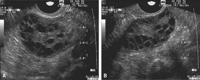
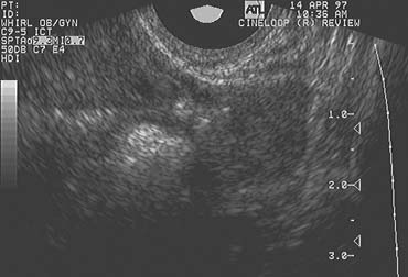
The population of ovarian follicles can be evaluated at any time during the menstrual cycle, but the interval of greatest interest spans the late follicular phase during the final stages of preovulatory follicular development. The dominant follicle destined to ovulate can be identified by its size on approximately cycle day 7 and thereafter grows at an average rate of 2 mm per day until it reaches full maturity at a diameter of approximately 22 mm (Fig. 1).2,4,5,6,7,8

Fig. 1. Dominant preovulatory follicle. The antrum of the follicle (black) is central to the image. The stigma, or site at which follicular rupture will occur, is on the right side of the antrum. The follicle wall, stratum granulosum, and theca interna are identifiable around the periphery of the antrum.

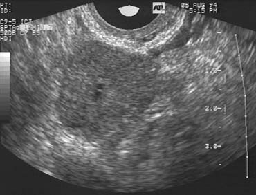
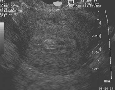
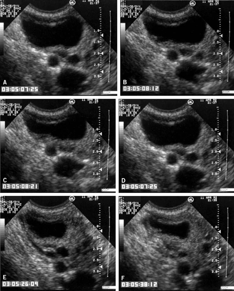
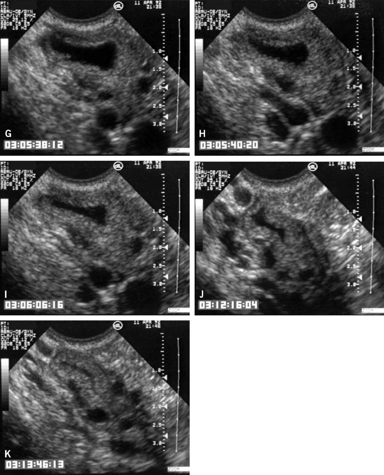
In the classically normal menstrual cycle, ovulation occurs on or at approximately cycle day 14 and menses begins approximately 14 days thereafter.2,4,5,6 Although ultrasound has been used to detect ovulation in women for a number of years,9 the ability to observe the physical rupture of the preovulatory follicle, escape of the follicular fluid, and extrusion of the cumulus/oocyte complex have only recently been clearly demonstrated (Fig. 2).10 The site at which ovulation will occur can be readily identified up to a week in advance.5,10 The sequence of ultrasonographic events that surrounds ovulation occurs over an average of approximately 10 minutes, but the interval varies, lasting from less than 1 minute to more than 20 minutes in duration. After ovulation, in normal cycles, the collapsed follicle gives rise to the corpus luteum. The corpus luteum then becomes the dominant ovarian structure and can be easily recognized across the remainder of the cycle (see Fig. 2).5,10

Fig. 2. Images of ovulatory failure. (A) The walls of the unruptured follicle are thin and only marginally luteinized. The antrum contains internal echoes consistent with a filamentous fibrin network. (B) Fully luteinized unruptured follicle. Note the thick, luteinized border surrounding the cloudy blood-filled antrum. The central clot appears to be well organized.

Use of Transvaginal Ultrasonography to Determine the Optimum Time for Artifical Insemination

Increasingly, TVUS monitoring of follicular development is being used to better and more precisely define the time of ovulation and the optimum time to perform IUI, when indicated, in both spontaneous and stimulated cycles. The accuracy of TVUS in defining the time of ovulation is far superior to estimates based on results of monitoring basal body temperature (BBT) or urinary luteinizing hormone (LH) excretion. Given the relatively brief interval for which sperm remain viable after IUI and the considerable costs involved, serial TVUS examinations may help to maximize both the effectiveness and efficiency of such advanced forms of treatment.

Abnormal Follicular Growth and Development

Careful serial TVUS examinations during the latter stages of preovulatory follicular development and during the interval immediately after ovulation in both normal fertile women and those with idiopathic infertility suggest that subtle but distinct abnormalities of follicular growth, development, and ovulation occur and may often go unrecognized. The luteinized unruptured follicle (LUF) and the follicular cyst are two distinctly abnormal patterns of follicular development that may represent subtle forms of subtle ovulatory dysfunction.

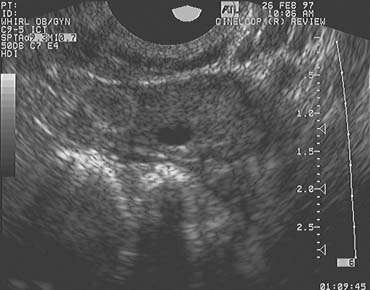
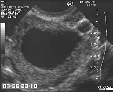
The LUF syndrome is characterized by a preovulatory follicle that grows and develops in a normal pattern but does not collapse at the expected time of ovulation. Ovulation does not, in fact, occur and the oocyte presumably remains trapped within the follicle. The follicular walls subsequently thicken and increase in density much like is seen after a normal ovulation, the follicular margins become less distinct, and the structure persists for the duration of the cycle, regressing in a manner very similar to that of a normally formed corpus luteum (Fig. 3).11,12 Other indices of ovulation (basal body temperature, serum progesterone concentration) remain normal; the time and character of the subsequent menses also are most often entirely normal.

Fig. 3. Sequence of images (A–I) recorded during ovulation in situ. The images in the sequence were taken to represent the times at which 90%, 80%, 70%, and so on of the follicle fluid was extruded from the follicle. Time code markers are displayed in the lower left portion of the images.

Fig. 3. Continued.

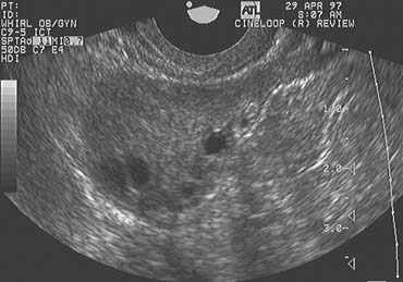
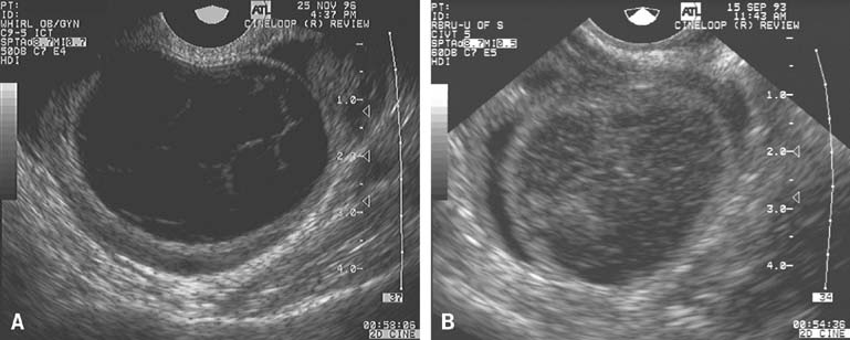
The follicular cyst is recognized when the preovulatory follicle grows to a diameter beyond those typically observed before ovulation but does not collapse and persists for an interval of several days before gradually regressing at a pace similar to that at which it grew. In contrast to the LUF, there is no sonographic evidence of luteinization. The walls of the follicular cyst do not thicken, increase in density, or become indistinct. Instead, they remain thin, and follicular margins are still sharply defined. Menses generally follow at or near the expected time, although the duration and character of flow is more variable.

Used in combination with serial TVUS examinations, serum estradiol determinations may provide even greater insights into the quality of follicular growth and development and reveal abnormalities that would otherwise go unrecognized by either method alone. TVUS examinations may demonstrate that normally rising serum estradiol concentrations reflect the combined contributions of a large number of small follicles measuring 5 to 10 mm diameter rather than the development of a single dominant preovulatory follicle. Conversely, a late follicular phase TVUS examination may reveal a follicle having the diameter normally expected immediately before ovulation, but an abnormally low estradiol concentration may indicate it is unlikely to contain a normal or mature oocyte and may be expected to regress (become irregular in contour and progressively decrease in size) over the ensuing week.

At present, such careful monitoring of follicular growth and development in natural cycles remains largely in the realm of ongoing clinical research and has limited applications in clinical care. Further research, to include investigations aimed at defining and correlating the microvascular characteristics of individual follicles with patterns of growth and ovulation or regression will surely expand our current understandings of follicular dynamics and ultimately may also help to explain the cause of reproductive failure in some women with otherwise idiopathic infertility. As technological capabilities and research continue to advance, TVUS may be expected to play an increasingly valuable role in the evaluation and treatment of infertility related to ovulatory dysfunction and failure.

Back to Top
EVALUATION OF THE OVARIAN FOLLICULAR RESERVE

TVUS also has been used to investigate follicular dynamics in aging women. Numerous studies in recent years have demonstrated that fertility declines progressively as age advances. In parallel, the ovarian response to exogenous gonadotropin stimulation also decreases, but the range of individual variation is extremely wide. Recent demographic trends (aging of the baby boom generation, increased interest in advanced education, and greater career orientation among women, later marriage, more frequent divorce) have combined to yield increasing numbers of women seeking pregnancy when they are older and less biologically fertile. Although detailed studies of ovarian kinetics during the latter reproductive years have not yet been conducted, the increased number of older women in pursuit of pregnancy provides ample justification for such investigations.

Dynamic endocrinologic tests of ovarian reserve, including the clomiphene citrate challenge test and the gonadotropin-releasing hormone (GnRH) agonist stimulation test, offer prognostic information valuable in the counseling of aging infertile women. Recent observations suggest that TVUS can provide additional useful information of clinical relevance. Early follicular phase antral follicle counts can help to predict the number of follicles likely to develop during ovarian stimulation with exogenous gonadotropins and appear to correlate with the outcome of cycles in which ovarian stimulation is combined with intrauterine insemination (IUI). Women having fewer than 5 follicles smaller than 10 mm in diameter before ovarian stimulation begins have a relatively poor prognosis for success. Studies to determine the extent to which antral follicle counts correlate with endocrinologic measures of ovarian reserve (e.g., cycle day 3 follicle-stimulating hormone [FSH] and estradiol concentrations) remains have not yet been conducted, but promise to provide additional important insights.

Back to Top
POLYCYSTIC OVARIAN SYNDROME

Chronic anovulation is a characteristic feature of polycystic ovarian syndrome (PCOS) and a common cause of infertility in affected women. The cause of the abnormal patterns of follicular growth, development, and regression typically observed in women with PCOS are poorly understood. Further research aimed at defining the mechanisms responsible for the aberrant patterns of follicular development in women with PCOS may, in turn, suggest new and more effective treatment strategies. Although the diagnosis of PCOS is based on clinical criteria (chronic anovulation, clinical or endocrinologic evidence of hyperandrogenism, and exclusion of related disorders), the ultrasonographic appearance of the ovaries in women with PCOS is also classic, if not specific for the disorder (Fig. 4).13

Fig. 4. (A, B) Ovaries with morphology typical of polycystic ovary syndrome. Note the peripheral location of many follicles in the 4 to 5 mm range in a characteristic string of pearls appearance.

Many women with PCOS present a difficult therapeutic challenge during attempts at ovulation induction. Whereas they often prove refractory to clomiphene treatment, women with PCOS also frequently exhibit an exquisite sensitivity to exogenous gonadotropin stimulation. The therapeutic range that will achieve progressive follicular development and ovulation but avoid frank superovulation of far more than a single ovum is often quite narrow. Consequently, serial TVUS examinations are essential to safe and effective treatment in women with PCOS.

Back to Top
CORPUS LUTEUM FUNCTION AND REGRESSION

In many ways, the corpus luteum is the forgotten gland in reproductive endocrinology. In each normal menstrual cycle, the corpus luteum forms from the luteinized granulosa and thecal layers of the recently ovulated follicle and rapidly develops a complex neovascular network that ensures it will receive the level of tropic support (LH) and supply of cholesterol substrate (derived from circulating low-density lipoprotein) necessary for normal levels of progesterone production. Normal corpus luteum function thus depends on the success of the neoangiogenic process. Not surprisingly, regression or deterioration of its neovascular network also appears to be one of the mechanisms responsible for the spontaneous regression of the corpus luteum as the cycle comes to a close. The dynamic cyclic proliferation and regression of the microvascular vasculature surrounding corpus luteum lends itself well to study with color-flow Doppler ultrasonography.

Color Doppler imaging, combined with high-resolution grayscale ultrasonography, provides the means to critically evaluate corpus luteum development and function (Fig. 5). Color Doppler imaging reveals blood flow in the microvasculature that cannot be seen with conventional grayscale imaging and spectral Doppler interrogation generates flow velocity waveforms that may then be analyzed. After corpus luteum regression, the corpus albicans remains visible, at least up until the time of the next ovulation (Fig. 6). Several such structures may occasionally be observed within the ovaries, depending on the proximity of newly emerging small follicles.

Fig. 5. Mature mid cycle corpus luteum with a small central cystic cavity.

Fig. 6. Corpus albicans. The corpus albicans is well-circumscribed by a ring of hyperechoic tissue on the left side of the image of the ovary. This image was recorded on the day before ovulation in the subsequent menstrual cycle.

In the 48 to 72 hours immediately after ovulation, the walls of the collapsed ovulatory follicle rapidly neovascularize with both blood and lymph vessels. These neovascular structures form a distinct ring that becomes more prominent when the maturing corpus luteum is subjected to color flow Doppler interrogation. The grayscale values of the corpus luteum mirror the vascular perfusion of the gland. Luteal tissue appears quite dark during the early stages of corpus luteum development and become a progressively lighter shade of gray during luteal regression. During the period of active progesterone secretion during the luteal phase of the cycle and in early pregnancy, resistance to vascular flow generally is low, as might be expected in an active endocrine organ. As luteal regression begins, vascular flow characteristics change dramatically. The color flow mapping patterns become much less pronounced and vascular resistance increases.

Color flow Doppler studies of corpus luteum vascular dynamics could prove useful in the evaluation of women with a history of recurrent early pregnancy loss. If performed early in a new pregnancy, however, it may still prove difficult to determine whether any abnormality observed is the cause, or the result, of a failing early pregnancy. Definitive studies comparing the vascular dynamics surrounding corpus luteum development and regression in normal women and those with infertility or recurrent early pregnancy loss have not yet been conducted, but such investigations may be expected to provide important new insights that may have clinical applications and suggest new treatment strategies.

Back to Top
IMAGING OF THE OVIDUCTS

The oviduct generally is difficult to visualize with ultrasonography under normal circumstances, but with recent improvements in technology, it is now possible for a skilled ultrasonographer to identify the oviduct throughout its course from the uterine cornu to the fimbria in most patients (Figs. 7 and 8). After ovulation, the collection of free-fluid in the cul-de-sac can make imaging of the oviduct relatively easy. However, in women with severely retroverted uteri or adjacent segments of distended bowel, imaging remains difficult.

Fig. 7. Oviduct. The oviduct is seen in its entirety from the fimbria to the uterus.

Fig. 8. Oviduct. The fimbria and ampulla are seen covering the superior aspect of the ovary.

The abnormal oviduct is more easily imaged than the normal oviduct. Hydrosalpinges, characterized by accumulated fluid within the oviduct, are easily identified by TVUS.14 Given recent evidence that implantation and clinical pregnancy after in vitro fertilization (IVF) and embryo transfer may be significantly reduced in women with demonstrable hydrosalpinges, and normal after their removal, TVUS before IVF can identify those who may benefit from preliminary surgical treatment. Typically, the dilated tubal ampulla and infundibulum can be seen immediately adjacent to the ipsilateral ovary. Smaller, physiologic collections of fluid occasionally may be observed in the oviduct immediately after ovulation, intrauterine insemination, or injection of saline through the cervix.

Saline-enhanced color-flow Doppler ultrasonography has been evaluated as an alternative to traditional hysterosalpingography as a means to demonstrate tubal patency during the evaluation of infertile women. Ultrasonohysterosalpingography, using one of the commercially available contrast media, is an accepted method for documenting tubal patency. The procedure is simple to perform, generally provides reliable results, may be performed in the office setting, and avoids exposure to ionizing radiation. With the ability to more reliably and consistently image the entire oviduct, TVUS is likely to become an even more essential part of the evaluation of women suspected of harboring an ectopic pregnancy.

Back to Top
IMAGING OF THE UTERUS

The endometrium undergoes dramatic morphologic changes across the normal menstrual cycle that can be easily and rapidly evaluated by TVUS (Fig. 9).15,16,17,18,19 The endometrium is the principal target of steroid hormones produced by the ovaries and time invested in learning how the two relate is well spent.

Fig. 9. Mid sagittal images of the uterus (A–D). A. Pattern A. The lumen is visualized as a thin hyperechoic line in the middle of the myometrium. The stratum basalis and stratum spongiosum of the endometrium are not seen. This morphology is typically observed immediately following menstruation. B. Pattern B. The lumen is visualized and thin stratum basalis and stratum spongiosum layers are observed superior and inferior to the lumen. This morphology is typically observed in the early to mid- follicular phase. C. Pattern C. Thick hypertrophied endometrium in which the lumen, stratum basalis, and stratum spongiosum are observed. This morphology is typical of the late follicular phase and estrogen exposure. D. Pattern D. This pattern is observed postovulation and is the result of progesterone exposure. The endometrium is homogeneous, the lumen is typically not detected, and distinction between stratum basalis and stratum spongiosum is faint, if observed at all.

The endometrial detail revealed by high-resolution TVUS is striking. The inner and outer strata of the endometrium can be easily identified, and observations of the changes in the echotexture of the intervening stratum spongiosum across the menstrual cycle provide the means to evaluate endometrial growth and development.19 Variations in the appearance of the endometrial stripe correlate with the cyclic patterns of circulating estrogen and progesterone and thus represent a bioassay of their actions. During the proliferative phase of the endometrial cycle (corresponding to the follicular phase of the ovarian cycle), when estrogen concentrations are rising progressively, the hypoechoic stratum functionale grows in thickness to yield a prominent triple-line or trilaminar configuration. After ovulation, the endometrium may thicken further and increases in echodensity. The patterns of ovarian follicular and corpus luteum development should be synchronous with the patterns of endometrial growth and maturation. Discordant patterns suggest the possibility of an abnormality in hormone production or action.

Several studies have examined the correlation between implantation rates and endometrial thickness in IVF cycles.20 Evidence suggests that endometrial thickness may have predictive value for treatment outcomes, but existing data are inconclusive and cannot yet be confidently applied in the clinical care of infertile couples.

Congenital abnormalities of the uterus that may be associated with increased risk for reproductive failure also can be identified with high-resolution TVUS.21,22 Although hysterosalpingography is the gold standard of methods for the diagnosis of uterine anomalies, a unicornuate, bicornuate, septate, or didelphic uterus may also be diagnosed using TVUS.22 Saline-enhanced ultrasonography provides additional detail on the contours of the uterine cavity and readily identifies other intrauterine pathology, including septae, polyps, adhesions, and submucous myomas (Fig. 10). Color-flow Doppler technology may be used to define the vascular supply to various regions of the uterus that may be important in planning reconstructive surgery.

Fig. 10. Transverse image taken at the level of the uterine fundus. A 7-mm polyp is observed in the center of the image.

The myometrium does not change in appearance in any recognizable way across the menstrual cycle. Its echotexture generally appears homogenous. The major vasculature that supplies the uterus is quite distinct, particularly at the interface with the broad ligaments. Blood vessels in women who have previously been pregnant generally are demonstrably larger than those in nulligravida women.

TVUS has proven value for the diagnosis of uterine pathology.21,22,23,24,25 Uterine leiomyomata arising within the myometrium are exceedingly common. Their location and size can be accurately mapped with TVUS, allowing for ready recognition of any significant changes that may occur over time. Their correlation with infertility or recurrent pregnancy loss remains unclear and longitudinal studies involving serial TVUS examinations may prove useful to better-define any role they may play.23 In those who may benefit from myomectomy, preoperative TVUS can help to define the number and location of the myomas to be removed. TVUS may also be used to assess the effectiveness of preoperative treatment with gonadotropin-releasing hormone agonists when such treatment is used to reduce the size of myomas to facilitate their removal or limit intraoperative blood loss.

TVUS has been advocated for the diagnosis of adenomyosis,24 but diagnosis of this enigmatic disorder remains difficult. Used alone or in combination with HSG, TVUS has not proven its value for this purpose. Color-flow mapping with TVUS may offer additional accuracy, but its application for diagnosis of adenomyosis remains investigational.27

Nabothian cysts of varying diameter are frequently observed and not particularly noteworthy. Small cystic inclusions may also be seen at higher locations, at the junction of the internal cervix and the lower uterine segment, and at various intrauterine locations. Their relevance when identified in women under evaluation for reproductive failure is unknown and currently undergoing study.

Back to Top
ECTOPIC PREGNANCY

Used together, highly sensitive assays for measurement of the -subunit of human chorionic gonadotropin and high-resolution TVUS have proven value for the diagnosis of ectopic pregnancy and often eliminate the need for diagnostic laparoscopy. Combined, they have 97% sensitivity and 95% specificity for diagnosis of ectopic pregnancy. With early diagnosis, medical management (treatment with methotrexate) is highly effective and can eliminate the need for surgical intervention.

Back to Top
OVULATION INDUCTION AND ASSISTED REPRODUCTIVE TECHNOLOGIES

In contrast to their limited applications in natural cycles, serial TVUS examinations, used in concert with serial serum estradiol determinations, are firmly established as an integral part of ovulation induction treatment regimens in anovulatory women, those with idiopathic infertility undergoing stimulation purposely intended to achieve ovulation of multiple ova (superovulation), and women pursuing pregnancy with any of the various forms of assisted reproductive technology. When follicular growth and development progress normally during stimulation, the serum estradiol concentration rises in parallel with the number and size of developing ovarian follicles.2,4 Both parameters help to guide and refine the treatment regimen.

The use of TVUS to monitor various ovarian stimulation regimens varies widely among physicians. It may be used to document the effectiveness of ovulation induction with clomiphene citrate, whether the drug is intended only to achieve ovulation or as a conservative method of superovulation, but often is unnecessary. When exogenous gonadotropin stimulation is required, however, the narrower margins of error and increased risks involved generally demand more careful monitoring. TVUS allows one to accurately determine the number and size of developing ovarian follicles and to administer exogenous HCG at the optimum time, at the peak of follicular maturity, to ensure that ovulation is achieved

Ovarian hyperstimulation syndrome (OHSS) is a potentially serious complication of ovarian stimulation with exogenous gonadotropins. Mild forms of the disorder may be seen after ovulation induction with clomiphene citrate, but risk of more serious disease is much higher when exogenous gonadotropins are used. In women with the disorder, TVUS often demonstrates grossly enlarged ovaries containing numerous large follicular cysts with thin, highly echogenic margins, and dramatically increased local blood flow. The ovaries may enlarge to diameters greater than 10 cm, and echotexture suggesting intrafollicular hemorrhage in some of the large cysts frequently may be seen. Serial TVUS during treatment with exogenous gonadotropins can help to limit the risk of OHSS. When excessive numbers of preovulatory follicles develop in association with markedly elevated serum estradiol concentrations and the risk of OHSS is high, treatment may be aborted. When OHSS does occur, torsion of an enlarged ovary is yet another complication of the illness that must be kept in mind. When torsion is suspected, color Doppler imaging can help to establish an early and accurate diagnosis.

Back to Top
INVASIVE ULTRASOUND-GUIDED PROCEDURES

The most common and visible use of ultrasound imaging in the treatment of couples is within the context of assisted reproductive technologies (Table 1).26,27,28,29 Effective controlled ovarian hyperstimulation with exogenous gonadotropins requires careful serial TVUS examinations, typically performed every 1 to 3 days, most frequently in the final stages of follicular maturation, so that hCG can be administered at the optimum time.2 In many IVF stimulation cycles, up to 20 follicles or more may develop, from which fertilizable oocytes may be retrieved.


Table 1. Invasive Ultrasonographically Guided Procedures in ART and Infertility Investigations


Transvaginal oocyte retrieval
Aspiration of ovarian cysts or follicles
Ultrasonographically guided embryo or gamete transfer
Drainage of ovarian abcesses
Aspiration of endometrioma
Ultrasonohysterosalpingography
Ultrasonographically guided trancervical baloon tuboplasty
Selective fetal reduction
Ultrasonographically guided tubal methotrexate injection for ectopic pregnancy


Retrieval of oocytes in IVF cycles is now routinely performed under TVUS guidance.26,27,29 An aspirating needle is introduced through a guide attached to a transvaginal probe and inserted into first one ovary, then the other, via the vaginal fornices. The path of the needle as it is guided into each ovarian follicle is accurately defined by a line imposed on the ultrasound screen. The needle tip can be observed directly as it is maneuvered within the ovaries and into each follicle. The follicular fluid containing the oocyte/cumulus complex is aspirated by application of gentle suction. Reported complication rates are extremely low and procedures are typically performed in an outpatient facility.30,31

After fertilization in vitro, one to four embryos are typically transferred into the uterine cavity via the cervix 3 to 5 days after oocyte retrieval. New techniques currently undergoing development include methods for transfer of embryos into the oviducts, rather than the uterus, under ultrasound guidance, but whether such a modification will improve treatment outcomes is unknown.

Potential uses for TVUS, in both diagnostic and therapeutic applications, continue to evolve.28,32 Transvaginal ultrasound-guided aspiration of persistent follicular cysts or ovarian endometriomas that may delay or otherwise interfere with treatment are now frequently aspirated before treatment begins, using techniques similar to those for oocyte retrieval. Although highly controversial, some have proposed ultrasound-guided, fine-needle biopsy using a double needle technique for diagnostic purposes in infertile women with pelvic masses.32 Ultrasound-guided direct injection of methotrexate into unruptured ectopic pregnancies has also been advocated as one means to increase the effectiveness of medical treatment.33 Another important, albeit unfortunate, application of ultrasound in the care of infertile couples is in guiding fetal reduction procedures in women who conceive high-order multifetal pregnancies after controlled ovarian hyperstimulation/IUI or IVF.

Back to Top
SUMMARY

Transvaginal diagnostic and interventional ultrasonography is a powerful tool for evaluation and therapy of female reproductive organs and detection of overt and subtle abnormalities that were previously imperceptible. Ultrasonographic imaging provides rapid noninvasive access to the morphology and physiologic state of the female reproductive organs, previously accessible only on surgical investigation. The techniques for assessing the reproductive organs in the same women over time have afforded an unprecedented depth to studies of dynamic processes such as ovarian follicular development, ovulation, luteogenesis, and endometrial response to hormonal stimulation. The potential for human reproductive research is unprecedented and the incorporation of research data into clinical reproduction programs are occurring rapidly. The ability to detect subtle pathologies affecting reproductive potential is great. Incorporation of ultrasonographic imaging into routine clinical investigations for infertility is rapidly occurring as the advantages of instantaneous access to reproductive information for individual patients are recognized. The best is yet to come as basic research using ultrasonography evolves and the imaging technology is incorporated into clinical and research programs.

Back to Top
ACKNOWLEDGMENTS

Original research in Dr. Pierson's laboratory is supported by the Canadian Institutes of Health Research. The assistance of Margaret Roebuck, RN to our ongoing studies is gratefully acknowledged. Portions of this manuscript have been published in the Journal of the Society of Obstetricians and Gynaecologists of Canada and the Proceedings of the Canadian Fertility and Andrology Society.

Back to Top
REFERENCES

1. Timor-Tritsch IE, Ruttem S: Transvaginal ultrasonography in the management of infertility. In: Kurjak A (ed): Ultrasound and Infertility. p 125, Boca Raton, CRC Press, 1989

2. Bomsel-Helmreich O: Ultrasound and the preovulatory human follicle. Oxford Rev Reprod Biol 7:1, 1985

3. Renaud RL, Macler J, Dervain I et al: Echographic study of follicular maturation and ovulation during the normal menstrual cycle. Fertil Steril 33:3:272, 1980

4. Hackelöer BJ, Fleming R, Robinson HP et al: Correlation of ultrasonic and endocrinologic assessment of human follicular development. Am J Obstet Gynecol 135:122, 1979

5. Hall DA, Hann LE, Ferrucci Jr JT, Black EB et al: Sonographic morphology of the normal menstrual cycle. Radiology 133:185, 1979

6. Lenz S: Ultrasonic study of follicular maturation, ovulation and development of corpus luteum during normal menstrual cycles. Acta Obstet Gynecol Scand 64:15, 1985

7. Meldrum DR, Chetkowski RJ, Steingold KA et al: Transvaginal ultrasound scanning of ovarian follicles. Fertil Steril 42:5:803, 1984

8. Ritchie GM: Sonographic evaluation of normal and induced ovulation. Radiology 161:1-10, 1986

9. Queenan JT, O'Brien GD, Bains LM et al: Ultrasound scanning of ovaries to detect ovulation in women. Fertil Steril 34:2:99, 1980

10. Pierson RA, Martinuk SD, Chizen DR et al: Ultrasonographic Visualization of human ovulation. Proc Reinier de Graaf Symposium--From Ovulation to Implantation. Maastricht, The Netherlands, 1990 (In Press)

11. Haines CJ: Luteinized unruptured follicle syndrome. Clin Reprod Fertil 5:321-332, 1987

12. Katz E: The luteinized unruptured follicle and other ovulatory dysfunctions. Fert Steril 50:839, 1988

13. Hann LE, Hall DA, McArdle CR et al: Polycystic ovarian disease: Sonographic spectrum. Radiol 150:531, 1984

14. Tessler FN, Perrella RR, Fleischer AC et al: Endovaginal sonographic diagnosis of dilated fallopian tubes. AJR Am J Roentgenol 153:523, 1989

15. Brandt TD, Levy EB, Grant TH et al: Endometrial echo and its significance in female infertility. Radiology 157:225, 1985

16. Fleischer AC, Kalemeris GC, Entman SS: Sonographic depiction of the endometrium during normal cycles. Ultrasound Med Biol 12:271, 1986

17. Fleischer AC, Kalemeris GC, Machin JE et al: Sonographic depiction of normal and abnormal endometrium with histopathologic correlation. J Ultrasound Med 5:445, 1986

18. Fleischer AC, Mendelson EB, Bohm-Velez M et al: Transvaginal and transabdominal sonography of the endometrium. Semin US CT MR 9:81, 1988

19. Lenz S, Lindenberg S: Ultrasonic evaluation of endometrial growth in women with normal cycles during spontaneous and stimulated cycles. Human Reprod 5:377, 1990

20. Casper RF, Gonen Y: Prediction of implantation by the sonographic appearance of the endometrium during controlled ovarian stimulation for in vitro fertilization (IVF). J IVF ET 7:146, 1990

21. Malini S, Valdes C, Malinak R: Sonographic diagnosis and classification of anomalies of the female genital tract. J Ultrasound Med 3:397, 1984

22. Reuter KL, Daly DC, Cohen SM: Septate versus bicornuate uteri: Errors in imaging diagnosis. Radiol 172:749, 1989

23. Bree RL: Transvaginal ultrasound in infertility. Syllabus of the Endovaginal Ultrasound Course, p 45. American Institute of Ultrasound in Medicine, 1989.

24. Siedler D, Laing FC, Jeffrey RB et al: Uterine adenomyosis, a difficult sonographic diagnosis. J Ultrasound Med 6:345, 1987

25. Hata T, Hata H, Daisaku S et al: Transvaginal doppler color flow mapping. Gynecol Obstet Invest 27:217, 1989

26. Stovall T: Imaging and conservative treatment of ectopic gestation. In: Jaffe, Pierson, Abramowicz (eds): Imaging in Infertility and Reproductive Endocrinology. p 369–380, Philadelphia, Lippincott, 1994

27. Stovall TG, Ling FW, Carson SA et al: Nonsurgical diagnosis and treatment of tubal pregnancy. Fertil Steril 54:537, 1990

28. Feichtinger W, Kemeter P: Transvaginal sector scan sonography for needle guided transvaginal follicle aspiration and other applications in gynecologic routine and research. Fertil Steril 45:722, 1986

29. Feldberg D, Goldman JA, Ashkenazi J et al: Transvaginal oocyte retrieval controlled by vaginal probe for in vitro fertilization: A comparative study. J Ultrasound Med 7:339, 1988

30. Schulman JD, Dorfmann AD, Jones SL et al: Outpatient in vitro fertilization using transvaginal ultrasound-guided oocyte retrieval. Obstet Gynecol 69:665, 1987

31. Kurjack A, Biljan M: In vitro fertilization and embryo transfer. In: Kurjak A (ed): Ultrasound and Infertility. p 137, Boca Raton, CRC Press, 1989

32. Jurkovic D, Kurjak A: Interventional ultrasound diagnosis and treatment of female infertility. In: Kurjak A (ed): Ultrasound and Infertility. p 159, Boca Raton, CRC Press, 1989

33. Menard A, Crequat J, Mandelbrot L et al: Treatment of unruptured tubal pregnancy by local injection of methotrexate under transvaginal sonographic control. Fertil Steril 54:47, 1990

Back to Top