Chapter 80
Male Pseudohermaphroditism Due to Androgen Insensitivity or 5α-Reductase Deficiency
Helen L. Ross and Joe Leigh Simpson
Main Menu   Table Of Contents

Search

Helen L. Ross, MD
Fellow, Reproductive Genetics, Departments of Obstetrics and Gynecology and Molecular and Human Genetics, Baylor College of Medicine, Houston, Texas (Vol 5, Chap 80)

Joe Leigh Simpson, MD
Ernst W. Bertner Chairman and Professor, Department of Obstetrics and Gynecology, Baylor College of Medicine, Houston, Texas (Vol 2, Chap 98; Vol 3, Chaps 69, 112; Vol 4, Chap 66; Vol 5, Chaps 79, 80, 84, 86, 87, 90; Vol 6, Chap 37)

INTRODUCTION
GENITAL DIFFERENTIATION
ANDROGEN BIOLOGY
THE ANDROGEN RECEPTOR
5α-REDUCTASE DEFICIENCY
ANDROGEN INSENSITIVITY SYNDROME
REFERENCES

INTRODUCTION

Male pseudohermaphrodites are persons with a Y-chromosome whose external genitalia fail to develop as expected for normal males. Causes of male pseudohermaphroditism include cytogenetic abnormalities, teratogenic causes, defects in testosterone biosynthesis, and defects in androgen action (Table 1).1 This chapter focuses on perturbations involving the androgen receptor, as well as on defects in androgen action caused by the failure of testosterone to convert to dihydrotestosterone via 5α-reductase (Table 2).

TABLE 1. Causes of Male Pseudohermaphroditism

  Teratogens

  Antiestrogens (cyproteronate acetate)


  Cytogenetic abnormalities

  45,X/46,XY
  45,X/47,XXY and 45,X/46XY/47,XYY mosaicism


  Mixed gonadal dysgenesis
  Autosomal abnormalities
  Defects in testicular activity

  Agonadia (testicular regression syndrome)
  Leydig-cell agenesis or hypoplasia
  Defects in androgen synthesis

  Cholesterol desmolase complex deficiency
  3β-Hydroxylase deficiency
  7α-Hydroxylase deficiency
  17,20-desmolase deficiency
  17-Hydroxysteroid dehydrogenase deficiency


  Defects in androgen action

  5α-Reductase deficiency
  Complete androgen insensitivity
  Partial androgen insensitivity


(Data from Simpson JL, Rebar RW: Normal and abnormal sexual differentiation and development. In Becker KL, Bilezikian JP, Bremner WJ et al: Principles and Practice of Endocrinology and Metabolism, 2nd ed, pp 788–822. Philadelphia, JB Lippincott, 1995)

TABLE 2. 5α-Reductase Deficiency and Androgen Insensitivity Syndromes


5α-Reductase Deficiency

Complete AIS

Partial AIS

Karyotype

46,XY

46,XY

46,XY

Inheritance

Autosomal recessive

X-linked recessive

X-linked recessive

Chromosome location

2 (p23)

X (q11–12)

X (q11–12)

Hormone levels


 Testosterone

Normal male

Normal male

Normal male

 Dihydrotestosterone

Decreased

Normal or increased

Normal or increased

 Luteinizing hormone

Increased

Increased

Increased

 Estrogens

Normal male

Slightly increased

Slightly increased

Anatomic development


 External genitalia


  Neonate

Female or ambiguous

Female

Ambiguous

  Puberty

Variable virilization male-like

Female habitus, decreased

Variable virilization


 breasts

 axillary/public hair, fe-


 male breast devel-


 opment


Internal structures

Tests, wolffian derivatives

Testes (variable location),

Testes, no wolffian


 (epididymides, vas defer-

 no wolffian derivatives,

 derivatives, no mülle-


 rens, seminal vesicles), no

 no müllerian derivatives

 rian derivatives


  müllerian derivatives


 (uterus, fallopian tubes)


Back to Top
GENITAL DIFFERENTIATION

If the embryo, specifically the gonadal stroma, is 46,XY, the indifferent gonads will develop into testes. This process begins approximately 43 days after conception. Testes become morphologically identifiable 7 to 8 weeks after conception (9 to 10 gestational or menstrual weeks).

Sertoli cells are the first cells to become recognizable in testicular differentiation. These cells organize the surrounding cells into tubules. Both Leydig cells2 and Sertoli cells3 function in dissociation from testicular morphogenesis, which is consistent with these cells' directing gonadal development, rather than the converse. These two cells secrete hormones that direct subsequent male differentiation (Fig. 1).

Fig. 1. Effect of gonadal hormones on phenotypic sex.

Fetal Sertoli cells produce antimüllerian hormone, which is also called müllerian-inhibiting substance. This glycoprotein diffuses locally to cause regression of müllerian derivatives (uterus and fallopian tubes). Anti müllerian hormone production and müllerian duct regression occurs by 8 weeks' fetal age, before secretion of testosterone and stimulation of the wolffian ducts.

Fetal Leydig cells produce an androgen testosterone, whose function is to stabilize wolffian ducts and to permit differentiation of the vas deferens, epididymides, and seminal vesicles.

Testosterone secreted by the fetal testes is also converted to dihydrotestosterone by 5α-reductase, an enzyme in the primordia of the external genitalia. Acting locally, dihydrotestosterone stimulates differentiation of the glans penis and corpora cavernosa from the genital tubercle; the corpus spongiosum (which surrounds the penile urethra) from the urethral folds; and the scrotum from the labioscrotal swellings (Fig. 2).4 Dihydrotestosterone also stimulates the formation of the prostate and Cowper's glands. Testosterone alone cannot accomplish these steps, although it can produce virilization at puberty.

Fig. 2. A and B. Diagrams illustrating development of the external genitalia during the indifferent stage (4 to 7 weeks). C, E, and G. Stages in the development of male external genitalia at 9, 11, and 12 weeks, respectively. To the left are schematic transverse sections ( C1, E1 to E3, and G1) of the developing penis, illustrating formation of the spongy urethra. D, F, and H. Stages in the development of female external genitalia at 9, 11, and 12 weeks, respectively.(Moore KL, Persand TVN: The Developing Human: Clinically Oriented Embryology, p. 291. Philadelphia, WB Saunders, 1993)

In the absence of testosterone, dihydrotestosterone, and anti müllerian hormone, the following occur: (1) the wolffian ducts regress; (2) the müllerian ducts develop into the uterus, fallopian tubes, and upper vagina; and (3) the external genitalia develop along female lines. The genital tubercle gives rise to a clitoris, the urethral folds to the labia minora, the labioscrotal swellings to the labia majora, and the urogenital sinus to the lower two thirds of the vagina and to Bartholin's and Skene's glands (see Fig. 2).4

Differentiation of the external genitalia begins in the fetus approximately 7 weeks after the last menstrual period, at which time the genital tubercle first becomes evident. Genital differentiation is completed by 14 weeks in the female fetus and by 16 weeks in the male fetus. Thus, pseudohermaphroditism arises as the result of an abnormality in genital differentiation before this time. In the female fetus, clitoral hypertrophy, labioscrotal fusion, and posterior displacement of the urethral orifice creating a urogenital sinus can occur by 14 weeks; clitoral hypertrophy is the only genital abnormality that can develop after 14 weeks' gestation. In the male fetus, hypospadias and undescended testicles can occur by 16 weeks; undescended testes is the only abnormality that can develop after 16 weeks' gestation. It is normal for premature infants to be born with undescended testes; in these infants, descent will occur in the first several weeks after birth.

Back to Top
ANDROGEN BIOLOGY

Secreted by the testes, testosterone is the principal androgen in adult and fetal male plasma. Bound to testosterone-binding globulin (sex hormone-binding globulin) and albumin, testosterone can be converted to dihydrotestosterone and to estradiol.

Testosterone is converted to estradiol by the enzyme aromatase. In normal men, the ratio of production of testosterone to estradiol is 100 to 1. An excess in absolute or relative estrogen, via either an increase in estrogen production or a decrease in testosterone (synthesis or action), will result in feminization.

Testosterone is converted to dihydrotestosterone by 5α-reductase. In addition to its effects on C19 androgen metabolism, 5α-reductase affects C21 steroid metabolism. However, only the C19 androgen metabolic changes are clinically relevant.5 5α-Reductase is a nicotinamide adenine dinucleotide phosphate- (NADPH) dependent, non-P450 enzyme associated with both the nuclear membrane and the endoplasmic reticulum membrane. 5α-Reductase activity has been demonstrated in tissue of the urogenital sinus, urogenital swellings, and urogenital tubercle, but not in that of the wolffian duct anlage.6 The enzyme has not yet been solubilized or purified in an active form; however, two isozymes of 5α-reductase are recognized: 5AR-1 and 5AR-2. These isozymes each contain approximately 250 amino acids.7 The 5AR-1 isozyme has an optimal pH range of 7.5 to 8.5, whereas 5AR-2 has an optimal pH of 5.5.8 5AR-2 is responsible for at least some forms of male pseudohermaphroditism.

Back to Top
THE ANDROGEN RECEPTOR

The androgen receptor is a member of the superfamily of transcription regulators. This receptor regulates transcription of androgen-responsive genes. Located intracellularly, the receptor is inactive unless bound to an androgen (testosterone or dihydrotestosterone). The receptor-androgen complex then governs DNA transcription in the cell nucleus of target tissues. Both testosterone and dihydrotestosterone bind to the same receptor, but they produce different physiologic effects. Testosterone regulates secretion of luteinizing hormone by the hypothalamic-pituitary axis and stimulates differentiation of the wolffian ducts to form the epididymides, vas deferens, and seminal vesicles. As noted above, dihydrotestosterone is required for male differentiation of the external genitalia (penis, scrotum), urethra, and prostate during embryogenesis. Dihydrotestosterone also contributes to male virilization at puberty.8 A partial explanation for the different physiologic effects of these two hormones is the fact that their interaction with the androgen receptor differs, dihydrotestosterone having a greater binding affinity to the receptor than testosterone. The testosterone-receptor complex is thus less stable and has a shorter half-life than the dihydrotestosterone-receptor complex. In addition, it is thought that the dihydrotestosterone-receptor complex is transformed to the DNA-binding state and thus activates the targeted gene more efficiently, producing an amplification of the androgenic signal.9,10

The androgen-receptor protein, which is 902 to 919 amino acids in length, consists of three domains (Fig. 3) (eight exons).11,12,13,14 The amino-terminal domain regulates transcription and includes motifs (21 glutamine residues, 8 proline residues, 24 glycine residues) that vary in length in the normal population (i.e., are polymorphic).15,16 Interestingly, an increase in the glutamine region, corresponding to an increase in CAG repeats, has been identified in Kennedy's disease (X-linked spinal and bulbar muscular atrophy).17 The central or DNA-binding domain consists of 66 to 68 amino acids and forms two zinc fingers. This DNA-binding domain interacts with hormone-responsive elements of the target genes. These hormone-responsive elements are usually located 5' to the gene and consist of 15 nucleotides forming a palindromic sequence.15 The C-terminal domain, composed of approximately 250 amino acids, is the site of androgen binding. As discussed above, the greater stability of the dihydrotestosterone-receptor complex is probably why dihydrotestosterone is a more potent androgen than testosterone.14

Fig. 3. Androgen receptor protein and gene. (Data compiled from multiple sources.11,12,13,14,38)

Back to Top
5α-REDUCTASE DEFICIENCY

For more than 20 years, it has been recognized that some persons who are genetically male, but who had ambiguous external genitalia at birth, proceed to virilize like normal males at puberty. The external genitalia of these male subjects consist of a phallus that bears a closer resemblance to a clitoris than to a penis, a perineal urethral orifice, and usually a separate blindly ending perineal orifice that resembles a vagina (pseudovagina)18 (Fig. 4).19 Testes are relatively normal in size and secrete testosterone in normal amounts. At puberty, affected males undergo phallic enlargement, increased facial hair, muscular hypertrophy, voice deepening, and no breast development. This phenotype is now known to be due to 5α-reductase deficiency. This is consistent with observations that virilization of the external genitalia during embryogenesis requires dihydrotestosterone, but wolffian differentiation requires only testosterone.18 Newborns with proven 5α-reductase deficiency present with ambiguous external genitalia, bilateral testes, and normally virilized wolffian structures that terminate in a vagina.

Fig. 4. A and B. External genitalia of patients with pseudovaginal perineoscrotal hypospadias (5α-reductase deficiency).(Opitz JM, Simpson JL, Sarto GE et al: Pseudovaginal perineoscrotal hypospadias. Clin Genet 3:1, 1972)

Hormone Levels

Serum testosterone levels are not only normal (i.e., male) but increase in response to human chorionic gonadotropin (hCG), indicating normal hypothalamic-gonadal feedback control.10 Dihydrotestosterone levels are decreased and, predictably, the testosterone-to-dihydrotestosterone ratio (T:DHT) increased. Luteinizing hormone levels are normal or slightly increased. Estradiol levels are in the normal male range.6,10

Genetics

Initially called “pseudovaginal perineoscrotal hypospadias (PPSH),” inheritance of the phenotype was shown in 1972 to be autosomal recessive.20 Studies in the Dominican Republic later verified that 5α-reductase deficiency was also an autosomal recessive trait.21 Familial aggregates (sibships) of 5α-reductase deficiency (PPSH phenotype) have also been observed in the United States (black and white populations), Northern Europe, Turkey,22 Latin America,23 and elsewhere.

The disorder is silent in females, who have not only a normal phenotype but a normal reproductive history.24 Of course, the chromosome analysis complement in affected males is 46,XY. The recurrence risk is one in four, which is expected for an autosomal-recessive trait, multiplied by the one-in-two risk of having a male offspring, yielding a total risk of one in eight.

As noted above, two 5α-reductase proteins, 5AR-1 (SRD5A1) and 5AR-2 (SRD5A2), have been identified and their respective genes have been cloned.25 Both genes consist of five exons and four introns; the two isozyme genes share 50% of their sequence identity.7 The 5AR-1 gene is located on the short arm of chromosome 5 (5p15), whereas the 5AR-2 gene is located on chromosome 2 (2p23).25 Only the type II isozyme is expressed in gonads, and predictably 5AR-2 has been shown responsible for the 5α-reductase deficiency syndrome.26 Several investigators have identified mutations in 5AR-2 affecting 22 ethnic groups. Twenty-eight mutations, mostly point mutations, have spanned the five exons and have included 23 amino acid substitutions (missense mutations), nonsense mutations, a splice-junction alteration, and two deletions24 (Table 3). A family in New Guinea has a deletion of the entire gene-coding sequence.24 Several deletions were detected in more than one ethnic group (see Table 3), suggesting a founder effect in some cases.7,25,26,27

TABLE 3. Some Mutations in the 5α-Reductase 2 Gene ( 5AR-2)


Exon

Dinucleotide

Mutation

Ethnic Group


Position


1

G34R

Missense

Mexican-American, Vietnamese, Sicilian


L55Q

Missense

Jordanian


Q56R

Missense

Jordanian


P59R

Missense

Algerian


Y91D

Missense

Russian, Latvian-American

2

R103

Nonsense

French


G115D

Missense

Jordanian


Q126R

Missense

Creole-Brazilian, Creole-American, Italian-American


 Brazilian, Portuguese


A145W

Missense

New Guinean


359,delta TC

Deletion

Maltese


(cDNA)


3

D164)V

Missense

Creole-Brazilian


R171S

Missense

Sicilian, Maltese


P181L

Missense

Brazilian


G183S

Missense

Italian-American

4

N193S

Missense

Brazilian


G196S

Missense

Greek-American


E197D

Missense

Russian Jew


A207D

Missense

Austrian


P212R

Missense

Mexican-American


L224P

Missense

Native American


R227

Nonsense

Creole-Brazilian, Mexican-American


H230P

Missense

Italian-American


H231R

Missense

White American, African-American


725,G-T

Splice junction

Pakistani

5

R246W

Missense

Dominican Republic, Brazilian,


 Egyptian, African-American


R246Q

Missense

Pakistani, Austrian

(Data from Thigpen AE, Davis DL, Milatovich A et al: Molecular genetics of steroid 5α-reductase 2 deficiency. J Clin Invest 90:799, 1992)

Like many other disorders of sex differentiation, the molecular heterogeneity precludes much diagnostic assistance at this time. Although screening at the molecular level is not feasible on a large scale, diagnosis via linkage analysis or direct mutation detection should be feasible in families where probands (and the mutation or linkage polymorphisms) have already been identified. Prenatal diagnosis of 5α-reductase deficiency has not been reported to date, but should be possible molecularly if mutations have been identified in both parents.

Diagnosis

5α-Reductase deficiency can be diagnosed during infancy or at puberty. Neonates would typically present with ambiguous genitalia, palpable testes and a vagina or pseudovagina. At puberty, patients often present with virilizing features, including phallic enlargement, increased body hair growth, and voice deepening. Affected males have a 46,XY complement, normal serum testosterone levels, and an increased T:DHT ratio.1 Differential diagnosis includes abnormalities of testosterone biosynthesis and partial (incomplete) androgen insensitivity syndrome. In infants, baseline testosterone and dihydrotestosterone levels are so low that it may be difficult to distinguish between normal and affected infants; however, diagnosis is readily achieved via hormonal assays (Fig. 5). The elevated T:DHT ratio becomes further accentuated after administration of either hCG or testosterone propionate,18 as does the ratio of the respective urinary metabolites of testosterone and dihydrotestosterone (i.e., etiocholanolone/androsterone).6,28

Fig. 5. Evaluation of male pseudohermaphroditism.

The hCG stimulation test requires that prestimulation plasma and urinary steroid levels be obtained. The patient is then given 2000 units hCG daily for 3 days. Alternatively, a single dose of 5000 units can be administered. Plasma and urinary steroid levels are obtained 24 hours after that last hCG dose. Normal infants (2 weeks to 6 months of age) have a mean prestimulation plasma T:DHT ratio of 3.9 ± 2.7 (SD) and a mean poststimulation ratio of 4.8 ± 2.7 (SD). In one study, infants with 5α-reductase deficiency (3 weeks to 3 months of age) had a prestimulation T:DHT ratio of 14 to 31.8 and a poststimulation ratio of 20 to 60.12,28 A recent report described a Pakistani boy with 5α-reductase deficiency who had a normal plasma T:DHT ratio at 3 days of age but an abnormal ratio at 9 months of age; however, the urinary metabolite ratios were abnormal at 3 days of age, which is indicative of 5α-reductase deficiency. The authors suggested that both plasma and urinary metabolite studies be performed, and that these studies be repeated at later ages.29

5α-Reductase activity is higher in some tissues than in others, which is why it is preferable to assay cells derived from genital tissue (e.g., foreskin). There is, however, considerable variability in 5α-reductase activity among control genital tissue, with near-overlap between controls and persons recognized on other grounds to have 5α-reductase deficiency. Thus 5α-reductase activity in cultured genital fibroblasts excludes the diagnosis of 5α-reductase deficiency, but absence of 5α-reductase offers less confidence in confirming this diagnosis.

Clinical heterozygote detection is difficult, at best. One study found obligate heterozygotes of two affected children to have urinary metabolite ratios, including etiocholanolone-to-androsterone, within the normal adult range; however, the ratio of 11β-hydroxyetiocholanole to 11β-hydroxyandrosterone was slightly increased in the two fathers.28

Management

Most patients with 5α-reductase deficiency are raised as females. Such persons require removal of the testes to prevent further virilization and to reduce the risk of tumors. At puberty, estrogen treatment is needed to produce feminization. Any of several forms of estrogen (e.g., conjugated estrogen, ethinyl estradiol, micronized estradiol, piperazine estrone sulfate) may be used. Because these patients do not have a uterus, one can prescribe continuous estrogen therapy (i.e., without progesterone). The clinician must tailor the estrogen dosage to the individual patient, balancing the benefits (e.g., feminization, prevention of osteoporosis) against the side effects (e.g., hypercoagulability, liver and gallbladder disease). Creation of an adequate vagina can be achieved via medical (use of vaginal dilators) or surgical means.

Some patients with 5α-reductase deficiency who are raised as males may require androgen supplementation for virilization. Because oral dihydrotestosterone is currently not available, supraphysiologic levels of testosterone must be administered. Androgens that do not require 5α-reductase, such as 19 nortestosterone, also can be given.10 The long-term effects of such high levels of androgens are not known. In addition to receiving androgen treatment, these boys must undergo urologic reconstructive surgery. Odame and colleagues29 reported on the application of topical dihydrotestosterone cream to the external genitalia before urologic reconstructive surgery. The dihydrotestosterone cream increased the virilization of the external genitalia, which facilitated the surgery.

Back to Top
ANDROGEN INSENSITIVITY SYNDROME

Formerly known as “testicular feminization syndrome,” androgen insensitivity syndrome (AIS) is an X-linked disorder in which a 46,XY shows a female phenotype. The prevalence of complete AIS has been reported to be 1 in 60,000.10,13 Diagnosis is usually not made until puberty, at which time normal linear growth and normal breast development have occurred, but menarche has not. Despite pubertal feminization, some persons with androgen insensitivity show clitoral enlargement and labioscrotal fusion. The term partial (incomplete) androgen insensitivity (formerly incomplete testicular feminization) is applied to these patients. Both complete and partial androgen insensitivity are inherited in an X-linked recessive fashion, and both involve the same gene; however, the two disorders are considered distinct because they clearly breed true in a given family.

Complete Androgen Insensitivity Syndrome

Persons with complete AIS may be quite attractive and show excellent breast development, and most are similar in appearance to unaffected females in the general population. Their breasts contain normal ductal and glandular tissue, but their areolae are often pale and underdeveloped. Pubic and axillary hair are usually sparse, but scalp hair is normal. The vagina terminates blindly. Sometimes vaginal length is shorter than usual, presumably because müllerian ducts fail to contribute to formation of the vagina. Rarely, the vagina is only 1 to 2 cm long or represented merely by a dimple (Fig. 6).30

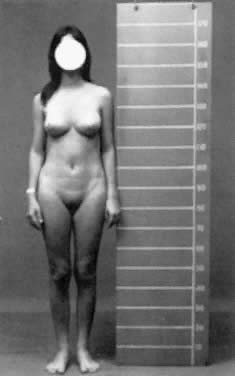
Fig. 6. Photograph of a patient with complete androgen insensitivity syndrome.(Courtesy Charles B. Hammond, Duke University. From Simpson JL: Disorders of Sexual Differentiation: Etiology and Clinical Delineation. New York, Academic Press, 1976)

Neither a uterus nor fallopian tubes are ordinarily present. Occasionally, fibromuscular remnants, rudimentary fallopian tubes, or rarely even a uterus are detected.31,32,33 The absence of müllerian derivatives is expected because anti müllerian hormone, which is secreted by the fetal Sertoli cells, is not an androgen; therefore, müllerian regression is expected to occur in males with androgen sensitivity, just as in normal males. The only other condition in which a uterus is absent in a phenotypic female is müllerian aplasia, which is readily distinguishable on the basis of pubic hair and a 46,XX complement.

In complete AIS, testes are usually normal in size and located in the abdomen, inguinal canal, or labia (i.e., anywhere along the path of embryonic testicular descent). If present in the inguinal canal, testes can produce inguinal hernias. It may therefore be worthwhile to determine cytogenetic status of prepubertal girls with inguinal hernias, although most will be 46,XX.

Height is slightly increased over that of normal women, but unremarkable compared with 46,XY males. Presumably the increased height reflects the influence of the Y chromosome. Consistent with this is the impression expressed by many clinicians that the hands and feet of these women are relatively large compared with those of normal women.

The frequency of gonadal neoplasia is increased, but probably less so than once believed. In 1953, Morris and Mahesh34 stated that 22% of affected patients had neoplasia. The actual risk is probably no greater than 5%.35,36 Most investigators now agree that the risk of neoplasia is low before 25 to 30 years of age. Benign tubular adenomas (Pick's adenomas) are especially common in postpubertal patients, probably as result of increased secretion of luteinizing hormone. The pathogenesis of complete AIS involves end-organ insensitivity to androgens.

HORMONE LEVELS.

Serum testosterone and dihydrotestosterone levels are normal or elevated in AIS. Luteinizing hormone and estrogen are elevated, suggesting abnormal gonadal-hypothalamic feedback. Consistent with this, Leydig cells are hyperplastic.18 Androgen binding is absent, decreased, or qualitatively abnormal. In a study of androgen binding of genital skin fibroblasts from 42 patients with complete AIS, 24 (57%) showed absent androgen binding, 15 (36%) decreased or qualitatively abnormal androgen binding, and 3 (7%) ostensibly normal androgen binding.8

GENETICS.

AIS is inherited as an X-linked recessive disorder, the result of a defect in the androgen-receptor gene. Chromosome analysis is 46,XY. 23 The androgen-receptor gene was cloned by several groups in 1988, and is 
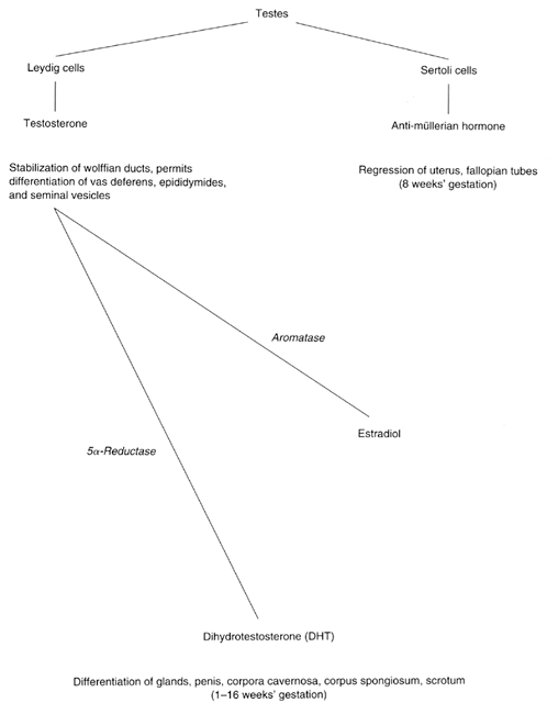
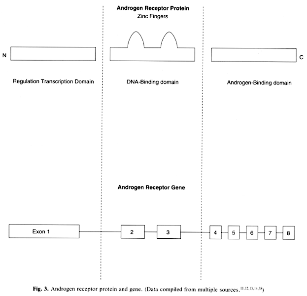
located on the X chromosome at Xq11–12.37 The gene is 90 kb in length and consists of eight exons that encode the three domains of the receptor (see Fig. 3).11,12,13,14,38 Exon 1 encodes the transcription regulation domain. This region includes polymorphic CAG repeats, which aid in restriction fragment length polymorphism (RFLP) diagnosis. Exons 2 and 3 encode the DNA-binding domains; exons 4 to 8 encode the androgen-binding domains. Many mutations involving the androgen-receptor gene have been identified.39,40,41,42,43 Like most genes, molecular heterogeneity exists among affected persons. Surprisingly, however, both receptor-positive and receptor-negative persons with complete AIS seem indistinguishable clinically.

Approximately 70% of cases studied have shown a mutation in the androgen-receptor gene. Deletions are rare, as are insertions.44 More commonly encountered are point mutations involving single nucleotide changes that result in either substitution of an unscheduled amino acid, deletion of three nucleotides with preservation of an open reading frame, or generation of an unscheduled stop codon to cause premature message termination of the message and production of a nonfunctional protein. Large deletions and mutations that result in premature termination (stop codon) yield no functional receptor and predictably cause complete AIS.45 Point mutations resulting from single nucleotide substitutions might be associated with the production of some androgen receptors. Sometimes the receptor may be unstable or characterized by poor binding.45

Clarification of the relationship between phenotype and specific androgen-receptor gene mutations is under way, with an emphasis on an understanding of the consequences of altered androgen-receptor kinetics on tertiary structure.46,47,48,49 Especially interesting are circumstances in which substitution resulting in one new amino acid produces complete AIS,43 but substitution of another produces only partial AIS. An example was reported by Kazemi-Esfarjani and associates48 in which valine-865 was substituted with either methionine or leucine, causing complete or partial AIS, respectively. Studies such as this have become increasingly important both academically and clinically because of the marked molecular heterogeneity of AIS, which has made it difficult to improve our diagnostic capability in sporadic cases. Of course various strategies (e.g., direct mutation detection, RFLP analysis) can still be devised to detect heterozygotes or hemizygotes once a mutant sequence is identified in a given family.49

DIAGNOSIS.

Patients with AIS have normal female external genitalia, feminize at puberty (develop breasts), and show primary amenorrhea. History may elicit prior inguinal hernias. Physical examination may reveal a shortened and blindly ending vagina, as well as an absent uterus and cervix (Fig. 7).50 An absent uterus and ovaries is found on ultrasound examination, and chromosome analyses reveal a 46,XY complement. Receptor studies or DNA studies are not routinely available except through laboratories involved 
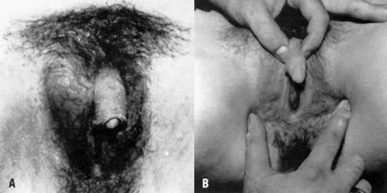
with AIS research, and thus are not considered obligatory.

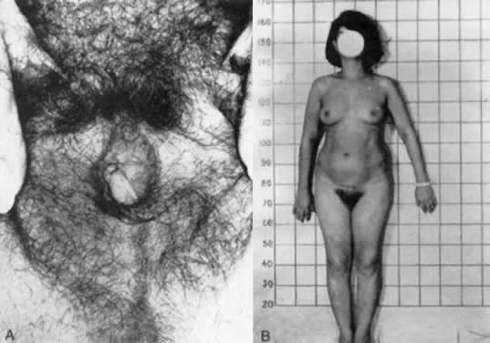
Fig. 7. A and B. Patient with partial androgen insensitivity syndrome.(Park IJ, Jones HW: Familial male hermaphroditism with ambiguous external genitalia. Am J Obstet Gynecol 108:1197, 1970)

Prenatal diagnosis for AIS can be achieved by the use of DNA obtained from chorionic villi sampling or amniocentesis. Direct mutation analysis or RFLP analysis can be used if the molecular defect is known.44

MANAGEMENT.

Treatment is straightforward. Affected persons are raised as female and act female. As previously mentioned, affected persons are at increased risk for gonadal neoplasia, and an orchiectomy is eventually needed. It is acceptable to leave the testes in situ until after pubertal feminization, but most surgeons would perform orchiectomies if herniorrhaphies prove necessary before puberty. There may also be psychologic benefit in prepubertal orchiectomies. Inguinal or intra-abdominal testes can sometimes be removed laparoscopically.51

After orchiectomy, estrogen replacement is necessary. Because these patients do not usually have a uterus, continuous therapy with conjugated estrogens or other estrogen forms may be prescribed. As mentioned above, the clinician must tailor the estrogen dosage to the individual patient, balancing its benefits against its side effects. Vaginoplasty is rarely necessary, but occasionally dilators may be required to increase vaginal length.

Partial Androgen Insensitivity Syndrome

Partial AIS is the result of a mutation of the same androgen-receptor gene as is involved in complete AIS.52,53 Persons with partial AIS (incomplete testicular feminization) feminize (i.e., exhibit breast development) despite having external genitalia characterized by phallic enlargement and partial labioscrotal fusion. Both partial and complete AIS share the following features:

  Bilateral testes with similar histologic findings
  No müllerian derivatives
  Pubertal breast development
  Lack of pubertal virilization
  Normal (male) plasma testosterone.

Partial AIS is an X-linked recessive condition that encompasses three entities that once were considered separate: Lubs' syndrome, Gilbert-Drefus syndrome, and Reifenstein syndrome. In fact, the partial AIS spectrum even extends to include some infertile but otherwise normal males. Traditionally, diagnosis of Reifenstein syndrome was applied to males whose phallic development was more nearly normal than that of males with traditionally termed incomplete androgen insensitivity. In the Reifenstein phenotype there was no vagina-like perineal orifice, testes were small,54 and decreased virilization was thought to be the result of inadequate testosterone secretion. The Lubs phenotype was considered intermediate between that of Reifenstein and that of traditionally termed incomplete androgen insensitivity.55

This complicated stratification later proved genetically incorrect. First, males with small testes and elevated gonadotropic levels sometimes were found to display signs of partial AIS.56 At around the same time, Wilson and co-workers57 observed the occurrence in a single kindred of the Reifenstein phenotype and that of partial AIS. Ten years later, Wilson and associates58 confirmed partial androgen-receptor deficiency in two persons with Lubs phenotype. Finally, males with Reifenstein syndrome and Lubs syndrome, and some infertile males, were observed in the same kindred.57 Thus, it was concluded that perturbation of a single gene is responsible for all three of these disorders.

The pathogenesis of partial AIS logically would appear to involve a decreased number of receptors or qualitative defects in the androgen receptors.44,45,46,57,58,59 Complete absence of receptors has been observed, but this is more likely to be associated with complete AIS. Surprisingly, poor correlation exists between receptor levels (or androgen-binding affinity) and the degree of masculinization, nor are precise correlations evident between a specific mutation and the phenotype. Irrespective, the clinical significance of partial AIS is that this disorder must be excluded before the decision is made to rear a child as a male. Presence of androgen receptors and demonstration of response to exogenous androgen is therefore necessary to exclude the diagnosis of partial androgen insensitivity.

GENETICS.

Like complete AIS, partial AIS is inherited in an X-linked recessive fashion. The chromosomal complement is 46,XY. As previously discussed, the androgen-receptor gene has been mapped to Xq11–12. Androgen binding studies of fibroblasts derived from the genital skin of patients with partial AIS tend to show decreased, qualitatively abnormal, or complete absence of androgen binding.10 Molecular analysis of the androgen-receptor gene has revealed several different mutations in partial AIS, predictably in the androgen-binding domains (exons 4 to 8) but sometimes in the DNA-binding domains (exons 2 and 3) (see Fig. 3).

Of note is that mutations in the androgen-receptor gene have specifically been detected in the Reifenstein phenotype,60 confirming molecularly that this phenotype is indeed the result of mutation in the androgen-receptor gene and that partial AIS is truly an X-linked recessive condition encompassing the entities historically considered separate, as reasoned above.

Complete deletions or point mutations resulting in premature (message) termination are more likely to cause complete AIS than partial AIS.44 Overall, however, a molecular defect in the androgen-receptor gene is found less often in partial AIS, suggesting that the defect may often involve a more distal step in androgen action. Lobaccaro and colleagues41 recently studied mutations in 25 patients with partial AIS and found that the mutations were probably the result of point mutations or microdeletions. In addition, they were able to identify carriers in 50% of 
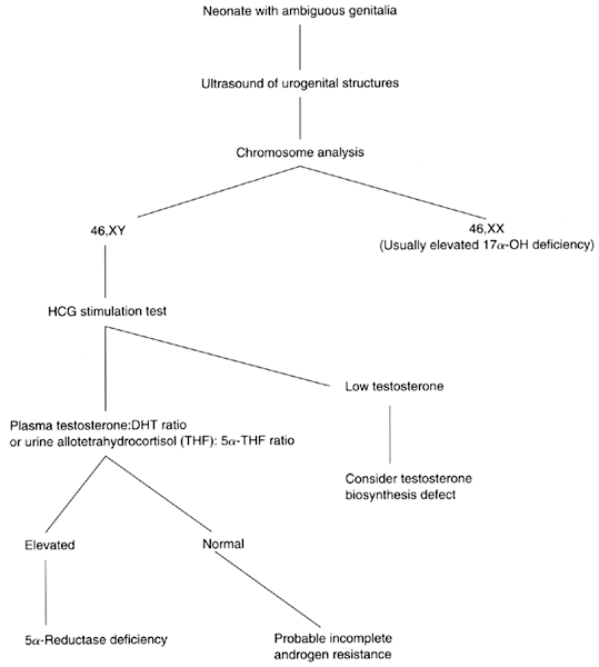
the families with the use of exon 1 Hind III polymorphisms.41,61

DIAGNOSIS.

Infants with partial AIS usually present with ambiguous genitalia. Adrenal 21-OH deficiency can be excluded readily by chromosome studies and 17α-OH progesterone levels.62 Ultrasound can aid in the evaluation of internal genitalia. Serum hormone levels are assessed after hCG is administered; the presence of normal testosterone excludes the presence of a defect in testosterone biosynthesis. An elevated T:DHT ratio indicates 5α-reductase deficiency. If a normal T:DHT ratio is present in an XY person with genital ambiguity, the clinician should consider the diagnosis of partial AIS (see Fig. 5). Prenatal diagnosis for complete AIS and partial AIS is now possible with molecular analyses of the androgen-receptor gene from trophoblastic or amnionic DNA, if the molecular defect is known. RFLP analyses can also be used, particularly those that utilize the polymorphic CAG repeats in exon 1.63

MANAGEMENT.

Treatment is difficult. If external genitalia are characterized by other than simple hypospadias, a female sex of rearing is preferable. These patients require orchiectomy and estrogen replacement (as with complete AIS patients). If no androgen receptors are present, one must assess the degree of androgen response in infants before considering male sex of rearing. Patients raised as males require androgen replacement, which may or may not be efficacious.64 Patients raised as males often require multiple urogenital reconstructive surgeries, sometimes still with poor results.64 Furthermore, little information is available on the long-term success of the corrective surgery and masculinization, sexual performance, and fertility of these patients.65 Overall, it is preferable for these patients to be raised as females.

Back to Top
REFERENCES

1. Simpson JL, Rebar RW: Normal and abnormal sexual differentiation and development. In Becker KL, Bilezikian JP, Bremner WJ et al (eds): Principles and Practice of Endocrinology and Metabolism, 2nd ed, pp 788–822. Philadelphia, JB Lippincott, 1995

2. Patsavoudi E, Magre S, Castinior M et al: Dissociation between testicular morphogenesis and functional differentiation of Leydig cells. J Endocrinol 105: 235, 1985

3. Magre S, Jost A: Dissociation between testicular morphogenesis and endocrine cytodifferentiation of Sertoli cells. Proc Natl Acad Sci USA 81: 7831, 1984

4. Moore KL, Persaud TVN: The Developing Human: Clinically Oriented Embryology, p 291. Philadelphia, WB Saunders, 1993

5. Imperato-McGinley I: 5α-Metabolism in finasteride-treated subjects and male pseudohermaphrodites with inherited 5α-reductase deficiency. Eur Urol 20 (suppl 2): 78, 1991

6. Imperato-McGinley I: 5α-Reductase deficiency: Human and animal models. Eur Urol 25 (suppl 1): 20, 1994

7. Wigley WC, Prihoha JS, Mowszowicz I et al: Natural mutagenesis: Study of the human steroid 5α-reductase 2 isozyme. Biochemistry 33: 1265, 1994

8. Wilson JD: Syndromes of androgen resistance. Biol Reprod 46: 168, 1992

9. Pinsky L, Kaufman M, Levitsky LL: Partial androgen resistance due to a distinctive qualitative defect of the androgen receptor. Am J Med Genet 27: 459, 1987

10. Griffin JE, McPhaul MJ, Russell DW, Wilson JD: The androgen resistance syndromes: 5α-Reductase 2 deficiency, testicular feminization, and related disorders. In: Scriver CR, Beaudet AL, Sly WS, Valle D (eds): The Metabolic and Molecular Bases of Inherited Disease, 7th ed, pp 2967–2991. New York, McGraw-Hill, 1994

11. Deslypere J-P, Young M, Wilson JD, McPhaul MJ: Testosterone and 5α-dihydrotestosterone interact differently with the androgen receptor to enhance transcription of the MMTV-CAT reporter gene. Mol Cell Endocrinol 88: 15, 1992

12. Lumbroso S, Lobaccaro JM, Belon C et al: A new mutation within the deoxyribonucleic acid-binding domain of the androgen receptor gene in a family with complete androgen insensitivity syndrome. Fertil Steril 60: 814, 1993

13. Kupfer SA, Quigley CA, French FS: Male pseudohermaphroditism. Semin Perinatol 16: 319, 1992

14. Batch JA, Williams DM, Davies HR et al: Role of the androgen receptor in male sexual differentiation. Horm Res 38: 226, 1992

15. Janne OA, Palvimo JJ, Kallio P et al: Androgen receptor and mechanism of androgen action. Ann Med 25: 83, 1993

16. Chong C, Kokontis J, Liao S: Structural analyses of cDNA and amino acid sequences of human and rat androgen receptor. Proc Natl Acad Sci USA 85: 7211, 1988

17. La Spada AR, Wilson EM, Lubahn DB et al: Androgen receptor gene mutations in X-linked spinal and bulbar muscular atrophy. Nature 352: 77, 1991

18. Simpson JL, Golbus MS: Genetics in Obstetrics and Gynecology, 2nd ed, pp 148–156. Philadelphia, WB Saunders, 1992

19. Opitz JM, Simpson JL, Sarto GE et al: Pseudovaginal perineoscrotal hypospadias. Clin Genet 3: 1, 1972

20. Simpson JL, New M, Peterson RE, German J: Pseudovaginal perineoscrotal hypospadias (PPSH) in sibs. Birth Defects 7 (6): 140, 1971

21. Imperto-McGinley J, Gurrero L, Gautier T, Peterson RE: Steroid 5α-reductase deficiency: An inherited form of male pseudohermaphroditism. Science 186: 1213, 1974

22. Akgun S, Ertel NH, Imperato-McGinley J et al: Familial male pseudohermaphroditism due to 5α-reductase. Am J Med 81: 267, 1974

23. McKusick VA: Mendelian Inheritance in Man, 10th ed, p 1160. Baltimore, The Johns Hopkins University Press, 1992

24. Wilson JD, Griffin JE, Russell DW: Steroid 5α-reductase 2 deficiency. Endocr Rev 14: 577, 1993

25. Thigpen AE, Davis DL, Milatovich A et al: Molecular genetics of steroid 5α-reductase 2 deficiency. J Clin Invest 90: 799, 1992

26. Labrie F, Sugimoto Y, Luu-The V et al: Structure of human type II 5α-reductase gene. Endocrinology 131: 1571, 1992

27. Andersson S, Berman DM, Jenkins EP et al: Deletion of steroid 5α-reductase 2 gene in male pseudohermaphroditism. Nature 354: 159, 1991

28. Imperato-McGinley J, Gautier T, Pichardo M, Shackleton C: The diagnosis of 5α-reductase deficiency in infancy. J Clin Endocrinol Metab 63: 1313, 1986

29. Odame I, Donaldson MDC, Wallace AM et al: Early diagnosis and management of 5α-reductase deficiency. Arch Dis Child 67: 720, 1992

30. Simpson JL: Disorders of Sexual Differentiation. New York, Academic Press, 1976

31. Ulloa-Aguirre A, Mendez JP, Angeles A et al: The presence of müllerian remnants in the complete androgen insensitivity syndromes: A steroid hormone-mediated defect? Fertil Steril 45: 302, 1986

32. Heller DS, Ranzini A, Futterweit et al: Müllerian remnants in complete androgen insensitivity syndrome. Int J Fertil 37: 283, 1992

33. Swanson ML, Coronel EH: Complete androgen insensitivity with persistent müllerian structures: A case report. J Reprod Med 38: 565, 1993

34. Morris JM, Mahesh VB: Further observations on the syndrome “testicular feminization.” Am J Obstet Gynecol 87: 731, 1953

35. Simpson JL, Photopulos G: The relationship of neoplasia to disorders of abnormal sexual differentiation. Birth Defects 12: 15, 1976

36. Verp MS, Simpson JL: Abnormal sexual differentiation and neoplasia. Cancer Genet Cytogenet 25: 191, 1987

37. Brown C, Gross S, Lubahn D et al: Androgen receptor locus on the human X chromosome: Regional localization to Xq11–12 and description of a DNA polymorphism. Am J Hum Genet 44: 264, 1989

38. Sultan C, Lobaccaro JM, Belon C et al: Molecular biology of disorders of sex differentiation. Horm Res 38: 105, 1992

39. Yong EL, Chua KL, Yang M et al: Complete androgen insensitivity due to a splice-site mutation in the androgen receptor gene and genetic screening with single-stranded conformation polymorphism. Fertil Steril 61: 856, 1994

40. Beitel LK, Paior L, Vasiliou DM et al: Complete androgen insensitivity due to mutations in the probable a-helical segments of the DNA-binding domain in the human androgen receptor. Hum Mol Genet 3: 21, 1994

41. Lobaccaro JM, Belon C, Chaussain JL et al: Molecular analysis of the androgen receptor gene in 52 patients with complete or partial androgen insensitivity syndrome: A collaborative study. Horm Res 37: 54, 1992

42. Quigley CA, Friedman KJ, Johnson A et al: Complete deletion of the androgen receptor gene: Definition of the null phenotype of the androgen insensitivity syndrome and determination of carrier. J Clin Endocrinol Metab 74: 927, 1992

43. Shkolny DL, Brown TR, Punnett HH et al: Characterization of alternative androgen receptor that cause complete androgen insensitivity in three families. Hum Mol Genet 4: 515, 1995

44. McPhaul MJ, Marcelli M, Aoppi S et al: Genetic basis of endocrine disease 4: The spectrum of mutations in the androgen receptor gene that causes androgen resistance. J Clin Endocrinol Metab 76: 17, 1993

45. Zoppi S, Wilson CM, Harbison MD et al: Complete testicular feminization caused an amino-terminal truncation of the androgen receptor with downstream initiation. J Clin Invest 91: 105, 1993

46. Sultan C, Lumbroso S, Poujol N et al: Mutations of androgen receptor gene in androgen insensitivity syndromes. J Steroid Biochem Mol Biol 46: 519, 1993

47. Adeyemo O, Kallio PJ, Palvimo JJ et al: A single-base substitution in exon 6 of the androgen receptor gene causing complete androgen insensitivity: The mutated receptor fails to transactivate but binds to DNA in vitro. Hum Mol Genet 2: 1809, 1993

48. Kazemi-Esfarjani P, Beitel LK, Trifiro M et al: Substitution of valine-865 by methionine or leucine in the human androgen receptor causes complete or partial androgen insensitivity, respectively with distinct androgen receptor phenotypes. Mol Endocrinol 7: 37, 1993

49. Trifiro M, Prior RL, Sabbaghian N et al: Amber mutation creates a diagnostic MaeI site in the androgen receptor gene of a family with complete androgen insensitivity. Am J Med Genet 40: 493, 1991

50. Park IF, Jones HW: Familial male hermaphroditism with ambiguous genitalia. Am J Obstet Gynecol 108: 1197, 1970

51. Lee RM, Peterson CM, Kreger DO et al: Digital blunt dissection technique to assist laparoscopic gonadectomy in inguinally located/adhered gonads. J Laparoendosc Surg 3: 229, 1993

52. DeBells A, Quigley CA, Marschke KB et al: Characterization of mutant androgen receptors causing partial androgen insensitivity syndrome. J Clin Endocrinol 78: 513, 1994

53. Lumbroso S, Lobaccaro JM, Belon C et al: Molecular prenatal exclusion of familial partial androgen insensitivity (Reifenstein syndrome). Eur J Endocrinol 130: 327, 1994

54. Reifenstein EC: Hereditary familial hypogonadism. Clin Res 3: 86, 1947

55. Lubs HA Jr, Vilar O, Bergenstal DM: Familial male pseudohermaphroditism of labial testes and partial feminization: Endocrine studies and genetic aspects. J Clin Endocrinol Metab 19: 1110, 1959

56. Amrhein JA, Klingsmith GJ, Walsh PC et al: Partial androgen insensitivity: The Reifenstein syndrome revisited. N Engl J Med 297: 350, 1977

57. Wilson JD, Harrod MJ, Goldstein JL et al: Familial incomplete male pseudohermaphroditism, type I. N Engl J Med 290: 1097, 1974

58. Wilson JD, Carlson BR, Weaver DD et al: Endocrine and genetic characterization of cousins with male pseudohermaphroditism: Evidence that the Lubs phenotype can result from a mutation that alters the structure of the androgen receptor. Clin Genet 26: 363, 1984

59. Sulton C: Androgen receptors and partial androgen insensitivity in male pseudohermaphroditism. Ann Genet 29: 5, 1986

60. Ris-Stalpers C, Verleun-Mooijman MC, de-Blaeij TJ et al: Differential splicing of human androgen receptor pre-mRNA in X-linked Reifenstein syndrome, because of a deletion involving a putative branch site. Am J Genet 54: 609, 1994

61. Lobaccaro J-M, Belon C, Lumbroso S et al: Molecular prenatal diagnosis of partial androgen insensitivity syndrome based on the Hind III polymorphism of the androgen receptor gene. Clin Endocrinol 40: 297, 1994

62. Hughes IA, Williams DM, Batch JA et al: Male pseudohermaphroditism: Clinical management, diagnosis and treatment. Horm Res 38 (suppl 2): 77, 1992

63. Lobaccaro JM, Belon C, Chaussain JL et al: Molecular analysis of the androgen receptor gene in 52 patients with complete or partial androgen insensitivity syndrome: A collaborative study. Horm Res 37: 54, 1992

64. Williams DM, Patterson MN, Hughes IA: Androgen insensitivity syndrome. Arch Dis Child 68: 343, 1993

65. Shah R, Wooley MM, Costin G: Testicular feminization: The androgen insensitivity syndrome. J Pediatr Surg 27: 757, 1992

Back to Top