Chapter 39
Surgical Procedures for Tubal Sterilization
John J. Sciarra
Main Menu   Table Of Contents

Search

John J. Sciarra, MD, PhD
Thomas J. Watkins Professor and Chairman, Department of Obstetrics and Gynecology, Northwestern University Medical School; Chairman, Department of Obstetrics and Gynecology, Prentice Women's Hospital and Maternity Center, Chicago, Illinois (Vol 1, Chap 36; Vol 6, Chap 39)

INTRODUCTION
PATIENT SELECTION AND COUNSELING
TYPE OF INCISION: MINILAPAROTOMY OR COLPOTOMY
SURGICAL PROCEDURES
THE POMEROY TECHNIQUE
THE MADLENER TECHNIQUE
THE IRVING PROCEDURE
THE OXFORD TECHNIQUE
THE UCHIDA TECHNIQUE
THE KROENER FIMBRIECTOMY
THE ALDRIDGE PROCEDURE
MECHANICAL OCCLUSIVE DEVICES
SHORT-TERM AND LONG-TERM COMPLICATIONS
REVERSIBILITY
REFERENCES

INTRODUCTION

Tubal sterilization is a relatively simple, very safe, and extremely effective method for preventing future pregnancy. The procedure may be performed on an outpatient or inpatient basis, by the abdominal or vaginal route, using local or general anesthesia, and in the postpartum or postabortal period or as an interval operation. The choice of a specific procedure, the surgical approach, and the type of anesthesia depends on the facilities available and the background and experience of the surgeon.

Back to Top
PATIENT SELECTION AND COUNSELING

Because most methods of tubal sterilization are designed to be permanent, proper patient counseling and informed consent is of paramount importance preoperatively in preparing the patient for a sterilization procedure. There must be no contraindications to elective surgery. The decision for sterilization should be made on an entirely voluntary basis following appropriate discussion regarding risks, benefits, and alternatives.

Various materials are available to assist the physician in patient counseling and in obtaining informed consent. One such pamphlet, which is widely distributed by the American College of Obstetricians and Gynecologists, includes a description of several different techniques of tubal sterilization, as well as a discussion of alternative methods of contraception.1 In discussing tubal sterilization, it is appropriate to discuss with the patient the benefits of this procedure, as well as some of the potential risks. The benefits are substantial. All methods of tubal sterilization are extremely effective with a low failure rate. Tubal sterilization involves a one-time surgical procedure that is immediately effective and does not require the continued motivation of other contraceptive methods. The failure rate of a properly performed sterilization operation is less than 1% and there are no consistent differences in efficacy documented among the standard techniques used today. If the procedure is performed on an ambulatory or a short-stay hospitalization basis and either minilaparotomy or colpotomy is used, the operation is both practical and cost-effective. Finally, the secondary effects are few.

The risks associated with voluntary sterilization, although present, are minimal. Female sterilization is more complex than male sterilization and often is performed under general anesthesia. Most complications occur secondary to general anesthesia. Thus, the risks of anesthesia, the general risks of elective surgery, and the possibility of failure, with subsequent intrauterine or ectopic pregnancy, should be explained to the patient preoperatively.2

Back to Top
TYPE OF INCISION: MINILAPAROTOMY OR COLPOTOMY

Elective tubal sterilization may be performed by the abdominal or the vaginal route. In either case, the usual preoperative preparation of the abdomen or vagina should be employed.

For minilaparotomy, either a vertical or a transverse incision is satisfactory, and both incisions provide easy access to the fallopian tubes. In many instances, a transverse incision made approximately 3 cm above the symphysis and approximately 6 cm in length is satisfactory. The fascia is divided transversely, the rectus muscles are retracted laterally, and the transversalis fascia and the underlying parietal peritoneum is incised in a vertical fashion. To avoid bladder injury, the bladder must be adequately emptied immediately before surgery. A device to elevate the uterus to the level of the anterior abdominal wall is sometimes helpful and allows the incision to be kept to a minimum. Several different types of uterine elevators are available, and all are satisfactory. Following the sterilization procedure, the incision is closed in the customary fashion. A subcuticular absorbable suture is often useful and has excellent patient acceptance.3,4

Although less common today than minilaparotomy, colpotomy is still a choice in selected patients. In this technique, the peritoneal cavity is entered through an incision made in the posterior vaginal fornix between the uterosacral ligaments. The incision may be either transverse or vertical and is made directly through the vagina into the cul-de-sac. The oviducts are then drawn into the surgical field and the selected sterilization procedure is performed. In some instances, the mobility of the fallopian tubes is limited by the pelvic anatomy and the size of the colpotomy incision, and these factors may limit the choice of surgical procedures. The colpotomy incision is usually closed in one layer with absorbable suture material. Since infection is a concern following colpotomy, the use of prophylactic antibiotics should be considered to minimize this risk.5

Back to Top
SURGICAL PROCEDURES

Tubal sterilization has been performed for over 100 years. A review of the historic milestones and of the most significant operative techniques in this field is presented in Table 1. Many modifications of the following techniques have periodically been introduced and all seem to be effective as long as the basic principles are followed.

TABLE 1. Selected Chronology of Tubal Sterilization


Year

Scientist

Event

1834

Blundell

First recommendation in the United States for incision and removal of a portion of fallopian tube


 for sterilization

1881

Lundgren

First report of tubal sterilization by simple ligation

1910

Madlener

Technique for crushing and ligation of fallopian tube; 89 procedures, 3 deaths, 0 pregnancies by


 1919

1924

Irving

Procedure of ligation, division, and burial of proximal stump in myometrium; modified technique


 described; 814 procedures, 0 failures by 1950

1930

Bishop & Nelms

Procedure for ligation and resection devised by their late associate, Pomeroy; 60 sterilizations

1934

Aldridge

Technique for temporary sterilization; 1 successful reversal and pregnancy

1935

Kroener

Fimbriectomy procedure; 200 fimbriectomies, 0 failures by 1969

1946

Uchida

Technique for tubal ligation, resection, and burial; 5000 sterilizations, 0 failures by 1961; 20,000


 sterilizations, 0 failures by 1975

Back to Top
THE POMEROY TECHNIQUE

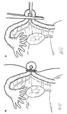
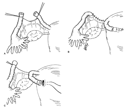
The Pomeroy technique for bilateral partial salpingectomy is the preferred method for interval surgical female sterilization (Fig. 1). The popularity of this technique is based on its inherent simplicity and its long-established efficacy. Following accurate identification of the fallopian tube, a Babcock clamp is placed around the proximal portion of the tubal ampulla and the tube is elevated to reveal the vascular supply of the mesosalpinx (see Fig. 1A). A single strand of absorbable suture material (1-0 plain catgut) is placed around the elevated loop of tube and firmly tied. The fallopian tube is thus ligated and the blood supply is occluded simultaneously (see Fig. 1B). A hemostat may now be placed on the suture strands immediately distal to the knot, and the excess suture may be excised. The hemostat now becomes a useful holder for the next step in the procedure. At this point, a second tie of the same suture material may be applied at the discretion of the surgeon, but this is not usually necessary. While gentle traction is maintained on the elevated section of tube, the open blade of the Metzenbaum scissors is used to pierce the mesosalpinx and approximately 1 cm of tube is excised (see Fig. 1C). The excised tube should be appropriately labeled and sent to the pathology laboratory for documentation. With the contraction of the muscularis, the white avascular endosalpinx appears as an elevated area in the center of each cut segment. The proximal and distal ends of the divided and ligated oviduct are now examined for bleeding and then the tube is returned to the abdominal cavity and the procedure is repeated on the opposite tube.

Fig. 1. Pomeroy technique. A. A loop of the proximal portion of the tubal ampulla is elevated to reveal the vascular supply of the mesosalpinx. B. A strand of absorbable suture material is used to bilaterally ligate the tube and simultaneously provide hemostasis. C. A hemostat is placed on the suture strands immediately distal to the knot to prevent the tube from retracting into the abdomen. The open blade of the Metzenbaum scissors is used to pierce the mesosalpinx and approximately 1 cm of the tube is excised. D. The end result following dissolution of the absorbable suture material with return of the proximal and distal portions of the tube to their normal anatomic position.

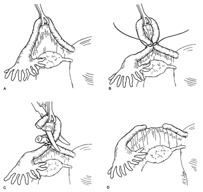
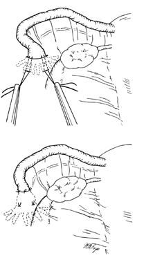
The end result following dissolution of the absorbable suture material and return of the proximal and distal portions of the tube to their normal anatomic positions is shown in Figure 1D. The use of absorbable suture material allows this separation to occur and is probably a critical factor in the development of the anatomic discontinuity. This factor is undoubtedly related to the low failure rate reported for this procedure. Accordingly, the newer synthetic absorbable suture materials with longer dissolution times are probably less desirable than simple plain catgut.

The major advantages of the Pomeroy technique are that it is easily taught, is simple to perform, and is highly effective. Its acceptance for both puerperal and interval sterilization is quite high. It can be performed either abdominally or vaginally, and the complications are minimal. It has no major disadvantages. The reported pregnancy rate is two to four pregnancies per 1000 procedures.6

Back to Top
THE MADLENER TECHNIQUE

The Madlener technique is less commonly performed than the Pomeroy technique (Fig. 2). A loop of tube is elevated and crushed before ligation. The original Madlener procedure used nonabsorbable suture material. This factor may have been responsible for failures due to recanalization and tuboperitoneal fistula formation. While no tube is excised, the ligated segment undergoes aseptic necrosis. The end result is similar to the laparoscopic sterilization procedure employing a Silastic band for occlusion as described elsewhere in this text.

Fig. 2. Madlener technique. A. A loop of the ampullary portion of the tube is elevated and then both segments are crushed with a hemostat. B. A strand of nonabsorbable suture material is used to ligate the tube over the crushed area. No tissue is excised. The devascularized loop of tube undergoes aseptic necrosis.

Back to Top
THE IRVING PROCEDURE

The Irving procedure was introduced as a technique for ligation and division of the oviduct at the time of cesarean section (Fig. 3). This technique was developed due to the perceived higher failure rates for traditional tubal sterilization when performed at cesarean section, possibly caused by increased hypertrophy and vascularity of the oviducts in the immediate postpartum period. In the technique as originally described, the tube is divided at approximately the ampullary-isthmic junction. With the ends of the suture left long, the proximal tube is buried within the substance of the myometrium on the anterior uterine surface just above the insertion of the round ligament. However, the tube may be buried posteriorly if this is more convenient (see Fig. 3B). The end of the distal portion of the tube is buried between leaves of the broad ligament. When the procedure is performed at the time of cesarean section, as the uterus undergoes involution, the buried proximal ends of the tubes become more and more compressed and eventually become obliterated. The Irving procedure is not recommended as an interval procedure, although when performed in the puerperal period, it is highly effective and has a low failure rate.6

Fig. 3. Irving procedure. A. The tube is divided in the region of the ampullary-isthmic junction, and the ends of the suture are kept long for traction and for use in the subsequent steps of the procedure. B. Using blunt dissection, a tunnel is made within the substance of the uterine myometrium and the proximal tube is pulled into this chamber and sutured in place. C. The distal tube is then buried within the substance of the broad ligament. Additional sutures may be necessary to close the defect within the mesosalpinx and adjacent broad ligament as a result of the previous dissection.

Back to Top
THE OXFORD TECHNIQUE

A somewhat similar procedure that can be applied in the interval situation is the Oxford technique as described by Williams.7 The oviduct is divided, as in the Irving procedure, and a segment of the isthmus is removed. The proximal end of the oviduct is carried under the round ligament and tied in this location. The distal tube is then tied on the opposite side of the round ligament. This procedure provides for appropriate tubal discontinuity with minimal destruction of tissue.

Back to Top
THE UCHIDA TECHNIQUE

The Uchida technique for tubal sterilization is shown in Figure 4. A saline-epinephrine solution is injected into the subserosal area of the tube, causing the muscular tube to separate from the serosa. The ballooned serosa is incised, and the muscular tube is withdrawn. A 5-cm section of the tube is then excised and the proximal end ligated. A purse-string suture is applied. The procedure may be extended so as to include fimbriectomy and removal of the isthmus and ampulla with another suture placed around the mesosalpinx.

Fig. 4. Uchida technique. A. A subserosal injection of a saline-epinephrine solution is made in the region of the tubal ampulla. B. The serosa is then incised with the scissors, exposing the muscular layer of the tube. A segment of the muscular layer is elevated while the serosa is simultaneously stripped back over the proximal and distal segments. C. The proximal portion of the muscular tube is ligated and excised. The proximal ligated segment then drops back beneath the serosa. D. A purse-string suture is placed around the distal tube and tied. Additional sutures may be needed to close the defect in the mesosalpinx and adjacent broad ligament involved in the earlier dissection.

The Uchida technique is more complex than the other procedures. Nevertheless, the technique is associated with relatively few failures.

Back to Top
THE KROENER FIMBRIECTOMY

The technique of fimbriectomy as described by Kroener employs the ligation of the distal ampulla of the tube with two permanent sutures and then division and removal of the infundibulum of the tube (Fig. 5). Ligation and hemostasis are accomplished simultaneously. The simplicity with which this excisional procedure is performed on the distal portion of the tube accounted for its early popularity, especially when the sterilization was being performed through a colpotomy incision. Presently, however, fimbriectomy does not appear to have many advocates.

Fig. 5. Kroener fimbriectomy. Top. A suture is anchored in the mesosalpinx and placed around the tube in the distal ampulla. A second suture may be placed adjacent to the first, and the infundibulum of the tube is excised. Bottom. The tube as it appears following excision of the distal segment.

Back to Top
THE ALDRIDGE PROCEDURE

The Aldridge procedure is of interest because the fallopian tube remains intact (Fig. 6). The fimbrial end of the fallopian tube is drawn into a pocket beneath the peritoneum of the broad ligament. The buried fimbrial end is then secured in place by several sutures of nonabsorbable suture material. This circle of sutures incorporates the serosal and muscular layers of the tube in the peritoneum of the broad ligament. Following the introduction of this procedure, numerous failures were reported and the procedure has not been popular in recent years.

Fig. 6. Aldridge procedure. Top. By blunt dissection, a pocket is developed within the substance of the broad ligament. Traction sutures are placed within the muscular layer of the distal tube and are used to draw the infundibulum into the peritoneal pocket. Bottom. Several sutures of nonabsorbable suture material are then used to anchor the infundibulum of the tube into the new anatomic subperitoneal location. Care must be taken to assure that the entire fimbriated portion of the tube is firmly held beneath the peritoneum.

Back to Top
MECHANICAL OCCLUSIVE DEVICES

In addition to the above surgical procedures for tubal occlusion, both Silastic bands and spring-loaded clips may be employed by either a minilaparotomy or a colpotomy incision. These mechanical occlusive devices, when used directly, are applied by a method that is similar to the laparoscopic application discussed in the chapter on laparoscopic sterilization in this text. There appears to be little advantage to the use of mechanical occlusive devices at the time of either minilaparotomy or colpotomy. Such devices are preferentially best reserved for endoscopic application.

Back to Top
SHORT-TERM AND LONG-TERM COMPLICATIONS

Immediate surgical complications of elective interval tubal sterilization procedures include both hemorrhage and infection. Incisional or vaginal bleeding is usually easily controlled with either pressure or additional sutures. The rare instance of intra-abdominal bleeding may necessitate a repeat laparotomy. Superficial or deep infection may require antibiotic therapy. The use of prophylactic antibiotics preoperatively, before colpotomy, should be considered.

All tubal sterilization procedures presently employed are effective but pregnancies do occur. The failure rate is in the range of two to four pregnancies per 1000 operations when follow-up is for a sufficient period. In many reported series, luteal phase pregnancies have occurred, indicating the patient was already pregnant when the procedure was performed. This can be minimized by scheduling the procedure during the proliferative phase of the menstrual cycle or by testing for pregnancy before surgery. A recent report of pregnancies that occurred following sterilization indicated that operator error was the main reason for sterilization failures.8 The literature indicates that sterilization procedures have a biologic failure rate. Intrauterine pregnancies tend to occur early, often during the first year or two following the sterilization procedure. Later pregnancies have a higher chance of presenting as tubal ectopic pregnancies. One report indicates that approximately 7% of total pregnancies following tubal sterilization are ectopic.9 Thus, it is apparent that careful follow-up is mandatory following female sterilization. Unexplained pelvic pain in a sterilized patient should suggest the possibility of ectopic pregnancy.

A recent control study of over 1000 women indicated that tubal sterilization does not affect most menstrual parameters. Menstrual cycles, duration of menstrual flow, and bleeding between periods were unchanged in the sterilization and comparison groups. However, noncyclic pelvic pain increased to a moderate but insignificant degree.10,11 Subsequent epidemiologic investigations have not found “post-tubal ligation syndrome” to be a serious problem.12

Accordingly, the weight of the evidence at the present time indicates that tubal sterilization does not significantly increase the risk to the patient for the development of subsequent gynecologic abnormalities.

Back to Top
REVERSIBILITY

All of the procedures described in this chapter are designed as permanent procedures to be offered to patients seeking permanent sterilization. Nevertheless, there are occasions when due to a change in personal life or social situation the patient may request reversal of a tubal sterilization procedure. In general, the potential for reversal is directly proportional to the amount of normal tube remaining. Thus, the amount of tube either removed or destroyed at the original procedure will, in large part, determine the success of a reversal operation, should one be attempted. This fact, at times, may be a consideration for the surgeon in the choice of a surgical procedure. The use of microsurgery for the reversal of tubal sterilization is presented elsewhere in this volume.

Several techniques of female sterilization are summarized in Table 2, with regard to the degree of tubal destruction, the failure rate (pregnancy), and the potential for reversal.

Table 2. Techniques of Female Sterilization


Tubal

Failure and/or

Reversal

Technique

Popularity*

Destruction

Pregnancy Rate

Potential

Uchida

1+

50%

Rare

Very poor

Fimbriectomy

1+ to 2+

40%


Poor

Irving

1+

30%


Poor

Pomeroy

5+

3–4 cm

2–4 : 1000 women

Good

Aldridge

Rarely done

None

Significant

Excellent


*Arbitrary scale of 1 (least popular procedure) to 5 (most common procedure).
(Sciarra JJ: Survey of tubal sterilization procedures. In Sciarra JJ, Zatuchni GI, Speidel JJ [eds]: Reversal of Sterilization, p 129. Hagerstown, MD, Harper & Row, 1978)
Back to Top
REFERENCES

1. Sterilization for Women and Men. Washington, DC, The American College of Obstetricians and Gynecologists, Publication No. APO11, April 1991

2. Rioux JE: Sterilization of women: benefits vs. risks. Int J Gynaecol Obstet 16: 488, 1979

3. Penfield AJ: Minilaparotomy for female sterilization. Obstet Gynecol 54: 184, 1979

4. Lee RB, Boyd JAK: Minilaparotomy under local anesthesia for outpatient sterilization: a preliminary report. Fertil Steril 33: 129, 1980

5. Miesfeld RR, Giarrantano RC, Moyers TG: Vaginal tubal ligation—is infection a significant risk? Am J Obstet Gynecol 137: 183, 1980

6. Sciarra JJ: Survey of tubal sterilization procedures. In Sciarra JJ, Zatuchni GI, Speidel JJ (eds): Reversal of Sterilization, p 129. Hagerstown, MD, Harper & Row, 1978

7. Williams EA: Results of reversal of female sterilization. In Brosens I, Winston R (eds): Reversibility of Female Sterilization. New York, Grune & Stratton, 1978

8. Chi I-C, Gardner SD, Laufe LE: The history of pregnancies that occur following female sterilization. Int J Gynaecol Obstet 17: 265, 1979

9. Wolf FC, Thompson NJ: Female sterilization and subsequent ectopic pregnancy. Obstet Gynecol 55: 17, 1980

10. Rulin MC, Davidson AR, Philliber SG et al: Changes in menstrual symptoms among sterilized and comparison women: a prospective study. Obstet Gynecol 74: 149, 1989

11. Wilcox LS, Martinez-Schnell B, Peterson HB et al: Menstrual function after tubal sterilization. Am J Epidemiol 135 (12): 1368, 1992

12. The American College of Obstetricians and Gynecologists Technical Bulletin, Number 113, February 1988

Back to Top