
This chapter should be cited as follows:
Teodorico E, Ciancia M, et al., Glob Libr Women's Med
ISSN: 1756-2228; DOI 10.3843/GLOWM.419723
The Continuous Textbook of Women’s Medicine Series – Gynecology Module
Volume 10
Ultrasound in gynecology
Volume Editors:
Professor Antonia Testa, Agostino Gemelli University Hospital, Rome, Italy
Professor Simona Maria Fragomeni, Agostino Gemelli University Hospital, Rome, Italy

Chapter
Ultrasound of the Uterine Cervix: From Anatomy to Clinical Application
First published: July 2026
Study Assessment Option
By answering four multiple-choice questions (randomly selected) after studying this chapter, readers can qualify for Continuing Professional Development points plus a Study Completion Certificate from GLOWM.
See end of chapter for details.

INTRODUCTION
Cervical cancer remains a major global health challenge and is currently the fourth most common malignancy among women worldwide, with an estimated 604,127 new cases and 341,831 deaths reported in 2020.1 Despite advances in prevention strategies, including human papillomavirus (HPV) vaccination and screening programs, the disease continues to affect women, above all in low- and middle-income countries (LMICs), in which the majority of cases and deaths occur.
Accurate staging is essential for determining prognosis and selecting the most appropriate therapeutic strategy in patients with cervical cancer. Traditionally, staging has relied on clinical examination combined with imaging.2 Among imaging modalities, magnetic resonance imaging (MRI) is widely regarded as the reference standard for local staging because of its high accuracy in assessing tumor size, stromal infiltration, parametrial extension and adjacent organ involvement. However, growing evidence and recent international guidelines suggest that endocavitary ultrasound, when performed by experienced operators, may be a reliable alternative to MRI for preoperative assessment of cervical cancer.3,4,5,6,7
The role of ultrasound becomes particularly relevant in LMICs, in which access to advanced imaging technologies such as MRI and positron emission tomography (PET) may be limited or unavailable. In these settings, ultrasound often represents the most accessible and cost-effective imaging modality for the initial evaluation of patients with cervical cancer, contributing substantially to disease staging and treatment planning. Beyond staging, ultrasound plays a pivotal role in identifying candidates for fertility-sparing treatment in those with early-stage cervical cancer (FIGO stages IA1–IB1 and selected IB2 cases) who wish to preserve fertility, with defined imaging criteria guiding conservative surgical management.8 Conversely, when the tumor appears locally advanced (FIGO stages IB3–IVB), treatment should be systemic rather than surgical (radiochemotherapy or chemotherapy).
Given the critical importance of precise disease assessment in guiding treatment decisions, ultrasound has emerged as a valuable tool in the diagnostic workup of cervical cancer, enabling accurate assessment of primary tumor characteristics, local extent of disease, nodal involvement and abdominal metastases.
DIAGNOSTIC PERFORMANCE OF IMAGING IN CERVICAL CANCER
Several studies have compared ultrasound to other imaging techniques for assessment of cervical cancer. Regarding ultrasound performance, Testa et al.9 reported a sensitivity of 93% for detecting cervical cancer, compared with 88% for MRI. Fischerova et al.5 reported that ultrasound performed by an expert ultrasonographer had higher accuracy than MRI for tumor detection (94% vs 83%). Concerning parametrial involvement, several studies have shown that ultrasound may achieve comparable diagnostic accuracy to that of MRI.5,6,10,11
A literature-based overview of the diagnostic performance of ultrasound and MRI in cervical cancer, including accuracy, sensitivity and specificity, is presented in Table 1.
Regarding lymph node metastases in cervical cancer, a recent multicenter study by Frühauf et al.12 showed that ultrasound was non-inferior to PET/CT and DW-MRI in overall diagnostic accuracy, with comparable sensitivity, thereby supporting ultrasound as a widely accessible and reliable option for preoperative pelvic lymph node assessment in cervical cancer.
Tian et al.13 reported that transvaginal ultrasound showed high specificity for detecting lymph node metastases (95%) but relatively low sensitivity (52%). However, no imaging modality is sufficiently accurate to exclude reliably small-volume metastases in para-aortic lymph nodes. Therefore, in patients with negative para-aortic lymph nodes on CT or PET-CT, surgical-pathological staging with para-aortic lymph node dissection may be considered. Conversely, to reduce the risk of false-positive imaging findings and avoid inappropriate treatment, ultrasound- or CT-guided Tru-Cut biopsy of any equivocal extrauterine lesion is recommended.4
1
Overview of the diagnostic performance of ultrasound and MRI in cervical cancer, including accuracy, sensitivity and specificity.
Author, year (number of patients) | Ultrasound | MRI | ||||||
Tumor detection | Parametrial involvement | Lymph node metastases | Tumor size | Tumor detection | Parametrial involvement | Lymph node metastases | Tumor size | |
Fischerova, 2008 (n = 95)5 | Accuracy 94% | Accuracy 99% | Accuracy 83% | Accuracy 95% | ||||
Testa, 2009 (n = 68)9 | Sens 93% | Sens 88% | ||||||
Kim, 2011 (n = 200)14 | Accuracy 79% | |||||||
Epstein, 2013 (n = 182)10 | Sens 90% | Sens 77% | Sens 67% | Sens 69% | ||||
Klerkx, 2012 (n = 68)15 | Sens 33% | |||||||
Fischerova, 2014 (n.a.)16 | Sens 40% | |||||||
Palsdottir, 2015 (n = 104)17 | Sens 43% | |||||||
Moloney, 2016 (n = 46)6 | Accuracy 89% | Accuracy 79% | ||||||
Yang, 2017 (n = 203)18 | Accuracy 77% | |||||||
Alcazar, 2020 (n.a.) 19 | Sens 78% | Sens 68% | ||||||
Stukan, 2021 (n = 50)20 | Accuracy 94% | Prediction of tumor stage based on size: accuracy 69% | Accuracy 76% | Prediction of tumor stage based on size: accuracy 42% | ||||
Tian, 2022 (n = 760)13 | Sens 62% | Sens 52% | ||||||
Imai, 2023 (n = 98)21 | Sens 92% | |||||||
Bizzarri, 2023 (n = 164)22 | Accuracy 79% | |||||||
Frühauf, 2026 (n = 141)12 | Sens 79.3% Spec 87.9% Accuracy 85.8% | Sens 70.8% Spec 90.5% Accuracy 86.1% | ||||||
n.a., not applicable as literature/systematic review; Sens, sensitivity; Spec, specificity.
ULTRASOUND ASSESSMENT OF THE CERVIX
Detailed images of the cervix can be obtained on transrectal5 and transvaginal3 ultrasound. The transrectal approach provides a better acoustic window for visualizing the distal part of the cervix,23 vaginal walls and bladder, and avoids vaginal bleeding associated with interaction between probe and lesion.
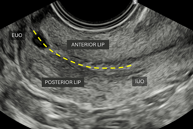
During the examination, the endocavitary probe should be gently introduced into the vaginal or rectal canal and positioned perpendicular to the cervix. This orientation allows clear identification of the anterior lip of the cervix, the cervical canal and the posterior lip (Figure 1).
1
Longitudinal transvaginal ultrasound scan of the cervix, showing cervical length measurement from the external uterine os (EUO) to the internal uterine os (IUO).
Detection and characterization of cervical cancer
The combination of transvaginal/transrectal and transabdominal ultrasound allows a complete assessment of the abdomen and pelvis for abdominal staging.23
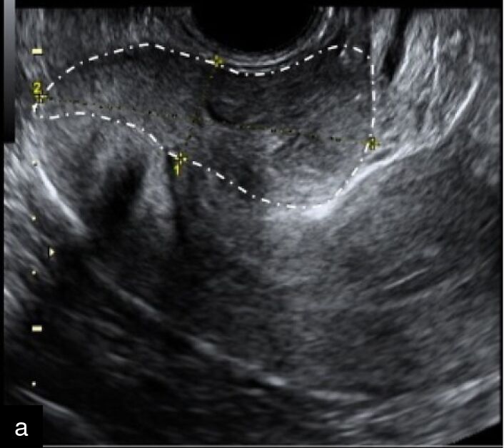
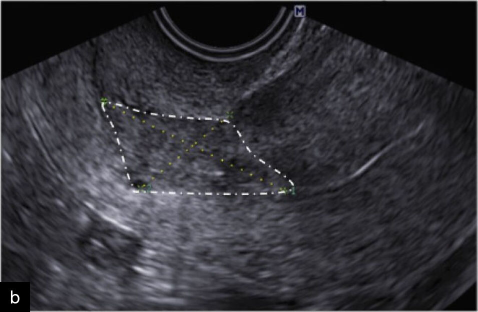
After introducing the probe transvaginally or transrectally, cervical cancers can be examined by gently applying pressure against the cervix (Video 1). The tumor generally appears as a non-compressible lesion with one of three growth patterns: (a) exophytic, appearing as a mushroom-shaped lesion (Figure 2a); (b) endophytic, appearing as an ovoid or conical lesion (Figure 2b); or (c) mixed endo-exophytic.3
1
Cervical cancer evaluation. During transvaginal or transrectal ultrasound, cervical cancers can be evaluated by applying pressure of the probe against the cervix.
|
|
2
Exophytic (a) and endophytic (b) growth patterns of cervical cancer lesions.
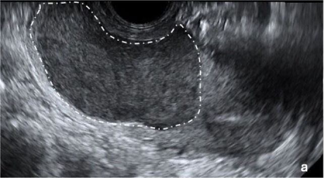
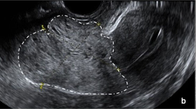
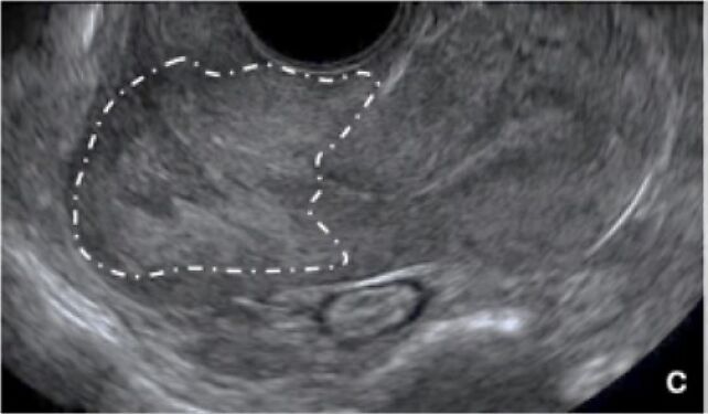
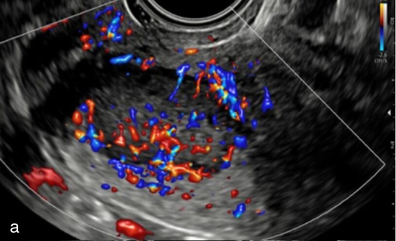
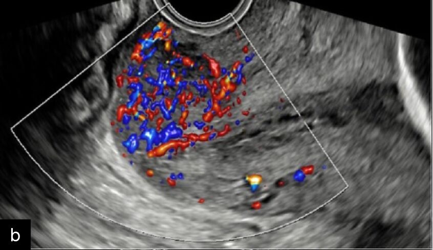
Cervical cancer typically presents as a solid lesion. In squamous cell carcinomas, it is usually hypoechoic relative to the surrounding cervical stroma, whereas in adenocarcinomas, it may appear isoechoic or hyperechoic24 (Figure 3). On color or power Doppler examination, cervical tumors usually appear moderately or richly vascularized (Figure 4).
|
|
| |
3
Echogenicity of cervical lesions relative to surrounding stroma: (a) hypoechoic, (b) hyperechoic, and (c) isoechoic.
|
|
4
(a,b) Cervical lesions richly vascularized on color Doppler examination.
Assessment of tumor size and eligibility for fertility-sparing treatment
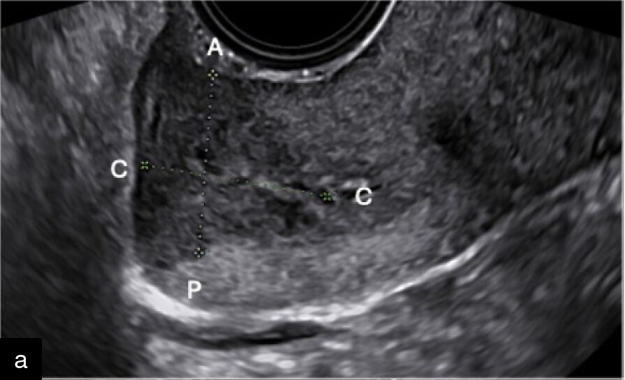
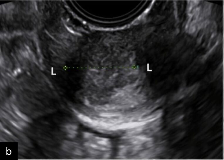
Once the lesion has been identified, it should be measured in three orthogonal planes (anteroposterior, craniocaudal and laterolateral) (Figure 5).
|
|
5
Cervical lesion diameters: craniocaudal and anteroposterior (a) and laterolateral (b).
The tumor borders should then be delineated to assess key prognostic parameters, including tumor size, depth of stromal invasion, minimum thickness of uninvolved cervical stroma and the cranial tumor-free margin. These parameters guide treatment selection and help determine eligibility for fertility-sparing treatment (FST).
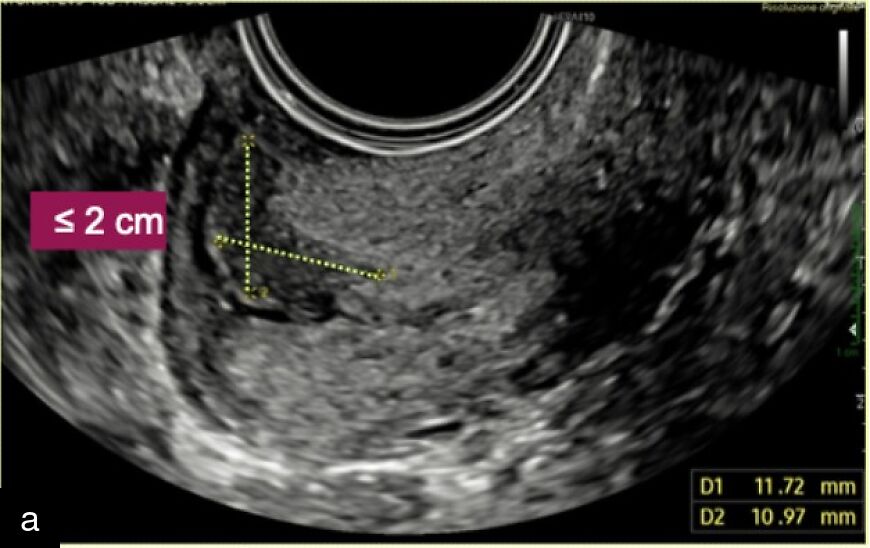
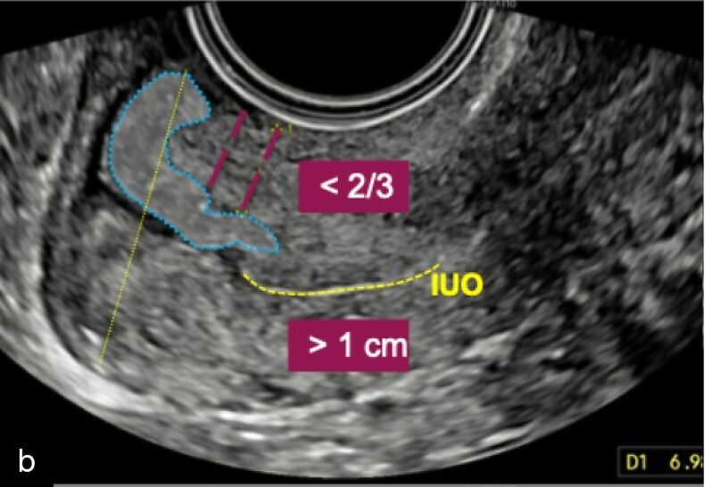
FST may be considered for patients affected by squamous cell carcinoma or human papillomavirus (HPV)-related adenocarcinoma, with early-stage cervical cancer (FIGO stage IA1–IB1 and selected IB2 cases), who wish to preserve fertility and who meet the imaging criteria for conservative management. These criteria include: (a) tumor size < 2 cm; (b) minimum distance of > 1 cm between the tumor and the internal uterine os; (c) absence of deep stromal invasion, defined as involvement of the outer third of the cervical stroma8,23 (Figure 6).
|
|
6
Ultrasound criteria for conservative management, which include: (a) tumor size < 2 cm; (b) minimum distance of > 1 cm between the tumor and internal uterine os; (c) cervical stromal invasion involving less than two-thirds of the stromal thickness.
Assessment of local extension
Anterior/ventral compartment
The endocavitary probe should be gently introduced into the vagina and advanced to the level of the anterior vaginal fornix while carefully assessing the vaginal walls. In this position, the vesicovaginal septum can be assessed in a midsagittal plane by gently pressing the uterus and vagina against the bladder (Video 2, Figure 7).25
2
Vesicovaginal and vesicouterine septum: the vesicovaginal septum can be assessed in a midsagittal plane by gently pressing the uterus and vagina against the bladder.
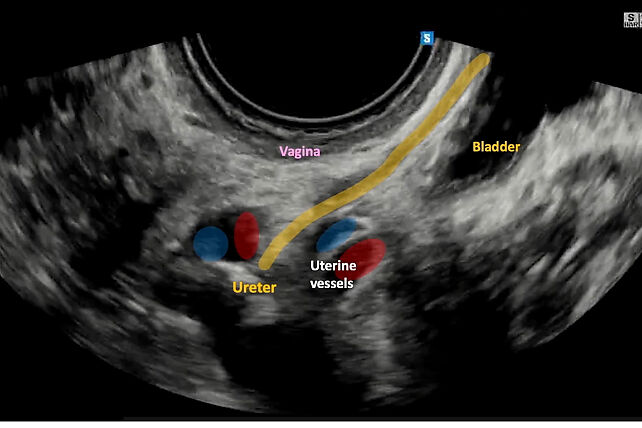
7
Longitudinal view of the anterior parametrium, comprising the vesicovaginal septum, bladder and ureter.
The bladder can be further evaluated by applying slight pressure to the vesicouterine septum to assess for tumor infiltration, which is typically associated with tissue fixation and peritumoral hyperechoic fibrosis (Video 3).
3
Bladder invasion by cervical cancer, with hyperechoic fibrosis surrounding the tumor.
Together, these maneuvers enable a thorough assessment of neoplastic extension into adjacent anterior pelvic structures.
The anterior parametrium, including the right and left vesicovaginal and vesicouterine ligaments, can be explored and assessed for possible infiltration, as described in another chapter in this volume: Ultrasound Assessment of Pelvic Anatomy. Potential infiltration may appear as thickened hyperechoic tissue or as hypoechoic nodular involvement of these structures (Figure 8).
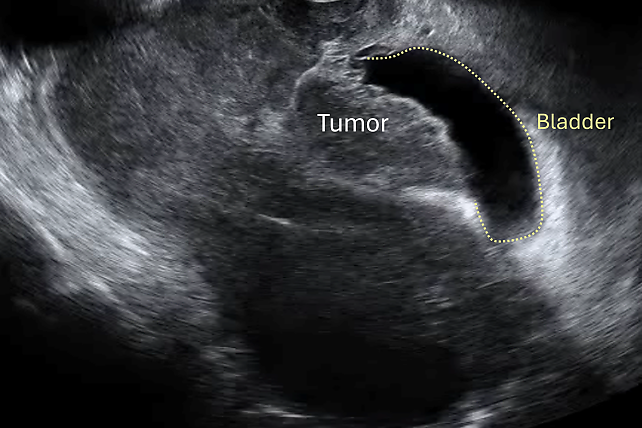
8
Tumor infiltration of the anterior parametrium, with thickened hyperechoic tissue extending to the bladder mucosa.
Posterior/dorsal compartment
The rectovaginal septum and its integrity can be assessed during transvaginal ultrasound by gently pushing the probe against the rectum and posterior wall of the vagina. Normal sliding motion indicates the absence of infiltration within the septum (Video 4). The rectum can be evaluated during transvaginal pelvic ultrasound examination, allowing identification of the different layers of the rectal wall (Figure 9).
4
The rectovaginal septum and its integrity can be assessed during transvaginal ultrasound, with a normal sliding motion, which indicates the absence of infiltration within the septum.
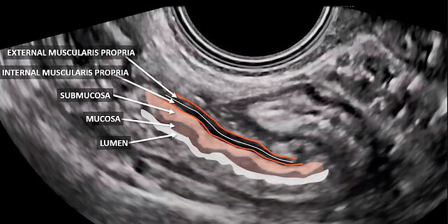
9
Transvaginal ultrasound image showing the rectal wall layers and lumen.
The posterior parametrium, including the right and left uterosacral and rectovaginal ligaments, can be explored and assessed for possible infiltration; this may appear as thickened hyperechoic tissue or as hypoechoic nodular involvement of these structures (Figure 10).
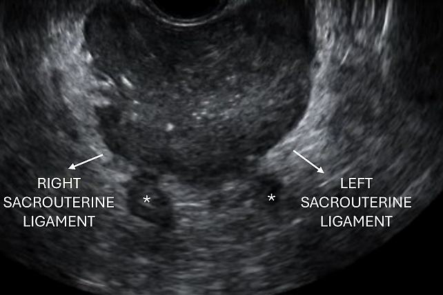
10
Dorsal parametrial infiltration by cervical cancer, with hypoechoic tumor spicules infiltrating the uterosacral ligaments (*) and surrounded by hyperechoic fibrotic tissue.
Lateral compartment
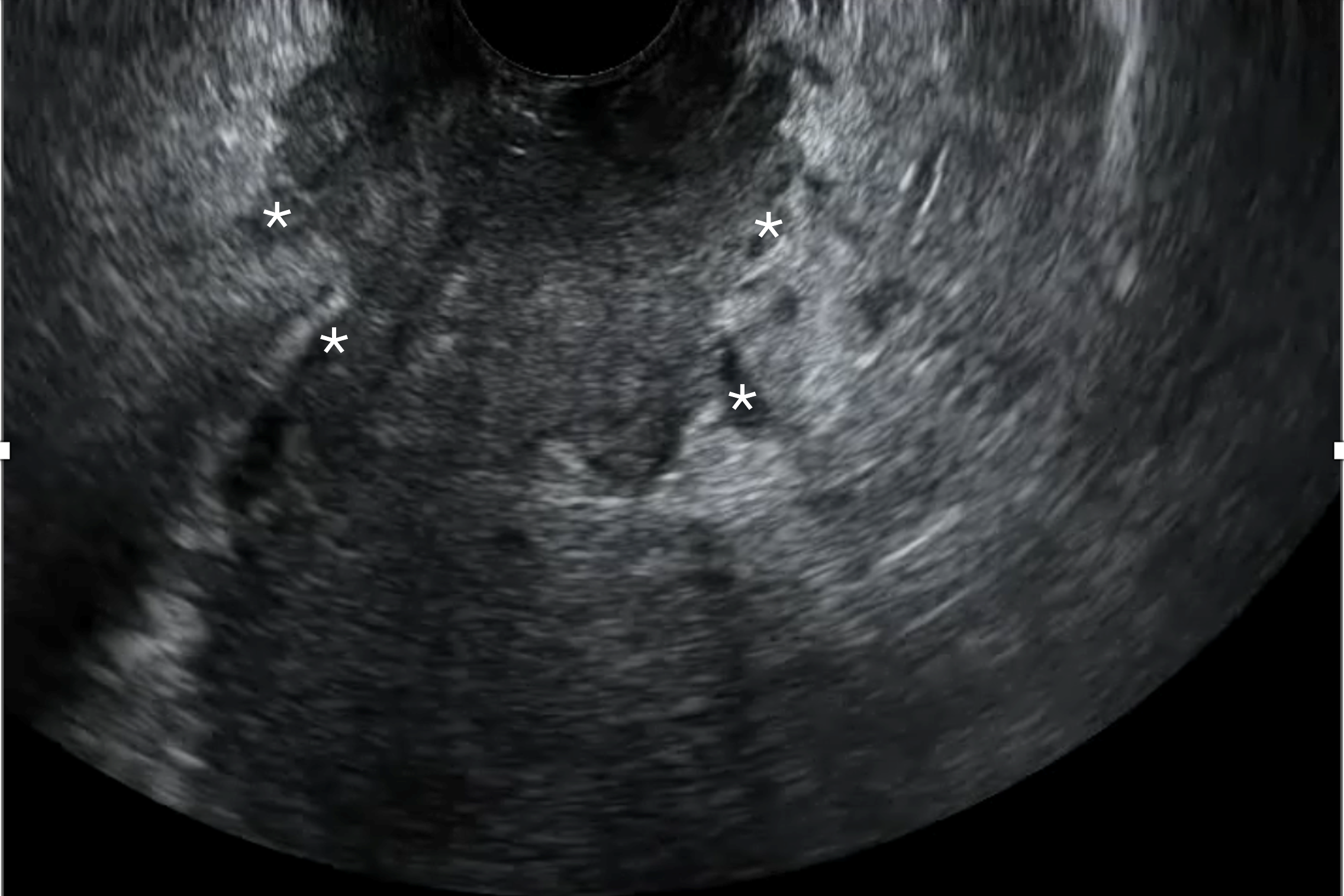
During ultrasound examination, the lateral parametrium can be assessed by placing the probe in the posterior vaginal fornix to visualize the midsagittal plane of the uterus. The probe can then be moved laterally towards the pelvic sidewall until the iliac vessels are visualized, providing a longitudinal view of the ureter and uterine artery. At the point at which the uterine vessels cross the ureter, the probe should be rotated 90° to obtain a transverse view of the ureter, allowing assessment of the medial portion of the lateral parametrium (i.e. from the cervix to the ureter) and the lateral portion of the lateral parametrium (i.e. from the ureter to the iliac vessels/pelvic wall) (Video 5). When the parametrium is infiltrated, cervical cancer typically appears as hypoechoic spicules radiating into the pericervical tissue (lateral parametrium), often accompanied by surrounding hyperechoic tissue representing reactive fibrosis (Figure 11, Video 6). Ureteral tumor infiltration is almost invariably associated with hydroureter (Video 7); therefore, assessment of the kidneys should also be performed to assess for hydronephrosis.
5
Evaluation of the lateral parametrium.
6
Infiltration of the lateral parametrium, with hypoechoic spicules infiltrating pericervical tissue.
11
Lateral parametrial infiltration by cervical cancer, with hypoechoic tumor spicules infiltrating the paracervix bilaterally (*) and the sacrouterine ligaments, surrounded by hyperechoic fibrotic tissue.
7
Hydroureter, due to tumor infiltration of the bladder and lateral parametrium.
Ultrasound features of local disease infiltration from cervical cancer are summarized in Table 2.
2
Ultrasound features of local disease infiltration in cervical cancer.
Structure | Ultrasound sign of infiltration |
Bladder | Loss of interface, fixation, irregular wall infiltration |
Rectum | Loss of sliding sign, disruption of rectal wall layers |
Vesicovaginal septum | Disruption/interruption of normal hyperechoic plane |
Paracervix | Hypoechoic spicules extending into pericervical tissue |
Ureter | Direct infiltration and/or hydroureter |
Assessment of lymph nodes and abdominal disease
Pelvic and para-aortic nodes
On ultrasound, a combination of endocavitary and transabdominal (convex and linear array) probes is used to detect regional (pelvic [para-iliac] and abdominal [para-aortic]) and distant nodal involvement (supraclavicular, inguinal, axillary and other lymph nodes).4
The lymph nodes most frequently involved in gynecological malignancy (apart from vulvar cancer) are parietal (retroperitoneal) and visceral abdominopelvic lymph nodes.
Using the systematic assessment of the pelvic sidewall described by Fischerova et al.25, it is possible to perform a systematic evaluation of the pelvic sidewall vessels to detect parietal lymph nodes located along the branches of the external and internal iliac vessels.
The first step is to identify the uterine vessels lateral to the cervix. The second step is to detect the branches of the anterior division of the internal iliac artery, including the obturator artery. The third step is to identify the branches of the posterior division of the internal iliac artery. The fourth and final step is to follow the anterior and posterior branches until the interiliac bifurcation between the internal and external iliac arteries.25
A convex-array transducer is recommended to complete the assessment of the abdominal parietal lymph nodes. The transducer is placed on the abdominal wall with the patient lying in a dorsal lithotomy position. The examination starts at the inguinal ligament to identify the femoral vessels running beneath it to become the external iliac vessels.26 The external iliac artery is visualized ventral to the external iliac vein on the psoas major muscle. The transducer is oriented in an oblique plane and moved along the large iliac vessels up to the aortic bifurcation.
Visceral lymph nodes are classified as pelvic or abdominal. Pelvic visceral lymph nodes are located around the pelvic viscera, whereas abdominal visceral lymph nodes are located around the celiac trunk and the superior and inferior mesenteric arteries and their branches.27
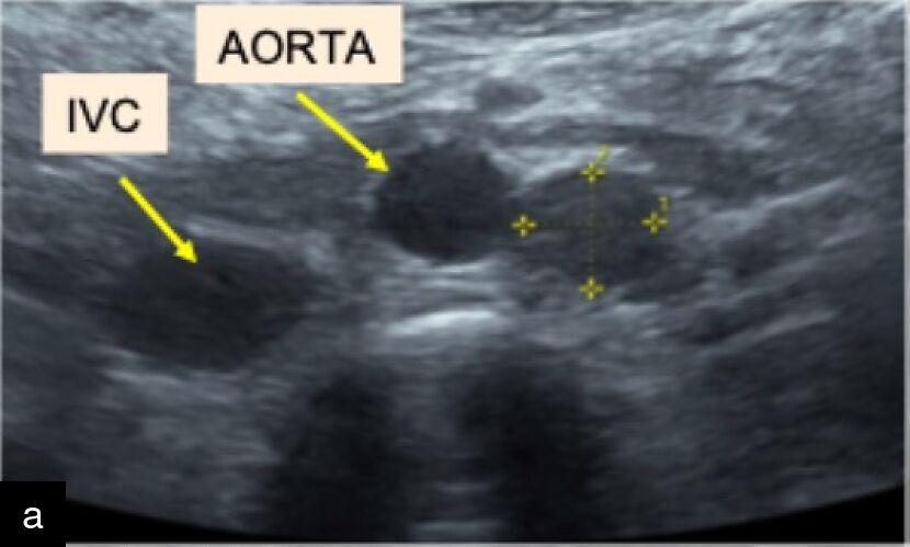
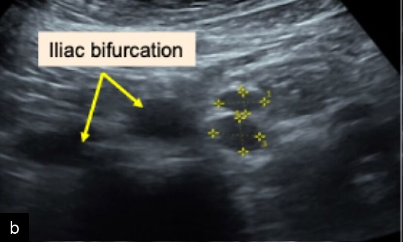
The lymph nodes should be described according to VITA (Vulvar International Tumor Analysis) terms.28 Metastatic lymph nodes typically appear as solid round masses near major vessels, with loss of the nodal core sign (defined as the functional unit formed by the nodal medulla, with or without a visible hilum) due to tumoral infiltration (see another chapter in this volume Ultrasound Evaluation of Inguinal and Pelvic Lymph Nodes). Additional features may include cystic necrosis and calcification. In cervical cancer, metastatic lymph nodes are usually markedly hypoechoic4 (Figure 12).
|
|
12
Para-aortic (a) and iliac (b) lymphadenopathy, visible as hypoechoic, rounded masses adjacent to the major vessels.
Distant nodal sites
Ultrasound can also be used for the detection of extrapelvic tumor spread to the abdominal cavity in the form of parietal or visceral carcinomatosis, omental and/or mesenteric infiltration.
Because of the lower spatial resolution of transabdominal ultrasound, miliary dissemination may be more difficult to detect than with transvaginal or transrectal ultrasound. However, image quality is often improved in the presence of ascites, which provides an acoustic window.
A systematic abdominal ultrasound examination is required for a complete and standardized evaluation of the patient. The examination should begin with assessment of the upper abdominal viscera, including the kidneys (with particular attention to hydronephrosis), adrenal glands, spleen, liver and pancreas.23 Any focal or diffuse intraparenchymal lesions, capsular infiltration or visceral lymphadenopathy should be documented. The parietal, visceral and mesenteric peritoneum, together with the omentum, should then be evaluated for evidence of tumour spread, including parietal (lateral paracolic gutters, diaphragm and anterior abdominal wall), omental, visceral (intestinal carcinomatosis and organ surfaces) and mesenteric (small-bowel mesentery or mesocolon) carcinomatosis. Finally, the peripheral (superficial and deep inguinal) and retroperitoneal (parietal) lymph nodes should be assessed.23
Carcinomatosis is usually accompanied by ascites, defined as fluid outside the pouch of Douglas,29 which could also be quantified by measuring each fluid pocket in the abdominal quadrants.
In the case of tumor spread to the omentum, infiltration may be either focal or diffuse. Focal nodules are found in both the lesser and greater omenta, are hypoechoic with irregular borders, and are vascularized. Diffuse omental infiltration (‘omental cake’) has a characteristic appearance, with a nodular, perfused tumor plate, underneath which the movements of intestinal loops can usually be observed.30
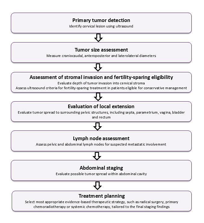
A schematic flowchart of the ultrasound steps for assessing and staging cervical cancer is presented in Figure 13.
13
Flowchart illustrating the main ultrasound steps for assessing and staging cervical cancer
ASSESSMENT OF CERVICAL CANCER IN LOW AND MIDDLE-INCOME COUNTRIES
Cervical cancer continues to represent a significant global health challenge, especially in low- and middle-income countries, where access to advanced diagnostic imaging and treatment options is frequently restricted. In these resource-limited settings, transvaginal ultrasound is a valuable tool for local staging and assessment of nodal and distant metastatic disease, thereby supporting appropriate therapeutic management. A practical checklist developed by Chilinda et al.7 defines eligibility for upfront surgery, the appropriate extent of hysterectomy, indications for neoadjuvant chemotherapy and contraindications to surgery according to the 2018 FIGO staging system (Figure 14). The proposed framework builds on existing guidelines by providing an ultrasound-based algorithm with clear decision-making pathways and representative imaging examples. It provides practical clinical guidance that is not fully detailed in current recommendations and is suitable for use in a range of resource settings. Successful implementation requires practical training, appropriate ultrasound equipment and an understanding of the strengths and limitations of ultrasound-based staging.
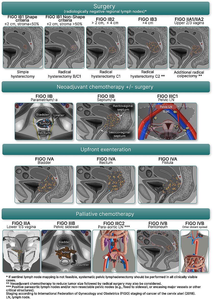
14
Ultrasound-based preoperative assessment for cervical cancer: a pragmatic staging and treatment-planning strategy adaptable to diverse resource settings. (Reproduced from Chilinda et al. 7, under the terms of the CC BY licence.)
ULTRASOUND AND NEW PERSPECTIVES FOR CERVICAL CANCER
Artificial intelligence (AI)-based approaches are emerging as promising tools to support image interpretation and reduce operator dependency. Machine learning (ML) and deep learning (DL) algorithms have already demonstrated encouraging results in gynecological oncology imaging, including tumor detection, segmentation and risk stratification, particularly in ultrasound- and MRI-based applications.13,31,19,32 Ultrasound evaluation of women with endometrial or cervical cancer enables an accurate assessment of tumor extent and invasion patterns, facilitating tailored surgical management based on imaging findings.33,34
In a recent systematic review by Moro et al.34 on the application of AI in ultrasound assessment of gynecological conditions, only five studies were identified that focused on cervical cancer, mostly conducted in early-stage disease and primarily focused on predicting lymph node involvement. In these studies,35,36,37,38,39 models built with features based on DL automatic segmentation had higher or similar performance as compared to models built with features based on manual segmentation in the validation set.
Regarding the prediction of response to treatment, Gui et al.40 developed a radiomics-based predictive model for pathological complete response using radiomic features extracted from pretreatment T2-weighted MRI in 183 patients undergoing neoadjuvant chemoradiotherapy followed by radical surgery. The best-performing random forest model achieved an AUC of 0.80, with a specificity of 98.4%, sensitivity of 50.2% and an overall accuracy of 74.5%. Although the model demonstrated excellent specificity, its limited sensitivity suggests that MRI radiomics can reliably identify non-responders but is less effective at detecting complete responders. Similarly, Chiappa et al. focused on response prediction after neoadjuvant chemotherapy, developing a baseline T2-weighted MRI radiomic model in 72 patients, and achieved promising results with an AUC of 0.83, sensitivity of 71%, and specificity of 75%.41
The application of AI and radiomics to ultrasound has the potential to expand its role in the personalized management of gynecological malignancies. For example, AI may facilitate the preoperative prediction of histological characteristics and molecular profiles to support more personalized treatment, including POLE mutations in endometrial cancer, PDL-1 expression in cervical cancer and lymphovascular space invasion in early-stage cervical cancer. It may also have other applications in predicting treatment response after chemotherapy and recurrence in ovarian and cervical cancers. The integration of advanced ultrasound techniques with AI-based tools into multidisciplinary decision-making pathways has the potential to support more personalized and effective management of patients with gynecological malignancies, including cervical cancer.
CONCLUSION
Ultrasound has evolved from a complementary imaging technique to a comprehensive staging tool for patients with cervical cancer, capable of evaluating the primary tumor, local extension, lymph node involvement and abdominal dissemination. When performed by adequately trained operators and integrated into multidisciplinary pathways, ultrasound can provide diagnostic information comparable to that of MRI for several key staging parameters while remaining widely accessible and cost-effective worldwide. Future integration of AI and standardized examination protocols may further enhance its role in personalized treatment planning and global cervical cancer care.
PRACTICE RECOMMENDATIONS
- Adopt ultrasound as a first-line imaging tool. Transvaginal and transrectal ultrasound should be considered first-line modalities for the local assessment of cervical cancer, particularly in settings in which MRI is not readily available.
- Ensure adequate operator training and standardization. Ultrasound evaluation should be performed by experienced operators following standardized methodology to minimize variability and improve diagnostic accuracy.
- Assess tumor size and local extent systematically. Ultrasound examination should include careful evaluation of tumor dimensions, stromal invasion, parametrial involvement and extension to adjacent structures (e.g. vagina, bladder, rectum).
- Use ultrasound for preoperative planning. Imaging findings should guide individualized surgical strategies, including fertility-sparing approaches when appropriate, if ultrasound criteria are fulfilled.
CONFLICTS OF INTEREST
Author(s) statement awaited.
Feedback
Publishers’ note: We are constantly trying to update and enhance chapters in this Series. So if you have any constructive comments about this chapter please provide them to us by selecting the "Your Feedback" link in the left-hand column.
REFERENCES
Global Cancer Observatory. Cancer over time. Accessed October 25, 2023. Available at: https://gco.iarc.fr/overtime/en/dataviz/tables?hide_tab_age_specific_numbers=1&mode=population&years=2012 | |
Sozzi G, Berretta R, Fiengo S, Ferreri M, Giallombardo V, Finazzo F, Messana D, Capozzi VA, Colacurci N, Scambia G, Chiantera V. Integrated pre-surgical diagnostic algorithm to define extent of disease in cervical cancer. Int J Gynecol Cancer. 2020 Jan;30(1):16–20. doi: 10.1136/ijgc-2019-000665. Epub 2019 Oct 23. PMID: 31645425. | |
Testa AC, Di Legge A, De Blasis I, Moruzzi MC, Bonatti M, Collarino A, Rufini V, Manfredi R. Imaging techniques for the evaluation of cervical cancer. Best Pract Res Clin Obstet Gynaecol. 2014 Jul;28(5):741–768. doi: 10.1016/j.bpobgyn.2014.04.009. Epub 2014 May 4. PMID: 24861248. | |
Fischerova D, Frühauf F, Burgetova A, Haldorsen IS, Gatti E, Cibula D. The Role of Imaging in Cervical Cancer Staging: ESGO/ESTRO/ESP Guidelines (Update 2023). Cancers (Basel). 2024 Feb 14;16(4):775. doi: 10.3390/cancers16040775. PMID: 38398166; PMCID: PMC10886638. | |
Fischerova D, Cibula D, Stenhova H, Vondrichova H, Calda P, Zikan M, Freitag P, Slama J, Dundr P, Belacek J. Transrectal ultrasound and magnetic resonance imaging in staging of early cervical cancer. Int J Gynecol Cancer. 2008 Jul–Aug;18(4):766–772. doi: 10.1111/j.1525-1438.2007.01072.x. Epub 2007 Sep 24. PMID: 17892456. | |
Moloney F, Ryan D, Twomey M, Hewitt M, Barry J. Comparison of MRI and high-resolution transvaginal sonography for the local staging of cervical cancer. J Clin Ultrasound. 2016 Feb;44(2):78–84. doi: 10.1002/jcu.22288. Epub 2015 Aug 21. PMID: 26294324. | |
Chilinda G, Moro F, Rijken MJ, Ciancia M, Teodorico E, Fagotti A, Fischerova D, Testa AC, Querleu D. Ultrasound-based preoperative assessment for cervical cancer: a pragmatic staging and treatment-planning strategy adaptable to diverse resource settings. Int J Gynecol Cancer. 2026 Feb;36(2):102885. doi: 10.1016.j.ijgc.2025.102885. Epub 2025 Dec 19. PMID: 41576735. ISSN: 1048-891X, https://doi.org/10.1016/j.ijgc.2025.102885 | |
NCCN Clinical Practice Guidelines in Oncology (NCCN Guidelines®) Cervical Cancer. Version 4.2025 – March 24, 2025. | |
Testa AC, Ludovisi M, Manfredi R, Zannoni G, Gui B, Basso D, Di Legge A, Licameli A, Di Bidino R, Scambia G, Ferrandina G. Transvaginal ultrasonography and magnetic resonance imaging for assessment of presence, size and extent of invasive cervical cancer. Ultrasound Obstet Gynecol. 2009 Sep;34(3):335–344. doi: 10.1002/uog.7325. PMID: 19705403. | |
Epstein E, Testa A, Gaurilcikas A, Di Legge A, Ameye L, Atstupenaite V, Valentini AL, Gui B, Wallengren NO, Pudaric S, Cizauskas A, Måsbäck A, Zannoni GF, Kannisto P, Zikan M, Pinkavova I, Burgetova A, Dundr P, Nemejcova K, Cibula D, Fischerova D. Early-stage cervical cancer: tumor delineation by magnetic resonance imaging and ultrasound – a European multicenter trial. Gynecol Oncol. 2013 Mar;128(3):449–453. doi: 10.1016/j.ygyno.2012.09.025. Epub 2012 Sep 28. PMID: 23022593. | |
Chiappa V, Di Legge A, Valentini AL, Gui B, Miccò M, Ludovisi M, Giansiracusa C, Testa AC, Valentin L. Agreement of two-dimensional and three-dimensional transvaginal ultrasound with magnetic resonance imaging in assessment of parametrial infiltration in cervical cancer. Ultrasound Obstet. Gynecol. 2015;45:459–469. doi: 10.1002/uog.14637. | |
Frühauf F, Fischerová D, Moro F, Kocián R, Ptáčník V, Wagnerová M, Lambert L, Testa AC, Bizzarri N, Perez RO, Teodorico E, Cibula D; Collaborators. Prospective comparison of diagnostic accuracy of ultrasound, PET/CT and DW-MRI for preoperative assessment of pelvic lymph nodes in cervical cancer patients: results of the CANNES trial. Ultrasound Obstet Gynecol. 2026 Jun;67(6):836–851. doi: 10.1002/uog.70241. Epub 2026 May 24. PMID: 42178785. | |
Tian Y, Luo H. Diagnostic accuracy of transvaginal ultrasound examination for local staging of cervical cancer: a systematic review and meta-analysis. Med Ultrason. 2022 Aug 31;24(3):348–355. doi: 10.11152/mu-3246. Epub 2021 Nov 3. PMID: 34762728. | |
Kim SH, Lee HJ, Kim YW. Correlation between tumor size and surveillance of lymph node metastasis for IB and IIA cervical cancer by magnetic resonance images. Eur J Radiol. 2012 Aug;81(8):1945-50. doi: 10.1016/j.ejrad.2011.04.053. Epub 2011 May 24. PMID: 21605954. | |
Klerkx WM, Veldhuis WB, Spijkerboer AM, van den Bosch MA, Mali WP, Heintz AP, Bipat S, Sie-Go DM, van der Velden J, Schreuder HW, Stoker J, Peeters PH. The value of 3.0Tesla diffusion-weighted MRI for pelvic nodal staging in patients with early stage cervical cancer. Eur J Cancer. 2012 Dec;48(18):3414-21. doi: 10.1016/j.ejca.2012.06.022. Epub 2012 Jul 24. PMID: 22835781. | |
Fischerová D. Staging zhoubného nádoru děložního hrdla (stanovení předoperačního rozsahu onemocnění) - přehled výsledků nejnovějších ultrazvukových studií [Cervical cancer staging - preoperative assessment of tumor extent (a review of the most recent ultrasound studies)]. Ceska Gynekol. 2014 Dec;79(6):436-46. Czech. PMID: 25585552. | |
Pálsdóttir K, Fischerova D, Franchi D, Testa A, Di Legge A, Epstein E. Preoperative prediction of lymph node metastasis and deep stromal invasion in women with invasive cervical cancer: prospective multicenter study using 2D and 3D ultrasound. Ultrasound Obstet Gynecol. 2015 Apr;45(4):470-5. doi: 10.1002/uog.14643. Epub 2015 Mar 1. PMID: 25092154. | |
Yang K, Park W, Huh SJ, Park BK, Kim CK, Kim BG, Bae DS, Lee JW. Parametrial Involvement on Magnetic Resonance Imaging Has No Effect on the Survival of Early-Stage Cervical Cancer Patients. Int J Gynecol Cancer. 2017 Mar;27(3):507-513. doi: 10.1097/IGC.0000000000000909. PMID: 28129242. | |
Alcazar JL, García E, Machuca M, Quintana R, Escrig J, Chacón E, Mínguez JA, Chiva L. Magnetic resonance imaging and ultrasound for assessing parametrial infiltration in cervical cancer. A systematic review and meta-analysis. Med Ultrason. 2020 Mar 1;22(1):85–91. doi: 10.11152/mu-2361. PMID: 32096793. | |
Stukan M, Buderath P, Szulczyński B, Gębicki J, Kimmig R. Accuracy of Ultrasonography and Magnetic Resonance Imaging for Preoperative Staging of Cervical Cancer-Analysis of Patients from the Prospective Study on Total Mesometrial Resection. Diagnostics (Basel). 2021 Sep 23;11(10):1749. doi: 10.3390/diagnostics11101749. PMID: 34679447; PMCID: PMC8534714. | |
Imai K, Chikazawa K, Kuwata T, Konno R. Volume Index Measured Using Magnetic Resonance Imaging for Diagnosing Cervical Cancer Tumors <2 cm. In Vivo. 2023 Jul-Aug;37(4):1786-1789. doi: 10.21873/invivo.13267. PMID: 37369483; PMCID: PMC10347931. | |
Bizzarri N, Arciuolo D, Certelli C, Pedone Anchora L, Gallotta V, Teodorico E, Carbone MV, Piermattei A, Fanfani F, Fagotti A, Ferrandina G, Zannoni GF, Scambia G, Querleu D. Ultrastaging of the Parametrium in Cervical Cancer: A Clinicopathological Study. Cancers (Basel). 2023 Feb 9;15(4):1099. doi: 10.3390/cancers15041099. PMID: 36831442; PMCID: PMC9954180. | |
Fischerova D. Ultrasound scanning of the pelvis and abdomen for staging of gynecological tumors: A review. Ultrasound Obstet. Gynecol. 2011;38:246–266. doi: 10.1002/uog.10054. | |
Epstein E, Di Legge A, Måsbäck A, Lindqvist PG, Kannisto P, Testa AC. Sonographic characteristics of squamous cell cancer and adenocarcinoma of the uterine cervix. Ultrasound Obstet Gynecol. 2010;36:512–516. | |
Fischerova D, Culcasi C, Gatti E, Ng Z, Burgetova A, Szabó G. Ultrasound assessment of the pelvic sidewall: methodological consensus opinion. Ultrasound Obstet Gynecol. 2025 Jan;65:94–105. https://doi.org/10.1002/uog.29122 | |
Fischerova D, Gatti E, Culcasi C, Ng Z, Szabó G, Zanchi L, Burgetova A, Nanka O, Gambino G, Kadajari MR, Garganese G; Collaborators. Ultrasound assessment of lymph nodes for staging of gynecological cancer: consensus opinion on terminology and examination technique. Ultrasound Obstet Gynecol. 2025 Feb;65(2):206–225. doi: 10.1002/uog.29127. Epub 2024 Nov 8. PMID: 39513930; PMCID: PMC12133214. | |
Lengele B, Nyssen-Behets C, Scalliet P. Anatomical bases for the radiological delineation of lymph node areas. Upper limbs, chest and abdomen. RadiotherOncol. 2007;84:335–347. | |
Fischerova D, Garganese G, Reina H, Fragomeni SM, Cibula D, Nanka O, Rettenbacher T, Testa AC, Epstein E, Guiggi I, Frühauf F, Manegold G, Scambia G, Valentin L. Terms, definitions and measurements to describe sonographic features of lymph nodes: Consensus opinion from the Vulvar International Tumor Analysis (VITA) group. Ultrasound Obstet Gynecol 2021 Jun;57(6):861–879. doi: 10.1002/uog.23617. PMID: 34077608 | |
Timmerman D, Valentin L, Bourne TH, Collins WP, Verrelst H, Vergote I; International Ovarian Tumor Analysis (IOTA) Group. Terms, definitions and measurements to describe the sonographic features of adnexal tumors: a consensus opinion from the International Ovarian Tumor Analysis (IOTA) Group. Ultrasound Obstet Gynecol. 2000 Oct;16(5):500–505. doi: 10.1046/j.1469-0705.2000.00287.x. PMID: 11169340. | |
Fischerova D, Smet C, Scovazzi U, Sousa DN, Hundarova K, Haldorsen IS. Staging by imaging in gynecologic cancer and the role of ultrasound: an update of European joint consensus statements. Int J Gynecol Cancer. 2024 Mar 4;34(3):363–378. doi: 10.1136/ijgc-2023-004609. PMID: 38438175; PMCID: PMC10958454. | |
Follen M, Levenback CF, Iyer RB, Grigsby PW, Boss EA, Delpassand ES, Fornage BD, Fishman EK. Imaging in cervical cancer. Cancer. 2003 Nov;98(9 Suppl):2028–2038. doi: 10.1002/cncr.11679. PMID: 14603539. | |
Esteva A, Robicquet A, Ramsundar B, Kuleshov V, DePristo M, Chou K, Cui C, Corrado G, Thrun S, Dean J. A guide to deep learning in healthcare. Nat Med. 2019 Jan;25(1):24–29. doi: 10.1038/s41591-018-0316-z. Epub 2019 Jan 7. PMID: 30617335. | |
Moro F, Ciancia M, Zace D, Vagni M, Tran HE, Giudice MT, Zoccoli SG, Mascilini F, Ciccarone F, Boldrini L, D'Antonio F, Scambia G, Testa AC. Role of artificial intelligence applied to ultrasound in gynecology oncology: A systematic review. Int J Cancer. 2024 Nov 15;155(10):1832–1845. doi: 10.1002/ijc.35092. Epub 2024 Jul 11. PMID: 38989809. | |
Moro F, Giudice MT, Ciancia M, Zace D, Baldassari G, Vagni M, Tran HE, Scambia G,Testa AC. Application of artificial intelligence to ultrasound imaging for benign gynecological disorders: systematic review. Ultrasound Obstet Gynecol. 2025 15 Mar;65(3):295–302. doi: 10.1002/uog.29171. Epub 2025 Jan 31. PMID: 39888598; PMCID: PMC11872345. | |
Jin X, Ai Y, Zhang J, Zhu H, Jin J, Teng Y, Chen B, Xie C. Noninvasive prediction of lymph nodestatus for patients with early-stage cervical cancer based on radio-mics features from ultrasound images. Eur Radiol. 2020 Jul;30(7):4117–4124. doi: 10.1007/s00330-020-06692-1. Epub 2020 Feb 20. PMID: 32078013. | |
Teng Y, Ai Y, Liang T, Yu B, Jin J, Xie C, Jin X. The effects of automatic segmentations on preoperative lymph node status prediction models with ultrasound radiomics for patients with early-stage cervical cancer. Technol Cancer Res Treat. 2022 Jan–Dec;21:15330338221099396. doi: 10.1177/15330338221099396. PMID: 35522305; PMCID: PMC9082739. | |
Yi J, Lei X, Zhang L, Zheng Q, Jin J, Xie C, Jin X, Ai Y. The influence of different ultrasonic machines on Radiomics models in prediction lymph node metastasis for patients with cervical cancer. Technol Cancer Res Treat. 2022 Jan–Dec;21:15330338221118412. doi: 10.1177/15330338221118412. PMID: 35971568; PMCID: PMC9386859. | |
Jin J, Zhu H, Teng Y, Ai Y, Xie C, Jin X. The accuracy and radiomics feature effects of multiple U-Net-based automatic segmentation models for transvaginal ultrasound images of cervical cancer. J Digit Imaging. 2022 Aug;35(4):983–992. doi: 10.1007/s10278-022-00620-z. Epub 2022 Mar 30. PMID: 35355160; PMCID: PMC9485324. | |
Zhou H, Wang S, Zhang T, Liu D, Yang K. Ultrasound image analysis technology under deep belief networks in evaluation on the effects of diagnosis and chemotherapy of cervical cancer. J Supercomput.2021;77:4151-4171. | |
Gui B, Autorino R, Micco M, Nardangeli A, Pesce A, Lenkowicz J, Cusumano D, Russo L, Persiani S, Boldrini L, Dinapoli N, Macchia G, Sallustio G, Gambacorta MA, Ferrandina G, Manfredi R, Valentini V, Scambia G. Pretreatment MRI Radiomics Based Response Prediction Model in Locally Advanced Cervical Cancer. Diagnostics (Basel). 2021 Mar;11(4):631. doi: 10.3390/diagnostics11040631. PMID: 33807494; PMCID: PMC8066099. | |
Chiappa V, Bogani G, Interlenghi M, Vittori Antisari G, Salvatore C, Zanchi L, Ludovisi M, Leone Roberti Maggiore U, Calareso G, Haeusler E, Raspagliesi F, Castiglioni I. Using Radiomics and Machine Learning Applied to MRI to Predict Response to Neoadjuvant Chemotherapy in Locally Advanced Cervical Cancer. Diagnostics (Basel). 2023 Oct;13(19):3139. doi: 10.3390/diagnostics13193139. PMID: 37835882; PMCID: PMC10572442. |
Online Study Assessment Option
All readers who are qualified doctors or allied medical professionals can automatically receive 2 Continuing Professional Development points plus a Study Completion Certificate from GLOWM for successfully answering four multiple-choice questions (randomly selected) based on the study of this chapter. Medical students can receive the Study Completion Certificate only.
(To find out more about the Continuing Professional Development awards program CLICK HERE)
