
This chapter should be cited as follows:
Ohno T, Glob Libr Women's Med
ISSN: 1756-2228; DOI 10.3843/GLOWM.422073
The Continuous Textbook of Women’s Medicine Series – Gynecology Module
Volume 13
Gynecological cancer
Volume Editors:
Professor Hextan Ngan, Department of Obstetrics and Gynaecology, The University of Hong Kong, Hong Kong
Professor Karen Chan, Department of Obstetrics and Gynaecology, The University of Hong Kong, Hong Kong

Chapter
Role of Radiotherapy in Cervical Cancer
First published: March 2026
Study Assessment Option
By answering four multiple-choice questions (randomly selected) after studying this chapter, readers can qualify for Continuing Professional Development points plus a Study Completion Certificate from GLOWM.
See end of chapter for details.

INTRODUCTION
Radiotherapy has been a cornerstone in the management of cervical cancer, with its well-established role evolving alongside advances in imaging, treatment planning and delivery techniques. When carefully planned and executed, radiotherapy achieves excellent pelvic tumor control and survival outcomes. For selected early-stage tumors, it offers efficacy comparable to that of radical surgery. Radiotherapy remains the primary treatment modality for patients with advanced disease, either alone or in combination with systemic therapies. The integration of concurrent cisplatin-based chemotherapy with radiation has significantly improved local control and survival in patients with locally advanced cervical cancer. Therefore, contemporary indications for radiotherapy span a wide range of disease stages. Optimal treatment selection requires an individualized, multidisciplinary approach that considers clinical, anatomic and social factors, and emphasizes close collaboration among gynecologic oncologists, radiation oncologists and referring physicians.
CLINICAL PRESENTATION
The signs and symptoms of cervical cancer often include abnormal vaginal bleeding, which is the most common early symptom experienced by patients. This abnormal bleeding may manifest during menstrual period, after sexual intercourse or after menopause. Other early symptoms can include offensive vaginal discharge and lower abdominal pain.1 Many patients do not perceive themselves to be at risk and tend to attribute these initial symptoms to normal bodily changes (e.g. menopause) or common illnesses (e.g. sexually transmitted infections), thus leading to delayed recognition and diagnosis.1
Additional symptoms that may arise as the disease progresses include heavy vaginal bleeding, which is life-threatening; persistent lower abdominal pain; and symptoms that are severe enough to interfere with daily activities or work routines. Persistent and worsening symptoms despite home-based treatment often prompt patients to seek professional help.1 Other notable symptoms include abnormal vaginal discharge with a foul odor, pelvic pain and symptoms related to metastasis or effects on other organs in advanced stages.2,3
Owing to inadequate knowledge of cervical cancer symptoms, particularly in low-resource settings, presentation and diagnosis are often late, thus leading to a more advanced disease at the time of detection.2,4 Psychological symptoms, such as anxiety and depression, can also be present in patients with cervical cancer and may be influenced by the level of social support and availability of coping resources; however, these symptoms are consequences related to the disease experience rather than the direct symptoms of the cancer itself.5,6
CLINICAL EVALUATION AND STAGING
Role of imaging in radiotherapy for cervical cancer
Imaging modalities play a crucial role in radiotherapy planning and cervical cancer management by providing the necessary anatomical and functional information for accurate tumor delineation, staging and treatment-response assessment.7
Computed tomography (CT) is primarily used to assess extent of disease, particularly lymph node involvement and presence of distant metastases. CT provides essential anatomical detail that aids radiotherapy treatment planning, particularly for dose calculation and targeting. However, its limited soft-tissue contrast restricts its accuracy in evaluating the extent of local tumors.
18F-fluorodeoxyglucose positron emission tomography with CT (FDG-PET/CT) combines metabolic and anatomical imaging, making it valuable for detecting nodal metastases and distant disease. In advanced disease, PET/CT is particularly useful for radiotherapy planning by identifying the involved lymph nodes and assessing treatment response, which may influence radiotherapy field design and systemic therapy decisions.
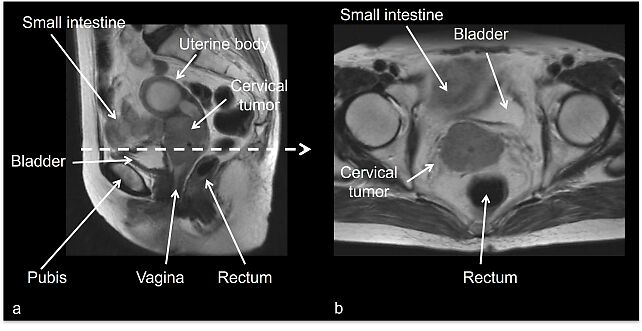
Magnetic resonance imaging (MRI) is the preferred modality for local staging of cervical cancer owing to its superior soft-tissue resolution (Figure 1). MRI accurately defines tumor size, parametrial invasion and involvement of adjacent pelvic structures, which is critical for precise radiotherapy planning, including the delineation of gross tumor volume and organs at risk (OARs). This accuracy improves targeting while minimizing damage to the surrounding tissues, particularly in image-guided adaptive brachytherapy.
1
T2-weighted magnetic resonance images of cervical cancer FIGO Stage IIB as seen in sagittal (a) and axial (b) views. The cervical tumor disrupts the hypointense peripheral stroma and extends into the parametrium bilaterally. No tumor invasion is observed in the bladder and rectum.
Ultrasound is used mainly as an initial, accessible tool to evaluate the cervix and pelvic organs and to guide biopsies. It has been suggested as a valid alternative to MRI in the primary diagnostic workup of cervical cancer if performed by an expert sonographer.7
For clinical staging, pelvic MRI or transvaginal/transrectal ultrasound enable tumor delineation and the precise assessment of its local extent, including the evaluation of bladder or rectal invasion. Both techniques have very high specificity for confirming involvement of metastatic pelvic lymph nodes. However, their sensitivity is insufficient for detecting small-volume metastases, similar to any currently available imaging modality. Contrast-enhanced CT or FDG-PET/CT are recommended for assessing the spread of extrapelvic tumors.7
International Federation of Gynecology and Obstetrics (FIGO) staging system
The FIGO stage is a clinical staging system for classifying gynecologic cancers, including cervical cancer, on the basis of tumor size, local extension and spread to adjacent or distant structures.8 It provides a standardized framework for disease assessment and is essential for treatment planning, prognostic evaluation and comparison of clinical outcomes across studies.
Table 1 shows the FIGO 2018 classification. A key update in the FIGO 2018 classification was the incorporation of imaging and pathology results in the FIGO stage. In addition, the size criteria for defining T1b were revised with the introduction of a new subcategory, namely, IB3, thus resulting in the following classifications: IB1 (≤ 2 cm), IB2 (> 2 to ≤ 4 cm) and IB3 (> 4 cm) in the greatest dimension. Furthermore, pelvic lymph node involvement was newly defined as Stage IIIC1, whereas para-aortic lymph node involvement was defined as Stage IIIC2 regardless of the primary tumor size or local extent. As a result, FIGO Stage IIIC disease is highly heterogeneous and associated with variable survival outcomes.9
1
FIGO 2018 classification of cervical cancer
Stage | Description |
I | Carcinoma strictly confined to the cervix |
IA | Invasive carcinoma that can be diagnosed only by microscopy, with maximum depth of invasion ≤ 5 mm* |
IA1 | Stromal invasion ≤ 3 mm in depth |
IA2 | Stromal invasion > 3 mm and ≤ 5 mm in depth |
IB | Invasive carcinoma with deepest invasion > 5 mm (greater than Stage IA); lesion limited to the cervix with size measured according to maximum tumor diameter† |
IB1 | Invasive carcinoma with > 5 mm depth of stromal invasion and ≤ 2 cm in greatest dimension |
IB2 | Invasive carcinoma > 2 cm and ≤ 4 cm in greatest dimension |
IB3 | Invasive carcinoma > 4 cm in greatest dimension |
II | Carcinoma invades beyond uterus, but has not extended to lower third of vagina or to pelvic wall |
IIA | Involvement is limited to upper two-thirds of vagina without parametrial invasion |
IIA1 | Invasive carcinoma ≤ 4 cm in greatest dimension |
IIA2 | Invasive carcinoma > 4 cm in greatest dimension |
IIB | Involves parametrial invasion but not up to pelvic wall |
III | Carcinoma involves lower third of vagina and/or extends to pelvic wall and/or causes hydronephrosis or non- functioning kidney and/or involves pelvic and/or para-aortic lymph nodes |
IIIA | Carcinoma involves lower third of vagina, with no extension to pelvic wall |
IIIB | Extension to pelvic wall and/or hydronephrosis or non-functioning kidney (unless known to be due to another cause) |
IIIC | Involvement of pelvic and/or para-aortic lymph nodes (including micrometastases)‡ irrespective of tumor size and extent (with r and p notations)§ |
IIIC1 | Pelvic lymph node metastasis only |
IIIC2 | Para-aortic lymph node metastasis‡ |
IV | Tumor extends beyond true pelvis or involves (biopsy proven) mucosa of bladder or rectum (bullous edema alone is insufficient to classify a case as Stage IV) |
IVA | Spread to adjacent pelvic organs |
IVB | Spread to distant organs |
*Imaging and pathology can be used when available to supplement clinical findings with respect to tumor size and extent in all stages. Pathological findings supersede imaging and clinical findings.
†Involvement of vascular/lymphatic spaces should not change staging. Lateral extent of the lesion is not considered.
‡Isolated tumor cells do not change the stage, but their presence should be recorded.
§Notations r (imaging) and p (pathology) were added to indicate the findings that are used to allocate the case to Stage IIIC. For example, if imaging indicates pelvic lymph node metastasis, the stage allocation would be Stage IIIC1r; if confirmed by pathological findings, it would be Stage IIIC1p. The imaging modality or pathological technique used should always be documented. When in doubt, the lower staging should be assigned.
TECHNICAL ASPECTS OF RADIOTHERAPY
Clinical workflow
When a patient is referred, the physician determines the indication for radiotherapy by confirming the patient’s preferences and values and by reviewing the medical history, physical and pelvic examination findings, imaging studies, and other relevant test results, including laboratory findings. In patients with a history of irradiation, the spatial relationship between the previously irradiated area and the requested treatment site should be carefully evaluated. In addition, because comorbidities may increase the risk of adverse events, the following should be confirmed: presence of diabetes mellitus, inflammatory bowel disease, connective tissue disease or renal dysfunction; history of pelvic surgery; and use of anticoagulants or antiplatelet medications.
After informed consent is obtained, immobilization devices are fabricated to ensure reproducibility of patient positioning. A treatment-planning CT scan is performed using the same patient position and pretreatment conditions as those used during irradiation. Thereafter, the CT images are imported into the treatment-planning system, where the beam arrangements are designed for the target tumor and surrounding normal tissues. After the approved treatment plan is verified through measurements and/or calculations, the actual radiation delivery is performed.
External beam radiotherapy (EBRT)
The definitive radiotherapy for cervical cancer requires a combination of EBRT to the pelvis and brachytherapy to the primary cervical tumor.10,11,12 In patients with FIGO Stage IIIC2 disease, extended-field radiotherapy encompassing the para-aortic region is performed.
The prophylactic irradiation of the para-aortic region is optional in cases with multiple positive pelvic lymph nodes or metastasis to the common iliac lymph nodes; however, there are no randomized controlled trials that strongly support this approach.13 By contrast, in early stage cervical cancer, pelvic irradiation that excludes the common iliac lymph nodes may be employed.13,14 However, determining the target volume according to individual risk factors remains an ongoing challenge.
Whole-pelvic irradiation for cervical cancer using 3D conformal radiotherapy (3D-CRT), intensity-modulated radiotherapy (IMRT) and volumetric-modulated arc therapy (VMAT) involves delivering radiation to the entire pelvic region, including the primary tumor site and regional lymph nodes, to address the potential spread of microscopic disease.
3D conformal radiotherapy (3D-CRT)
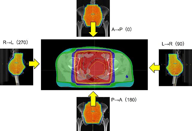
In 3D-CRT, radiation beams are shaped on the basis of imaging to conform to the 3D anatomy of the pelvis, allowing targeted dose delivery to the primary tumor site and pelvic lymph node region while sparing some normal tissues. However, 3D-CRT uses static beam arrangements, which may increase radiation delivery to adjacent organs, such as the bladder, rectum and bowels (Figure 2).
2
3D conformal radiotherapy for cervical cancer, showing uniform-intensity beams from multiple directions. Numbers in parentheses are beam angles. A, anterior; L, left; P, posterior; R, right.
Intensity-modulated radiotherapy (IMRT)
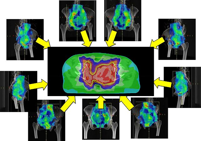
IMRT improves upon 3D-CRT by modulating the intensity of individual radiation beams, thus enabling more precise dose sculpting that conforms tightly to the complex pelvic anatomy (Figure 3). This results in better sparing of critical OARs such as the bladder, rectum and small bowel, thus reducing acute and late toxicities. The ability of IMRT to deliver differential doses within the pelvic volume allows the simultaneous integration of boosts to high-risk areas while treating elective nodal regions.
3
Intensity-modulated radiotherapy for cervical cancer. By modulating the intensity across each beam, a high dose that perfectly fits the complex shape of the tumor can be delivered while significantly sparing the surrounding healthy organs. Beam direction is shown by yellow arrows.
Volumetric-modulated arc therapy (VMAT)
VMAT further advances whole-pelvic irradiation by delivering radiation continuously as the treatment machine rotates around the patient, thus dynamically adjusting the beam shape and intensity in real time. This technique enhances dose conformity and homogeneity across pelvic target volumes and reduces treatment time compared with static IMRT. VMAT maintains or improves OAR sparing, contributes to lower toxicity profiles and improves patient comfort during treatment.
Stereotactic body radiotherapy (SBRT)
SBRT is a highly precise form of radiotherapy that delivers large doses in few fractions to limited metastatic sites, with minimal damage to the surrounding tissues. SBRT is increasingly being used in gynecological oncology, including cervical cancer, for treating oligometastatic diseases or in patients unsuitable for brachytherapy. It offers good local control and is favorable for patients who are not candidates for intracavitary brachytherapy. SBRT is generally considered when definitive local control of limited metastases is desired or as a boost in patients ineligible for brachytherapy. The recommended SBRT dose for cervical cancer boost has been evaluated in clinical studies, with doses of 22.5 Gy delivered in three fractions showing acceptable toxicity profiles.15
Particle beam therapy
Proton and carbon ion radiotherapy for gynecological malignancies, including cervical cancer, utilizes charged particles that deliver radiation with distinct physical and biological properties compared with conventional X-rays. These particles exhibit a characteristic Bragg peak, allowing for maximal energy deposition at a specific depth with a minimal exit dose beyond the target. Carbon ion radiotherapy offers additional potential advantages because of its higher linear energy transfer, resulting in increased relative biological effectiveness.16 This can enhance tumor cell kill, particularly in photon-resistant tumors such as adenocarcinoma, malignant melanoma and hypoxic tumors, thus potentially improving local control.17,18
Image-guided brachytherapy
Intracavitary brachytherapy is an essential component in definitive radiotherapy for cervical cancer in FIGO 2018 clinical Stages IA2–IVA. Except in cases in which brachytherapy cannot be performed because of patient- or tumor-related factors, the use of highly conformal external beam radiation techniques, such as IMRT or SBRT, as substitutes for brachytherapy is not recommended.
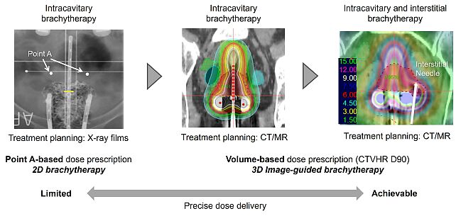
Brachytherapy for cervical cancer has evolved from conventional treatment planning based on prescription to point A using orthogonal radiographs to 3D image-guided brachytherapy (3D-IGBT), which employs imaging modalities, such as CT and MRI, acquired with the applicator in place (Figure 4). In 3D-IGBT, target volumes and OARs are delineated on 3D images, and dose prescription and evaluation are performed on the basis of dose distribution and dose–volume histogram parameters.19 A major advantage of 3D-IGBT is the ability to develop treatment plans while visualizing the target organs, including the uterus, bladder and rectum, thereby enabling greater individualization of treatment.
4
Evolution of brachytherapy. Traditionally, 2D brachytherapy was performed using X-ray films, where ‘Point A’ was the standard reference for dose prescription. Nowadays, CT or MRI is used to create treatment plans; this is called 3D image-guided brachytherapy (3D-IGBT). 3D-IGBT includes the standard intracavitary technique using tandem and ovoids, as well as the ‘IC/IS’ technique, which combines intracavitary and interstitial brachytherapy. IC/IS is superior at delivering the appropriate dose according to the size and shape of the tumor.
More recently, hybrid brachytherapy, which is also referred to as combined intracavitary and interstitial brachytherapy, has been developed to improve dose distribution by adding a limited number of interstitial needles to intracavitary brachytherapy (Figure 4).20 Hybrid brachytherapy is particularly indicated when adequate dose coverage of the high-risk clinical target volume cannot be achieved with intracavitary brachytherapy alone or when compliance with dose constraints for adjacent OARs is difficult.
Advances in brachytherapy techniques have led to improved local control of cervical tumors (90–95% with CT- or MRI-guided brachytherapy) without increasing the incidence of severe adverse events.21,22 Table 2 summarizes the clinical outcomes of MRI-guided brachytherapy for cervical cancer. At the same time, these advances require a higher level of technical expertise and careful attention to medical safety from clinical staff. In addition to cervical cancer, 3D-IGBT can be applied to other gynecological malignancies requiring brachytherapy, such as endometrial and vaginal cancers.
2
Clinical outcomes of MRI-guided brachytherapy for cervical cancer.
FIGO stage | Number of patients (n) | 5-year local control (%) | 5-year pelvic control (%) | 5-year disease-free survival (%) | 5-year overall survival (%) |
IB1 | 124 | 98 | 95 | 76 | 83 |
IB2 | 119 | 92 | 84 | 65 | 73 |
IIA1 | 38 | 91 | 88 | 75 | 80 |
IIA2 | 31 | 89 | 77 | 65 | 74 |
IIB | 693 | 91 | 88 | 73 | 78 |
IIIA | 13 | 100 | 100 | 76 | 76 |
IIIB | 190 | 92 | 86 | 59 | 64 |
IVA | 34 | 91 | 81 | 47 | 52 |
IVB | 98 | 89 | 81 | 48 | 61 |
Total | 1341 | 92 | 87 | 68 | 74 |
Data from EMBRACE-I22
Overall treatment time (OTT) considerations
Prolonged OTT after radiotherapy for cervical cancer is consistently associated with poor survival outcomes, including reduced overall survival (OS), disease-free survival (DFS) and local tumor control. This negative effect is mainly attributed to accelerated tumor cell repopulation during treatment delays or interruptions, which diminishes the effectiveness of radiotherapy.23,24 Clinical evidence shows that shorter, uninterrupted treatment courses lead to better survival and local control, whereas OTT prolongation increases the risk of recurrence and treatment failure. Maintaining an OTT of approximately 7 weeks or less during radiotherapy for cervical cancer is recommended to maximize efficacy.11
In resource-limited settings, delays before and during radiotherapy often result in more advanced disease at treatment initiation, thus further worsening prognosis ('stage migration').25 Overall, there is a clear inverse relationship between OTT duration and survival in cervical cancer, underscoring the importance of timely, continuous radiotherapy (preferably combined with chemotherapy) to optimize patient outcomes.
ROLE OF CHEMOTHERAPY/IMMUNOTHERAPY IN COMBINATION WITH RADIOTHERAPY
Concurrent chemoradiotherapy (CCRT)
In 1999, the USA National Cancer Institute issued a significant clinical alert recommending CCRT with cisplatin-based chemotherapy and radiation as the new standard of care for locally advanced cervical cancer, replacing radiation alone, based on compelling trial results showing improved survival.26 Since then, CCRT for cervical cancer has been widely adopted worldwide as a standard treatment. The chemotherapy regimen most commonly used for CCRT is single-agent cisplatin administered at a dose of 40 mg/m² weekly. To date, cisplatin-based combination chemotherapy has not been shown to be superior to single-agent cisplatin in this setting. As shown in Table 3, clinical trials have demonstrated that CCRT improves intrapelvic tumor control.27 In contrast, the reduction in distant metastases likely reflects not only the direct antitumor effects of chemotherapy but also a secondary effect resulting from improved local control and reduced local recurrence. Notably, the response rate to single-agent cisplatin in cervical cancer is limited to approximately 20–30%, and its curative potential when used alone is modest.
3
Clinical trials showing improvement in pelvic recurrence rates following chemoradiotherapy
Clinical study | Chemoradiotherapy arm | Control arm | ||
Total patients | Pelvic recurrence | Total patients | Pelvic recurrence | |
GOG85 | 177 | 44 | 191 | 58 |
GOG120 | 176 | 33 | 177 | 53 |
GOG120 | 173 | 35 | ||
RTOG9001 | 193 | 37 | 193 | 68 |
GOG123 | 183 | 29 | 186 | 73 |
SWOG8797 | 127 | 11 | 116 | 20 |
NCIC | 126 | 34 | 123 | 41 |
Total | 1155 | 223 (19.3%) | 986 | 313 (31.7%) |
According to a meta-analysis by Green et al. comparing CCRT with radiotherapy, CCRT significantly reduced both local and distant recurrence and resulted in significant improvements in OS and DFS.28 The benefit in 5-year survival was 8% overall for patients with Stages IB–IIA disease (hazard ratio (HR), 0.78). However, when analyzed by stage, the survival benefit decreased with advancing disease stage: 10% for Stages IB–IIA, 7% for Stage IIB and 3% for Stages III–IVA.29 Nevertheless, even in patients with Stage IIIB cervical squamous cell carcinoma, a more recent report shows that CCRT provides a significant survival advantage compared with radiotherapy alone.30
With regard to toxicity, the above meta-analysis demonstrated that CCRT was associated with a 3.6-fold increase in Grades-3–4 hematologic toxicity, including a 2.2-fold increase in leukopenia and a 2.4-fold increase in thrombocytopenia, as well as a 2.0-fold increase in Grades-3–4 acute gastrointestinal toxicity.28 By contrast, no significant increase in Grades-3–4 acute toxicity was observed in the urinary tract or skin.
Subsequent clinical trials focused on the development of new combination therapies based on an established CCRT framework. The representative clinical trials are described below.
Induction chemotherapy followed by CCRT
The INTERLACE trial was a Phase-III randomized study that compared CCRT alone with induction chemotherapy followed by CCRT in patients with locally advanced cervical cancer.31 Patients in the experimental arm received weekly carboplatin and paclitaxel for six cycles prior to standard CCRT.
Among the 500 enrolled patients, the majority had Stage-II disease and negative lymph node status. At a median follow-up of 64 months, induction chemotherapy followed by CCRT was associated with improved 5-year DFS and OS compared with CCRT alone.
However, the details of the radiotherapy component are limited. IMRT and 3D-IGBT were used in a subset of patients, and the prescribed dose to point A (equivalent dose in 2-Gy fractions [EQD2] of ≥ 78 Gy) was lower than that reported in contemporary studies, such as EMBRACE. Although treatment completion rates were high, a careful interpretation of the results is warranted owing to the incomplete reporting and potential variability in the radiotherapy dose and technique.
CCRT and adjuvant chemotherapy
The OUTBACK trial was a multicenter, randomized Phase-III study in which adults with locally advanced cervical cancer were assigned to receive standard cisplatin-based CCRT alone or the same CCRT followed by adjuvant carboplatin and paclitaxel.32 The trial evaluated whether four cycles of adjuvant chemotherapy administered after definitive CCRT could improve survival compared with CCRT alone. Radiotherapy consisted of 45.0–50.4 Gy of pelvic external beam irradiation with weekly cisplatin, followed by intracavitary brachytherapy according to standard practice. After a median follow-up of 5 years, adjuvant chemotherapy did not improve OS (72% vs 71%; HR, 0.90), and no benefit in disease progression or survival was observed. By contrast, adjuvant chemotherapy was associated with increased hematologic toxicity and serious adverse events, thus suggesting that the routine use of adjuvant carboplatin and paclitaxel after CCRT is not beneficial in unselected patients with locally advanced cervical cancer.
Immunotherapy and CCRT
In the CALLA randomized, double-blind Phase-3 trial, adults with previously untreated, locally advanced cervical cancer (FIGO 2009 Stages IB2–IIB with nodal involvement or Stages III–IVA) were assigned to receive standard CCRT with or without the programmed death-ligand 1 antibody durvalumab.33 Radiotherapy consisted of 45 Gy EBRT delivered in 25 fractions with weekly cisplatin or carboplatin, followed by image-guided brachytherapy using high-dose-rate or low-/pulse-dose-rate techniques to deliver a total dose of 27.5–30 Gy or 35–40 Gy, respectively. After a median follow-up of approximately 18 months, progression-free survival (PFS) did not differ significantly between the groups (HR, 0.84), although treatment was generally well tolerated with comparable rates of Grade-3–4 toxicities. These findings indicate that the addition of durvalumab to CCRT did not improve PFS.
The KEYNOTE-A18 trial was a Phase-III randomized study evaluating the addition of pembrolizumab to CCRT in patients with cervical cancer.34 This trial enrolled patients with FIGO 2014 Stages IB2–IIB with positive lymph node status and those with Stages III–IVA and conducted a comparison between CCRT alone and CCRT combined with pembrolizumab. In the experimental arm, pembrolizumab was administered concurrently with CCRT at a dose of 200 mg every 3 weeks for five cycles, followed by maintenance therapy with pembrolizumab at 400 mg every 6 weeks for up to 15 cycles. At a median follow-up of 17.9 months, the 2-year PFS was 57.3% in the CCRT-alone group (n = 531) and 67.8% in the pembrolizumab plus CCRT group (n = 529), thus demonstrating that a statistically significant improvement was achieved with the addition of pembrolizumab (HR = 0.70; P = 0.002). The incidence of Grade ≥ 3 diarrhea was 4% in both treatment groups. Highly conformal external beam radiation techniques, such as IMRT and VMAT, were used in nearly 90% of the patients. The total combined dose from EBRT and brachytherapy reached approximately 87 Gy EQD2 in both treatment arms, indicating that high-quality radiotherapy was delivered.
COMPLICATIONS OF RADIOTHERAPY
Adverse events
Adverse events caused by radiotherapy are generally classified into acute adverse events and late adverse events. Acute adverse events occur within 90 days of the start of treatment, whereas late adverse events occur at 91 days or later. Acute reactions are primarily caused by inflammation and are often reversible. By contrast, late reactions are mainly due to fibrosis (overgrowth of connective tissue) and thickening of the vascular intima, which leads to impaired blood flow. These changes are typically difficult to reverse and often become chronic.
Bone marrow
Lymphocytes, a type of white blood cell, are highly sensitive to radiation. When a large area such as the entire pelvis is irradiated, lymphocyte counts decrease early during treatment. The combination of whole-pelvic irradiation with concurrent chemotherapy and/or para-aortic irradiation increases the risk of leukopenia. A recent systematic review reported a significant increase in hematologic toxicity for whole-pelvic-bone doses of V10Gy > 95–75%, V20Gy > 80–65% and V40Gy > 37–28%.35
Skin
Acute skin reactions usually appear 2–3 weeks after the start of radiotherapy and include dryness, redness (erythema) and pigmentation in the irradiated area. If the damage reaches the dermis, moist desquamation with fluid discharge may occur. Skin reactions tend to be more severe when the radiation field includes the perineal region owing to lower vaginal extension or when it includes areas such as the intergluteal cleft and anal region.
Maintaining the cleanliness of the affected skin is essential. Heparinoid-containing moisturizers are commonly used to treat dryness, and dimethyl isopropylazulene ointment may be applied for inflammation. Topical corticosteroids are administered when itching or redness is severe. Physical irritation, such as friction, and chemotherapy use are risk factors for worsening skin reactions.
Late skin effects include hyperpigmentation, hypopigmentation, telangiectasia (small blood vessels) and skin atrophy.
Nausea and vomiting
According to the MASCC/ESMO 2023 guidelines, the risk of nausea and vomiting during pelvic radiotherapy is classified as low risk, whereas irradiation involving to the upper abdomen is considered moderate risk.36 For prevention and treatment, 5-HT3 receptor antagonists, dexamethasone and dopamine antagonists are recommended.
The concurrent administration of chemotherapy increases the risk of nausea and vomiting, and antiemetic therapy should be selected on the basis of the chemotherapy risk classification. For patients with cervical cancer receiving concurrent weekly cisplatin and radiotherapy, a three-drug combination (5-HT3 receptor antagonists, dexamethasone, and aprepitant/fosaprepitant) is recommended.
Gastrointestinal tract
During pelvic radiotherapy, diarrhea is a common acute adverse event that usually improves after treatment completion. Probiotics and intestinal regulators are used for basic treatment, and loperamide hydrochloride is added when necessary.37
Late adverse events include radiation proctitis (urgency and rectal bleeding), small bowel injuries such as intestinal obstruction, and rectovaginal fistula, each occurring in less than 5% of patients.
During endoscopic examination for rectal bleeding, biopsy of the irradiated area should be avoided unless a new tumor is suspected because biopsy may increase the risk of bowel perforation in the irradiated tissue. A history of abdominal or pelvic surgery increases the risk of bowel obstruction, whereas the use of anticoagulants or antiplatelet agents increases the risk of bleeding. In cases with rectal bleeding, constipation should be avoided. Mild bleeding often improves with conservative treatment involving carbazochrome sodium sulfonate or tranexamic acid.
If bleeding is severe or persistent, endoscopic therapy (argon plasma coagulation), hyperbaric oxygen therapy or surgery is required.
Bladder
Radiation cystitis is caused by damage to the bladder mucosa, similar to radiation proctitis. The acute symptoms include frequent urination, pain during urination and incomplete bladder emptying. Medications are commonly used for an overactive bladder, and antibiotics are prescribed when an infection is present.
Late adverse events include reduced bladder capacity due to fibrosis, ureteral strictures and vesicovaginal fistulas (> 5%). Hematuria (blood in the urine) may occur more than 5 years after treatment.38 Most cases respond to conservative treatment; however, severe hemorrhagic cystitis may require total cystectomy. Hyperbaric oxygen therapy has also been demonstrated to be effective.
Ovaries
Oocytes (egg cells) are highly sensitive to radiation exposure. Ovarian failure typically occurs when curative doses are administered to the pelvis. In young patients, menopausal symptoms may appear early, and fertility cannot be preserved.
Vagina
Vaginal stenosis and sexual dysfunction represent ongoing clinical challenges following radiotherapy. Incremental radiation doses have been associated with a higher incidence of vaginal stenosis, and 45–50% of patients experience vaginal stenosis at approximately 45 Gy.39 Vaginal dilatation therapy is widely recommended to prevent radiation-induced vaginal stenosis, and higher adherence is associated with a lower prevalence of this complication. However, patient adherence remains limited owing to the discomfort, lack of personalization and physical rigidity of the current dilators.39
Bones
Late bone complications after pelvic radiotherapy include insufficiency fractures, which occur in approximately 14% of patients undergoing irradiation.40 A meta-analysis showed that, among cases with pelvic insufficiency fractures, the proportion of symptomatic patients was 61%. IMRT was associated with a significantly lower fracture rate. Insufficiency fractures most commonly occurred in the sacrum (40%), followed by the pubis (13%) and lumbar vertebrae (7%). The median time to fracture was 7.1–19 months after radiotherapy.40 Risk factors identified in another meta-analysis included postmenopausal status, osteoporosis and diabetes mellitus.41
Secondary cancer
Secondary cancer after radiotherapy refers to any new cancer that develops after treatment. After radiotherapy for cervical cancer, the relative risk of secondary cancer is approximately 1.2–1.3 times that of the general population overall.42,43 Compared with the general population, risks for HPV-related cancers (pharynx, genital sites and rectum/anus) and smoking-related cancers (pharynx, trachea/bronchus/lung, pancreas and urinary bladder) were elevated in both radiotherapy and non-radiotherapy groups. Additionally, cervical cancer patients receiving radiotherapy were at an increased risk for cancers at heavily irradiated sites (colon, rectum/anus, urinary bladder, ovary and genital sites) compared with the general population.42 However, the long-term survival benefits of radiotherapy outweigh these risks.
MANAGEMENT OF ANEMIA
Etiology of anemia
Patients with cervical cancer frequently present with anemia, which can arise from multiple factors. Tumor-related bleeding, particularly genital bleeding, tends to worsen as the disease progresses, thus resulting in more severe anemia in the advanced stages. In addition, comorbid conditions (e.g. chronic inflammation and infection) and anticancer treatments (e.g. radiotherapy and cisplatin-based chemotherapy) may contribute to the development of anemia.
Prognostic effect
Anemia has long been reported as a poor prognostic factor after definitive radiotherapy for cervical cancer. One proposed explanation is that anemia leads to tumor hypoxia, which results in increased radioresistance. However, the correction of anemia does not necessarily translate into improved clinical outcomes, and the underlying biological mechanisms affecting radiation response remain incompletely understood.
Treatment-related anemia and bone marrow toxicity
With regard to treatment-related effects on anemia, attention should be paid to bone marrow suppression associated with pelvic EBRT and combined chemotherapy as well as bleeding related to applicator insertion during brachytherapy. Miszczyk et al. reported the results of a meta-analysis evaluating the effect of bone marrow-sparing radiotherapy on hematologic toxicity.44 EBRT delivered with highly conformal techniques, such as IMRT or VMAT, resulted in significant reductions in grade ≥ 3 leukopenia and grade ≥ 2 neutropenia. However, no significant reduction in the incidence of anemia or thrombocytopenia was observed.
Red blood cell transfusion
Clear criteria for red blood cell transfusion in patients with anemia and cervical cancer undergoing radiotherapy have not yet been established. Zayed et al. conducted a systematic review addressing this issue and reported wide variability in transfusion thresholds, with hemoglobin levels of 8 to 12 g/dL.45 Regarding oncologic outcomes, six studies found that there were no survival benefits associated with red blood cell transfusion, whereas three studies suggested a potential improvement.
Evidence from head and neck cancer
Anemia has also been reported as a poor prognostic factor for head and neck cancer. Hoff et al. performed a pooled analysis of the DAHANCA 5 and DAHANCA 7 trials, in which patients with low pretreatment hemoglobin levels (women < 13.0 g/dL, men < 14.5 g/dL) were randomized to receive or not receive red blood cell transfusion.46 Regardless of transfusion status, patients with low hemoglobin levels had worse local control, disease-specific survival and OS, and no prognostic benefit from transfusion was observed.
Overall, there is currently no clear evidence that red blood cell transfusion improves oncological outcomes in patients with anemia undergoing radiotherapy.
POSTOPERATIVE RADIOTHERAPY
Indications for adjuvant radiotherapy
In cervical cancer, the decision to administer adjuvant therapy (treatment after surgery) is based on the pathological findings from the hysterectomy and lymphadenectomy. Careful patient selection and multidisciplinary coordination are essential to balance therapeutic benefits against potential complications and ensure an improved quality of life after treatment. These patients were categorized into high- and intermediate-risk groups.
Postoperative radiotherapy (PORT) concurrent with cisplatin-based chemotherapy after hysterectomy for cervical cancer is indicated primarily in patients with high-risk pathological features, such as positive surgical margins, lymph node involvement, and parametrial invasion.47 These factors are associated with an increased likelihood of residual microscopic disease and local recurrence. PORT aims to eradicate residual disease, thereby improving local control and potentially enhancing long-term survival outcomes. Adding chemotherapy to PORT appeared to provide a smaller absolute benefit when only one node is positive or when the tumor size is < 2 cm.48
If the surgical margins and lymph nodes are negative but the tumor has aggressive local features, the patient is classified as intermediate risk.49 These features may include deep stromal invasion, lymphovascular space invasion and large tumor size. Adjuvant radiotherapy or CCRT has been used for intermediate-risk patients. In the GOG263/KGOG1008 study, although recurrence-free survival and OS favored CCRT, the addition of weekly cisplatin during radiotherapy did not result in a statistically significant improvement in the intermediate-risk group.50 CCRT increased Grade-3 and -4 adverse events with a transient decline in quality of life.
Advances in PORT
The severe adverse events associated with PORT for cervical cancer include bowel obstruction and ureteral strictures resulting from tissue fibrosis. Furthermore, treatment often leads to lower-extremity lymphedema, which may worsen recurrent cellulitis. Radiation techniques used in postoperative settings have evolved, particularly with the availability of IMRT and 3D-CRT. In cervical cancer, IMRT has shown equivalent clinical outcomes (pelvic recurrence-free survival and DFS) to those of 3D-CRT but with significantly reduced gastrointestinal and genitourinary toxicities, thus improving the therapeutic ratio.51,52 In addition, patient-reported sexual function was similar between the treatment groups. After radiotherapy, fear of sex decreased and interest in sex improved over time.53
TREATMENT OF RECURRENT AND METASTATIC DISEASE
Pelvic recurrence
The management of recurrent gynecological cancer requires the centralization of care within experienced centers and the close collaboration of a multidisciplinary team, with an emphasis on individualized decision making, patient counseling, clinical trial participation and early palliative care integration. The diagnostic workup aims to define the extent of locoregional and metastatic disease, preferably with histologic confirmation, and to distinguish patients suitable for radical, potentially curative treatment, particularly those with oligorecurrent disease, from those with multifocal or widely metastatic disease for whom radical approaches are inappropriate. Prognostic factors must be carefully weighed against the risk of major treatment-related morbidities.
According to the guidelines of the European Society of Gynaecological Oncology, European Society for Radiotherapy and Oncology, and European Society of Pathology,11 definitive chemoradiotherapy with IGBT is the treatment of choice for central recurrence after primary surgery in radiotherapy-naïve patients. Pelvic sidewall recurrence may be managed with definitive chemoradiotherapy in radiotherapy-naïve patients, whereas extended pelvic surgery aimed at complete resection may be considered when radical radiotherapy is not feasible. In patients previously treated with radiotherapy, pelvic exenteration is recommended for selected central recurrences without sidewall or extrapelvic involvement, whereas re-irradiation with IGBT or extended surgery may be considered in selected cases at specialized centers. Patients who are not candidates for radical local treatment should receive systemic chemotherapy with additional therapies guided by treatment response.
Distant metastatic disease
Palliative radiotherapy is typically used to relieve symptoms, such as pain, bleeding or obstruction caused by primary tumors or metastatic sites. Specifically, it is used to relieve pain due to bone metastases or soft-tissue invasion by tumors, neurological deficits resulting from brain metastases or spinal cord compression due to bone metastases, obstructive or stenotic symptoms involving the airway or esophagus, and tumor-related exudation or bleeding. Palliative radiotherapy aims to maintain or improve a patient’s quality of life.
The standard of care for Stage-IVB cervical cancer primarily involves systemic therapy, including cytotoxic chemotherapy, anti-angiogenic therapy and immunotherapy. Radiotherapy in Stage-IVB cervical cancer plays a palliative role and, in selected cases, may have a curative role depending on the disease burden and patient factors.
Several retrospective studies demonstrated that the use of definitive pelvic radiotherapy in patients with Stage-IVB cervical cancer was associated with better oncologic outcomes, such as PFS, cause-specific survival and OS.54,55 In addition, Li et al. reported that 60 patients who received curative-intent irradiation for primary and metastatic sites showed a 5-year OS rate of 51% and a 5-year PFS of 26%. In their study, lymphatic metastases had a better OS than hematogenous metastases, and patients with one metastasis site showed a more favorable prognosis than patients with ≥ 2 metastases sites.56 The prospective evaluation of systemic treatment with or without definitive pelvic radiotherapy is highly warranted to identify patients who benefit from the radiotherapy.
SBRT can be considered for patients with cervical cancer who develop synchronous or metachronous oligometastatic disease. In a cohort of 215 patients with gynecologic oligometastases, SBRT achieved excellent local control, with a 5-year local recurrence rate of only 18.5%, despite a high 5-year distant recurrence rate of 73.1%. Notably, the treatment provided a 5-year OS of 33.1% and a median chemotherapy-free survival of 21.7 months, thus highlighting the role of SBRT in delaying systemic therapy and maintaining quality of life.57
PRACTICE RECOMMENDATIONS
- The key signs and symptoms of cervical cancer include abnormal vaginal bleeding, malodorous vaginal discharge and persistent pelvic or lower abdominal pain.
- MRI is the preferred modality for local staging because it accurately defines the tumor size, parametrial invasion and involvement of adjacent pelvic structures.
- The FIGO staging system provides a standardized framework for disease assessment and is essential for treatment planning, prognostic evaluation and clinical outcome comparisons across studies.
- Definitive radiotherapy for cervical cancer requires a combination of external beam radiotherapy to the pelvis and brachytherapy to treat primary cervical tumors.
- Brachytherapy is an indispensable component of definitive radiotherapy for patients with FIGO 2018 clinical Stages IA2–IVA.
- Prolonged overall treatment time in radiotherapy for cervical cancer is consistently associated with inferior local control and survival, therefore, this situation should be avoided.
- Concurrent chemoradiotherapy (CCRT) with cisplatin-based chemotherapy is the standard of care for patients with locally advanced cervical cancer.
- The addition of pembrolizumab to CCRT for locally advanced cervical cancer has been shown to significantly improve progression-free survival and overall survival.
- Postoperative intensity-modulated radiotherapy with concurrent cisplatin-based chemotherapy after hysterectomy is primarily indicated for patients with high-risk pathological features.
- Palliative radiotherapy plays an important role in symptom control, including pain relief, bleeding and obstruction caused by primary or metastatic disease.
CONFLICTS OF INTEREST
The author(s) of this chapter declare that they have no interests that conflict with the contents of the chapter.
Feedback
Publishers’ note: We are constantly trying to update and enhance chapters in this Series. So if you have any constructive comments about this chapter please provide them to us by selecting the "Your Feedback" link in the left-hand column.
REFERENCES
Mwaka AD, Okello ES, Wabinga H, Walter FM. Symptomatic presentation with cervical cancer in Uganda: a qualitative study assessing the pathways to diagnosis in a low-income country. BMC Womens Health. 2015;15:15. doi: 10.1186/s12905-015-0167-4. Epub 2015 Feb 18. PMID: 25783641; PMCID: PMC4337099. | |
Gyenwali D, Pariyar J, Onta SR. Factors associated with late diagnosis of cervical cancer in Nepal. Asian Pac J Cancer Prev. 2013;14(7):4373–4377. doi: 10.7314/apjcp.2013.14.7.4373. PMID: 23992006. | |
Ouasmani F, Hanchi Z, Haddou Rahou B, Bekkali R, Ahid S, Mesfioui A. Determinants of Patient Delay in Seeking Diagnosis and Treatment among Moroccan Women with Cervical Cancer. Obstet Gynecol Int. 2016;2016:4840762. doi: 10.1155/2016/4840762. Epub 2016 Nov 2. PMID: 27882055; PMCID: PMC5110862. | |
Binka C, Doku DT, Awusabo-Asare K. Experiences of cervical cancer patients in rural Ghana: An exploratory study. PLoS One. 2017 Oct 11;12(10):e0185829. doi: 10.1371/journal.pone.0185829. PMID: 29020099; PMCID: PMC5636100. | |
Li Q, Liu L, Gu Z, Li M, Liu C, Wu H. Sense of coherence mediates perceived social support and depressive and anxiety symptoms in cervical cancer patients: a cross-sectional study. BMC Psychiatry. 2023 May 4;23(1):312. doi: 10.1186/s12888-023-04792-y. PMID: 37143028; PMCID: PMC10157999. | |
Kobayashi M, Ohno T, Noguchi W, Matsuda A, Matsushima E, Kato S, et al. Psychological distress and quality of life in cervical cancer survivors after radiotherapy: do treatment modalities, disease stage, and self-esteem influence outcomes? Int J Gynecol Cancer. 2009 Oct;19(7):1264–1268. doi: 10.1111/IGC.0b013e3181a3e124. PMID: 19820389. | |
Fischerova D, Frühauf F, Burgetova A, Haldorsen IS, Gatti E, Cibula D. The Role of Imaging in Cervical Cancer Staging: ESGO/ESTRO/ESP Guidelines (Update 2023). Cancers (Basel). 2024 Feb 14;16(4):775. doi: 10.3390/cancers16040775. PMID: 38398166; PMCID: PMC10886638. | |
Salvo G, Odetto D, Pareja R, Frumovitz M, Ramirez PT. Revised 2018 International Federation of Gynecology and Obstetrics (FIGO) cervical cancer staging: A review of gaps and questions that remain. Int J Gynecol Cancer. 2020 Jun;30(6):873–878. doi: 10.1136/ijgc-2020-001257. Epub 2020 Apr 1. PMID: 32241876. | |
Tomizawa K, Kaminuma T, Murata K, Noda SE, Irie D, Kumazawa T, et al. FIGO 2018 Staging for Cervical Cancer: Influence on Stage Distribution and Outcomes in the 3D-Image-Guided Brachytherapy Era. Cancers (Basel). 2020 Jul 2;12(7):1770. doi: 10.3390/cancers12071770. PMID: 32630799; PMCID: PMC7408064. | |
Cibula D, Pötter R, Planchamp F, Avall-Lundqvist E, Fischerova D, Haie Meder C, et al. The European Society of Gynaecological Oncology/European Society for Radiotherapy and Oncology/European Society of Pathology guidelines for the management of patients with cervical cancer. Radiother Oncol. 2018 Jun;127(3):404–416. doi: 10.1016/j.radonc.2018.03.003. Epub 2018 May 1. PMID: 29728273. | |
Cibula D, Rosaria Raspollini M, Planchamp F, Centeno C, Chargari C, Felix A, et al. ESGO/ESTRO/ESP Guidelines for the management of patients with cervical cancer – Update 2023. Radiother Oncol. 2023 Jul;184:109682. doi: 10.1016/j.radonc.2023.109682. Epub 2023 May 1. PMID: 37336614. | |
Chino J, Annunziata CM, Beriwal S, Bradfield L, Erickson BA, Fields EC, et al. Radiation Therapy for Cervical Cancer: Executive Summary of an ASTRO Clinical Practice Guideline. Pract Radiat Oncol. 2020 Jul-Aug;10(4):220–234. doi: 10.1016/j.prro.2020.04.002. Epub 2020 May 18. PMID: 32473857; PMCID: PMC8802172. | |
Pötter R, Tanderup K, Kirisits C, de Leeuw A, Kirchheiner K, Nout R, et al. ; EMBRACE Collaborative Group. The EMBRACE II study: The outcome and prospect of two decades of evolution within the GEC-ESTRO GYN working group and the EMBRACE studies. Clin Transl Radiat Oncol. 2018 Jan 11;9:48–60. doi: 10.1016/j.ctro.2018.01.001. PMID: 29594251; PMCID: PMC5862686. | |
Yoshimoto Y, Murata K, Irie D, Ando K, Adachi A, Aoki H, et al. A retrospective study of small-pelvis radiotherapy plus image-guided brachytherapy in stage I-II non-bulky cervical squamous cell carcinoma. J Radiat Res. 2022 Mar 17;63(2):290–295. doi: 10.1093/jrr/rrac001. PMID: 35152292; PMCID: PMC8944301. | |
Ito K, Nakajima Y, Ogawa H, Furusawa A, Murofushi KN, Kito S, et al. Phase I/II study of stereotactic body radiotherapy boost in patients with cervical cancer ineligible for intracavitary brachytherapy. Jpn J Radiol. 2024 Aug;42(8):909–917. doi: 10.1007/s11604-024-01566-8. Epub 2024 Apr 16. PMID: 38625476; PMCID: PMC11286639. | |
Oike T, Niimi A, Okonogi N, Murata K, Matsumura A, Noda SE, et al. Visualization of complex DNA double-strand breaks in a tumor treated with carbon ion radiotherapy. Sci Rep. 2016 Mar 1;6:22275. doi: 10.1038/srep22275. PMID: 26925533; PMCID: PMC4772097. | |
Nakano T, Suzuki Y, Ohno T, Kato S, Suzuki M, Morita S, et al. Carbon beam therapy overcomes the radiation resistance of uterine cervical cancer originating from hypoxia. Clin Cancer Res. 2006 Apr 1;12(7 Pt 1):2185–2190. doi: 10.1158/1078-0432.CCR-05-1907. PMID: 16609033. | |
Murata K, Okonogi N, Ando K, Tsuchida K, Fukunishi K, Irie D, et al. Carbon-Ion Radiation Therapy for Adenocarcinoma of the Uterine Cervix: Clinical Outcomes of a Multicenter Prospective Registry-Based Study in Japan (2016–2020). Int J Radiat Oncol Biol Phys. 2025 Mar 1;121(3):703–710. doi: 10.1016/j.ijrobp.2024.10.003. Epub 2024 Oct 17. PMID: 39424081. | |
Pötter R, Haie-Meder C, Van Limbergen E, Barillot I, De Brabandere M, Dimopoulos J, et al. ; GEC ESTRO Working Group. Recommendations from gynaecological (GYN) GEC ESTRO working group (II): concepts and terms in 3D image-based treatment planning in cervix cancer brachytherapy-3D dose volume parameters and aspects of 3D image-based anatomy, radiation physics, radiobiology. Radiother Oncol. 2006 Jan;78(1):67–77. doi: 10.1016/j.radonc.2005.11.014. Epub 2006 Jan 5. PMID: 16403584. | |
Murakami N, Ohno T, Toita T, Ando K, Ii N, Okamoto H, et al. Japanese Society for Radiation Oncology Consensus Guidelines of combined intracavitary and interstitial brachytherapy for gynecological cancers. J Radiat Res. 2022 May 18;63(3):402–411. doi: 10.1093/jrr/rrac011. PMID: 35349700; PMCID: PMC9124614. | |
Ohno T, Noda SE, Okonogi N, Murata K, Shibuya K, Kiyohara H, et al. In-room computed tomography-based brachytherapy for uterine cervical cancer: results of a 5-year retrospective study. J Radiat Res. 2017 Jul 1;58(4):543–551. doi: 10.1093/jrr/rrw121. PMID: 27986859; PMCID: PMC5766167. | |
Pötter R, Tanderup K, Schmid MP, Jürgenliemk-Schulz I, Haie-Meder C, Fokdal LU, et al. ; EMBRACE Collaborative Group. MRI-guided adaptive brachytherapy in locally advanced cervical cancer (EMBRACE-I): a multicentre prospective cohort study. Lancet Oncol. 2021 Apr;22(4):538–547. doi: 10.1016/S1470-2045(20)30753-1. PMID: 33794207. | |
Perez CA, Grigsby PW, Castro-Vita H, Lockett MA. Carcinoma of the uterine cervix. I. Impact of prolongation of overall treatment time and timing of brachytherapy on outcome of radiation therapy. Int J Radiat Oncol Biol Phys. 1995 Jul 30;32(5):1275–1288. doi: 10.1016/0360-3016(95)00220-S. PMID: 7635767. | |
Grigiene R, Valuckas KP, Aleknavicius E, Kurtinaitis J, Letautiene SR. The value of prognostic factors for uterine cervical cancer patients treated with irradiation alone. BMC Cancer. 2007 Dec 22;7:234. doi: 10.1186/1471-2407-7-234. PMID: 18154683; PMCID: PMC2241630. | |
Deressa BT, Assefa M, Tafesse E, Kantelhardt EJ, Soldatovic I, Cihoric N, et al. Contemporary treatment patterns and survival of cervical cancer patients in Ethiopia. BMC Cancer. 2021 Oct 13;21(1):1102. doi: 10.1186/s12885-021-08817-1. PMID: 34645407; PMCID: PMC8515694. | |
Green J, Kirwan J, Tierney J, Symonds P, Fresco L, Williams C, et al. Concomitant chemotherapy and radiation therapy for cancer of the uterine cervix. Cochrane Database Syst Rev. 2001;(4):CD002225. doi: 10.1002/14651858.CD002225. Update in: Cochrane Database Syst Rev. 2005 Jul 20;(3):CD002225. doi: 10.1002/14651858.CD002225.pub2. PMID: 11687152. | |
Rose PG. Concurrent chemoradiation for locally advanced carcinoma of the cervix: where are we in 2006? Ann Oncol. 2006 Sep;17 Suppl 10:x224–x229. doi: 10.1093/annonc/mdl264. PMID: 17018728. | |
Green J, Kirwan J, Tierney J, Vale C, Symonds P, Fresco L, et al. Concomitant chemotherapy and radiation therapy for cancer of the uterine cervix. Cochrane Database Syst Rev. 2005 Jul 20;2005(3):CD002225. doi: 10.1002/14651858.CD002225.pub2. PMID: 16034873; PMCID: PMC10634661. | |
Chemoradiotherapy for Cervical Cancer Meta-Analysis Collaboration. Reducing uncertainties about the effects of chemoradiotherapy for cervical cancer: a systematic review and meta-analysis of individual patient data from 18 randomized trials. J Clin Oncol. 2008 Dec 10;26(35):5802–5812. doi: 10.1200/JCO.2008.16.4368. Epub 2008 Nov 10. PMID: 19001332; PMCID: PMC2645100. | |
Shrivastava S, Mahantshetty U, Engineer R, Chopra S, Hawaldar R, Hande V, et al. ; Gynecologic Disease Management Group. Cisplatin Chemoradiotherapy vs Radiotherapy in FIGO Stage IIIB Squamous Cell Carcinoma of the Uterine Cervix: A Randomized Clinical Trial. JAMA Oncol. 2018 Apr 1;4(4):506–513. doi: 10.1001/jamaoncol.2017.5179. PMID: 29423520; PMCID: PMC5885185. | |
McCormack M, Eminowicz G, Gallardo D, Diez P, Farrelly L, Kent C, et al. ; INTERLACE investigators. Induction chemotherapy followed by standard chemoradiotherapy versus standard chemoradiotherapy alone in patients with locally advanced cervical cancer (GCIG INTERLACE): an international, multicentre, randomised phase 3 trial. Lancet. 2024 Oct 19;404(10462):1525–1535. doi: 10.1016/S0140-6736(24)01438-7. Epub 2024 Oct 14. Erratum in: Lancet. 2025 Feb 8;405(10477):468. doi: 10.1016/S0140-6736(25)00207-7. PMID: 39419054. | |
Mileshkin LR, Moore KN, Barnes EH, Gebski V, Narayan K, King MT, et al. Adjuvant chemotherapy following chemoradiotherapy as primary treatment for locally advanced cervical cancer versus chemoradiotherapy alone (OUTBACK): an international, open-label, randomised, phase 3 trial. Lancet Oncol. 2023 May;24(5):468–482. doi: 10.1016/S1470-2045(23)00147-X. Epub 2023 Apr 17. PMID: 37080223; PMCID: PMC11075114. | |
Monk BJ, Toita T, Wu X, Vázquez Limón JC, Tarnawski R, Mandai M, et al. Durvalumab versus placebo with chemoradiotherapy for locally advanced cervical cancer (CALLA): a randomised, double-blind, phase 3 trial. Lancet Oncol. 2023 Dec;24(12):1334–1348. doi: 10.1016/S1470-2045(23)00479-5. PMID: 38039991. | |
Lorusso D, Xiang Y, Hasegawa K, Scambia G, Leiva M, Ramos-Elias P, et al. ; ENGOT-cx11/GOG-3047/KEYNOTE-A18 investigators. Pembrolizumab or placebo with chemoradiotherapy followed by pembrolizumab or placebo for newly diagnosed, high-risk, locally advanced cervical cancer (ENGOT-cx11/GOG-3047/KEYNOTE-A18): overall survival results from a randomised, double-blind, placebo-controlled, phase 3 trial. Lancet. 2024 Oct 5;404(10460):1321–1332. doi: 10.1016/S0140-6736(24)01808-7. Epub 2024 Sep 14. Erratum in: Lancet. 2024 Nov 2;404(10464):1730. doi: 10.1016/S0140-6736(24)02371-7. PMID: 39288779. | |
Corbeau A, Kuipers SC, de Boer SM, Horeweg N, Hoogeman MS, Godart J, et al. Correlations between bone marrow radiation dose and hematologic toxicity in locally advanced cervical cancer patients receiving chemoradiation with cisplatin: a systematic review. Radiother Oncol. 2021 Nov;164:128–137. doi: 10.1016/j.radonc.2021.09.009. Epub 2021 Sep 21. PMID: 34560187. | |
Herrstedt J, Clark-Snow R, Ruhlmann CH, Molassiotis A, Olver I, Rapoport BL, et al. ; participants of the MASCC/ESMO Consensus Conference 2022. Electronic address: clinicalguidelines@esmo.org. 2023 MASCC and ESMO guideline update for the prevention of chemotherapy- and radiotherapy-induced nausea and vomiting. ESMO Open. 2024 Feb;9(2):102195. doi: 10.1016/j.esmoop.2023.102195. Epub 2024 Jan 11. PMID: 38458657; PMCID: PMC10937211. | |
Ohno T, Kato S, Wakatsuki M, Noda SE, Murakami C, Nakamura M, et al. Incidence and temporal pattern of anorexia, diarrhea, weight loss, and leukopenia in patients with cervical cancer treated with concurrent radiation therapy and weekly cisplatin: comparison with radiation therapy alone. Gynecol Oncol. 2006 Oct;103(1):94–99. doi: 10.1016/j.ygyno.2006.01.048. Epub 2006 Mar 9. PMID: 16527337. | |
Nakano T, Kato S, Ohno T, Tsujii H, Sato S, Fukuhisa K, et al. Long-term results of high-dose rate intracavitary brachytherapy for squamous cell carcinoma of the uterine cervix. Cancer. 2005 Jan 1;103(1):92–101. doi: 10.1002/cncr.20734. PMID: 15540233. | |
Torigoe R, Dennen C, Mayadev J, Makale MT. Current challenges in the assessment and management of radiotherapy-induced vaginal stenosis. Int J Gynecol Cancer. 2026 Jan;36(1):102801. doi: 10.1016/j.ijgc.2025.102801. Epub 2025 Nov 17. PMID: 41317693. | |
Sapienza LG, Salcedo MP, Ning MS, Jhingran A, Klopp AH, Calsavara VF, et al. Pelvic Insufficiency Fractures After External Beam Radiation Therapy for Gynecologic Cancers: A Meta-analysis and Meta-regression of 3929 Patients. Int J Radiat Oncol Biol Phys. 2020 Mar 1;106(3):475–484. doi: 10.1016/j.ijrobp.2019.09.012. Epub 2019 Sep 30. PMID: 31580930. | |
Razavian N, Laucis A, Sun Y, Spratt DE, Owen D, Schonewolf C, et al. Radiation-Induced Insufficiency Fractures After Pelvic Irradiation for Gynecologic Malignancies: A Systematic Review. Int J Radiat Oncol Biol Phys. 2020 Nov 1;108(3):620–634. doi: 10.1016/j.ijrobp.2020.05.013. Epub 2020 May 19. PMID: 32442476. | |
Chaturvedi AK, Engels EA, Gilbert ES, Chen BE, Storm H, Lynch CF, et al. Second cancers among 104,760 survivors of cervical cancer: evaluation of long-term risk. J Natl Cancer Inst. 2007 Nov 7;99(21):1634–1643. doi: 10.1093/jnci/djm201. Epub 2007 Oct 30. PMID: 17971527. | |
Ohno T, Kato S, Sato S, Fukuhisa K, Nakano T, Tsujii H, et al. Long-term survival and risk of second cancers after radiotherapy for cervical cancer. Int J Radiat Oncol Biol Phys. 2007 Nov 1;69(3):740–745. doi: 10.1016/j.ijrobp.2007.04.028. PMID: 17889265. | |
Miszczyk M, Wu T, Kuna K, Stankiewicz M, Staniewska E, Nowicka Z, et al. Clinical outcomes of pelvic bone marrow sparing radiotherapy for cervical cancer: A systematic review and meta-analysis of randomised controlled trials. Clin Transl Radiat Oncol. 2024 May 31;47:100801. doi: 10.1016/j.ctro.2024.100801. PMID: 38946805; PMCID: PMC11214291. | |
Zayed S, Nguyen TK, Lin C, Boldt G, Beriwal S, Creutzberg CL, et al. Red Blood Cell Transfusion Practices for Patients With Cervical Cancer Undergoing Radiotherapy. JAMA Netw Open. 2021 Apr 1;4(4):e213531. doi: 10.1001/jamanetworkopen.2021.3531. PMID: 33818620; PMCID: PMC8022218. | |
Hoff CM, Lassen P, Eriksen JG, Hansen HS, Specht L, Overgaard M, et al. Does transfusion improve the outcome for HNSCC patients treated with radiotherapy? – results from the randomized DAHANCA 5 and 7 trials. Acta Oncol. 2011 Oct;50(7):1006–1014. doi: 10.3109/0284186X.2011.592650. Epub 2011 Jul 26. PMID: 21790306. | |
Peters WA 3rd, Liu PY, Barrett RJ 2nd, Stock RJ, Monk BJ, Berek JS, et al. Concurrent chemotherapy and pelvic radiation therapy compared with pelvic radiation therapy alone as adjuvant therapy after radical surgery in high-risk early-stage cancer of the cervix. J Clin Oncol. 2000 Apr;18(8):1606–1613. doi: 10.1200/JCO.2000.18.8.1606. PMID: 10764420. | |
Monk BJ, Wang J, Im S, Stock RJ, Peters WA 3rd, Liu PY, et al. ; Gynecologic Oncology Group; Southwest Oncology Group; Radiation Therapy Oncology Group. Rethinking the use of radiation and chemotherapy after radical hysterectomy: a clinical-pathologic analysis of a Gynecologic Oncology Group/Southwest Oncology Group/Radiation Therapy Oncology Group trial. Gynecol Oncol. 2005 Mar;96(3):721–728. doi: 10.1016/j.ygyno.2004.11.007. PMID: 15721417. | |
Sedlis A, Bundy BN, Rotman MZ, Lentz SS, Muderspach LI, Zaino RJ. A randomized trial of pelvic radiation therapy versus no further therapy in selected patients with stage IB carcinoma of the cervix after radical hysterectomy and pelvic lymphadenectomy: A Gynecologic Oncology Group Study. Gynecol Oncol. 1999 May;73(2):177–183. doi: 10.1006/gyno.1999.5387. PMID: 10329031. | |
Ryu SY, Deng W, Albuquerque K, Koh WJ, Mayadev J, Heugel A, et al. Randomized phase III trial of adjuvant radiation versus chemoradiation in intermediate-risk, early-stage cervical cancer following radical hysterectomy and lymphadenectomy: results from NRG Oncology/GOG-263/KGOG 1008. Ann Oncol. 2025 Dec;36(12):1514–1524. doi: 10.1016/j.annonc.2025.09.003. Epub 2025 Sep 12. PMID: 40947016; PMCID: PMC12721713. | |
Chopra S, Gupta S, Kannan S, Dora T, Engineer R, Mangaj A, et al. Late Toxicity After Adjuvant Conventional Radiation Versus Image-Guided Intensity-Modulated Radiotherapy for Cervical Cancer (PARCER): A Randomized Controlled Trial. J Clin Oncol. 2021 Nov 20;39(33):3682–3692. doi: 10.1200/JCO.20.02530. Epub 2021 Sep 10. PMID: 34506246. | |
Yeung AR, Pugh SL, Klopp AH, Gil KM, Wenzel L, Westin SN, et al. Improvement in Patient-Reported Outcomes With Intensity-Modulated Radiotherapy (RT) Compared With Standard RT: A Report From the NRG Oncology RTOG 1203 Study. J Clin Oncol. 2020 May 20;38(15):1685–1692. doi: 10.1200/JCO.19.02381. Epub 2020 Feb 19. PMID: 32073955; PMCID: PMC7238486. | |
Corrigan KL, Paulus R, Klopp AH, Wenzel LB, Yeung AR, Thompson JS, et al. Patient-Reported Outcomes During Pelvic Radiation Therapy: A Secondary Analysis on Sexual Function From NRG-RTOG 1203. JCO Oncol Adv. 2025 May 12;2(1):e2400088. doi: 10.1200/OA-24-00088. PMID: 40370492; PMCID: PMC12071503. | |
Wang Y, Farmer M, Izaguirre EW, Schwartz DL, Somer B, Tillmanns T, et al. Association of Definitive Pelvic Radiation Therapy With Survival Among Patients With Newly Diagnosed Metastatic Cervical Cancer. JAMA Oncol. 2018 Sep 1;4(9):1288–1291. doi: 10.1001/jamaoncol.2018.2677. PMID: 30054609; PMCID: PMC6143011. | |
Viveros-Carreño D, Vieira-Serna S, Grillo-Ardila CF, Rodriguez J, Mora-Soto N, Jhingran A, et al. Definitive pelvic radiotherapy for patients with newly diagnosed stage IVB cervical cancer: a systematic review. Int J Gynecol Cancer. 2023 Jul 3;33(7):1057–1062. doi: 10.1136/ijgc-2023-004465. PMID: 37192760. | |
Li J, Wang Y, Huo L, Huang X, Shi L, Huang L, et al. Definitive irradiation as a first treatment strategy for primary and metastatic sites of newly diagnosed IVB cervical cancer that presented with synchronous oligometastases. Radiat Oncol. 2023 Sep 26;18(1):159. doi: 10.1186/s13014-023-02320-6. PMID: 37752606; PMCID: PMC10521549. | |
Donovan EK, Lo SS, Beriwal S, Chen H, Cheung P, Keller A, et al. Stereotactic Ablative Radiotherapy for Gynecological Oligometastatic and Oligoprogessive Tumors. JAMA Oncol. 2024 Jul 1;10(7):941–948. doi: 10.1001/jamaoncol.2024.1796. Erratum in: JAMA Oncol. 2024 Sep 1;10(9):1294. doi: 10.1001/jamaoncol.2024.3507. PMID: 38869888; PMCID: PMC11177214. |
Online Study Assessment Option
All readers who are qualified doctors or allied medical professionals can automatically receive 2 Continuing Professional Development points plus a Study Completion Certificate from GLOWM for successfully answering four multiple-choice questions (randomly selected) based on the study of this chapter. Medical students can receive the Study Completion Certificate only.
(To find out more about the Continuing Professional Development awards program CLICK HERE)
