
This chapter should be cited as follows:
Belci D, Glob Libr Women's Med
ISSN: 1756-2228; DOI 10.3843/GLOWM.422153
The Continuous Textbook of Women’s Medicine Series – Obstetrics Module
Volume 20
Surgical Techniques in Obstetrics
Volume Editors:
Professor Michael Stark, New European Surgical Academy (NESA) and Charité University Hospital, Berlin, Germany
Professor Sergej Barinov, Omsk State Medical University, Ministry of Health of Russia
Professor Gian Carlo Di Renzo, PREIS International School, Florence, Italy
Chapter
Obstetric Surgery: Universal Principles, Safety and Instrumentation
First published: September 2026
Study Assessment Option
By answering four multiple-choice questions (randomly selected) after studying this chapter, readers can qualify for Continuing Professional Development points plus a Study Completion Certificate from GLOWM.
See end of chapter for details.

INTRODUCTION
Obstetric surgery ranks among the most frequently performed types of major surgery worldwide, with Cesarean delivery (CD) being the most frequently performed abdominal operation.1,2 The steady rise in CD rates over the past three decades has made it increasingly important to define operative methods that are safe, reproducible, efficient and evidence-based, rather than left to individual habit.1,3,4,5 Yet wide variation in abdominal entry, hysterotomy creation, uterine closure and peritoneal handling persists, not only between countries but between surgeons working in the same department, a pattern observed in both high- and low-income settings.1,6
This variation is not a harmless quirk of practice. Postoperative outcomes depend on which surgical steps are performed and how, which is why adopting a universal, evidence-based method validated in comparative studies matters so much.6,5
This chapter uses CD as the primary model for discussing universal obstetric surgical principles, while also showing how those principles extend to peripartum hysterectomy, uterine rupture repair, relaparotomy for hemorrhage, and other major obstetric procedures.7,8 The aim is not to prescribe a single, inflexible technique for every clinical situation, but to set out a coherent, evidence-based and teachable framework that can be adapted across resource levels, case complexity, and surgeon experience.1,6,7
WHY STANDARDIZATION MATTERS IN OBSTETRIC SURGERY
One of the most important concepts in modern obstetric surgery is that outcomes cannot be meaningfully interpreted unless the operation being studied is clearly defined.9 In a seminal paper, Stark et al. argued that surgical methods cannot be compared across institutions unless operative steps are rigorously standardized, because differing techniques introduce noise that obscures both benefits and harms.9 Their analysis, which distilled an optimized vaginal hysterectomy technique from six previously described variants, showed that standardization is both scientifically necessary and clinically achievable.9
In CD specifically, marked variation persists in abdominal incision type, bladder flap management, hysterotomy expansion, placental removal, number of uterine closure layers, peritoneal closure, and skin approximation, even when surgeons describe themselves as performing the same operation.1,3,10
Standardization should not be confused with rigidity.1,9 Its purpose is to define the best default technique for routine cases, to make explicit the rationale behind each step, and to identify the situations that justify deliberate modification.1,6,7 Far from diminishing surgical judgment, this actually sharpens it, because it forces the surgeon to distinguish which parts of an operation are evidence-based constants and which need to be adapted to anatomy, urgency, placental position, prior surgery or hemorrhage risk.7,9
Once the routine operation is standardized, several things follow: training becomes more coherent, scrub teams work more efficiently, instrument requirements become clearer, audits become more meaningful, and unexpected complexity is easier to recognize as a genuine deviation from established practice.6,7,11 Standardization also lays the groundwork for unit protocols, checklists and simulation scenarios that reflect actual local practice rather than an idealized version of it.11,12
THE STARK (MISGAV LADACH) METHOD: A MODEL FOR UNIVERSAL PRINCIPLES
The clearest embodiment of evidence-based standardization in obstetric surgery is the Stark (Misgav Ladach) cesarean section, developed in Jerusalem over a systematic 3-year process in which every operative step was scrutinized for necessity and refined to its most efficient form, as needed.1,5,6 First presented at the FIGO Congress in Montreal in 1994, the method has since been compared with traditional techniques in scores of studies across different countries and continents, consistently showing advantages.6
The comparative literature points to shorter skin-to-delivery and overall operative times, reduced blood loss, less need for postoperative analgesics, lower febrile morbidity, less postoperative adhesion formation, and reduced suture material requirements.1,6,13 The method has since been implemented across Africa, Asia, Europe and South America, confirming that its benefits are not confined to any single resource setting.6,14
Drawing on 15 years of institutional experience and more than 4000 cesarean sections at the Department of Gynaecology and Obstetrics, General Hospital Pula, Croatia, Belci and colleagues described the Stark approach as transforming ‘a complicated operative technique into a logical, fast and simple one’ by eliminating unnecessary operative steps, thereby saving time and reducing complications.13 Their comparative findings also indicated benefits in both short- and long-term outcomes, including chronic and neuropathic pain and quality of life.13
A key difference from the Pfannenstiel incision lies in the location of the Stark abdominal incision above the arcuate line, which removes the need to separate the fascia from the rectus muscles and keeps the muscle separation away from the iliohypogastric and ilioinguinal nerves, reducing the risk of nerve damage and chronic neuropathic pain.13 The Pfannenstiel incision, by contrast, sits roughly 3 cm above the symphysis, directly within the territory of both nerve trunks, and has been linked to prolonged numbness, dysesthesia and long-term radiating pain in a proportion of women.13
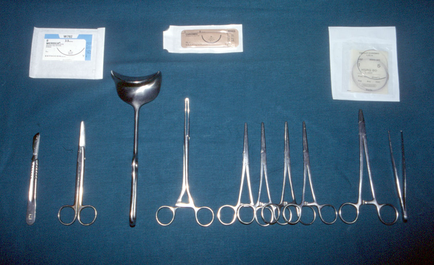
The minimalistic instrument set used in the Stark method is itself an expression of these same principles. A complete operation requires only 10 instruments: scalpel, straight surgical scissors with blunt/rounded tips, hand-held abdominal retractor (Doyen-type retractor or equivalent), four straight hemostatic clamps/forceps, surgical tissue forceps (thumb forceps/pincette), needle holder and ring forceps (sponge-holding forceps).6,13 This economy reduces cognitive clutter, simplifies counting, speeds up setup, and works just as well in high-income institutions as in resource-limited ones.6
GLOBAL APPLICABILITY: THE AFRICAN EXPERIENCE
The universal applicability of evidence-based obstetric surgical principles has been demonstrated in studies from Africa. Stark et al. reported on the introduction of the Misgav Ladach method across several African countries, with the aim of facilitating its wider adoption across the continent.6
The results have consistently mirrored the global evidence. In Dar es Salaam, the operation produced shorter operative time, less suture material use, reduced blood loss and faster mobilization.6 At Nazareth Hospital in Kenya, average operative time fell significantly (20.4 min vs 30.4 min), with fewer wound infections, markedly less need for analgesics, and fewer hypertrophic scars at 6-week follow-up, prompting the authors to recommend the method as the standard for low-income countries and rural hospitals.6 In Senegal, investigators reported shorter skin-incision-to-delivery time, reduced suture material and lower operative costs, and suggested the method as an alternative to the traditional approach.6 In Burkina Faso, where 86.5% of obstetricians and 95.3% of nurses performing cesarean sections had already adopted the Misgav Ladach technique, a comparative study concluded it was 'a reliable technique, fast and simple which eliminates unnecessary time of surgery and limits the risk of complications' and recommended its dissemination nationwide.6
The All-African Surgical Database, developed by the New European Surgical Academy together with the Marchuk Institute of Numerical Mathematics of the Russian Academy of Sciences, was built specifically to allow standardized electronic reporting of individual operative steps and postoperative follow-up, making it possible to evaluate precisely how each operative detail influences outcome.6 It reflects a broader truth: quality improvement in surgery depends on structured data collection, comparison and feedback.6,11
The African experience makes one point unmistakably clear: universal operative principles are not a privilege of well-resourced settings. A standardized, evidence-based technique reduces complications, shortens operative time, lowers costs and improves outcomes precisely where the margin for error is smallest.6,14
CHRONIC PAIN AND LONG-TERM MORBIDITY
One of the most important, and historically underappreciated, consequences of CD is long-term postoperative pain. Systematic reviews have reported chronic postsurgical pain (CPSP) after CD at varying rates, including 15.2% at 3 months, 9.5% at 6 months and 5.0% at 12 months in one review, with a neuropathic component documented in a proportion of affected women.15,16 Individual studies report wider variation, with chronic pain persisting beyond 2 months ranging from 4% to 41.8%, depending on the definition used, follow-up interval and patient population.17 Chronic pain can impair daily function and affect breastfeeding, maternal-infant bonding, return to work and overall quality of life.15,16,17
The comparative study by Belci and colleagues provides direct evidence that operative technique may influence long-term morbidity.14 In women assessed at a mean of 5 years after surgery, neuropathic pain, assessed using the Leeds Assessment of Neuropathic Symptoms and Signs (LANSS), was reported in 16% of those who underwent the traditional Pfannenstiel operation compared with 2% of those who underwent the Misgav Ladach (Stark) method.14 Chronic pain persisting beyond 2 months affected 44% of women in the traditional group compared with 12% in the Stark group, while Visual Analog Scale pain scores were also significantly better in the Stark group.13,14 Pain in both groups was located mainly in and around the scar and was considered most likely attributable to involvement of the iliohypogastric nerve.14,18,19
The anatomical basis for this difference is relevant to surgical technique. The Pfannenstiel incision is positioned approximately 3 cm above the pubic symphysis, within the cutaneous territory of the iliohypogastric and ilioinguinal nerves, whereas the Stark modified Joel-Cohen incision is placed higher, above the arcuate line and away from these nerve distributions.13 The Pfannenstiel approach therefore carries a risk of nerve entrapment, stretch injury and ischemic neuropathy, which may manifest as numbness, dysesthesia, radiating inguinal pain and hyperalgesia persisting long after surgery.13,14
These findings support a broader principle in obstetric surgery: operative decisions should be evaluated in terms of both short- and long-term outcomes. Blood loss, operative time and early postoperative complications are important, but a fuller assessment should also consider chronic pain, nerve injury, adhesion formation, patient-reported quality of life and the implications for subsequent surgery.13,14 This is particularly relevant in obstetric patients who may undergo subsequent pregnancies or abdominal procedures involving the same scar and altered anatomy.13,14
The evidence therefore supports treating chronic pain as a potentially modifiable component of surgical morbidity rather than an inevitable consequence of CD. Incision site selection, minimizing suture material, avoiding unnecessary dissection and using validated minimalistic techniques may reduce long-term morbidity.13,14,15 Severe acute postoperative pain on the first day has also been identified as a contributing factor to CPSP, reinforcing the importance of multimodal analgesia and adequate acute pain management within the perioperative framework.17
UNIVERSAL OPERATIVE PRINCIPLES APPLIED TO OBSTETRIC SURGERY
Correct indication and operative planning
The first universal principle of obstetric surgery is a sound indication.7,11 No amount of technical skill compensates for a poorly justified operation, or for a decision delayed when intervention is clearly needed.7,11,12 For CD, establishing the right indication means weighing maternal condition, fetal status, labor progress, prior uterine surgery, placental characteristics, and a realistic assessment of the balance between benefit and risk.7,12
Once the indication is settled, operative planning begins: determining urgency, anticipating technical difficulty, deciding whether senior surgical presence is needed, agreeing an anesthesia strategy, confirming neonatal preparedness, antibiotic prophylaxis, a thromboprophylaxis plan, blood product availability, and the likelihood of escalation beyond a standard CD.7,11,12 Planning is particularly important when performing repeat CD, when placenta accreta spectrum is suspected, in patients with major obesity, and in any setting in which significant hemorrhage can reasonably be anticipated.7,8
Respect for anatomy in the pregnant patient
Pregnancy changes the size, position, vascularity and mechanical relationships of the pelvic organs, abdominal wall, bladder and adnexa, making reliance on rote surgical maneuvers potentially hazardous.1,7 The lower uterine segment may be thin and well-formed in one patient and thick or poorly developed in another. The bladder reflection may sit high or be obscured by adhesions. Placental location can determine the safest point of uterine entry.7,8 A universal principle, then, is active reorientation to anatomy at every stage rather than reliance on habitual movement alone.1,7
This becomes especially critical in abnormal placentation and hysterectomy, where pelvic tissues are highly vascular and edematous, tissue planes may be poorly defined, and the ureters and bladder are at real risk.7,8 Under these conditions, deliberate exposure, vascular awareness and continuous orientation to known and uncertain structures matter more than speed.7
Gentle tissue handling and minimization of trauma
Gentle tissue handling is supported by both biological rationale and comparative evidence in obstetric surgery.1,10,13 Repetitive traction, unnecessary sharp dissection, excessive electrocautery, forceful blunt spreading, and prolonged manipulation all contribute to bleeding, edema, pain, adhesion formation and slower recovery.1,10 The Stark approach evaluates every operative step for its necessity, replacing tradition-based steps with evidence-based ones and systematically reducing tissue trauma along the way.1,6,13
Minimizing trauma has both immediate and long-term value. In the short term, it reduces blood loss, analgesic requirement, postoperative fever and delays in return of bowel function.1,10,13 Over time, it shapes adhesion formation, chronic pelvic pain, nerve injury, the complexity of any repeat surgery and future obstetric risk.13,14 Because many obstetric patients go on to have further pregnancies or abdominal procedures, tissue preservation matters well beyond the first postoperative week.13,14
Surgical exposure and hemostasis
Adequate exposure is a prerequisite for safe obstetric surgery: poor exposure leads to uncertain incisions, poorly directed hysterotomy expansion, missed bleeding points and a higher risk of structural injury.7,10 Surgeons should aim for the least traumatic approach that still provides a clear field, while remaining ready to widen exposure whenever anatomy or bleeding demands it.7
Hemostasis in obstetric surgery has to be approached proactively rather than reactively. Postpartum hemorrhage associated with CD is frequently underestimated and arises from uterine atony, abnormal placentation, uterine trauma, and occasionally sepsis or coagulopathy.20,21 The underlying hemostatic principle is sequential: improve exposure, identify the likely source, obtain temporary control, then proceed to definitive repair, devascularization, compression sutures, tamponade or hysterectomy, depending on cause and maternal stability.7,21 Blind suturing into a bleeding field can fail to control hemorrhage while creating additional injury.7
Patient safety systems and the operative team
Modern surgical safety literature is consistent on one point: adverse outcomes rarely arise from technical execution alone.11,12 Failures of communication, equipment readiness, infection prevention, counting, escalation and postoperative surveillance repeatedly contribute to preventable harm.11,12 The WHO’s Safe Surgery Saves Lives initiative responded to these realities by defining core safety objectives and formalizing the Surgical Safety Checklist.11
That checklist remains directly applicable to obstetric surgery, and its value grows further when it is specifically adapted to the maternity environment: incorporating fetal considerations, urgency classification, neonatal team readiness, hemorrhage risk, uterotonic strategy, antibiotic prophylaxis timing, and thromboprophylaxis planning.7,11,12 A systematic study of an adapted WHO checklist for cesarean sections demonstrated measurable improvements in team communication and adverse event rates.12
The briefing before CD should never be treated as a box-ticking ritual.11,12 It is the moment when the surgeon communicates the operative indication, anticipated entry difficulties, placental concerns, expected fetal extraction issues, likely blood loss, necessary instruments, and the threshold for calling additional help.7,11 Non-technical skills matter just as much: communication should be explicit, closed-loop and unambiguous, and authority gradients should never prevent nursing staff, anesthetists or scrub personnel from voicing a safety concern.11,12
FIGO’s good practice recommendations on cesarean surgical technique extend this systems approach by emphasizing maternity-specific safety checks, hemorrhage anticipation and readiness for conversion to more complex surgery.7 In obstetric operations, where both maternal and neonatal patients are affected and deterioration can happen abruptly, systems thinking is inseparable from technical excellence.7,11
Infection prevention, thromboprophylaxis and recovery pathways
Prophylactic antibiotics given before skin incision reduce infectious morbidity in CD and are backed by high-certainty evidence.10,12 Skin preparation, maintaining asepsis, minimizing tissue trauma, meticulous hemostasis, and appropriate postoperative surveillance are equally relevant whether the operation is routine or complex.7,10,11
Venous thromboembolism prevention needs to be built into perioperative planning from the outset.7 Pregnancy, surgery, immobility, hemorrhage, obesity, infection and medical comorbidity can all coexist in the same patient, requiring an individualized assessment that is revisited after the operation as bleeding risk and the need for anticoagulation are re-evaluated.7
Enhanced Recovery After Cesarean (ERAC) shows how perioperative care and surgical technique work best as complementary domains rather than separate ones.22 Multimodal analgesia, reduced preoperative fasting, early feeding and early mobilization improve maternal recovery and may shorten length of stay without compromising safety.22 Early hydration and early ambulation are specifically recommended within the Stark method framework too, reflecting a natural alignment between minimalistic surgical technique and an accelerated recovery philosophy.6,13
Instrumentation
Instrumentation should make an operation efficient, safe and reproducible; it should never clutter the field, confuse the team or stand in for clarity of technique.1,9 Classical operative obstetrics texts remain important for teaching the underlying logic of tissue forceps, clamps, needle holders, scissors, retractors, suction devices and suture selection.3 Even so, contemporary practice increasingly recognizes that routine instrument trays often contain a large number of items that are rarely or never used in a standard CD.23,24
A recent assessment of instrument utilization in cesarean births found that a substantial proportion of tray items go unused in routine operations, and a systematic review spanning 45 studies on surgical tray optimization found that 19–89% of instruments could be removed from trays through evidence-based rationalization, with direct benefits for operating room workflow, counting accuracy, reprocessing costs and environmental sustainability.24
The Stark method puts this principle into practice by requiring only 10 instruments for a complete standard CD (Figure 1), as described above.6,13 Far from compromising safety, this rationalization improves it by giving both surgeon and scrub team a predictable, manageable and immediately familiar instrument set.6,13
1
The 10 essential instruments required for Cesarean delivery.
For routine CD, each unit should define a standardized core tray that covers the great majority of operations.6,23 Supplementary sets for difficult abdominal entry, major adhesiolysis, major hemorrhage, vessel ligation, compression sutures, tamponade and hysterectomy should be immediately available without disrupting the standard workflow of routine cases.7,23 This tiered approach reconciles efficiency with preparedness.23,24
Suture selection deserves the same explicit attention.3 The right material depends on tissue type, vascularity, infection risk, required duration of support, and the balance between security and tissue reactivity.3 Uterine closure, fascial repair, hemostatic sutures and skin approximation each place different demands on the surgeon, and needle geometry and handling characteristics directly affect how easily and accurately a repair can be performed in a deep or distorted field.3
Ergonomics and room organization round out the instrumentation domain. Patient positioning, operating table height, retractor selection, lighting, suction access, electrosurgical safety and the physical arrangement of team members all influence both performance and safety.25 Poor ergonomics increase fatigue, impair precision and can contribute to both technical error and musculoskeletal strain, effects that become most pronounced during prolonged or complex procedures.25
Long-term outcome as a surgical consideration
The principles described above have implications that extend beyond the immediate operation. Operative decisions should be judged not only by blood loss, operative time and early complications, but also by their potential long-term effects. Incision placement, nerve proximity, tissue handling, suture volume and adhesion formation may influence chronic pain, subsequent surgery and quality of life.13,14 This is particularly relevant in obstetric patients who may undergo subsequent pregnancies or abdominal procedures. Surgical technique should therefore aim to minimize unnecessary tissue trauma and other potentially modifiable contributors to long-term morbidity, alongside achieving immediate operative safety.13,14,15,17
APPLICATION OF UNIVERSAL PRINCIPLES TO CESAREAN DELIVERY
Abdominal entry
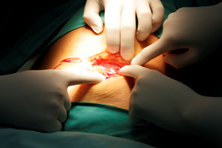
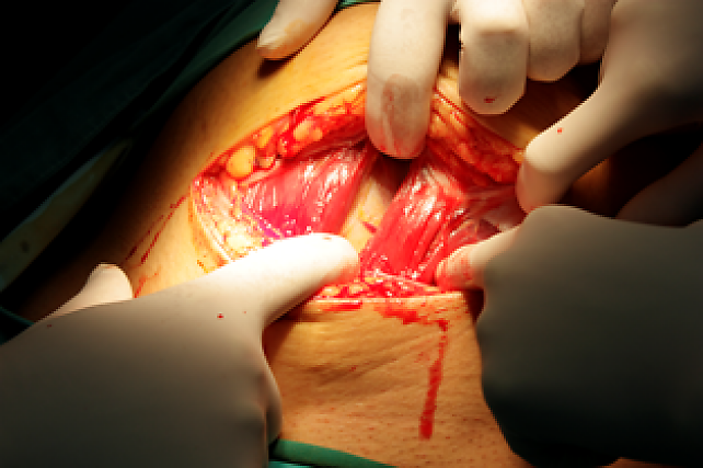
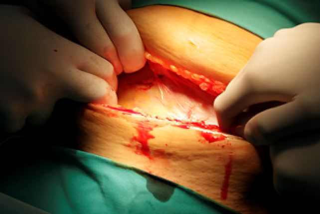
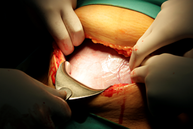
Abdominal entry should provide adequate exposure with minimum trauma (Figure 2).1,7 In the Stark approach, the skin incision is made approximately 3 cm below the line connecting both anterior superior iliac spines, above the arcuate line, in a straight line through the transverse skin creases (Video 1).1,6,13 Deepening the incision is confined to the midline, where no significant blood vessels are located, which eliminates the need for hemostasis in most cases.6,13 The fascia is opened with straight scissors using a controlled spreading technique that avoids injury to blood vessels and nerves.6,13
|
|
|
|
|
|
|
|



2
Key operative considerations when opening the abdomen: appropriate level and depth of the incision; preservation of the lateral compartments; avoid detaching the pyramidalis muscle; avoid separating the fascia from the rectus abdominis muscle; gently stretch the rectus abdominis muscle, taking care to protect the inferior epigastric vessels; avoid the use of retractors, as they may increase tissue trauma and reduce the effective operative space.
1
Misgav Ladach technique for opening the abdomen.
The muscles are separated by blunt finger dissection rather than fascial detachment, and the peritoneum is opened by repeated digital stretching rather than sharp incision, to protect underlying structures.6,13 This eliminates the need for fascial dissection from the rectus muscles, a step common to the Pfannenstiel method that adds time and bleeding risk and creates the conditions for nerve entrapment and chronic pain.13
In repeat CD, abdominal entry needs to be adapted to the degree of adhesions, the risk of bladder or bowel injury, and how well exposure can be achieved.8 Slower entry, clearer exposure and a willingness to diverge from standard technique are the right response when difficulty is unexpected.7,8
Uterine incision and fetal delivery
The vesicouterine fold is opened and the bladder pushed downward to expose the lower uterine segment, which is then incised transversely with a superficial 4-cm cut in the midline.1,6,13 The uterine wall is then opened bluntly by finger extension, right thumb to the left, left index finger to the right, minimizing muscle cutting and avoiding sharp injury to the presenting fetal part (Video 2).6,13
2
Misgav Ladach technique for uterine incision and single-layer repair.
Fetal extraction is not merely an obstetric maneuver but a coordinated surgical event that demands awareness of incision limits, fetal position, and clear communication with anesthesia and neonatal teams.7 In impacted or deeply engaged presentations, ad-hoc force can worsen extensions, increase blood loss and delay neonatal delivery.7 Rehearsed protocols and familiarity with alternative extraction strategies are essential parts of operative safety.7
Placental management and uterine closure
Spontaneous placental delivery is preferred; manual removal is reserved for cases in which spontaneous detachment fails to occur within the first few minutes, given its association with a slight increase in bleeding.6,13 After the placenta is removed, the uterus is exteriorized and compressed bimanually to reduce hemorrhage.6,13
Uterine closure should be performed in a single layer with a large needle (preferably 80 mm), using locked sutures to achieve immediate hemostasis.6,13 A second layer has not been shown to add value and simply increases the foreign body burden, since every suture left in situ can provoke tissue reaction, pain, adhesion and impaired healing.6,13
The peritoneum should not be sutured: new peritoneal tissue regenerates within days, and closure has been associated with increased adhesion formation.6,13 Only the fascia and skin require closure, with the fascia sutured to include both lateral layers above the arcuate line to prevent herniation (Video 3).6,13
3
Misgav Ladach technique for closure of the abdomen.
Abdominal wall closure and completion
Skin closure uses as few sutures as possible, typically one midline Donati suture with a large needle incorporating the subcutaneous tissue, followed by two additional Donati stitches at the quarter points.6,13 Minimal skin sutures allow better drainage and less foreign body reaction.6,13
Before the operation is concluded, the universal principles of completion apply: accurate instrument and swab counts, verification of hemostasis, confirmation of bladder and ureteric integrity where relevant, specimen labeling, and clear documentation of operative findings and any deviations from the planned technique.11,12
APPLICATION OF UNIVERSAL PRINCIPLES TO COMPLEX OBSTETRIC SURGERY
Complex and repeat cesarean delivery
Repeat CD with dense adhesions raises the risk of bladder, bowel and vascular injury during entry, calling for slower, more deliberate dissection and senior involvement whenever difficulty exceeds expectation.26 The FIGO guidelines for complex CD offer a standardized surgical approach for reducing the risk of intraoperative complications and postpartum hemorrhage, with an emphasis on dissection principles, bladder mobilization, pelvic access and hemorrhage control strategies that can be adapted across resource settings.8
Placenta previa and placenta accreta spectrum
Placenta previa and placenta accreta spectrum turn CD from a routine operation into one of the highest-risk procedures in obstetrics, demanding preoperative multidisciplinary planning, anticipation of distorted anatomy and avoidance of maneuvers likely to trigger uncontrollable hemorrhage.7,8 In placenta accreta spectrum, forced separation of an adherent placenta can be catastrophic; management should be guided by preoperative imaging and institutional capacity rather than reflex adherence to routine cesarean practice.7,8
Uterine rupture and hysterotomy extension
Effective repair of uterine rupture depends on identifying the full extent of the defect, controlling the vascular source and restoring anatomy in layers.7,20 Hurried, blind suturing into a distorted, bleeding field can worsen the injury without achieving hemostasis.7 Where repair is unsafe, or where preserving fertility is no longer the primary goal, timely hysterectomy is preferable to repeated conservative attempts that only prolong hemorrhage.7,20
Peripartum hysterectomy
Peripartum hysterectomy is a life-saving procedure when conservative measures fail, but it carries major morbidity, including massive transfusion, intensive care requirements and urological injury.20,21 Once the decision to proceed has been made, any delay caused by continuing ineffective conservative repair only worsens maternal instability.20,21 The operative principles here are the same throughout: clear exposure, anatomical orientation, early consideration of vascular control, protection of the ureters and bladder, and decisive teamwork.7,20
BMJ Best Practice guidance on postpartum hemorrhage emphasizes that early recognition depends on accurate quantification of blood loss combined with ongoing assessment of risk factors, vital signs and symptoms, and that surgical intervention should not be delayed once medical management is clearly failing.21
Postoperative hemorrhage and relaparotomy
Maternal deterioration after CD should raise suspicion of concealed intra-abdominal bleeding even when vaginal loss appears limited.20 Persistent tachycardia, hypotension, falling hemoglobin, oliguria or escalating abdominal pain after CD should prompt urgent reassessment, and relaparotomy should not be delayed when ongoing intra-abdominal hemorrhage is the most likely explanation.20,21
TRAINING, SIMULATION AND QUALITY IMPROVEMENT
Universal principles only work when they are embedded in departmental culture.6,9,11 Every obstetric unit should define its default CD technique, standard instrument tray, hemorrhage protocol, checklist process and escalation thresholds, and teach these explicitly to trainees and new staff rather than leaving them to informal apprenticeship.6,9
Simulation is especially valuable in operative obstetrics because some of the most dangerous events are simply too rare to be learned reliably from real clinical experience alone.7,11 Training should cover impacted fetal head extraction, major hemorrhage during CD, conversion to hysterectomy, uterine rupture repair, bladder injury management and postoperative deterioration requiring relaparotomy.7,11 It also needs to combine technical and non-technical skills, since communication failures and gaps in leadership remain recurrent contributors to major obstetric harm.11,12
Audit of process and outcomes closes the quality improvement loop. Units should track CD indications, quantified blood loss, antibiotic timing, checklist completion, transfusion rates, surgical site infection, return to theater, bladder and bowel injury, hysterectomy rate, chronic pain incidence, and patient-reported recovery outcomes.6,11,24 The data from General Hospital Pula, built up over 15 years of institutional standardization, illustrate how a department’s commitment to evidence-based technique produces a meaningful and analyzable outcome record.13,14
THE FUTURE OF OBSTETRIC SURGERY
The future of obstetric surgery will be shaped by the convergence of several trends: evidence-based simplification of technique, maternity-specific integration of safety systems, instrument rationalization, wider adoption of enhanced recovery pathways, and a stronger link between training, audit and measurable outcomes.1,7,11,22,23 CD will remain the principal platform for testing these ideas, precisely because it is common, resource-intensive and practiced across the full spectrum of health systems.1,6
The global experience with the Stark (Misgav Ladach) method shows that a standardized, evidence-based, minimalistic approach to CD works from sub-Saharan Africa to European tertiary centers, delivering gains in operative time, blood loss, pain, chronic morbidity and cost.1,6,13,14 Its underlying logic to interrogate each step, eliminate the unnecessary, respect the tissue and standardize evidence-based practice, extends to every obstetric surgical procedure and every level of the health system..5,6,9,13,27
Obstetric surgery is at its best when routine cases are reproducible, complex cases are anticipated and any deviation from standard practice is conscious, explainable and evidence-informed.1,6,9 Universal principles do not constrain good surgeons; they create the conditions in which judgment, teamwork and technical skill can be exercised to greatest effect.6,9,11
PRACTICE RECOMMENDATIONS
- Each obstetric unit should define and teach a standardized default technique for routine cesarean delivery (CD), specifying the rationale for each operative step and the circumstances that justify deliberate modification.
- Every obstetric operation should begin with explicit confirmation of indication, urgency, placental location, prior uterine surgery, hemorrhage risk and anticipated need for escalation to more complex surgery.
- A maternity-adapted surgical safety checklist should be used for all cesarean sections and major obstetric procedures, with both a pre-incision briefing and a post-closure debriefing.
- Gentle tissue handling, blunt rather than sharp dissection where the evidence supports it, and minimization of unnecessary operative steps should be treated as core quality principles.
- Surgeons should keep in mind that operative technique, including incision site and nerve proximity, is a modifiable determinant of chronic postoperative pain, and should choose technique with long-term as well as short-term outcomes in mind.
- Prophylactic antibiotics should be administered before skin incision in all cesarean deliveries, with antibiotic timing monitored as a quality indicator.
- Instrument trays should be standardized and optimized: a core tray of 10 instruments for routine CD, with supplementary packs immediately available for hemorrhage, hysterectomy or difficult entry.
- Peritoneal closure should not be performed routinely in CD, since the evidence supports non-closure to reduce adhesion formation and operative time.
- Postoperative deterioration after CD should trigger immediate consideration of concealed intra-abdominal or retroperitoneal hemorrhage, even in the absence of significant vaginal bleeding.
- Early oral hydration and early mobilization should be encouraged after uncomplicated CD, consistent with both ERAC principles and the recommendations within the Stark method framework.
- Departments should use simulation to train both technical and non-technical responses to rare, high-risk obstetric surgical emergencies, including impacted fetal delivery, massive hemorrhage and peripartum hysterectomy.
- Ongoing audit of technique, complications, chronic morbidity and instrument utilization should inform a culture of continuous quality improvement.
CONFLICTS OF INTEREST
The author(s) of this chapter declare that they have no interests that conflict with the contents of the chapter.
Feedback
Publishers’ note: We are constantly trying to update and enhance chapters in this Series. So if you have any constructive comments about this chapter please provide them to us by selecting the "Your Feedback" link in the left-hand column.
REFERENCES
Stark M. The Stark (Misgav Ladach) cesarean delivery – a streamlined surgical technique: development, rationale, and clinical outcomes. Am J Obstet Gynecol. 2026;233(6S):S55–S68. | |
World Health Organization. WHO Guidelines for Safe Surgery 2009: Safe Surgery Saves Lives. Geneva: WHO Press; 2009. | |
Cunningham FG, Gilstrap LC. Cunningham and Gilstrap’s Operative Obstetrics. 3rd ed. New York: McGraw-Hill; 2017. | |
Stark M. An evidence-based cesarean section suggested for universal use. J Perinat Med. 2021;49(7):806–808. | |
Stark M. Evidence-Based Cesarean Section for Universal Use. Glob Libr Women's Med. 2026. ISSN: 1756–2228; DOI: 10.3843/GLOWM.422183. https://www.glowm.com/article/heading/vol-20-surgical-techniques-in-obstetrics-evidencebased-cesarean-section-for-universal-use/id/422183 | |
Stark M, Mynbaev O, Belci D, Danilov A, Vassilevski Y, Ogutu O. Toward universal unified cesarean section method in Africa. East Afr Sch J Med Sci. 2020;3(10):5–9. | |
Nunes I, Nicholson W, Theron G; FIGO Childbirth and PPH Committee. FIGO good practice recommendations on surgical techniques to improve safety and reduce complications during cesarean delivery. Int J Gynaecol Obstet. 2023;163(Suppl 2):21–33. | |
Nieto-Calvache AJ, Palacios-Jaraquemada JM, D’Antonio F, Bhide A, Calì G, Collins SL, et al. Complex cesarean section: surgical approach to reduce the risks of intraoperative complications and postpartum hemorrhage. Int J Gynaecol Obstet. 2025;168(3):987–998. | |
Stark M, Gerli S, Di Renzo GC. The importance of analyzing and standardizing surgical methods. J Minim Invasive Gynecol. 2009;16(2):122–125. | |
Dahlke JD, Mendez-Figueroa H, Rouse DJ, Berghella V, Baxter JK, Chauhan SP. Evidence-based surgery for cesarean delivery: an updated systematic review. Am J Obstet Gynecol. 2013;209(4):294–306. | |
World Health Organization. The WHO Surgical Safety Checklist. In: WHO Guidelines for Safe Surgery 2009. Geneva: WHO Press; 2009. | |
Parra-Saavedra M, et al. Adaptation of the WHO Safe Surgery Checklist for cesarean sections: impact on team communication and adverse events. BMC Health Serv Res. 2024. PMCID: PMC11214655. | |
Belci D, Di Renzo GC, Zoričić D, Tinelli A, Malvasi A, Stark M. Less is more – a minimal approach technique for cesarean section. Clin Exp Obstet Gynecol. 2021;48(3):478–482. | |
Belci D, Di Renzo GC, Stark M, Ðurić J, Zoričić D, Belci M, Peteh LL. Morbidity and chronic pain following different techniques of caesarean section: a comparative study. J Obstet Gynaecol. 2015;35(5):442–446. PMID: 25384067. | |
Wang LZ, Huang JY, Hu HJ, Xia F. Incidence of chronic postsurgical pain after caesarean delivery: a systematic review and meta-analysis. Anaesthesia. 2025;80(7):834–845. PMID: 40070006. | |
Borges NC, de Deus JM, Guimarães RA, Conde DM, Bachion MM, de Moura LA, et al. The incidence of chronic pain following Cesarean section and associated risk factors: a cohort of women followed up for three months. PLoS One. 2020;15(9):e0238634. PMCID: PMC7473578. | |
Loos MJA, Scheltinga MRM, Mulders LGM, Roumen RMH. The Pfannenstiel incision as a source of chronic pain. Obstet Gynecol. 2008;111(4):839–846. PMID: 18378742. | |
Kalsi J, Shovelton C, O’Dwyer S, Walker S, Collis R. Chronic pain after cesarean delivery: a systematic review. Obstet Gynecol. 2024 (Epub). | |
Sng BL, Sia ATH, Quek KH, Woo D, Lim Y. Incidence and risk factors for chronic pain after caesarean section under spinal anaesthesia. Anaesth Intensive Care. 2009;37(5):748–752. | |
Madar H, Froeliger A, Kayem G, Bouchghoul H, Sentilhes L. Management of immediate and delayed postpartum hemorrhage with cesarean delivery. Am J Obstet Gynecol. 2026;233(6S):S314–S330. | |
Weeks A, Fawcus S. Postpartum hemorrhage. BMJ Best Practice. Updated June 2026. Available from: https://bestpractice.bmj.com/topics/en-us/3000329 | |
Macones GA, Caughey AB, Wood SL, Wrench IJ, Huang J, Norman M, et al. Enhanced Recovery After Cesarean (ERAC) Consensus Statement. Anesth Analg. 2021;132(5):1110–1120. | |
Belderes M, Subbiah S, Payne D, Cresswell K. Assessment of instrument utilization in cesarean births: a quality improvement study. Jt Comm J Qual Patient Saf. 2026;52(2):75–79. | |
Wong YT, Haddad FS, Konopka C. Reducing surgical instrument usage: a systematic review of tray optimization strategies. BJS Open. 2025;9(3):zraf030. | |
Venneri F, Caporale V, Scorzolini M, Baldazzi G. Safe Surgery Saves Lives. In: Donaldson L, Ricciardi W, Sheridan S, Tartaglia R, editors. Textbook of Patient Safety and Clinical Risk Management. Springer; 2021. p. 285–300. | |
Jauniaux E, Hussein AM, Nieto-Calvache A. Complex Cesarean Section. Glob Libr Women's Med. 2026 ISSN: 1756–2228; DOI: 10.3843/GLOWM.422193. https://www.glowm.com/article/heading/vol-20-surgical-techniques-in-obstetrics-complex-cesarean-section/id/422193 | |
Stark M. Evidence-based cesarean section for universal use. In: The Continuous Textbook of Women’s Medicine Series – Surgical Techniques in Obstetrics. Vol. 20. London: GLOWM; 2026. |
Online Study Assessment Option
All readers who are qualified doctors or allied medical professionals can automatically receive 2 Continuing Professional Development points plus a Study Completion Certificate from GLOWM for successfully answering four multiple-choice questions (randomly selected) based on the study of this chapter. Medical students can receive the Study Completion Certificate only.
(To find out more about the Continuing Professional Development awards program CLICK HERE)
