
This chapter should be cited as follows:
Barinov S, Kochev D, et al., Glob Libr Women's Med
ISSN: 1756-2228; DOI 10.3843/GLOWM.422133
The Continuous Textbook of Women’s Medicine Series – Obstetrics Module
Volume 20
Surgical Techniques in Obstetrics
Volume Editors:
Professor Michael Stark, New European Surgical Academy (NESA) and Charité University Hospital, Berlin, Germany
Professor Sergej Barinov, Omsk State Medical University, Ministry of Health of Russia
Professor Gian Carlo Di Renzo, PREIS International School, Florence, Italy
Chapter
Zhukovsky Double-Balloon Tamponade in Uterus-Preserving Surgery for Placenta Accreta Spectrum: A Technical Description
First published: September 2026
Study Assessment Option
By answering four multiple-choice questions (randomly selected) after studying this chapter, readers can qualify for Continuing Professional Development points plus a Study Completion Certificate from GLOWM.
See end of chapter for details.

INTRODUCTION
This chapter describes the authors' operative approach, developed from their clinical experience, to distal hemostatic techniques and Zhukovsky double-balloon tamponade in conservative surgery for placenta accreta spectrum (PAS). Comparative evidence supporting superiority over alternative techniques is currently limited, and the described methods should be interpreted as expert technical guidance rather than evidence-based recommendations.
Massive postpartum hemorrhage remains a major challenge in modern obstetrics and significantly contributes to maternal morbidity and mortality.1 Retained placental tissue and secondary uterine atony are among the leading causes of obstetric hemorrhage, particularly in the postpartum period.2 Placenta previa, a major risk factor for abnormal placental attachment, complicates approximately 0.2–3.0% of pregnancies worldwide.3,4
In recent decades, the incidence of PAS has increased significantly, becoming a major and potentially life-threatening complication for both mother and child.5,6 Numerous studies report that the prevalence of PAS is strongly associated with rising cesarean section rates and continues to increase globally.7,8 This trend is largely attributed to the growing number of uterine scars, which predispose patients to abnormal placental attachment.5,6 Uterus-preserving surgery for PAS is increasingly being explored. However, a conservative approach requires a high level of surgical expertise.7 Despite advances in management, up to 75% of PAS cases undergo hysterectomy.8,9
Surgical management of women with PAS presents significant technical challenges that depend on intraoperative findings. Successful outcomes rely on detailed knowledge of pelvic anatomy and retroperitoneal vascular plexuses, as well as advanced surgical skills, including specialized techniques for hemostasis and dissection. Therefore, comprehensive training in understanding pelvic vascular anatomy and retroperitoneal structures is essential for improving outcomes,10 and management strategies should be individualized.
Currently, both proximal and distal hemostatic techniques are used in the surgical management of women with PAS. Proximal hemostasis typically involves ligation or occlusion of the internal iliac arteries. However, this approach is associated with certain surgical risks and may be insufficient to control bleeding from multiple cervico-isthmic collaterals and paravesical plexuses. Endovascular balloon occlusion of the pelvic vessels has been proposed to reduce intraoperative blood loss and facilitate hemostasis, while providing a clearer surgical field and improved visualization.11,12 However, PAS is often associated with a wide network of vascular collaterals, which can be a source of deep bleeding after occlusion of some pelvic vessels.13 Current evidence remains inconsistent, and several professional bodies express caution about routine use of temporary balloon tamponade of the iliac arteries in women with suspected PAS.14,15 It has been suggested that ligation of the uterine arteries (a distal hemostatic approach) prior to placental separation may reduce intraoperative and postpartum blood loss and reduce the need for hysterectomy.16 However, it should be noted that this technique targets only uterine arterial flow and may be less effective in advanced PAS, especially in cases with placental protrusion beyond the uterus.
Thus, the need arose to develop a simple, practical and widely accessible intraoperative method to limit blood loss during cesarean section in pregnant women with PAS.
INTRAUTERINE BALLOON TAMPONADE
Current World Health Organization guidance recommends uterine balloon tamponade for the management of postpartum hemorrhage unresponsive to first-line treatment, including uterotonics, provided it is used in settings with immediate access to further supportive interventions, including surgery.17 FIGO (2022) similarly recommends uterine balloon tamponade as part of the management of postpartum hemorrhage.18 Various balloon-based techniques have been described in the literature,19,20 including Zhukovsky balloon tamponade. In our experience, when used promptly by a trained specialist in a surgical setting with immediate access to definitive intervention, it can contribute to hemorrhage control.
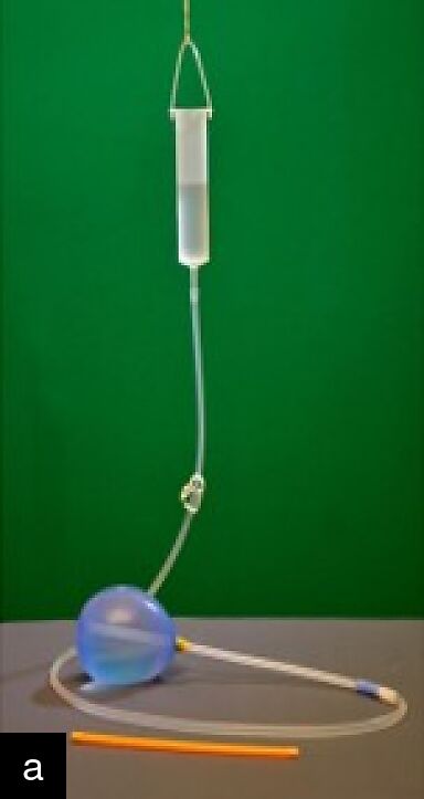
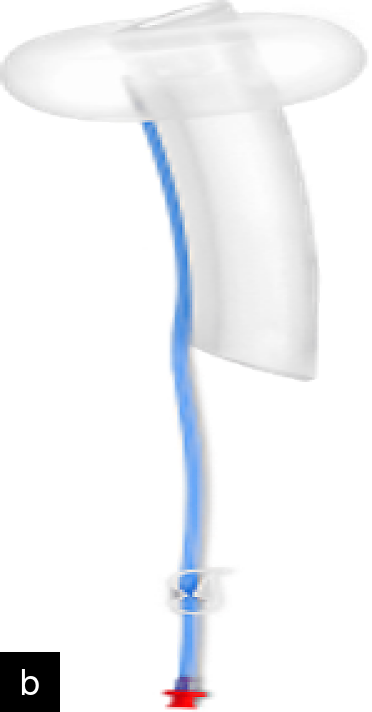
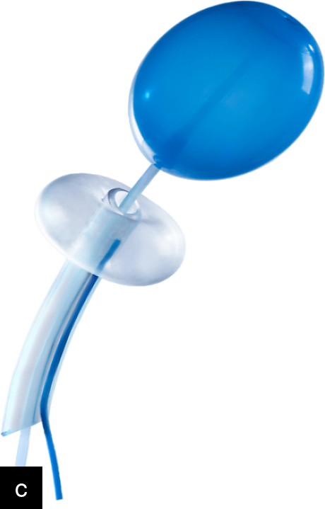
The Zhukovsky balloon system is available in three configurations: a uterine balloon, a vaginal balloon and a double-balloon system involving both a uterine and vaginal balloon21 (Figure 1). The double balloon has been shown to be effective in the management of postpartum hemorrhage following both vaginal delivery and cesarean section.22 Since 2014, the Perinatal Center of the Regional Clinical Hospital in the Omsk Region of Russia, has used a combined approach to cesarean section in patients with PAS using Zhukovsky double-balloon tamponade. This approach has been applied during delivery in pregnant women with abnormal placental attachment.
|
|
|
1
Zhukovsky balloon tamponade devices for uterine (a), vaginal (b) and double- (c) balloon tamponade.
Unlike other methods of distal hemostasis in the management of obstetric hemorrhage, the combined use of uterine and vaginal balloons in Zhukovsky’s method aims to provide simultaneous compression of multiple collateral vessels within the cervico-isthmus plexus as well as at the placental site (Figure 2). Specifically, the vaginal balloon exerts mechanical pressure on vascular territories supplied by the uterine and vaginal arteries, as well as branches of the internal pudendal artery, facilitating safe resection of the affected uterine wall. The uterine balloon compresses vessels at the placental site, maintaining prolonged tamponade of the lower uterine segment (typically for 10–14 hours). Following placement of the double-balloon device, absence of detectable blood flow in the lower uterine segment should be confirmed by Doppler examination. In our experience, this combined mechanism reduces the risk of bleeding in the parametrial tissue, pelvic hematoma formation and other hemorrhagic complications, thereby contributing to improved surgical outcomes.22
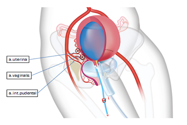
2
Mechanism of action of Zhukovsky double-balloon tamponade. Figure reproduced from Barinov and Di Renzo,23 under the terms of the CC BY licence.
PAS classification and operative decision-making
The operative strategy described in this chapter is based on the FIGO classification of PAS, together with assessment of the depth and extent of myometrial invasion. The principal features of each PAS grade and the corresponding operative approach are summarized in Table 1.
1
The summarized principal features of each grade of placenta accreta spectrum (PAS) and the corresponding operative approach.
FIGO PAS grade | FIGO definition | Extent of myometrial invasion | Operative approach |
Grade 1 (Accreta) | Chorionic villi are attached directly to the myometrium without intervening decidua. | No myometrial invasion. | Local excision of the placental bed, ligation of bleeding vessels, figure-of-eight sutures and external supraplacental compression suture. |
Grade 2 (Increta) | Chorionic villi invade the myometrium. | Myometrial invasion ≤ 1/3 thickness: local excision and repair. Myometrial invasion ≥ 2/3 thickness or defect > 5 cm: segmental uterine resection recommended. | Local excision and repair or segmental uterine resection with uterine reconstruction, according to depth and extent of invasion. |
Grade 3a (Percreta) | Chorionic villi penetrate through the myometrium to the uterine serosa. | Full-thickness invasion to the serosa. | Retrovesical dissection, vascular control, resection of the involved uterine wall and reconstruction. |
Grade 3b (Percreta) | Placental tissue invades the urinary bladder. | Full-thickness invasion with bladder involvement. | As for Grade 3a, with bladder dissection and repair if required. |
Grade 3c (Percreta) | Placental tissue invades adjacent pelvic tissues or organs (e.g. parametrium). | Extension beyond the uterus and bladder. | Hysterectomy is generally indicated if there is cervical or parametrial invasion or uncontrolled hemorrhage. |
Double-balloon Zhukovsky tamponade: surgical steps
Our approach to management of pregnant women with PAS, using double-balloon tamponade, includes surgical hemostasis (ligation of uterine vessels, including the descending branches of the uterine arteries, and placement of an external supraplacental uterine compression suture) and mechanical tamponade of bleeding sites using vaginal and uterine balloons.
The surgical technique described here is based on the approach previously reported by our group,23 with additional technical detail and practical considerations.
The approach consists of several sequential steps.
Step 1
Following bladder catheterization and before operative delivery, an uninflated vaginal Zhukovsky balloon catheter is inserted (Figure 3, Video 1).
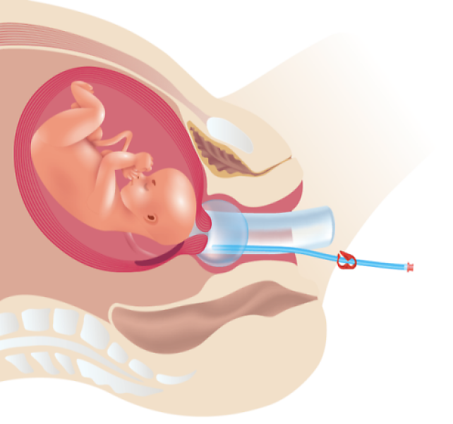
3
Insertion of Zhukovsky vaginal balloon before cesarean delivery. Reproduced from Barinov and Di Renzo,23 under the terms of the CC BY licence.
1
Insertion of the Zhukovsky vaginal balloon in a non-pregnant model.
Step 2
After preparation of the surgical field, a laparotomy is performed. A lower midline incision of the anterior abdominal wall is preferred, as, in our experience, it facilitates careful extraction of the fetus and optimal accessibility of the surgical field. On opening the abdominal cavity, the condition of the lower segment is assessed and the extent of placental invasion determined. In the presence of PAS, characteristic findings may include thinning and distension of the lower segment, lateral extension of the placenta toward the pelvic walls and increased vascularization (Figure 4).
|
|
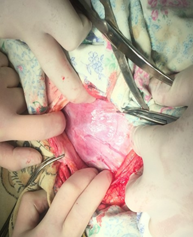
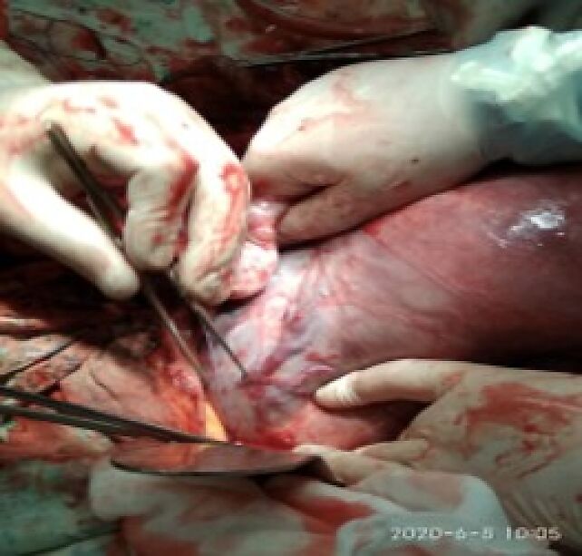
4
Evaluation of the condition of the lower uterine segment.
Step 3
Uterine incision is performed. The site and type of uterine incision should be individualized according to the location and extent of placental invasion, with the aim of avoiding transection of the placenta whenever possible.24 Our preference, when delivering a pregnant woman with PAS, is to perform a high transverse incision, as we have found that it does not lead to increased blood loss, and it may improve myometrial healing and reduce the risk of subsequent scar dehiscence.
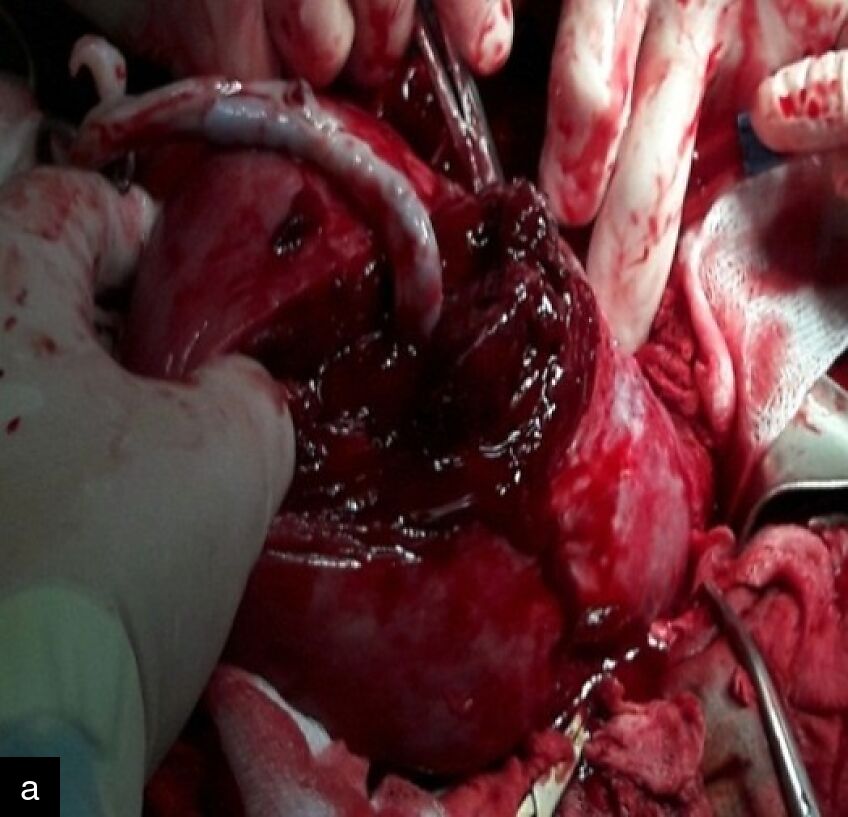
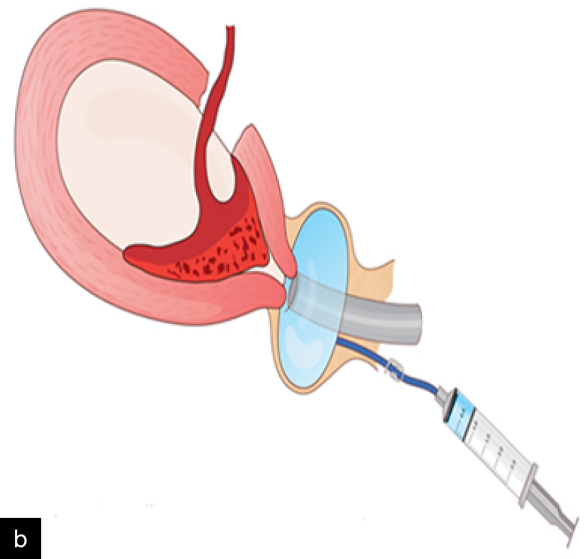
The transverse incision is made 4–5 cm above the presumed upper margin of the placenta (Video 2). The fetus is then delivered, the umbilical cord clamped and divided, and the stump ligated with a lavsan suture. Following fetal extraction, the vaginal catheter is filled with 180 ml of saline, and the procedure continued with the catheter in place (Figure 5).
2
High transverse incision.
|
|
5
(a) Surgical photo showing high transverse uterine incision and umbilical cord ligation. (b) Diagram showing inflation of a vaginal balloon catheter. Figure reproduced from Barinov and Di Renzo,23 under the terms of the CC BY licence.
Step 4
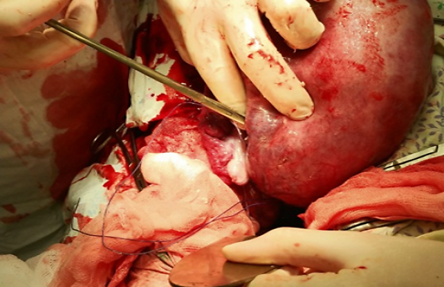
The uterus is exteriorized and the descending branches of the uterine arteries are ligated along the posterior aspect of the uterus (Figure 6, Video 3). In this approach, uterine vessel ligation is performed immediately after fetal delivery and placental separation. The exteriorized uterus is positioned toward the surgeon, while the assistant elevates the ovaries, to provide access to the blood vessels. The surgeon places his/her index finger along the posterior leaf of the broad uterine ligament to identify the vessels, which are then tied below the level of the internal cervical os with a needle and suture. The needle should be handled extremely carefully so as not to damage any parametrial vascular structures.
6
Uterine artery ligation. Figure reproduced from Barinov and Di Renzo,23 under the terms of the CC BY licence.
3
Uterine artery ligation.
Step 5
After separation and isolation, the placental site is examined. Further surgical steps are determined according to the FIGO PAS grade and the depth of myometrial invasion (Table 1).
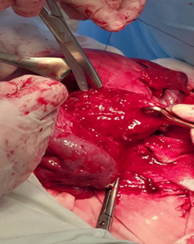
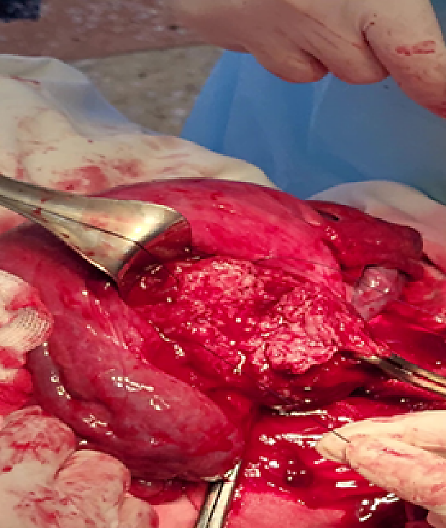
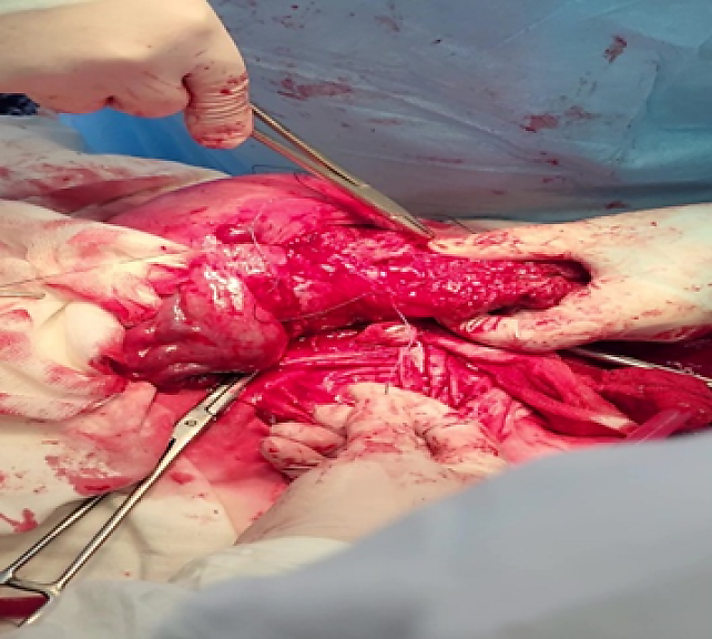
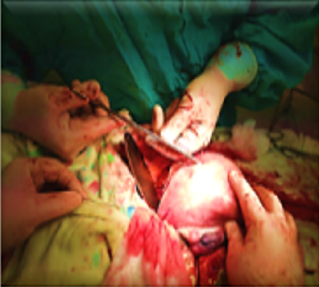
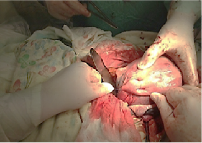
If placental invasion involves up to one-third of the myometrial thickness (PAS grades 1 and 2), excision of the affected area is performed with simultaneous ligation of bleeding vessels and suturing of the placental bed using figure-of-eight sutures. Subsequently, an external supraplacental uterine compression suture is applied (Figure 7).
|
|
|
|
|
7
Excision of the accretic area in a case of Grade 2 PAS (top row). Application of external supraplacental uterine compression suture (bottom row). Fourth image reproduced from Barinov and Di Renzo,23 under the terms of the CC BY licence.
External supraplacental uterine compression suture
The method we developed involves achieving hemostasis through local application of a supraplacental suture at the bleeding site i.e. the placental bed. In general surgery, hemorrhage control is commonly achieved by reducing inflow using a tourniquet above the bleeding site, and by direct compression of the bleeding vessel. Sustained compression of the vessel wall leads to its collapse and facilitates thrombosis, which provides a reliable stop to bleeding. By contrast, our proposed method avoids extensive longitudinal compression of the myometrium and whole intrauterine vascular network and, instead, targets the arcuate arteries supplying the placental bed, with minimal impact on the surrounding myometrial tissue. The technique is performed from the serosal surface of the uterus. The myometrium at the placental site is sutured using a half purse-string suture that does not penetrate the uterine cavity (Figure 8). The suture is placed within approximately one-third of the myometrial thickness and tightened transversely with a single thread to achieve hemostasis. The suture is secured with sufficient tension to control bleeding. This localized approach aims to minimize compression–ischemic damage to the myometrium and reduce intraoperative blood loss, surgical trauma and risk of intrauterine adhesions. In addition, it could potentially decrease the risk of bowel entrapment associated with long external sutures during uterine involution, and preserve normal lochial drainage in the postpartum period.
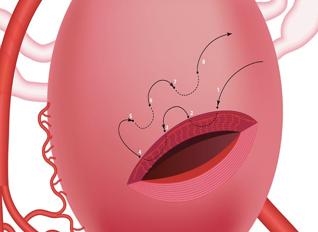
8
Diagram showing application of an external uterine supraplacental compression suture when placental invasion does not exceed one-third of the myometrial thickness. Figure reproduced from Barinov and Di Renzo,23 under the terms of the CC BY licence.
If placental invasion involves two-thirds or more of the thickness of the myometrium, or the area of invasion exceeds 5 cm in diameter, the affected portion of the uterus, including the invaded placental tissue, is excised. The uterine wall is then reconstructed using interrupted Vicryl sutures (Figure 9).
|
|
|
|
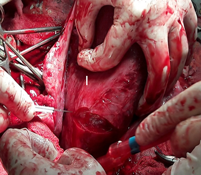
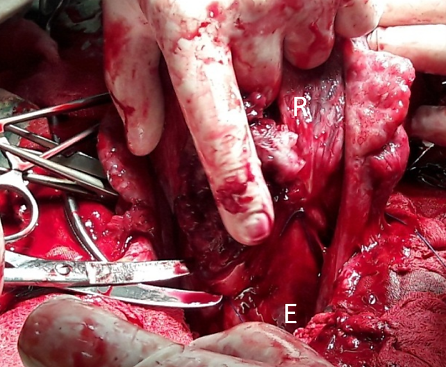
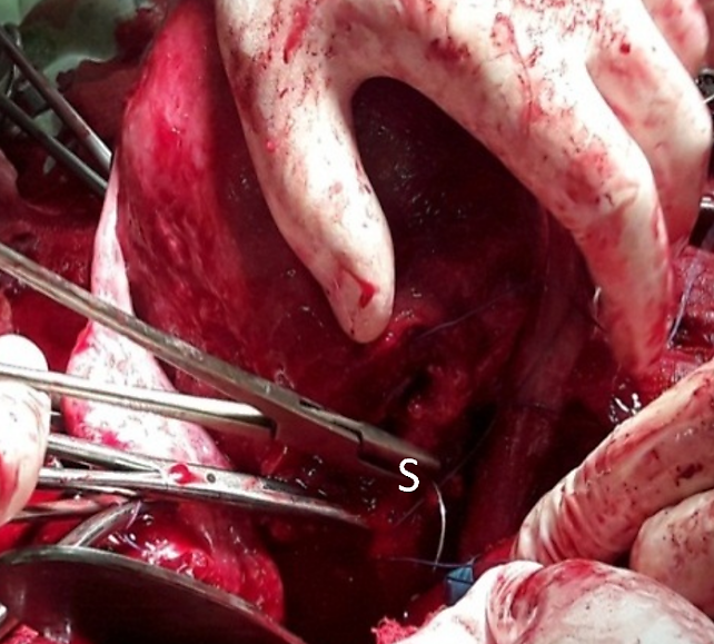
9
Resection of the uterine wall with an ingrown area of the placenta in a case of PAS 2. I, Uterine incision; R, Uterine wall resection; E, External supraplacental uterine compression suture; S, Uterine incision suturing.
Step 6
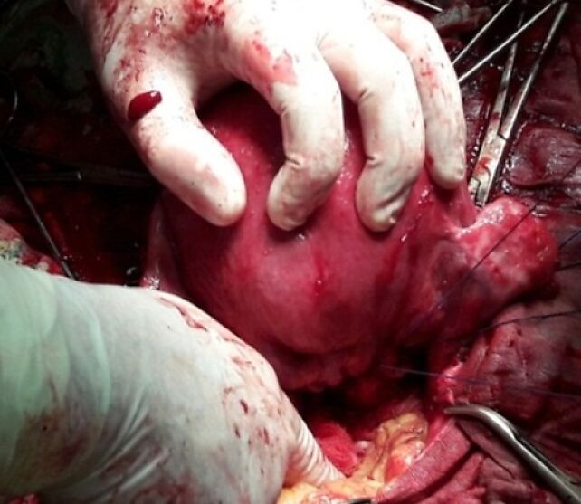
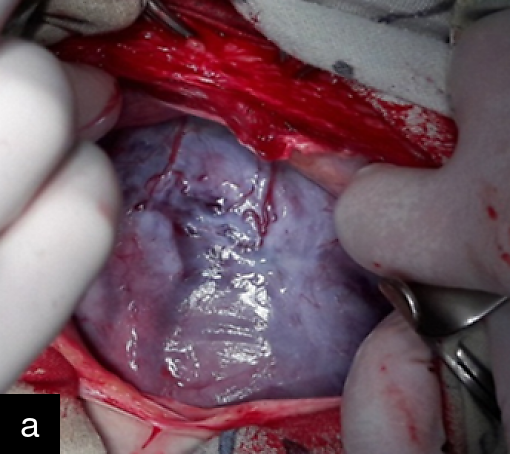
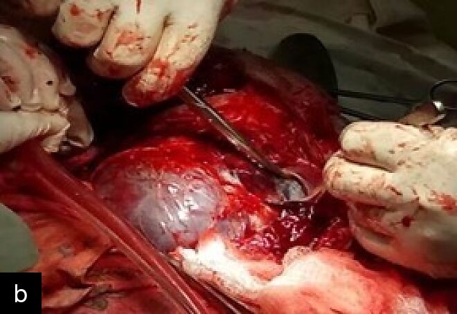
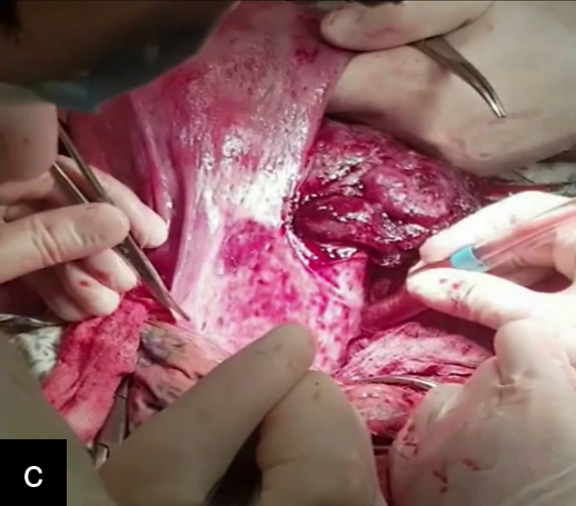
In cases of PAS Grade 3a or 3b, in which placental tissue protrudes beyond the uterine contour, dissection of the protruding sac and separation from the bladder are performed. The round uterine ligament is grasped with atraumatic clamps, and the anterior leaf of the broad uterine ligament is incised with scissors to access the space between the anterior uterine wall and the bladder (Figure 10). To create a working space, a retrovesical digital dissection (Pelosi maneuver) is performed (Video 4), elevating the vesicouterine fold and creating an ‘air cushion’, which facilitates less traumatic dissection. This approach allows assessment of both the depth of placental invasion and extension beyond the uterus. At the same time, ligation of the uterine artery at a higher level is performed. Additionally, when there are dense adhesions between the bladder and uterine walls (PAS 3b) resection of the bladder wall is performed.
|
|
|
10
(a) Placental protrusion beyond the uterine contour. (b) Opening of the broad uterine ligament. (c) Pelosi maneuver. Part (b) reproduced from Barinov and Di Renzo,23 under the terms of the CC BY licence.
4
Pelossi maneuver.
Step 7
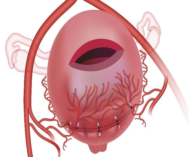
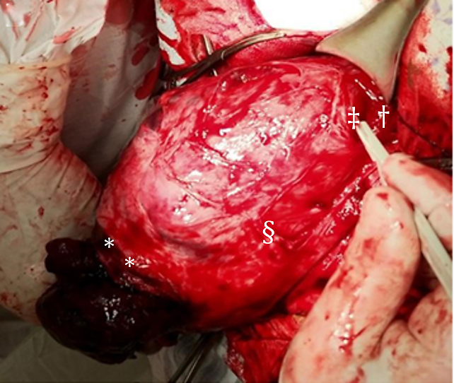
Following separation of the placental tissue from the bladder, vascular anastomoses along the anterior uterine wall and posterior bladder wall are ligated using Vicryl-0 sutures. After reaching the lower margin of placental invasion, additional ligation of the descending branch of the uterine artery is performed using Vicryl-1 sutures (lower level). Below the planned incision on the anterior uterine wall, an external supraplacental uterine compression suture is placed using a continuous suture, incorporating the vascular plexuses, and engaging approximately one-third of the myometrial thickness (Figure 11, Video 5). This is followed by sharp resection of the affected uterine wall containing the invasive placental tissue. As the uterine wall is resected (Figure 12, Video 6), hemostatic clamps are applied to the wound edges.
11
Application of external supraplacental uterine compression suture in case of PAS Grade 3. Numbers indicate sequence of needle insertions.
5
Placement of external supraplacental uterine compression suture
12
Resection of affected portion of uterus. Figure reproduced from Barinov and Di Renzo,23 under the terms of the CC BY licence. *, Uterine incision; †, Hemostatic external uterine assembly supraplacental suture; ‡, Uterine wall resection; §, Uterine wall with placenta accreta.
6
Resection of the uterine wall.
Step 8
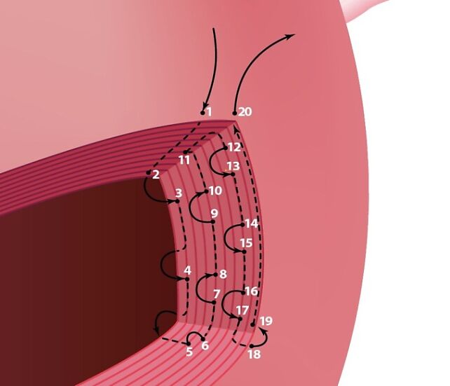
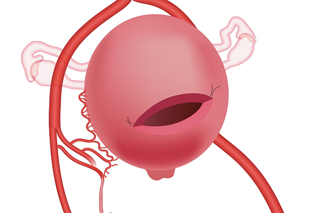
Restoration of uterine wall integrity is performed. Due to the large, resected area, approximation of the wound angles is the most challenging step. To address this, a continuous Vicryl-1 suture is used. A full-thickness suture is placed through the superior angle of the wound. The needle is then passed through the inner third of the incision margin using traction loops to advance towards the inferior angle. Finally, the needle is passed through the outer myometrial and seromuscular layers. After tightening the suture to achieve hemostasis and alignment, the process is repeated at the contralateral angle (Figure 13, Video 7).
|
|
13
Placement of continuous suture to reapproximate the uterine wound angles. Numbers indicate sequence of needle insertions. Figure reproduced from Barinov and Di Renzo,23 under the terms of the CC BY licence.
7
Reapproximation of uterine wound angles.
Step 9
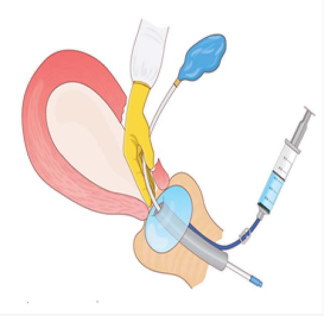
After suturing one-third of the wound on both sides of the uterus, an intrauterine catheter is inserted through the hysterotomy incision using a guidewire. The catheter is advanced manually using the surgeon’s index finger through the cervical canal into the central channel of the vaginal catheter (Figure 14, Video 8).
14
Placement of intrauterine catheter. Figure reproduced from Barinov and Di Renzo,23 under the terms of the CC BY licence.
8
Placement of Zhukovsky balloon.
Step 10
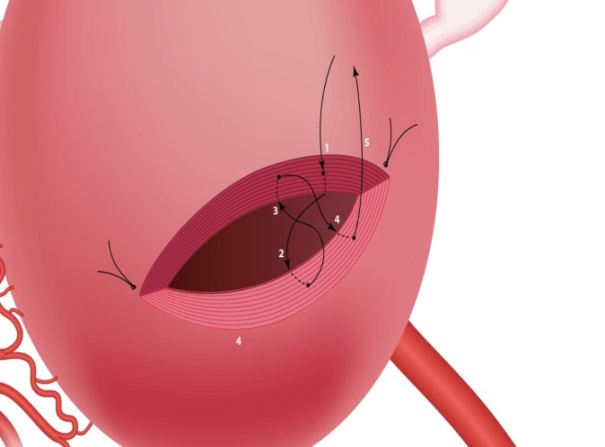
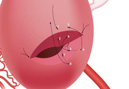
The edges of the uterine wound are approximated using two-layer single horizontal figure-of-eight sutures (Figure 15, Video 9).
|
|
15
Approximation of wound edges using two-layer single horizontal figure-of-eight sutures. Figure reproduced from Barinov and Di Renzo,23 under the terms of the CC BY licence.
9
Closure of uterine incision with two-layer single horizontal figure-of-eight sutures.
Step 11
Restoration of the anterior uterine wall is continued. A 10-cm sheet of regenerated cellulose (SurgicelTM, Ethicon) is placed as a local hemostatic and anti-adhesive barrier between the uterus and bladder.
Step 12
After suturing the wound on the uterus, the intrauterine catheter balloon is inflated with saline and the anterior abdominal wall is then closed. The balloon is left in place for 10–14 hours.
A critical consideration in clinical practice is determining the threshold for hysterectomy. In our opinion, indications for hysterectomy include invasion of the placenta into the cervix or parametrium, as well as development of uncontrollable intraoperative bleeding.
CONCLUDING REMARKS
We have presented the steps involved in our surgical approach to cesarean delivery in selected patients with PAS using distal hemostasis techniques and Zhukovsky double-balloon tamponade. The described uterine suturing technique is designed to promote the development of a well-healed scar and ultimately help preserve future fertility. In our experience, the technique appears to be effective, with the potential to reduce intraoperative blood loss and need for blood transfusion, need for hysterectomy, and duration of postoperative hospital stay, although comparative studies are required to support these findings.
Limitations and potential complications
We describe a technique of distal hemostasis in PAS disorders but do not compare it with methods of proximal hemostasis, such as temporary balloon occlusion of the iliac arteries or resuscitative endovascular balloon occlusion of the aorta (REBOA). Comparative multicenter studies are therefore needed to evaluate the relative effectiveness and safety of these approaches. Long-term follow-up is also required to assess uterine scar healing, subsequent fertility and pregnancy outcomes. In addition, late postoperative complications, including uterine ischemia, thromboembolic events and other adverse outcomes, have not yet been systematically evaluated. These complications will be studied in subsequent trials.
PRACTICAL CONSIDERATIONS
- Early multidisciplinary planning is essential in PAS management. Successful treatment of PAS requires experienced surgeons, detailed knowledge of pelvic anatomy, and individualized operative planning to reduce maternal morbidity and mortality.
- Uterus-preserving surgery should be considered in selected PAS cases. Conservative surgical approaches may reduce hysterectomy rates and help preserve future fertility when performed by appropriately trained teams.
- Double-balloon tamponade is an option to consider for distal hemostasis. Combined uterine and vaginal balloon tamponade aims to compress multiple collateral pelvic vessels simultaneously and improve hemorrhage control during PAS surgery.
- Balloon tamponade should complement surgical hemostatic techniques. Optimal hemorrhage control may be enhanced by combining balloon tamponade with uterine vessel ligation, compression sutures and targeted resection when required.
- A high transverse uterine incision may minimize surgical complications. In PAS cases, a high transverse incision above the placental edge may reduce blood loss and improve myometrial healing compared with lower-segment incisions.
- Early ligation of uterine vessels can reduce intraoperative bleeding. Ligation of descending uterine artery branches immediately after fetal delivery helps decrease blood loss during placental dissection and uterine reconstruction.
- Targeted compression of the placental bed using supraplacental compression sutures may help to achieve hemostasis while minimizing ischemic damage, intrauterine adhesions and distortion of uterine anatomy.
- The extent of placental invasion should guide surgical strategy. Limited invasion may be managed with local excision and repair, while extensive invasion requires segmental uterine resection and reconstruction.
- Careful bladder and retrovesical dissection is critical in advanced PAS. Techniques such as retrovesical digital dissection (Pelosi maneuver) can facilitate safer separation of placental tissue from the bladder and reduce operative trauma.
- Hysterectomy remains necessary in cases of uncontrolled bleeding or extensive invasion. Placental invasion into the cervix or parametrium, or failure to control hemorrhage should prompt timely conversion to hysterectomy for maternal safety.
CONFLICTS OF INTEREST
D.K. owns OU KODI OÜ and Pentcroft Pharma, which were involved in the distribution of the Zhukovsky double-balloon tamponade catheter described in this chapter.
Feedback
Publishers’ note: We are constantly trying to update and enhance chapters in this Series. So if you have any constructive comments about this chapter please provide them to us by selecting the "Your Feedback" link in the left-hand column.
REFERENCES
Society for Maternal-Fetal Medicine (SMFM). Electronic address: pubs@smfm.org, Gyamfi-Bannerman C. Society for Maternal-Fetal Medicine (SMFM) Consult Series #44: Management of bleeding in the late preterm period. Am J Obstet Gynecol. 2018;218(1):B2–B8. doi: 10.1016/j.ajog.2017.10.019. | |
Sentilhes L, Merlot B, Madar H, Sztark F, Brun S, Deneux?Tharaux C. Postpartum haemorrhage: Prevention and treatment. Expert Rev Hematol. 2016;9:1043–1061. | |
Fox KA, Lee W. Prenatal Diagnosis and evaluation of abnormal placentation. Clin Obstet Gynecol. 2017; 60(3):596–607; doi: 10.1097/GRF.0000000000000297. | |
Mogos MF, Salemi JL, Ashley M, Whiteman VE, Salihu HM. Recent trends in placenta accreta in the United States and its impact on maternal-fetal morbidity and healthcare-associated costs, 1998–2011. J Matern Fetal Neonatal Med. 2016; 29(7):1077–1082; doi: 10.3109/14767058.2015.1034103. | |
Thurn L, Lindqvist PG, Jakobsson M, Colmorn LB, Klungsoyr K, Bjarnadóttir RI, Tapper AM, Børdahl PE, Gottvall K, Petersen KB, Krebs L, Gissler M, Langhoff-Roos J, Källen K. Abnormally invasive placenta-prevalence, risk factors and antenatal suspicion: results from a large population-based pregnancy cohort study in the Nordic countries. BJOG. 2016;123(8):1348–1355. doi: 10.1111/1471-0528.13547. | |
Creanga AA, Bateman BT, Butwick AJ, Raleigh L, Maeda A, Kuklina E, Callaghan WM. Morbidity associated with cesarean delivery in the United States: is placenta accreta an increasingly important contributor? Am J Obstet Gynecol. 2015;213(3). doi: 10.1016/j.ajog.2015.05.002. | |
American College of Obstetricians and Gynecologists; Society for Maternal-Fetal Medicine. Obstetric Care Consensus No. 7: Placenta Accreta Spectrum. Obstet Gynecol. 2018;132(6):e259–e275. doi: 10.1097/AOG.0000000000002983. | |
Society of Gynecologic Oncology; American College of Obstetricians and Gynecologists and the Society for Maternal–Fetal Medicine, Cahill AG, Beigi R, Heine RP, Silver RM, Wax JR. Placenta Accreta Spectrum. Am J Obstet Gynecol. 2018;219(6):B2–B16. doi: 10.1016/j.ajog.2018.09.042. | |
Jauniaux E, Bhide A, Hussein AM, Lees C, D'Antonio F, Robson SC, Kaelin Agten A, Jurkovic D; Royal College of Obstetricians and Gynaecologists. Placenta Praevia and Placenta Accreta Spectrum: Diagnosis and Management: Green-Top Guideline No. 27a. BJOG. 2026 Sep;133(10):e93–e128. doi: 10.1111/1471-0528.70239. | |
Palacios-Jaraquemada JM, Fiorillo A, Hamer J, Martínez M, Bruno C. Placenta accreta spectrum: a hysterectomy can be prevented in almost 80% of cases using a resective-reconstructive technique. J Matern Fetal Neonatal Med. 2022;35(2):275–282. doi: 10.1080/14767058.2020.1716715. | |
Chen D, Xu J, Tian Y, Ling Q, Peng B. Clinical evaluation of the effect for prophylactic balloon occlusion in pregnancies complicated with placenta accreta spectrum disorder: A systematic review and meta-analysis. Int J Gynecol Obstet. 2024;167:109–127. doi: 10.1002/ijgo.15704 | |
Iskander R, Massalha M, Remer C, Izhaki I, Salim R. A systematic review and meta-analysis of randomized controlled trials on the effects of internal iliac artery balloon occlusion in placenta previa and placenta accreta spectrum in reducing perioperative bleeding. Int J Gynaecol Obstet. 2025 Sep;170(3):1061–1070. doi: 10.1002/ijgo.70118. Epub 2025 Apr 8. PMID: 40198092; PMCID: PMC12374018. | |
Morlando M, Collins S. Placenta Accreta Spectrum Disorders: Challenges, Risks, and Management Strategies. Int J Womens Health. 2020;12:1033–1045. Published 2020 Nov 10. doi: 10.2147/IJWH.S224191. | |
Collins SL, Alemdar B, van Beekhuizen HJ, Bertholdt C, Braun T, Calda P, Delorme P, Duvekot JJ, Gronbeck L, Kayem G, Langhoff-Roos J, Marcellin L, Martinelli P, Morel O, Mhallem M, Morlando M, Noergaard LN, Nonnenmacher A, Pateisky P, Petit P, Rijken MJ, Ropacka-Lesiak M, Schlembach D, Sentilhes L, Stefanovic V, Strindfors G, Tutschek B, Vangen S, Weichert A, Weizsäcker K, Chantraine F; International Society for Abnormally Invasive Placenta (IS-AIP).. Evidence-based guidelines for the management of abnormally invasive placenta: recommendations from the International Society for Abnormally Invasive Placenta. Am J Obstet Gynecol. 2019;220(6):511–526. doi: 10.1016/j.ajog.2019.02.054. | |
Allen L, Jauniaux E, Hobson S, Papillon-Smith J, Belfort MA; FIGO Placenta Accreta Diagnosis and Management Expert Consensus Panel. FIGO consensus guidelines on placenta accreta spectrum disorders: Nonconservative surgical management. Int J Gynaecol Obstet. 2018;140(3):281–290. doi: 10.1002/ijgo.12409. | |
Nabhan AE, AbdelQadir YH, Abdelghafar YA, Kashbour MO, Salem N, Abdelkhalek AN, Nourelden AZ, Eshag MME, Shah J. Therapeutic effect of Internal iliac artery ligation and uterine artery ligation techniques for bleeding control in placenta accreta spectrum patients: A meta-analysis of 795 patients. Front Surg. 2022 Sep 1;9:983297. doi: 10.3389/fsurg.2022.983297. PMID: 36117806; PMCID: PMC9474733. | |
WHO recommendation on Uterine balloon tamponade for the treatment of postpartum haemorrhage [Internet]. Geneva: World Health Organization; 2021. | |
Escobar MF, Nassar AH, Theron G, Barnea ER, Nicholson W, Ramasauskaite D, Lloyd I, Chandraharan E, Miller S, Burke T, Ossanan G, Andres Carvajal J, Ramos I, Hincapie MA, Loaiza S, Nasner D; FIGO Safe Motherhood and Newborn Health Committee. FIGO recommendations on the management of postpartum hemorrhage 2022. Int J Gynaecol Obstet. 2022;157(Suppl 1):3–50. doi: 10.1002/ijgo.14116. | |
Chen F, Zhang C, Hu Y. Efficacy of Bakri Intrauterine Balloon in Managing Postpartum Hemorrhage: A Comparative Analysis of Vaginal and Cesarean Deliveries with Placenta Accreta Spectrum Disorders. Med Sci Monit. 2024 Mar 4;30:e943072. doi: 10.12659/MSM.943072. PMID: 38433445; PMCID: PMC10921967. | |
Ozgen G, Aydin GA. Effectiveness of Intrauterine Bakri Balloon® Tamponade for Placenta Previa and Placenta Accreta Spectrum. J Coll Physicians Surg Pak. 2020 Jul;30(7):707–712. doi: 10.29271/jcpsp.2020.07.707. PMID: 32811599. | |
Barinov S, Tirskaya Y, Medyannikova I, Shamina I, Shavkun I. A new approach to fertility-preserving surgery in patients with placenta accreta. J Matern Fetal Neonatal Med. 2019;32(9):1449–1453. doi: 10.1080/14767058.2017.1408066. | |
Barinov SV, Shmakov RG, Medyannikova IV, Tirskaya YI, Kadtsyna TV, Lazareva OV, et al. Efficacy of distal haemostasis during caesarean delivery in women with placenta accreta spectrum disorders. J Matern Fetal Neonatal Med. 2022;35(25):8778–8785. doi: 10.1080/14767058.2021.2005019. | |
Barinov SV, Di Renzo GC. A new technique to preserve the uterus in patients with placenta accreta spectrum disorders. Am J Obstet Gynecol. 2024;230(3S):S1107–S1115. doi: 10.1016/j.ajog.2023.07.012. | |
Palacios-Jaraquemada JM. Caesarean section in cases of placenta praevia and accreta. Best Pract Res Clin Obstet Gynaecol. 2013;27(2):221–232. doi: 10.1016/j.bpobgyn.2012.10.003. |
Online Study Assessment Option
All readers who are qualified doctors or allied medical professionals can automatically receive 2 Continuing Professional Development points plus a Study Completion Certificate from GLOWM for successfully answering four multiple-choice questions (randomly selected) based on the study of this chapter. Medical students can receive the Study Completion Certificate only.
(To find out more about the Continuing Professional Development awards program CLICK HERE)
