This chapter should be cited as follows:
Salcedo AMC, Guarin CB, et al., Glob Libr Women's Med
ISSN: 1756-2228; DOI 10.3843/GLOWM.415713
The Continuous Textbook of Women’s Medicine Series – Obstetrics Module
Volume 6
Pregnancy complaints and complications
Volume Editor: Professor Gian Carlo Di Renzo, PREIS International School, Florence, Italy
Chapter
Surgery and Laparoscopy in Pregnancy: Feasibility and Safety
First published: August 2021
Study Assessment Option
By answering four multiple-choice questions (randomly selected) after studying this chapter, readers can qualify for Continuing Professional Development points plus a Study Completion Certificate from GLOWM.
See end of chapter for details.

INTRODUCTION
Minimally invasive surgery during pregnancy has represented one of the great advances in the care of obstetric patients requiring surgical management. Laparoscopy was considered a contraindicated technique in pregnant patients given the possible risks of gestational loss, preterm delivery, fetal hypoxia, and decreased uterine flow due to increased intra-peritoneal pressure1 in addition to the effect of pneumoperitoneum on fetal physiology2 and the potential risk of entry injuries, such as uterine perforation, fetal injury, or premature membranes' rupture. In recent years, data have shown that the multiple benefits of laparoscopic surgery are also applicable to the pregnant population.
Annually in the United States, around 1 in 200 to 1 in 500 pregnant women undergo non-obstetric abdominal surgeries. The vast majority constitute emergency abdominal surgical pathologies, such as appendicitis, cholelithiasis, and adnexal/torsion masses. Laparoscopy is the most frequently used approach, with very low complication rates.3
The objective of this review is to describe the recommendations for the use of the laparoscopic technique in pregnant patients, the advantages and disadvantages of the technique, the physiological changes of pregnancy that must be taken into account when choosing this route, and its potential complications and the relevant ways to prevent them.
PHYSIOLOGY OF PREGNANCY AND ANESTHESIOLOGY CONSIDERATIONS
Pregnant women have several physiological adaptations, initially generated by hormonal changes, and later related to mechanical factors, such as increased uterine size, fetal metabolic needs, and low resistance of placental circulation.4
Respiratory System
There is a 20% increase in oxygen consumption and a 20% decrease in pulmonary functional residual capacity, both of which contribute to the rapid decrease in maternal PaO2 that is observed even during a brief apnea.5 The risk of hypoxemia associated with general anesthesia induction can be accentuated by additional conditions, such as maternal obesity and pathologies specific to pregnancy, such as hypertensive disorders of pregnancy.
Another respiratory change includes maternal hyperventilation, mediated by progesterone-enhanced sensitivity of the brainstem to PaCO2. This effect is counteracted in the patient under general anesthesia by a greater sensitivity of the central nervous system to general anesthetics.
In addition, the pregnant woman's airway presents oropharyngeal friability and edema contributing to a decrease in the size of the glottis opening, starting from the middle of the second trimester and being more significant at the end of pregnancy. All these changes can cause difficulties for orotracheal intubation and for ventilation of the unconscious patient.
Rocke et al.6 published a study of 1500 patients undergoing cesarean section under general anesthesia. In women with Mallampati classification class III–IV, the relative risk of difficult intubation was 7.5 (compared with 11.3 in those with class I). In conclusion, the Mallampati classification is a better predictor of difficulty for orotracheal intubation in pregnant patients compared to non-pregnant ones.
Pilkington et al.7 took photographs of the airway evaluation in 242 pregnant women and found that from 12 to 38 weeks of gestation, the incidence of Mallampati classification class IV increased by 34%, findings that were also correlated with maternal weight gain.
The high incidence of failed intubation in pregnant women is a matter of debate in the literature. Not all pregnant women are difficult to intubate, but the main concern is as more pregnant patients get regional anesthesia techniques, the incidence of failed intubation has increased.8,9
Loss of airway control is the most frequent cause of maternal mortality associated with anesthesia.10
Hemodynamic Changes
Pregnant patients have a 40–50% increase in blood volume and cardiac output, and hematocrit reduction by 20% due to hemodilution.11
Physiological anemia begins during the first trimester of pregnancy and is more evident in the second trimester, after which it is balanced by an increase in red blood cell production depending on whether iron stores are adequate.12
Aortocaval compression is more important for the anesthesiologist after the second trimester. The supine position can predispose to maternal hypotension, especially after 20 weeks of gestation.
Additionally, uterine growth can lead to a decrease in venous return from the lower limbs, predisposing tibial edema and an increased risk of developing venous thromboembolism.
Surgical teams need to be aware of these changes so that left uterine displacement in advanced pregnancies during surgery and anesthesia is carried out.13
Gastrointestinal System
The risk of bronchial aspiration of gastric contents increases during pregnancy. Gastric emptying has been shown to be normal during pregnancy, but there is a lower esophageal sphincter pressure reduction14,15 and a gastric and pyloric anatomy distortion. The risk of regurgitation of the gastric contents and aspiration pneumonia are increased in general anesthesia when performed in pregnant women compared to the general population.16
After 20 weeks of gestation, more caution should be exercised with the unprotected airway, when general anesthesia is required. A laryngeal mask should only be used in carefully chosen patients. The combination of antacids with a H2 receptor antagonist and the general anesthesia rapid induction sequences are recommended.16
Obesity and a history of gastroesophageal reflux add additional risks for aspiration and cause a life-threatening event and increase the risk of post-operative lung infection.
Hematologic and Immune System Changes
There is an increase in plasma volume between 40 and 50% with a decrease in the mass of red cells by 20%, which leads to the development of physiological anemia, associated with a gradual increase in the leukocyte count at the expense of polymorphonuclear cells,17 which explains the increase in the severity of infections.
Pregnancy is a state of hypercoagulability as an adaptation process to the expected blood losses at the time of delivery. The risk of venous thromboembolism is up to 10 times during pregnancy and 25 times in the postpartum period, due to the increase in factors II, V, XII, and XIII, elevated levels of fibrin degradation, reduction in prothrombin time, and activated thromboplastin time.17
Central Nervous System
Cerebral blood flow is increased in pregnant women due to a decrease in cerebrovascular resistance. The permeability of the blood–brain barrier increases, leading to an increase in pain thresholds at the end of gestation, probably due to the effect of endorphins and progesterone.17 This translates into an increase in physiological sensitivity to sedative agents and anesthetic inducers. There is also a decrease between 30 and 50% of the needed doses of local anesthetics for spinal anesthesia.17
Renal System
The increase in renal flow leads to an increase in the glomerular filtration rate, with the consequent decrease in serum creatinine and urea nitrogen. There is a reduction in the response to vasopressors due to an altered expression of vascular receptors. Hydronephrosis and hydroureter are common findings because of the effect of progesterone and the mechanical compression by the gravid uterus.
Low levels of albumin lead to an increase in the bioavailability of protein-bound drugs.17
Endocrine System
The increase in thyroid transport globulin caused by estrogens leads to an increase of up to 50% in T3 and T4 levels.
Placental lactogen causes a reduction in insulin sensitivity with direct effects on blood glucose levels, and placental lactogen together with dopamine lead to hyperprolactinemia and increased oxytocin deposits.17
Musculoskeletal System
The effect of progesterone on the musculoskeletal system is given by joint hypermobility; there is a widening increased mobility of the sacroiliac joints and pubic symphysis, and also a joint laxity in the anterior longitudinal ligaments of the lumbar spine in preparation for childbirth.17,18
Transplacental Transfer of Drugs and Safety
The pharmacology of drugs in their bioavailability, distribution, and clearance can be affected during pregnancy by physiological, hormonal, and placental changes.
Liver enzyme changes affect absorption, metabolism, and may increase the activation of prodrugs. The availability of medications with the first hepatic step may be reduced. The increase in maternal weight leads to an increase in the medication distribution volume, requiring higher doses than in non-pregnant women. In addition, the increase in plasma volume induces the concentration of proteins in plasma and an increase in the volume of distribution of water-soluble drugs.16
The increase in cardiac output leads to an increase in glomerular filtration rate with its consequent clearance increase and, finally, decrease in tissue concentration of circulating medications.
At the placental level, passive transfer depends on fat solubility, molecular weight, and concentrations of the intermembrane gradient. Drugs that cross the blood–brain barrier will cross the placenta and the placenta expresses some enzymes that induce an increase in metabolism.16
Although anesthetic agents are teratogenic in animal studies, teratogenicity in humans has not been proven. Anesthetic agents, muscle relaxants, and opioids may not be teratogenic when used in clinical doses.19 Studies suggest that its use during pregnancy does not increase birth defects or the risks of gestational loss, growth restriction, or preterm delivery.19
SAFETY OF LAPAROSCOPIC SURGERY IN PREGNANCY
The complications' rate of laparoscopy during pregnancy seems to be comparable to the non-pregnant population. And benefits are also similar: less post-surgical pain, shorter hospital stay, earlier restart of bowel function, more rapid return to normal activity, and lower rates of post-surgical wound infection and hernias.19,20
There is no proven additional risk of fetal malformation or stillbirth in women who undergo non-obstetric laparoscopic surgery compared to controls. Maternal conditions may be associated with risk of abortion or preterm delivery.19
In a study carried out by Shigemi et al.,21 maternal–fetal outcomes of laparotomy vs. laparoscopy in 6018 patients performed between the first and second trimesters were compared. It was found that laparoscopic surgery (in terms of adverse fetal events in the 7 post-surgical days and preterm delivery) compared with laparotomy was safer and more cost-effective (−9.5% CI −1.39 to −0.52). In addition, the laparoscopy group showed less transfusions (2.3% vs. 0.41%, P = 0.002), shorter operative time (115 vs. 95 min, P <0.001), shorter hospital stay (9.2 vs. 5.9 days, P <0.001), and better maternal outcomes: Minimally invasive surgery was performed safely in most cases. Surgical indications were benign pathologies, such as appendicitis, cholecystitis, adnexal masses, and uterine fibroids.
A meta-analysis published by Liu et al.22 compared the safety of laparoscopy vs. laparotomy for the management of adnexal masses during the second trimester of pregnancy. Four studies were included. At conclusion, laparoscopy had a lower risk of post-operative adverse events (RR 0.20, 95% CI 0.06–0.72; P = 0.01). No differences were found in terms of post-surgical miscarriage risk between the groups (RR 0.28, 95% CI 0.03–2.53; P = 0.26). Laparoscopy is also associated with lower blood loss, lower post-surgical pain scores, and shorter hospital stay. Longer operative time (mean difference 13.70 min, 95% CI 12.58–14.82; P <0.001) was seen in the laparoscopy group. The final risk analysis suggested that laparoscopy is associated with a fifth lower risk of adverse events than laparotomy.
SURGICAL TECHNIQUE AND PERIOPERATIVE AND PERINATAL CARE
Patient Selection
The indication for surgery should be the same as in non-pregnant patients. Surgery should not be delayed due to pregnancy since it has been shown that the delay can affect the prognosis of both the mother and the fetus.23
Any surgical intervention during pregnancy, whether laparoscopic or open, can increase the risk of adverse results that may be secondary to the procedure or due to the initial condition causing the procedure.23
Laparoscopic treatment in acute abdominal pathology has similar benefits in pregnant and non-pregnant women compared to laparotomy (recommendation +++). The decision has to be based on the training and skills of the surgeon and the availability of the necessary staff and equipment.24
The benefits of minimal invasive surgery are well known. And during pregnancy, the benefits of laparoscopic surgery are crucial to obstetric morbidity, including less post-operative pain, less post-operative ileus, decrease in hospital stay, and faster recovery. In addition to lower risk of complications associated with surgical trauma, less maternal hypoventilation, reduction of thromboembolic events due to early mobilization. Laparoscopy also improves surgical field, requiring less uterine manipulation with less risk of developing uterine activity and preterm delivery.23,24 It is necessary to discuss with the patients the risk and benefits depending on the type of procedure.
Laparoscopy According to the Time of Pregnancy
Laparoscopy can be performed in any trimester of pregnancy when surgery is necessary (+++ strong). But there are data suggesting to avoid non-urgent procedures during the first and third trimesters of pregnancy, to minimize the risk of miscarriage and preterm delivery.24 But, urgent procedures can be performed at time gestational age.
Recent studies show laparoscopy is safe all through pregnancy and there is no increased risks to the mother or fetus.
In a case series published by Cohen et al., where the feasibility, immediate complications, and short-term results according to gestational age were studied. 12 patients underwent laparoscopic surgery in the third trimester with a gestational age range between 27 and 39 weeks, (7 appendectomies, 4 adnexal torsions, 3 cystectomies, and 1 diagnostic laparoscopy). 11/12 completed the surgery via laparoscopy. No complications related to entry were reported. Only two patients had immediate obstetric complications, one had premature contractions after a laparoscopic appendectomy that resolved with tocolytics and another had spontaneous rupture of the membranes after adnexal detorsion with aspiration cyst at 39 weeks. The length of hospital stay was 2 to 6 days.2
Patient Positioning
After the first trimester, patients should be placed in the left lateral decubitus or partial lateral decubitus position to minimize aortocaval compression. As described before, when the patient is placed in a supine position, the gravid uterus puts pressure on the inferior vena cava, resulting in a decrease in venous return, and leading to a reduction in cardiac output with the consequent hypotension and decreased placental perfusion during surgery. When placed in lateral decubitus, the uterus will be displaced from the vena cava, avoiding the described effects.24
Entry Techniques, Positioning of Ports and Lenses
Any abdominal entry technique can be safely used during pregnancy. Open technique (Hasson), Veress needle, or direct entry are equally safe and the decision should be based on the surgeon's training and experience, surgical volume, and individual patient's conditions, including uterine height.19,23,24 Complications associated with entry techniques have been described, where inadvertent uterine perforation with the Veress needle is the most common.23
The location of the primary port will depend on the level of the uterine fundus. It is suggested that, during the second and third trimesters, the use of alternative entry points, such as the Lee-Huang, Palmer, or Jain point.23 See figure 1 (positioning of trocars during pregnancy).
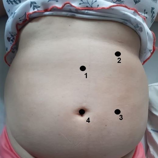
1
Positioning of trocars during pregnancy. 1, Lee Huang point; 2, Palmer's point; 3, Jain point; 4, supraumbilical point; 5, umbilical point.
When the Lee-Huang or the Palmer point is used, prior gastric decompression using an orogastric tube is recommended. The location of the uterine fundus must be identified prior to insufflation and the use of transabdominal ultrasound guidance is suggested in obese patients and other conditions where uterine fundus is not clear before surgery.19
Accessory ports will be placed according to the condition to be treated and uterine size. Accessory trocars should always be placed under direct visualization to avoid unnoticed injury to the uterine fundus and other intra-abdominal structures.19
The choice of the diameter of the laparoscope will depend on the surgical needs and availability of equipment. The 10 mm lenses give better visualization and allow the removal of large specimens through the main site. 5 mm lenses, smaller incisions, and the choice of the degree of the lens, will be defined according to the surgeon's preferences. The choice of the 30 ° lens can give better operative field visualization in the presence of a pregnant uterus.19
Insufflation Pressure
It is recommended to keep intra-abdominal pressure under 12 mmHg, after the main and accessory ports are placed. No increase in adverse maternal–fetal outcomes has been demonstrated.23 Intra-abdominal pressure should be adjusted to the physiology of the patient and the gestation trimester, due to the displacement of the diaphragm by the pneumoperitoneum associated with physiological pulmonary conditions. Intra-abdominal pressure under 12 mmHg has demonstrated to provide the same visualization as higher pressures, and less post-surgical pain.19 There are insufficient data to demonstrate the negative effect of CO2 pneumoperitoneum in human fetuses.19
Intra-Operative CO2 Monitoring
During laparoscopic surgery, capnography for intra-operative CO2 monitoring is mandatory. In pregnant patients undergoing laparoscopy, pressures between 32–34 mmHg throughout surgery should be maintained.16,24
Tissue Extraction Techniques
Choosing the more feasible tissue extraction technique will depend on the surgeon's training, preferences, and skills. In potentially malignant or contaminated specimens, contained extraction using a bag is recommended. There are no data on potentially negative effects of mechanical morcellation for tissue retrieval during laparoscopic surgery in pregnancy. Its use is discouraged due to insufficient published data about potential adverse effects in this specific population and the risk of uterine trauma.19
Energy Sources
Ultrasonic, bipolar, and monopolar energy sources are used safely during pregnancy. Just as in the rest of the population, the electrosurgical safety principles need to be considered during laparoscopic surgery in pregnant patients.19
Port-Site Closure Techniques
In non-obstetric patients, port-site incisional hernias after laparoscopic surgery can occur in 1% of cases. They are related to trocar diameter, trocar type and shape, previous fascial defects, and vectors and direction of trocar insertion. More than 90% of these defects occur in 10 mm or larger ports.25 No data were identified on port closure after laparoscopic during pregnancy.19 Conventional port-site closure indications and techniques are suggested in pregnant patients undergoing laparoscopic surgery. It is recommended that, as in non-obstetrical patients, a larger than 5 mm accessory port or a larger than 11 mm principal port should be properly closed at the end of the surgery.
Venous Thromboembolism (VTE) Prophylaxis
Pregnancy is associated with hypercoagulability and higher risk of venous thromboembolism. Also, pneumoperitoneum favors venous stasis and surgical trauma is an additional risk factor for VTE. Laparoscopic surgery seems to imply lesser risk, as patients normally mobilize earlier and have shorter hospital stay. Patients undergoing laparoscopic surgery during pregnancy need to be assessed and prophylactic measures as early mobilization, compression stockings, and intermittent pneumatic compression devices are recommended in all pregnant patients going through surgery. Pharmacological thromboprophylaxis is recommended in intermediate and high VTE risk patients. The patient's history and associated clinical factors should be evaluated to define time and length of use.16,19,26
Post-Surgical Pain Management
Paracetamol is the drug of choice for post-surgical pain management in pregnant women.16 It should be used in the lowest possible effective dose and for short periods of time. Low statistical power studies have reported association between paracetamol use during pregnancy and childhood asthma, cognitive disorders (decreased IQ, attention deficit/hyperactivity disorder), and shortening of the anogenital distance in male infants have been reported.27 Opioids can be used for post-surgical pain control in pregnancy. Oxycodone, fentanyl, and morphine are category B. Tramadol and codeine are category C and should be avoided in the first trimester.16
Epidural is a viable option for analgesia after thorax, abdomen, and lower extremities surgeries. The use of NSAIDs should be avoided, especially after 32 weeks due to their cardiovascular fetal effects.16
Fetal Well-Being Tests and Obstetric Care
Tocolysis
Prophylactic tocolysis should not be systematically used in pregnant asymptomatic women undergoing surgery. Only patients with signs of preterm labor (or threat of preterm labor) should receive tocolysis and complete testing should be done to rule out any other related/predisposing conditions.19,24
If there is a risk of preterm delivery, antenatal corticosteroids and neuroprotection should be administered between weeks 24 and 35.6 and urgent surgical conditions are not a contraindication for their use.19
Fetal Well-Being Assessment
Pre- and post-surgical evaluation of fetal heart rate is recommended. In previable fetus, Doppler assessment can be sufficient. In viable fetus, electronic fetal heart rate and contraction monitoring should be performed before and after the procedure to assess fetal well-being and the absence of contractions. The decision to use continuous fetal monitoring during surgery should be individualized and, if used, should be based on gestational age, type of surgery, and facilities available. Current published data shows no increase in fetal morbidity during and after laparoscopic surgery. Maternal and fetal morbidity and mortality are related to individual conditions and comorbidities.16,24,28
Obstetrical ultrasound can be useful to document fetal well-being after surgery and can be recommended based on facilities and healthcare providers' availability.28
Perioperative Obstetrical and Maternal Fetal Medicine (MFM) Assessment
When a non-obstetrical procedure is performed, an obstetric care provider with cesarean delivery privileges should be notified and readily available and surgery should be done at an institution with neonatal and pediatric services.25,29
The obstetrical and MFM evaluation will be limited to the acute condition of the patient and its evaluation cannot delay the care of the non-obstetrical surgical acute pathology, since it has been shown that the delay of surgery can increase maternal and perinatal morbidity and mortality.
Post-surgical MFM evaluation depends on the surgical extent, the intra- and post-surgical complications, and the previous maternal and fetal comorbidities.24 It is recommended to assure that pregnant patients having (obstetrical or non-obstetrical) surgery receive obstetrical evaluation after the procedure.
Prophylaxis with Anti-D Immunoglobulin
Laparoscopy is not considered a sensitizing event; therefore the administration of anti-D immunoglobulin is not necessary in patients at risk of isoimmunization – rhesus disease.19
Antibiotic Therapy
Antibiotics should be used only if an infectious process is confirmed. Antibiotics' prescription should be based on the patient’s condition, severity of the infection, and the safety of use during pregnancy.19
Laparoscopic surgery has a low risk of post-surgical infection. No strong data has been published about the effectiveness of antibiotic prophylaxis before laparoscopic surgery in pregnant patients. Antibiotic prophylaxis will depend on the institutional infectious profile and surgical protocols and should be the same as that used for non-pregnant patients.19,29
MORBIDITY AND MORTALITY OF LAPAROSCOPIC SURGERY DURING PREGNANCY
When performed by well-trained personnel, laparoscopic surgery during pregnancy is safe and does not present higher maternal and fetal risks.
Regarding the fetal repercussion, it has been found that fetuses from mothers that underwent surgery have a higher risk of having a birth weight under 2500 g, a preterm delivery before 37 weeks, and a greater number will be small for the gestational age when compared with the general population. There is no significant differences between laparoscopy and laparotomy in any of these variables or in cumulative infant survival up to 1 year (OR 0.85, 95% CI 0.48–1.51) and fetal malformation rate (RR 1.09, 95% CI 0.90–1.11).30
CONCLUSIONS
Laparoscopic surgery during pregnancy has proven to be a safe and effective option for managing acute surgical pathologies. In recent years, it became the standard of care, due to its safety, efficacy, maternal–fetal benefits, and low morbidity. Personnel trained and experienced in minimally invasive surgery, as well as multidisciplinary teams, adequate facilities, and appropriate surgical instruments are key for its successful implementation. Institutional surgical safety protocols and adequate training for health providers must be taken into consideration to assure feasible and reproducible results.
PRACTICE RECOMMENDATIONS
Before Surgery | During Surgery | After Surgery |
Laparoscopic surgery is safe in any trimester of pregnancy. If possible, the second trimester is the best time to perform it. | After the first trimester of pregnancy, patients should be positioned in left lateral or partial decubitus to minimize aortocaval compression. | Paracetamol is recommended for post-surgical pain control. Opioids, oxycodone, or fentanyl should be used with caution, mainly during the third trimester of pregnancy. |
For non-urgent/non-priority procedures, deferring the surgery until 6 weeks post-partum is recommended. | Entry technique should be chosen according to experience and preferences of the surgeon. No entry technique has shown to be safer than the other during pregnancy. | Antibiotic prophylaxis should be prescribed in the same manner as in non-pregnant patients undergoing surgery. Special consideration should be made of the safety profile of specific antibiotics during pregnancy. |
A specific informed consent needs to be read and fulfilled before surgery, describing risks and benefits according to type of procedure and gestational age. Laparoscopic surgery during pregnancy needs to be performed by trained and experienced surgical teams. Multidisciplinary teams show better results and seem to be safer. Surgery in pregnant patients should be performed in facilities where obstetric and pediatric support can be reached. Fetal heart rate should be confirmed before starting surgery. | The main surgical port should be placed according to uterine size, gestational age, and type of surgery to be performed. Alternative entry sites (such as Lee-Huang, Palmer, or Jain points) can be considered. Accessory ports should be placed according to uterine size, gestational age, and type of surgery to be performed. Accessory ports always need to be placed under direct vision. 0° or 30° optics can be used during pregnancy. 30° optics seem to provide better surgical field control and visualization. Gastric decompression is recommended using a nasogastric/orogastric tube before the placement of ports. During surgery, maintaining intra-abdominal pressure under 12 mmHg is recommended. Higher intra-abdominal pressure can be used momentarily after main-port insertion and for safer accessory trocar placement. | Prophylactic tocolysis is not recommended after laparoscopic surgery. Preterm delivery risk needs to be assessed according to an individual patient's conditions and comorbidities. Tocolysis, antenatal corticosteroids, and neuroprotection should be administered between weeks 24 and 35.6 in patients with preterm delivery risk. Urgent surgical conditions are not a contraindication for their use. Anti-VTE prophylactic measures, such as early mobilization, compression stockings, and intermittent pneumatic compression devices are recommended in all pregnant patients undergoing laparoscopic surgery. Pharmacological thromboprophylaxis is recommended in intermediate and high VTE risk patients. Obstetric post-surgical evaluation in pregnant patients having (obstetrical or non-obstetrical) surgery is recommended. |
Capnography for CO2 monitoring is mandatory and maintaining CO2 pressures of 32–34 mmHg is recommended. | Fetal heart rate should be confirmed after surgery. Obstetrical ultrasound can be useful to document fetal well-being after surgery and can be recommended based on facilities and healthcare providers' availability. | |
Closure of midline ports 10 mm or bigger and accessory ports 6 mm or bigger is recommended, depending on the entry technique, type of surgery. Continuous fetal monitoring during surgery should be individualized and should be based on gestational age, type of surgery, and available resources. |
CONFLICTS OF INTEREST
The author(s) of this chapter declare that they have no interests that conflict with the contents of the chapter.
Feedback
Publishers’ note: We are constantly trying to update and enhance chapters in this Series. So if you have any constructive comments about this chapter please provide them to us by selecting the "Your Feedback" link in the left-hand column.
REFERENCES
Chohan L, Kilpatrick CC. Laparoscopy in pregnancy: a literature review. Clin Obstet Gynecol 2009;52(4):557–69. | |
Cohen SB, Watad H, Shapira M, et al. Urgent Laparoscopic Surgeries during the Third Trimester of Pregnancy: A Case Series. J Minim Invasive Gynecol 2020;27(4):909–14. | |
Dizon AM, Carey ET. Minimally invasive gynecologic surgery in the pregnant patient: considerations, techniques, and postoperative management per trimester. Curr Opin Obstet Gynecol 2018;30(4):267–71. | |
Reitman E, Flood P. Anaesthetic considerations for non-obstetric surgery during pregnancy. r J Anaesth 2011;107(Suppl 1):i72–8. | |
Hegewald MJ, Crapo RO. Respiratory physiology in pregnancy. Clin Chest Med 2011;32(1):1–13. | |
Rocke DA, Murray WB, Rout CC, et al. Relative risk analysis of factors associated with difficult intubation in obstetric anesthesia. Anesthesiology 1992;77(1):67–73. | |
Pilkington S, Carli F, Dakin MJ, et al. Increase in Mallampati score during pregnancy. Br J Anaesth 1995;74(6):638–42. | |
Hawthorne L, Wilson R, Lyons G, et al. Failed intubation revisited: 17-yr experience in a teaching maternity unit. Br J Anaesth 1996;76(5):680–4. | |
Djabatey EA, Barclay PM. Difficult and failed intubation in 3430 obstetric general anaesthetics. Anaesthesia 2009;64(11):1168–71. | |
Hawkins JL, Chang J, Palmer SK, et al. Anesthesia-related maternal mortality in the United States: 1979–2002. Obstet Gynecol 2011;117(1):69–74. | |
Iscan ZH, Mavioglu L, Vural KM, et al. Cardiac surgery during pregnancy. J Heart Valve Dis 2006;15(5):686–90. | |
Shields RC, Caric V, Hair M, et al. Pregnancy-specific reference ranges for haematological variables in a Scottish population. J Obstet Gynaecol J Inst Obstet Gynaecol 2011;31(4):286–9. | |
Cheek TG, Baird E. Anesthesia for nonobstetric surgery: maternal and fetal considerations. Clin Obstet Gynecol 2009;52(4):535–45. | |
Wyner J, Cohen SE. Gastric volume in early pregnancy: effect of metoclopramide. Anesthesiology 1982;57(3):209–12. | |
Wong CA, Loffredi M, Ganchiff JN, et al. Gastric emptying of water in term pregnancy. Anesthesiology 2002;96(6):1395–400. | |
Bonnet M-P. Sedation and anaesthesia for non-obstetric surgery. Anaesth Crit Care Pain Med 2016;35(Suppl 1):S35–41. | |
Bhatia P, Chhabra S. Physiological and anatomical changes of pregnancy: Implications for anaesthesia. Indian J Anaesth 2018;62(9):651–7. | |
Soma-Pillay P, Nelson-Piercy C, Tolppanen H, et al. Physiological changes in pregnancy. Cardiovasc J Afr 2016;27(2):89–94. | |
Ball E, Waters N, Cooper N, et al. Evidence-Based Guideline on Laparoscopy in Pregnancy: Commissioned by the British Society for Gynaecological Endoscopy (BSGE) Endorsed by the Royal College of Obstetricians & Gynaecologists (RCOG). Facts Views Vis ObGyn 2019;11(1):5–25. | |
Al-Fozan H, Tulandi T. Safety and risks of laparoscopy in pregnancy. Curr Opin Obstet Gynecol 2002;14(4):375–9. | |
Shigemi D, Aso S, Matsui H, et al. Safety of Laparoscopic Surgery for Benign Diseases during Pregnancy: A Nationwide Retrospective Cohort Study. J Minim Invasive Gynecol 2019;26(3):501–6. | |
Liu Y-X, Zhang Y, Huang J-F, et al. Meta-analysis comparing the safety of laparoscopic and open surgical approaches for suspected adnexal mass during the second trimester. Int J Gynaecol Obstet Off Organ Int Fed Gynaecol Obstet 2017;136(3):272–9. | |
Estadella J, Español P, Grandal B, et al. Laparoscopy during pregnancy: Case report and key points to improve laparoscopic management. Eur J Obstet Gynecol Reprod Biol 2017;217:83–8. | |
Pearl JP, Price RR, Tonkin AE, et al. SAGES guidelines for the use of laparoscopy during pregnancy. Surg Endosc 2017;31(10):3767–82. | |
Singal R, Zaman M, Mittal A, et al. No Need of Fascia Closure to Reduce Trocar Site Hernia Rate in Laparoscopic Surgery: A Prospective Study of 200 Non-Obese Patients. Gastroenterol Res 2016;9(4–5):70–3. | |
Thrombosis and Embolism during Pregnancy and the Puerperium, Reducing the Risk (Green-top Guideline No. 37a) [Internet]. Royal College of Obstetricians & Gynaecologists. [citado 28 de octubre de 2020]. Disponible en: https://www.rcog.org.uk/en/guidelines-research-services/guidelines/gtg37a/. | |
Toda K. Is acetaminophen safe in pregnancy? Scand J Pain 2017;17:445–6. | |
ACOG Committee Opinion No. 775: Nonobstetric Surgery During Pregnancy. Obstet Gynecol 2019;133(4):e285–6. | |
Clifford V, Daley A. Antibiotic prophylaxis in obstetric and gynaecological procedures: a review. Aust N Z J Obstet Gynaecol 2012;52(5):412–9. | |
Reedy MB, Källén B, Kuehl TJ. Laparoscopy during pregnancy: A study of five fetal outcome parameters with use of the Swedish Health Registry. Am J Obstet Gynecol 1997;177(3):673–9. |
Online Study Assessment Option
All readers who are qualified doctors or allied medical professionals can automatically receive 2 Continuing Professional Development points plus a Study Completion Certificate from GLOWM for successfully answering four multiple-choice questions (randomly selected) based on the study of this chapter. Medical students can receive the Study Completion Certificate only.
(To find out more about the Continuing Professional Development awards program CLICK HERE)