<< back to Pathology Atlas menu
Pathology Atlas: Vagina
Squamous Cell Carcinoma
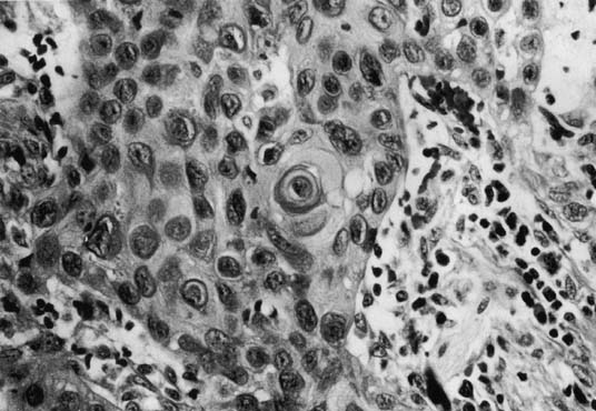 Squamous cell carcinoma
of the vagina (X200 magnification) demonstrating keratin pearl formation.
Squamous cell carcinoma
of the vagina (X200 magnification) demonstrating keratin pearl formation.
Back to Top
Clear Cell Carcinoma
 Vaginal clear-cell adenocarcinoma (X200 magnification) from a DES-exposed
patient.
Vaginal clear-cell adenocarcinoma (X200 magnification) from a DES-exposed
patient.
Back to Top
Vaginal Melanoma
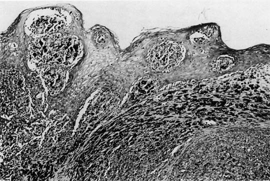 Vaginal melanoma (X100 magnification). Junctional melanocytic proliferation
and infiltrate of pigmented (epithelioid) melanoma. Vaginal melanoma (X100 magnification). Junctional melanocytic proliferation
and infiltrate of pigmented (epithelioid) melanoma.
Back to Top
Sarcoma Botryoides
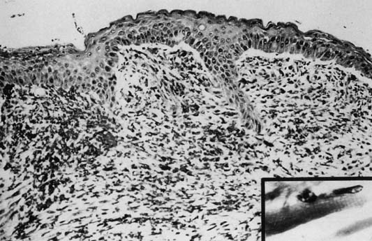 Sarcoma botryoides (X100). Cambium layer beneath epithelium. Loose spindle cell infiltrate. Inset (X400) demonstrates cross-striation in cytoplasm of spindle cells. Sarcoma botryoides (X100). Cambium layer beneath epithelium. Loose spindle cell infiltrate. Inset (X400) demonstrates cross-striation in cytoplasm of spindle cells.
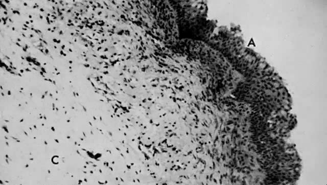 Sarcoma botryoides arising from the vagina of a 2-year-old girl, showing the squamous epithelium ( A) and the subepithelial cambium zone ( B ). Notice the central myxoid loose stroma ( C) (× 360)
Back to Top |