Operations on the Abdominal Wall
Authors
INTRODUCTION
Incision and closure of the abdominal wall is one of the most frequently performed, yet least discussed, of surgical procedures. This chapter will cover the fundamental principles of anatomy and wound physiology related to this topic, as well as the basic types of incisions used in gynecologic and obstetric surgery. Prevention and treatment of common complications will also be discussed.
RELEVANT ANATOMY OF THE ANTERIOR ABDOMINAL WALL
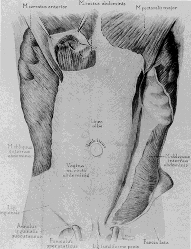
The structural integrity of the anterior abdominal wall depends upon the rectus abdominis muscles, the muscles of the flank, and the conjoined tendons of the flank muscles that combine to form the rectus sheath. The rectus abdominis muscle is found on either side of the midline with the pyramidalis muscle lying superficial to the rectus muscle just above the pubis. Lateral to these are the flank muscles: the external oblique, internal oblique, and transversus abdominis (Fig. 1 and Fig. 2). The broad sheet-like tendons of these latter muscles form aponeuroses that unite with their corresponding members of the other side, forming a dense white covering of the rectus abdominis muscle, properly called the rectus sheath (sometimes referred to in surgical writings as the rectus “fascia”),
|
|
Rectus Abdominis and Pyramidalis Muscles
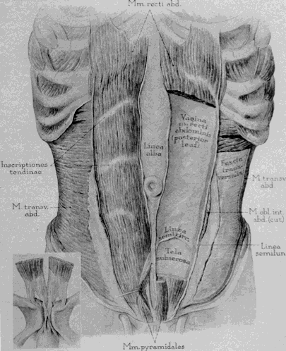
There are three tendinous inscriptions within each rectus abdominis muscle. These are fibrous interruptions within the muscle that firmly attach it to the rectus sheath. They are confined to the region above the umbilicus1 but can occasionally be found below. When found below the umbilicus, the rectus sheath is attached firmly to the rectus muscle at the inscription, causing difficult separation during Pfannenstiel incision. In addition, these points of fixation keep the muscle in place when it is transected during Maylard incision. The pyramidalis muscles arise from the pubic bones and insert into the linea alba in an area several centimeters above the symphysis pubis. The pointed insertion of the pyramidalis muscles into the linea alba can be used to assist in locating the midline.
Flank Muscles
The most superficial of the flank muscles is the external oblique. It runs diagonally anteriorly and inferiorly, from its origin on the lower 8 ribs and lilac crest to fuse with the rectus sheath. The fibers of the internal oblique fan out from their origin on the anterior two thirds of the iliac crest and the thoracolumbar fascia. In most areas, they are perpendicular to the fibers of the external oblique, but in the lower abdomen, their fibers arch somewhat more caudally, and run in a direction similar to those of the external oblique. The deepest of the three layers, the transversus abdominis, has fibers that run in a primarily transverse orientation. The caudal portion of the transversus abdominis muscle is fused with the internal oblique muscle. This explains why only two layers are discernible in the lateral part of a transverse lower abdominal incision. (The superficial layer is formed by the external oblique and the deep layer by the fused internal oblique and transversus abdominus muscles.)
Rectus Sheath
The rectus sheath is formed by the conjoined aponeuroses of the flank muscles. The line of demarcation between the muscular and aponeurotic portions of the external oblique occurs along a vertical line through the anterior superior iliac spine (see Fig. 1). The internal oblique and transversus abdominis muscles extend farther toward the midline, coming closest at their inferior margin, at the public tubercle. Therefore, the muscular fibers of the internal oblique are found underneath the aponeurotic portion of the external oblique.2
There are several specialized aspects of the rectus sheath that are important to the surgeon. In forming the rectus sheath, the conjoined aponeuroses of the flank muscles are separable lateral to the rectus muscles, but as they reach the midline, they fuse and lose their separate directions. As a consequence of this midline fusion, these layers are usually incised together in the midline during any transverse fascial incision until separate layers are identified laterally, where they can be individually incised parallel to the direction of their fibers.
The lower one fourth of the rectus sheath lies entirely anterior to the rectus muscle, while in the upper three fourths it splits to lie both ventral and dorsal to it forming both an anterior and posterior rectus sheath. The lower margin of the posterior rectus sheath is recognized as the semicircular or arcuate line, occurring midway between the umbilicus and the pubes. Cranial to this line, the midline ridge of the rectus sheath, the linea alba, unites the anterior and posterior sheathes. Sharp dissection is usually required to separate these layers when elevating the rectus sheath during Pfannenstiel incision. When the peritoneum is opened vertically and past the arcuate line, the posterior rectus sheath is divided along with the peritoneum and must be repaired during closure.
Transversalis Fascia, Peritoneum, and Bladder Reflection
Deep to the muscular layers, and superficial to the peritoneum, lies a layer of fibrous tissue called the transversalis fascia, which lines the abdominal cavity. It is visible during abdominal incisions as the layer just underneath the rectus abdominis muscles. It is separated from the peritoneum by a variable layer of adipose tissue and is frequently incised or bluntly dissected off the bladder prior to opening the peritoneum. The peritoneum itself is a single layer of serosa with a thin subserosal layer of connective tissue. It is thrown into five vertical folds by underlying ligaments or vessels that converge toward the umbilicus (Fig. 3). The single median umbilical fold is caused by the presence of the urachus. Lateral to this are paired umbilical ligaments raised by the obliterated umbilical arteries that connect the internal iliac vessels to the umbilicus. Finally, the lateralmost ridge is caused by the deep inferior epigastric arteries and veins.
|
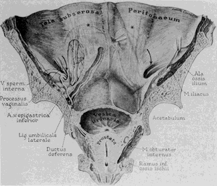
The reflection of the bladder onto the abdominal wall is triangular in shape, with its apex blending into the medial umbilical ligament. Because the bladder extends highest in the midline, incising the peritoneum somewhat off the midline is less likely to result in bladder injury and can provide more exposure.
Vessels of the Abdominal Wall
Beginning as a single artery that branches extensively, the superficial epigastric vessels run a diagonal course in the subcutaneum from the femoral vessels toward the umbilicus. Its position can be anticipated on a line between the palpable femoral pulse and the umbilicusjust superficial to Scarpa's fascia. The deep inferior epigastric artery and its accompanying veins originate lateral to the rectus muscle frorrt the external iliac vessels. They run diagonally toward the umbilicus and cross the muscle's lateral border midway between the pubis and the umbilicus (see Fig. 3). Below the point at which the vessels intersect the rectus, they are found laterad to the rectus muscle deep to the transversalis fascia. After crossing the lateral border of the muscle, they lie on its dorsal surface, between the muscle and the posterior rectus sheath. As the vessels enter the rectus sheath, they branch extensively so that they no longer represent a single trunk. The angle between the vessel and the border of the rectus muscle forms the apex of Hesselbach's triangle (inguinal triangle) whose base is the inguinal ligament.
A summary of some clinical applications for specific anatomic points is presented in Table 1.
TABLE 1. Clinical Importance of Anatomical Structures
Structure | Significance |
Superficial inferior epigastric vessels | Isolate and ligate on line from femoral vessels to umbilicus |
Insertion of pyramidal muscle | Identifies midline. |
Deep inferior epigastric vessels | Isolate and ligate during Maylard incision |
Tendinous inscriptions | Sharp dissection required to elevate rectus sheath during Pfannenstiel incision |
Arcuate line | Repair posterior rectus sheath above this line |
Bladder reflection onto peritoneum | Enter lateral to midline |
Hypogastric and ilioinguinal nerves | Identify and spare |
Femoral and genitofemoral nerves | Avoid retractor injury |
WOUND HEALING
Most wound complications can be traced to a failure of the healing process to eliminate the bacteria that are invariably introduced in some quantity into the wound, or failure of the healing process to synthesize adequate quantities of collagen to restore abdominal wall strength. Understanding the fundamental processes that are responsible for these functions is necessary to best create and close an abdominal incision. For a detailed description of these important events the reader is directed to Hunt and Dunphy's excellent book on this subject.3 The basic principles of healing are summarized here.
There is a common misconception that infectious complications are primarily related to sterile technique. Information concerning sterile technique has received a great deal of attention, yet surgical technique continues to play a critical role.4 The wound-healing process is a balance between the amount of damage done to the tissue during an operation, and the ability of the body to decontaminate and repair it. The surgeon stands in a position to influence this balance significantly and to affect both the rate of wound infection and dehiscence. Studies have shown that when a surgeon is made aware of his or her wound-infection rate when it is compared with the rates of peers in their institution, the surgeon with a higher infection rate can decrease it simply by altering his or her management of abdominal incisionsfi Understanding how wounds heal is critical to minimizing postoperative wound complications.
With the initial incision, exposure of blood and platelets to connective tissue begins the inflammatory response that will sterilize and heal the wound. During the initial phases of this process, the small vessels in the region of the injury become permeable to both molecular and cellular mediators of the inflammatory response. These elements are essential to eliminating bacteria through opsonization, phagocytosis, and cellular killing, as well as to recruiting wandering tissue macrophages that direct subsequent events. This is a decisive phase because it establishes the inflammatory process that is to follow. Clinical studies have shown that injection of vasoconstrictive agents at the time of surgery limits this response and is associated with increased numbers of infections.6 This may seem paradoxical because the infect:ions do not appear for several days--well after the vasoactive effects are gone. The vasoconstric-tion prevents the outpouring of the factors that initiate the inflammatory response. This creates a period of time for bacteria to multiply exponentially and become established in numbers that will later overwhelm host defense, thus explaining this phenomenon and calling attention to the importance of these early events.
After this initial phase, the polymorphonuclear neutrophils (PMNNs) and wandering tissue macrophages begin their work of digesting damaged tissue, killing bacteria, and synthesizing the chemotactic factors that direct wound repair. These cells lay the groundwork for the later appearance of the fibroblast that will reestablish wound strength. Although these cells are capable of limited activity in an anaerobic environment, their proper function in the wound depends upon the oxygen supply to tissue and, therefore, upon a lack of surgical damage adjacent to the wound. This emphasizes one of the key surgical applications of wound healing; namely, protecting the capacity of adjacent tissues to perfuse the healing wound after the operation by avoiding unnecessary damage to this tissue.
The next critical factor in proper healing is the amount of necrotic tissue created. Actual repair must begin from healthy tissue. If a ligature is placed around a piece of adipose tissue, it will become necrotic. Healing must then begin from the uninjured tissue behind the area of damage. Prior to reaching the edge of an incision, the healing process must disinfect, digest, and remove the dead tissue, before healing can begin. During this delay, bacteria in the ischemic tissues can multiply, further increasing the need for cleanup, delaying repair, and increasing the likelihood of infection. Hemostasis, either by ligation or electrocautery, abrasion, and desiccation of tissue are all injuries that occur in any incision. The more of these damaging elements present, the more necrotic tissue the body must eliminate before joining the edges of the wound. This allows more time and space for bacteria to multiply and overwhelm the host. Multiple knife strokes made when incising the subcutaneous tissue, leave more damaged tissue behind than a single, clean stroke and can be shown to increase the risk of wound infection.7
Although the minute details of repair cannot be covered here, an understanding of several points may influence the surgeon's management of abdominal incisions. First, healing is under the direction of the inflammatory response, especially the macrophage, and agents that influence inflammation also influence healing. For example, the: anti-inflammatory effect of steroid hormones can impair both PMNNs and macrophage function and will influence the development of wound strength.
The re-establishment of abdominal wall strength depends upon the synthesis of new connective tissue. This is accomplished by fibroblasts and requires, not only the protein precursors for collagen synthesis, but also occurs most rapidly in a normally oxygenated environment where the enzymes and cofactors needed for collagen synthesis are present. Factors limiting the availability of these critical substances and conditions will delay or impair the development of wound strength and increase the likelihood of wound disruption. Ischemia caused by tight sutures, foreign bodies, lack of nutritional factors such as protein or ascor-bic acid, or inhibitors of cell division can adversely influence found healing.
Collagen, the primary structural protein of the body, is synthesized by the fibroblast. It begins to appear in the wound on the second day, as an amorphous gel devoid of strength. Maximum collagen synthesis occurs around the fifth day. It depends especially upon the presence of oxygen, vitamin C, and amino acid precursors. Deficiency of these factors in the wound can inhibit healing, resulting in an increased incidence of wound dehiscence. Maximum strength development does not occur for several months and depends upon the interconnection of the collagen subunits. Approximately 80% of original strength is reached in about 6 weeks and can be significantly delayed if the normal factors for wound repair are not present.
It is important to recognize that perfusion of the wound is the most important factor in wound healing. Integrity of the microvasculature and flow is responsible for the oxygenation needed for cellular metabolism. Damage to tissue that impairs the delivery of oxygen to the wound increases the number of wound infections and the likelihood of dehiscence. Adopting an attitude in the operating room where tissue damage is minimized has been shown to decrease complications in obstetric and gynecologic surgery.8,9
OPENING THE ABDOMEN: SKIN PREPARATION, INCISION, AND HEMOSTASIS
Skin Preparation
The concept of cleaning the skin for surgery began with Maimonides in the 11th century and has evolved significantly during this century. The choice of approach and agent for cleansing is often derived from tradition and salesmanship rather than proven efficacy. Widely accepted goals, however, include cleansing away dirt and contaminants by physical means and rapid antisepsis to reduce bacterial density, followed by application of a long-acting bactericidal agent to deal with resident bacte:ria brought to the skin's surface by sweat.
Handwashing has been shown to reduce bacterial counts significantly, but to an inadequate degree. In fact, prolonged handwashing with plain soap may increase bacterial density due to chapped skin. Nonetheless, handwashing remains an important element of preoperative preparation by removing gross contaminants and dirt.
Numerous antiseptics are available, but with variable properties and effectiveness. Alcohol has repeatedly been shown to be an excellent choice due to its immediate and broad activity against gram-positive and gram-negative organisms. Although not sporicidal, alcohols also act against many fungi and viruses as well as mycobacteria. A 1-minute scrub with alcohol has been shown to be as effective, as a 4- to 7-minute scrub with other antiseptics.10 A 70% solution is most commonly used as a compromise between effectiveness and desiccation of the skin. Ethanol, normal propyl, and isopropyl are all effective. The World Health Organization's draft guidelines in 1987 designated alcohol as the gold standard against which all other skin antiseptics should be compared.10 Surgeons must be aware of alcohol's highly flammable nature, however, taking special precautions to assure complete drying where electrocautery or laser will be used.
Other popular scrubs include the iodophors, hexachlorophene, and chlorhexidine gluconate. The iodophors are highly effective, but their anti-microbial action declines rapidly upon drying. Hexachlorophene is active against gram-positive bacteria but less so against gram-negative bacteria, mycobacteria, and viruses. Chlorhexidine glu-conate has a broad spectrum of antibacterial activity, but is relatively more effective against gram-positive bacteria than gram-negative bacteria, with fair activity against the tubercle bacteria and poor activity against viruses. It does have extended effectiveness, remaining chemically active for approximately 5 hours. It is also available as an alcohol-based hand-rinse, combining the rapid and effective action of alcohol with the long action of chlorhexidine gluconate.
Preparation of the patient's skin involves similar considerations to those noted above. If necessary, hair removal should be accomplished immediately prior to surgery by clipping, not shaving, as the latter has been shown to damage the skin's defenses and increases the risk of wound infection.5
Incision
The incision should be accomplished with the least possible tissue damage. A scalpel should be used, and the fewest possible strokes will limit tissue damage.7 Electrocautery tends to produce much larger zones of damage and increases infection rates.12 Hemostasis can be obtained with well-directed cautery and fine ligatures (4-0 absorbable suture is adequate), taking care to isolate bleeding vessels and to exclude any unnecessary tissue from the ligature. In general, when discrete vessels are encountered, isolation with a hemostat and ligation will provide the least volume of necrotic tissue. Absorbable sutures such as polyglycolic acid or polyglactin are preferred to catgut, which causes inflammation.
Wound drainage has a long and varied history.12 In cases where diffuse oozing persists or wound contamination is greater than normal, drains may be considered. Seromas and hematomas significantly delay approximation and healing of the subcutaneous tissue. Because a surgical drain is a foreign body, it has the potential to increase wound infection by its presence. 13 In addition, it can provide access for bacteria to enter the wound after the skin has been closed.14 Subcutaneous drains, therefore, should be used only when there is sufficient risk of hematoma or seroma formation so that it can do more good than harm, as is true in the massively obese patient. 15 The best choice is a closed suction drain brought out through a separate stab incision since this option offers a lower infection rate than Penrose drains, or drains brought out through the incision.11
SPECIFIC INCISIONS: CHOICES AND TECHNIQUES
In choosing a particular approach to the pelvis or abdomen, specific operative goals should be weighed. Considerations include the need for .speed, potential difficulties with hemostasis, expo-.sure requirements, cosmetic concerns, the presence of a previous incision, and the patient's overall nutrition and health. The various advantages and disadvantages of abdominal incisions are summarized in Table 2. Although there is a temptation to become inflexible in the type of incision chosen, one must guard against habit or face the possibility of surgical compromise and complications.
TABLE 2. Characteristics of Lower Abdominal Incisions
Incisions | Pfannenstiel | Cherny | Maylard | Vertical |
Pelvic exposure | ++ | +++ | ++++ | +++ |
Upper abdomen exposure |
| + | ++ | ++++ |
Potential blood loss | ++ | ++ | +++ | + |
Potential hernia | + | + | ++ | +++ |
Evisceration risk |
| + | ++ | +++ |
Speed | ++ | +++ | + | ++++ |
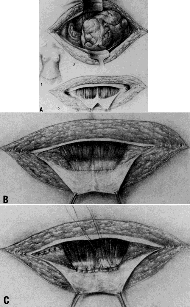
Pfannenstiel Incision
The Pfannenstiel incision is perhaps the most frequently used incision in both obstetrics and gynecology, offering satisfactory exposure of the pelvis, excellent postoperative strength, and pleasing cosmetic results (Fig. 4). Limitations include lack of upper abdominal exposure, increased risk of hematoma or seroma formation--especially in the face of abnormal coagulation--due to the extent of dissection required, and greater operating time. Great care should be taken preoperatively to make certain that exposure out of the pelvis will not be required, and that the incision will provide enough room to remove the expected structure safely (e.g., a large fibroid uterus).
|
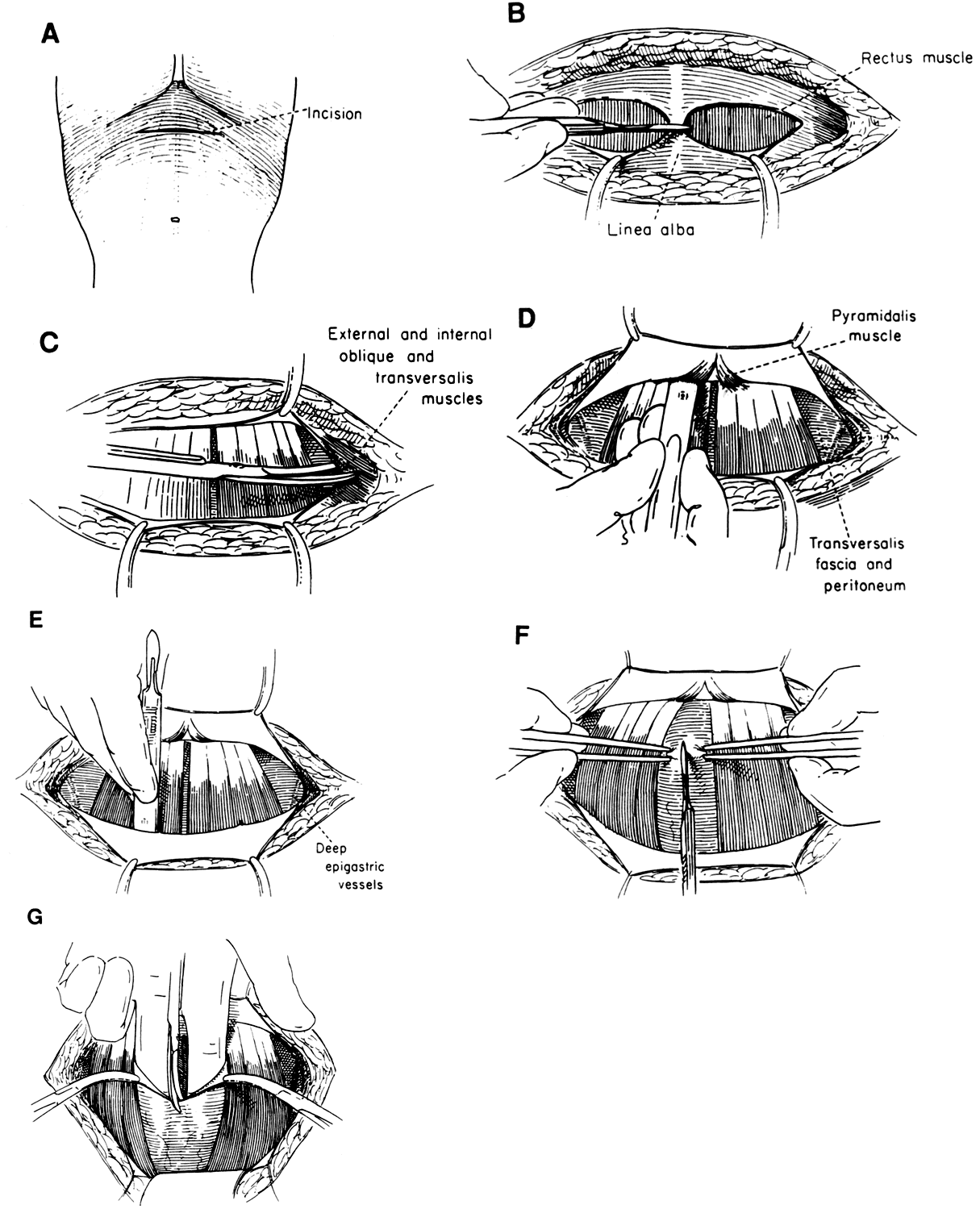
The skin incision is made transversely, approximately 4 cm above the superior border of the pubis, and is carried through the subcutaneous fat. When the rectus sheath is encountered, it is divided transversely in the midline with the knife, very often encountering the superior extent of the pyramidalis muscles. Once the incision is lateral to these structures, the rectus sheath is seen to consist of two layers: the aponeuroses of the external oblique and the combined internal oblique-transversus abdominus muscles. Each of these layers is separately divided laterally on each side with the scissors, following the fiber directions in each of the layers. Of equal importance to the skin and fascial incisions in providing adequate exposure is the next step, separating the rectus muscles from the sheath superiofiy and inferiorly. The sheath is elevated on each side of the midline using sharp and blunt dissection, separating the rectus sheath and muscle for a total distance above and below equal to the length of the fascial incision. If a tendinous inscription exists below the umbilicus, making elevation difficult, the muscle and sheath must be sharply separated, taking care not to cut through either the muscle or the sheath. Perforating blood vessels should be clamped, cut, and ligated only if bleeding occurs, otherwise preserving the nerve that accompanies the vessel. If the nerve is transected, some patients will develop an area of cutaneous anesthesia in this area that can be annoying for the patient. The pyramidalis muscles may be left attached to the undersurface of the fascia, or left on the rectus muscles and divided in the midline with the next step.
The rectus muscles are separated in the midline; this may be initiated by spreading the points of a hemostat between the muscles until the transversalis fascia is encountered. The separation of the muscles can usually be carried out superiorly and inferiorly, using blunt dissection with the exception of the insertion of the pyramidalis muscles that must be incised. The peritoneum is opened, initiating the entry at the superior extent of exposure, off of the midline, in order to minimize the risk of entering the urinary bladder. Final exposure is obtained by spreading the entire incised abdominal wall laterally. If exposure is inadequate, the skin, fascial, and peritoneal incisions should be extended, along with further dissection between the rectus muscles and their sheath. The four points that limit the size of the incision are the two lateral corners of the incision in the flank muscles and the superior and inferior extent to which the fascia has been elevated and the rectus muscles separated. The former points can be extended to the iliac crests, and the latter, to the pubis and umbilicus. If further room is needed once these limits have been reached, conversion to a Cherney incision can be made by simply cutting the rectus abdominus tendons from the pubic bones. Although surgeons frequently hesitate to take this step, the danger from operating with inadequate exposure is far greater than the possible risk of difficulty with wound healing from the extended incision.
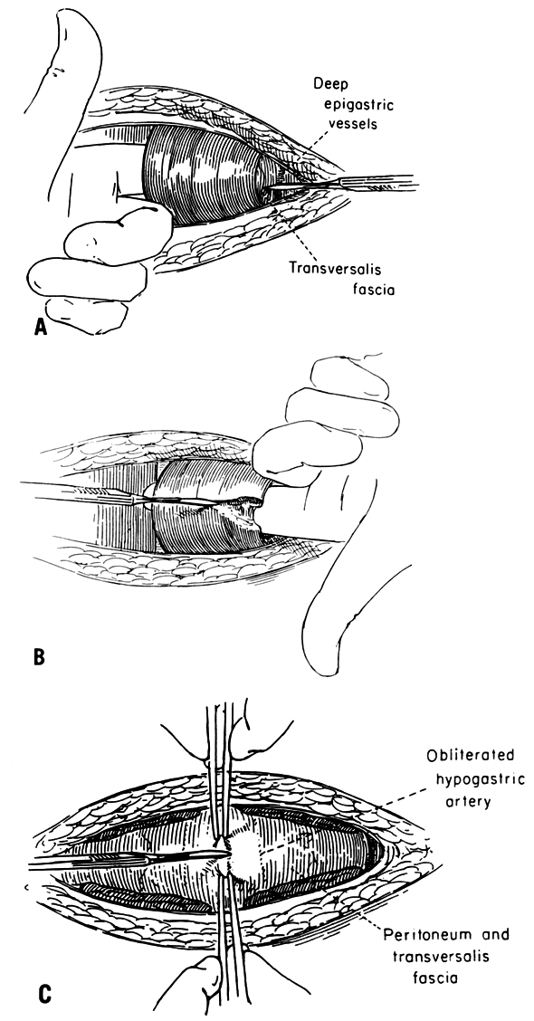
Cherney Incision
The Cherney incision17 combines the excellent exposure of a Maylard incision and the strength of a Pfannenstiel incision (Fig. 5). Unlike the Pfan-nenstiel incision, wherein the rectus muscles are separated, the Cherney permits the detachment of these muscles from their insertion on the pubes, and allows them to retract upward “like a window shade.” It begins with a low transverse incision of the abdominal skin and rectus sheath similar to the Hhnnenstiel incision. The sheath is then elevated off the rectus abdominis muscle inferior to the fascial incision until the pubic bone is reached; superior dissection of the fascia need not be done. Leaving the pyramidalis muscle attached to the rectus sheath minimizes unnecessary bleeding.
|
Next, the rectus muscles are severed from their insertion on the pubic bone. This is accomplished by perforating the transversalis fascia lateral to the muscle, but medial to the deep inferior epigastric vessels in Hesselbach's triangle. The surgeon's linger is then used to dissect under the tendons of the rectus muscle. The tendons are cut approximately 0.5 cm above their insertion to free them from the pubic bones. If the tendon is shaved directly from the periosteum, there is no tissue to grasp and ligate should bleeding from the bone occur. Care should be exercised in dissecting around the lateral border of the muscle to avoid damage to the adjacent deep inferior epigastric vessels. If exposure is found to be limited by these vessels during the procedure, they can be ligated and the peritoneal and fascial incisions extended. Since the rectus muscles are no longer attached, lateral exposure can be extensive, and the incision can be carried above the anterior iliac spines into the flank, if needed.
At the completion of the operation, the tendons are reattached to the under surface of the rectus sheath, rather than to the pubic bone. This is accomplished by placing horizontal mattress sutures (see Fig. 5). Usually, three 2-0 delayed absorption sutures on each half of the muscle suffice, but if delayed healing is anticipated, permanent sutures should be employed. The re-establishment of these attachments provides for excellent postoperative strength.
Maylard Incision
This incision requires more time to accomplish and includes more potential blood loss than other incisions, but offers great pelvic exposure and can be used at any level of the abdomen (Fig. 6). For pelvic operations requiring this degree of exposure, the skin incision is generally made at approximately the level of the superior iliac spine. In order to achieve adequate exposure of the pelvic sidewall, it is extended to about 5 centimeters medial to the iliac spine. In obese patients with a pendulous “apron” of skin, the incision may not need to be made in the moist crease of the skin, but can be made below the umbilicus, as long as it lies above the pubic bone, and the underlying crease is not traversed.18 Again., skin, subcutaneous tissue, and rectus sheath are divided transversely as noted above for the Pfannenstiel incision, carrying the incision past the lateral border of the rectus muscle. However, instead of separating the rectus muscles and fascia, the rectus muscles are cut transversely.
|
The deep inferior epigastric vessels are usually ligated before transecting the muscles, but not all authors have felt this is necessary. To accomplish this ligation, recall the course of the inferior epigastric vessels as described above. They will be found by gently retracting the lateral border of each rectus muscle, exploring the region for their location. Near the pubis, in the area of Hesselbach's triangle, they are found lateral to the muscle, whereas above that level, they are found on its undersurface. Note that numerous branches may exist arid should be carefully identified, isolated, clamped, cut, and ligated. After blunt dissection of the muscles from the peritoneum, they may then be divided safely with the knife or electrocautery. Elevation of the muscle off the peritoneum with the hand or an Army-Navy or similar retractor is required if the electrocautery is to be used safely. If the vessels are difficult to isolate, then the muscles can be cut before ligating the vessels. This approach is used to expose the vessels that lie between the muscle and the peritoneum. To complete the incision, the transversalis fascia and peritoneum are incised transversely.
Vertical Incision
The midline or paramedian vertical incision is the simplest of abdominal incisions, and it offers the greatest ease of extension into the upper abdomen as well as the least blood loss (Fig. 7). Although the pararectal approach that goes lateral to the rectus muscle might be acceptable in terms of exposure, the resultant denervation of the rectus muscle that occurs weakens this incision, 19 leaving the midline vertical and paramedian incision for discussion in the context of gynecologic procedures.
|
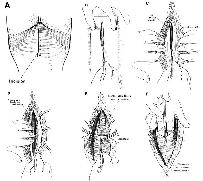
The main considerations in choosing the vertical incision include the need for speed, a relatively bloodless approach, and possible need for exposure of the upper abdomen. In addition, because of its lack of dead space, the vertical incision is preferable in patients taking anticoagulants or in the presence of disseminated intravascular coagulation. Also, for patients with cirrhosis of the liver who may have greatly enlarged abdominal wall vessels that follow a longitudinal course, vertical incision minimizes the number of vessels that must be transected.
In the lower abdomen, the incision is made from just above the pubis to below the umbilicus in the midline. Although it is customary to carry the incision lateral to the umbilicus if extension to the upper abdomen is required, the incision can equally well be made through the umbilicus without any increased risk of disruption and is technically simpler to perform.20 Below the umbilicus the linea alba tends to be narrow, and the rectus sheath is usually entered on one side or the other, thus making this a paramedian incision rather than a true midline approach. The transversalis fascia and peritoneum are also opened in a vertical direction; entry should begin at the superior extent of the incision to obviate the possibility of bladder entry. As is true of all peritoneal entry, great care must be taken against the possibility of encountering adherent bowel. Closure considerations are discussed below.
PRINCIPLES OF ABDOMINAL WALL CLOSURE
Regardless of the type or direction of incision, the factors involved in closure are similar and will be discussed together. Maintenance of tissue per-fusion, minimizing necrosis, creating good initial strength, protection against late hernia formation, and assuring a cosmetic result are factors all incisions share in common.
Tight Sutures and Ischemia
All sutures used to close the musculofascial wall must be tied with enough tension to approximate the edges of the incision. If greater tension is applied, the tissue will become ischemic, and a certain amount of necrosis will develop. If the extent of necrosis is marked, the tissue will not hold the suture, resulting in dehiscence or hernia formation. This is illustrated by the fact that dehiscence rarely occurs immediately after the surgery but is usually delayed for several days,21 during which interval the tissues' weakness from ischemia develops.
The choice of suture technique and the way that the sutures are tied determine the extent of necrosis that will occur. In much the same way that a suture tied around the base of a pedunculated skin lesion will allow it to become necrotic and fall off, abdominal wall sutures create ischemia, necrosis, and tissue disruption. Experiments studying the difference in the strength of wounds closed with tightly tied sutures, as opposed to those tied just tightly enough to coapt wound edges demonstrate that the wounds with more loosely tied sutures are stronger.22,23 These same studies demonstrate that tightly tied sutures create lower breaking strength, increasing the likelihood of disruption. Therefore, whatever suture is chosen, it should not be placed so tightly a to cause ischemia.
Suture Placement
A second element that is important to wound strength is the distance between the wound edge and suture placement. First, the inflammatory process at the wound edge produces collagenases to help with removal of necrotic debris. This zone of collagen degradation extends for approximately 1.5 cm from the edge. 24 The fascia in this region is partially digested during the immediate postopera-tive period. Secondly, there is a purely mechanical factor: The farther from the edge the suture is placed, the greater the amount of fascia the suture would have to tear through in order to pull free25 and the more secure the closure would be. Therefore, sutures should be placed at least 1 to 1.5 cm fi-om the wound edge. In patients at increased risk of wound disruption, sutures should be placed 2 cm from the edge.
Choice of Closure
There are several techniques that can be used to suture the wound edges together. In general, these can be divided into running and interrupted closures. Running sutures have the advantage of speed, since knots need only be tied at two or three points. In the past, these had often been considered to be weak closures because disruption of any portion of the suture would open the entire wound. More recently, it has been appreciated that these can be strong closures.26,27,28,29 Compared to the nature of interrupted sutures, the helical nature of an unlocked running stitch evenly distributes tension along the entire wound and allows for superior perfusion. Large studies of gynecology patients who are at high risk for dehiscence have demonstrated the safety of this type of closure.30
Interrupted and figure-of-eight sutures have an advantage; if one is insecurely tied, or breaks, the whole incision will not come apart. If tied only tightly enough to approximate the tissue, but loosely enough to permit adequate perfusion of the fascia within the suture, such sutures can provide a secure closure without necrosis. There is, however, an inherent tendency to pull forcefully on these sutures as they are being tied. This is unquestionably the reverse of what is best. If interrupted sutures are not done properly, they can be a greater impediment to a strong closure than a running suture that produces inherently less ischemia. Further aspects of wound closure will be discussed in the section on wound dehiscence below.
Peritoneum
Closure of the peritoneum has been a topic of controversy, but several points are now clear. The peritoneal mesothelium does not heal like skin.31 Rather than healing only from the edges toward the center of a defect, as is true of epidermis and dermis, a new layer arises from the exposed bed of connective tissue. Therefore, it makes little sense to bring edges of peritoneum together to hasten healing. If the underlying tissue is undamaged, adhesions do not form in the absence of mesothelium before it can regrow; moreover, this process of regrowth occurs rapidly (usually within 48–72 hours).
On the other hand, rarely is the mesothelium alone incised or damaged. In opening the abdomen, the peritoneum is incorporated in the lower abdomen with the transversalis fascia. Above the arcuate line, the posterior rectus sheath also lies under the rectus abdominus muscles, and these structures are incised along with what is clinically referred to as the peritoneum. Closure of the transversalis fascia and posterior rectus sheath can add to overall wound strength and should be accomplished to make a more secure wound.32
Experimental studies of adhesions frequently use a suture tied around a piece of tissue as a reliable stimulus for the formation of adhesions. Therefore, if a ligature is exposed to the intraperitoneal contents as it would be in a Maylard incision, where the ligatures on the deep inferior epigastric vessels are exposed, covering them by approximating peritoneum with a fine or 4-0 non-reactive suture seems preferable to leaving this nidus for adhesions to remain exposed to bowel.
WOUND COMPLICATIONS
Dehiscence
Dehiscence is defined as the separation of the sutured layers of the abdominal wall and may be classified as partial or complete. In the case of a partial dehiscence, one or more, but not all of the sutured layers may separate. This situation may also be referred to as wound disruption. Complete dehiscence is marked by separation of all layers resulting in exposure of the peritoneal cavity. Synonyms include evisceration and burst abdomen. The incidence of this complication has been quoted as 0.3% to 3% of all pelvic surgeries, but it is currently thought to occur in less than 1% of cases.33,34,35 Historically, the incidence was thought to be greater for vertical as opposed to transverse incisions, but more recent studies have shown them to be equal.35,36
ETIOLOGY AND PREVENTION.
The main causes of dehiscence include failure of the suture to remain anchored in the fascia, suture breakage, and knot failure. Of these, tissue failure and improper suture choice are the most common.35,37 Since closure techniques involving permanent suture and wide bites of tissue exist that effectively prevent dehiscence, the central problem becomes one of recognizing the patient in whom the extra time taken to use a more secure closure is justified.
Risk factors for dehiscence are listed in Table 3. Inherent strength of abdominal wall tissue affects the risk of dehiscence and, in turn, is also affected by such factors as age, sex, metabolic disease, and the presence of malignancy. Patients over the age of 60 are at increased risk, as are males with a ratio of 2.6 to 6.7: 1 over females.36 Uremia and diabetes are associated with poor healing, as is vitamin C deficiency in the malnourished patient. These underlying conditions should be corrected if possible. White and co-workers found half of their cases of burst abdomen occurred in patients with malignancy.38 The presence of these risk factors indicates the need for a closure that is resistant to disruption such as a mass closure, Smead-Jones closure, or placement of retention sutures.
TABLE 3. Risk Factors for Dehiscence
Systemic factors
Malnutrition
Hypoproteinemia
Chronic anemia
Vitamin C deficiency
Systemic steroids
Malignancy
Advanced age
Obesity
Infection
Previous radiation therapy
Chemotherapy
Increased stress on wound
Chronic coughing
Ileus with GI distention
Ascites
Local wound factors
Infection
Hematoma
Improper closure
Intraoperative technique
Incision type
Suture type
Closure technique
Pressure necrosis
The method of closure plays an important part in wound security (Fig. 8). In layered closure each layer--peritoneum, fascia, subcutaneous tissue, and skin--is closed separately, as opposed to mass closure where all layers, usually excluding skin, are closed in a single unit. Ellis cites mass closure as one of the most significant advances in reducing the risk of burst abdomen. One of the key elements in mass closure is the obligatory use of wide tissue bites (1.5–2 cm from the wound edge) when placing the suture. Recall that the edge of the fascial incision is often necrotic to some degree, resulting in tenuous tissue strength and increased risk of disruption. if sutures are placed near the cut edge. In a review, Wadstrom and Getdin27 found no studies proving an advantage to layered closure. Similar arguments are forwarded in concluding that a continuous closure is superior to interrupted sutures; no clinical advantage of interrupted closure has been shown in the large majority of studies comparing the two. In view of the :shorter operating time for continuous closure, it would seem to be the obvious choice. Additionally, one could argue that it is easier to tie two or three knots precisely than to tie precisely the many knots required in interrupted closure. As noted above, to prevent necrosis and subsequent wound disruption, sutures must not be placed under undue tension.
|

In patients at unusually high risk of wound dehiscence, special consideration should be given to using a closure that is, perhaps, somewhat more time-consuming, but lessens the risk of disruption.39,40 When a wound dehiscence occurs, and permanent suture has been used, the separation usually occurs where the sutures are inserted into the tissue. Therefore, unless a weak or absorbable suture has been used, it is usually not the suture that is at fault, but rather the way that the sutures are anchored in the tissue.37 As previously mentioned, the further a suture is placed from the edge of the wound, the more force is required to pull it out, making wide suture placement a stronger technique. An additional factor that can be used to increase the strength with which the suture can be anchored to the tissue comes from distributing the tension that the suture places on the tissue between at least two points. The Smead-Jones suture takes advantage of this distribution by placing two bites on each side of the wound edge in a far-near near-far arrangement as shown in Figure 8. Originally described, this technique was a mass closure that incorporated both the muscle, fascia, and peritoneum of the abdominal wall. This closure, incorporating all layers, is extremely strong, but it is usually modified to have a separate peritoneal closure, with the Smead-Jones stitches including only the musculofascial layer. This suture gains its strength from the fact that before the suture can tear out of the tissue, it would have to rupture the tissue at two points rather than just one41 and can be done either as an interrupted technique or running suture.
The most secure closure of the abdomen in-dudes both closure of the musculofascial layers and placement of retention sutures through all layers of the abdominal wall, including the skin and usually (although not always) the peritoneum. It is virtually impossible for dehiscence to occur while these sutures are in place because of the great amount of tissue they would have to disrupt in order to pull out. They are usually placed with a number 1, or greater, suture, and are especially useful in treating a wound that has already undergone dehiscence.
Proper suture selection will decrease the risk of dehiscence in patients at normal risk. Clinical studies have shown that wound disruption is most likely to occur in the early postoperative period, usually 5 to 8 days after surgery. Both theoretical concerns about maintenance of suture strength and clinical studies agree that there is no place for catgut sutures in fascial closure. Other absorbable sutures, such as polyglycolic acid (Dexon) and polyglactic acid (Vicryl), even losing up to 80% of its tensile strength in 2 weeks,42 seem to compare favorably to permanent sutures, such as Prolene, in healthy patients undergoing elective surgery who are at no unusual risk for dehiscencefi As we will see below, there may be concern about the more distant complication of wound hernia. In patients at risk for dehiscence, permanent sutures are needed.
In addition to the way in which a wound is closed, the stresses placed upon it are important. Mechanical factors such as abdominal distension from ileus, vomiting, and chronic cough may also play a role and should, therefore, be treated when present or prevented when possible in the patient already at risk due to other risk factors.
Other factors that may weaken a wound include the presence of hematoma, wound infection, and obesity. A hematoma will disrupt tissues, preventing approximation as well as providing an excellent nidus for infection. More often than not, wound dehiscence is associated with a combination of events that, when recognized, calls for meticulous preoperative preparation, wound closure, and postoperative care.
DIAGNOSIS AND TREATMENT.
Due to the increased morbidity and mortality associated with dehiscence, diagnosis and treatment should be prompt. Mortality as high as 15% to 20% has been reported in the literature, although more recent studies demonstrate a rate around 10%. Mortality is not solely due to the dehiscence, however; these patients are often ill with other chronic disease and undergo a second anesthetic.
Wound disruption usually occurs on the sixth to eighth postoperative day. Evisceration will be apparent on simple inspection. When this is the case, the intestines should be covered with a saline-moistened towel and immediate steps taken to close the incision in the operating room. Although lesser degrees of disruption may be asymptomatic, many patients have a sense of something “giving way.” The most common complaint is that of a profuse serosanguineous discharge from the wound. When disruption is strongly suspected, careful exploration may best be accomplished in the operating room with suitable anesthetic. In any event, the wound should be opened as necessary to aid in diagnosis and the fascial closure critically evaluated for disruption. A broad-spectrum antibiotic should be started as soon as cultures have been obtained.
Once the patient is in the operating room, the wound must be cleansed carefully and thoroughly. Debridement of the subcutaneous tissue and fascia should be accomplished as necessary. Strict attention must be paid to closure--in this case to mass closure. Permanent material of suitable size (number 1 or larger) should be used along with, possibly, retention sutures, depending on the patient's general health and other etiologic factors. Undue suture tension must be avoided to reduce the possibility of necrosis, and bites at least 2 cm from the edge should be used. Retention sutures may be left in place for 14 to 21 days. Underlying conditions should, of course, be treated (e.g., an NG tube placed if ileus is present). When malnutrition is present or develops, hyperalimentation should be considered and followed by careful dietary support once oral alimentation is resumed.
Wound Hernia
Wound herniation is defined as an incomplete dehiscence in which the peritoneum, subcutaneous tissue, and skin remain intact, but the muscle or fascia do not. As opposed to dehiscence that occurs and is recognized in the early postoperative period, wound herniation follows apparently satisfactory healing only to present with an incisional defect at a later date. Although it has been said that most such defects occur before 6 months and nearly all before 1 year, several studies with longer patient follow-up have found occurrences up to 5 years after the initial operation. For low midline incisions, the most often quoted incidence is about 1% in uncomplicated cases; after wound infection, the risk rises to about 10% and, after repair of a dehiscence, to about 30%. Ellis maintains that the reported low incidence of wound hernias is due in part to the lack of prolonged follow-up in most studies. In a review of their own patients for up to 5 years after operation in a group free from hernia at 1 year, an additional 5.8% incidence was found.44 These defects range from the small and insignificant to the large and unsightly. When the fascial defect is small, the risk of volvu-lus and infarction of the hernial contents is increased, but still uncommon.
ETIOLOGY AND PREVENTION.
Late wound separation is more common in the lower abdomen because of increased hydrostatic pressure and the lack of the posterior rectus sheath below the arcuate line and, for vertical incisions, the greater lateral forces provided by the bulkier oblique muscles inferiorly. The underlying cause is inadequate healing of the fascial layer--perhaps more related to degree than representing a true difference in etiology when compared to dehiscence. Causes may include fascial necrosis from initial excessive suture tension or, secondarily, from abdominal distension associated with ileus, postoperative nausea and vomiting, or pulmonary disease resulting in chronic cough. Necrosis may, in turn, be followed by suture pull-through due to the inadequate tissue strength. These causes of late wound separation can be largely eliminated by placing wide tissue bites and by approximating the tissue without undue tension. Poor tissue vitality is also the common factor in wound hernias associated with wound infections and after repair of dehiscence. One element in reducing the risk of wound infections would be selection of a permanent monofilament suture. Another strong association is fascial closure with catgut suture due to its inadequate continuation of strength during the period critical to healing 45,46
DIAGNOSIS AND TREATMENT.
Consideration should be given to the diagnosis of hernia when, with the patient lying supine and legs raised, an incisional bulging is noted. The hernia is often asymptomatic, although the patient may complain of a bulging, or even note apparent peristalsis with resolution upon lying down followed, in turn, by recurrence when standing. The bulge may increase with a Valsalva maneuver. On examination the fascial defect is often palpable. Because torsion and infarction are uncommon, colic, distension, nausea, and vomiting are unusual symptoms, and repair is usually elective. Small, asymptomatic hernias need not be repaired; those that are symptomatic, large, or disfiguring deserve operation. The principles of repair are:
- Meticulous isolation of the hernia sac2. Wide exposure of the fascia3. Careful closure, possibly with use of a graft in the case of very large defects.
Usually, the old skin scar is excised followed by careful dissection of the subcutaneous tissue until the hernia sac is encountered. Wide isolation of the sac is continued until the fascial edges are encountered and adequately undermined. The sac may then be opened, and attention turned to any adhesions of peritoneum, bowel, or omentum that are carefully separated until the hernia sac can be closed with a purse string suture, and excess sac excised. This dissection can be confusing, with apparent secondary hernia sacs formed by multiple bowel adhesions which, in turn, must be separated until sufficient normal anatomy can be restored to permit a safe closure. The next task is isolation of the fascial edges and freshening of them if the size of the defect will allow primary closure. Several satisfactory approaches may be used, including Smead-Jones closure, mass closure, and overlapping of fascia (pants-and-vest closure). Theoretical considerations would point to the selection of a permanent monofilament suture. Appropriate selections would include 0 or 1 Prolene, nylon, or other permanent sutures. Wire, although important historically, has no advantages over the newer, nonreactive, monofilament sutures.
Large defects may need to be closed with the use of prosthetic mesh (e.g., Merselene, Marlex, Gortex) to bridge the hiatus where approximation of the fascial edges is not possible or causes undue tension. Other indications for the use of a graft would include repair of a recurrent hernia, grossly attenuated tissues, and fascia that is too weak for adequate repair. Most commonly, the graft is placed anterior to the peritoneum and transversalis fascia, and posterior to the rectus muscles. The material must be anchored to the posterior aspect of the recti “on stretch” to prevent folding when the muscles are approximated in the midline. The anterior rectus sheath is approximated as closely as possible. In some instances, dissection between the hernial sac and the fascia may prove exceedingly difficult or impossible, in which case the Marlex mesh may be anchored to the anterior aspect of the rectus muscles and, again, the anterior rectus sheath would be approximated as closely as possible. This location of the dissection is not as satisfactory, however, in that the already increased risk of wound infection due to the presertce of the graft is further increased by its proximity to subcutaneous tissue and skin.
Postoperatively, predisposing conditions for wound failure should be addressed fastidiously, including control of nausea and vomiting, aggressive and early treatment of ileus and pulmonary complications, and attention to adequate nutrition.
Wound Infection
Wound infection has been reported to occur in 2% to 4% of all clean abdominal incisions and up to 35% of all grossly contaminated incisions. Clean incisions are defined as those initiated on prepared skin without entering a contaminated viscus or encountering infection. Clean contaminated wounds are the same as clean incisions, but a contaminated viscus, such as the vagina that has been prepared, is entered without gross spillage. A wound is classified as contaminated if an infected genitourinary tract is entered or gross gastrointestinal spillage occurs. A dirty wound is one that occurs when pus from an abscess is spilled intraoperatively, or previously ruptured bowel is present. The rate of infection varies not only according to increasing severity, but also according to patient socioeconomic status, surgical technique, operating time, obesity, age, and sex.
ETIOLOGY AND PREVENTION.
Infection is often initiated by direct inoculum of bacteria into the wound from the patient's or surgeon's skin and is potentiated by the presence of necrotic tissue. Proper preparation of both is necessary to ensure the lowest possible rate of infection. If hair removal is required, clipping immediately before surgery is preferable to shaving, and either is preferable to shaving the evening before, which has been associated with higher rates of wound infection. For the surgeon's scrub, both initial antisepsis and the use of a long-acting antibacterial agent is recommended; 20% to 40% of all gloves are punctured during the course of an average operation 47 Although not currently the most popular choice, an initial scrub followed by a 1-minute application of alcohol in an emollient base or the chlorhexidine gluconate-alcohol hand rinse (Hibistat) would represent the best possible preparation. After adequate skin antisepsis, multiple intraoperative factors come to bear.
Since devitalized tissue offers increased opportunity for poor wound healing and infection, every effort should be made to minimize infection's presence, including meticulous incisional technique with a stainless steel scalpel and precise hemostasis with cautery or fine, nonreactive suture. These same considerations hold true while operating (i.e., creation of the smallest possible pericles, only precise use of the electrocautery, avoidance of ischemic closure of the vaginal cuff). Mass closure of the abdominal wall with continuous mortoff-lament suture would seem preferable in theory, although clinical studies have not yet supported this view (other considerations, such as decreased risk of dehiscence, may suggest this combination).27
Other closure considerations include copious irrigation with nonirritating physiologic solutions, especially in wounds other than those classified as clean. Even in clean wounds, however, irrigation removes fragments of free tissue and fat globules from separated adipose cells that will prolong inflammation and delay repair. Drains may be placed in the subcutaneous tissue when diffuse oozing resistant to hemostatic efforts is present. Soft drains, such as the Penrose, have been replaced by closed suction drains brought out through a separate stab wound with improved results (i.e., decreased rates of infection and hematoma). A trial of closed, subcutaneous drains alternately placed in suction and irrigated every 8 hours for 3 days with an antibiotic solution showed possible benefit in grossly infected wounds, but probably are not justified in clean contaminated wounds.48 A more traditional and proven approach to the contaminated wound is delayed primary closure, either closed in delayed primary fashion or allowed to heal by secondary intention. With delayed primary closure, Verrier and colleagues showed a decrease in infection rates in contaminated wounds from 11.1% to 4.8% and in infected wounds from 33% to 6.6%.49
Primary closure of the skin and subcutaneous tissues is defined as closure at the time of the initial operation, and secondary closure indicates closure after granulation tissue has formed either with suturing or spontaneously as healing by secondary intention. A delayed primary closure is one in which the subcutaneous tissue and skin are not closed at the time of initial surgery, but covered by a :sterile dressing and then closed some days later (usually on the fourth day), but before the formation of granulation tissue. Sutures can be placed during the original operation and left to be tied later, or the wound can be sutured under local anesthesia in the patient's room. During this time, the body's immune response has had a chance to clean the wound, and microscopic capillary formation has begun, creating excellent oxygenation of the wound edge. Closure of the wound on the fourth day greatly decreases the chance of infection, allowing patients to avoid the potentially serious problem of sepsis associated with wound infection. This approach is most helpful during treatment of pelvic infection, especially in patients with poor healing characteristics. In these patients, delayed primary closure has resulted in an extremely low complication rate. 50,51
DIAGNOSIS AND TREATMENT.
Wound infections may present in several ways, depending on the extent of the infection, host resistance, and the etiologic microorganisms. Early, mild infections may be associated with only scant exudate from the incision and, upon exploration of the wound, poor healing. Hemolytic streptococcal organisms may cause erysipelas, an infection marked by a rapidly extending erythematous cutaneous border. Deeper infections may be found during the process of evaluation for postoperative fever and may additionally be associated with erythema, induration of skin and subcutaneous tissues or, possibly, fluctuation. One must be alert for the rare but devastating signs of necrotizing infections, including brawny edema, cutaneous sensory loss, and obvious necrosis. Patients with necrotizing fasciitis need prompt and aggressive debridement under general anesthesia to avoid death.
In cases of contaminated and infected wounds, consideration should be given to delayed primary or secondary closure. In these situations characteristics of each case should be taken into account, such as the amount of infected tissue left behind, nutritional status of the patient, presence of diabetes, malignancy, or obesity--factors associated with poor wound-healing. When the decision is made to proceed with delayed closure, retention sutures may be placed. Permanent, monofilament suture would be the best choice. Cultures, of course, should be obtained. Postoperatively, the incision can be left covered until the fourth day, at which time the attending physician assesses whether the wound is clean enough to close. If there is any infected or necrotic tissue, then regular dressing changes and debridement can be commenced postoperatively until the wound is ready to close. Delayed primary closure may be done using one of several techniques: closure with sutures placed, but not tied, in the operating room; placement of sutures with local anesthesia; or application of sterile adhesive strips. In the high-risk patient, when coaptation of the wound is difficult, or if the wound does not appear clean in a reasonable period of time, the wound may be allowed to heal by secondary intention. Perhaps surprisingly, the cosmetic result in such a case is equal to that of delayed primary closure. Careful instruction prior to discharge and follow-up by a visiting nurse will be very helpful to the patient and her family.
Treatment for superficial and minor infections may consist only of application of moist heat. Erysipelas usually responds rapidly to such local treatment with the addition of penicillin. When discharge from the wound is prominent, or fluctuation is thought to be present, the wound should be explored and all areas presenting little resistance to separation opened fully. Cultures should be obtained, appropriate antibiotics should be started and the wound should be debrided and packed. Secondary closure may be desirable and possible if the wound reveals healthy granulation tissue 3 to 5 days after opening. Again, the patient may be sent home with follow-up by a visiting nurse.
Nerve Injury
Nerve injury associated with abdominal incision can pose a distressing, and often unexpected ending to an otherwise successful operation. Two types of injury occur. First, the incision and closure may transect or damage the nerves of the abdominal wall. Second, a retractor used during the operation can cause injury to nerves on the posterior body wall.
The most serious nerve damage is that to the fiemoral nerve, because of the loss of innervation to the quadriceps muscle in the leg and loss of the ability to extent the leg at the knee joint. This damage is usually caused by the blades of a self-retaining retractor. The lateral blades of these instruments can press upon the nerve as it emerges from the lateral border of the psoas muscle before passing under the inguinal ligament (Fig. 9). The frequency of femoral neuropathy after gynecologic surgery is surprisingly common, occurring in approximately 10% of pelvic laparotomies.52,53,54 Fortunately, most of these cases resolve spontaneously, yet when a case does not, it poses a significant problem. Damage to the nerve should be suspected with loss of sensation in the anteromedial thigh, diminished knee jerk, and weakness of extension of the knee, which creates a specific problem climbing stairs.
|

In addition to damage to the main femoral nerve, retractor blades can compress the genito-femoral nerve that emerges from the body of the psoas muscle to lie on its muscle-belly. Although this situation creates no motor abnormality, the loss of sensation in the upper medial thigh and labium majus can be quite distressing.
The risk of these complications is higher in thin individuals and when retractors with deep blades have been used. Simply placing a laparotomy pack over the retractor blades will not diminish the amount of force that impinges on the nerve, and a space between the blade and nerve should always be confirmed, remembering that some downward pressure will unavoidably be placed on the retractor during surgery. Although the nerve itself can not readily be palpated in the operating room, the psoas muscle can be. It lies lateral to the external iliac artery, and identification of the vessel by its pulse will lead the examining finger laterally to the muscle.
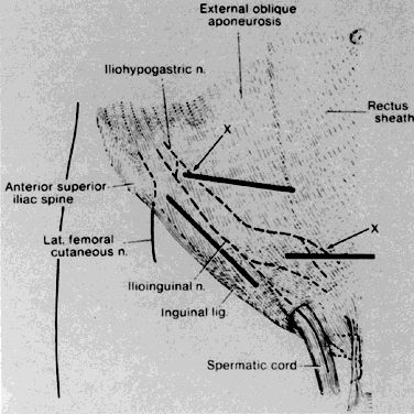
An additional type of injury that can occur is entrapment of the iliohypogastric or ilioinguinal nerves in the lateral closure of a transverse incision (Fig. 10)55 Transection of the nerve can lead to an area of anesthesia in the distribution of the damaged nerve, and trapping them in a suture can give rise to pain in the lower abdomen or groin. These nerves lie medial to the anterior superior iliac spine, first, between the layers of the transversus abdominus and internal oblique muscles, and then, more medially, come to lie between the internal oblique and external oblique. Although most surgeons fail to notice them during the lateral extension of a transverse incision, they are sometimes visible in the lateral aspects of the wound and should be looked for and avoided when seen.
|
Minor sensory abnormalities can arise when the nerve that innervates the abdominal skin and that accompanies the blood vessels that run between the rectus muscle and its sheath to reach the skin is transected during elevation of the fascia off of the muscle in a Pfannenstiel incision. Because of the extensive overlap of dermatomes here, this transection is usually not a problem, but can cause troublesome loss of sensation above the incision.
REFERENCES
Milloy FJ, Anson BJ, McAfee DK: The rectus abdominus muscle and the epigastric arteries. Surg Gynecol Obstet 110: 293, 1960 |
|
Anson BJ: An Atlas of Human Anatomy, p 241. Philadelphia, WB Saunders, 1950 |
|
Hunt TK, Dunphy JE: Fundamentals of Wound Management. Nev¢ York, Appleton-Century-Crofts, 1979 |
|
Condie JD, Ferguson DJ: Experimental wound infections: Contamination versus surgical technique. Surgery 50: 367, 1961 |
|
Cruse PJE, Ford R: The epidemiology of wound infection: A 10-year prospective study of 62,939 wounds. Surg Clin North Am 60: 27, 1980 |
|
Stevenson TR, Rodeheaver GT, Golden GT et al: Damage to tissue defenses by vasoconstrictors. J Am Coil Emerg Phys 4: 532, 1975 |
|
Edlich RF, Rodeheaver G, Thacker JG et al: Technical factors in wound management. In Hunt TK, Dunphy JE: Fundamentals of Wound Management, pp 364–454. New York, Appleton-Century-Crofts, 1979 |
|
Lyon JB. Richardson AC: Careful surgical technique can reduce infectious morbidity after cesarean section. Am J Obstet Gynecol 157: 557, 1987 |
|
Richardson AC, Lyon JB, Grahm EE: Abdominal hysterectomy: Relationship between morbidity and surgical technique. Am J Obstet Gynecol 145: 514, 1973 |
|
Laufman H: Current use of skin and wound cleansers and antiseptics. Am J Surg 157:359, 198911.Madden JE, Edlich RF, Custer JR et al: Studies in themanagement of the contaminated wound. IV. Resistance to infection of surgical wounds made by knife, electrosur-gery and laser. Am J Surg 119:222, 1970 |
|
Madden JE, Edlich RF, Custer JR et al: Studies in the management of the contaminated wound. IV. Resistance to infection of surgical wounds made by knife, electrosurgery and laser. Am J Surg 119: 222, 1970 |
|
Moss JP: Historical and current perspectives on surgical drainage. Surg Gynecol Obstet 152: 518, 1981 |
|
Magee C, Rodehearer GT, Golden GT et al: Potentiation of wound infection by surgical drains. Am J Surg 131: 547, 1976 |
|
Nora PF, Vanecko RM, Bransfield J J: Prophylactic abdominal drains. Arch Surg 105: 173, 1972 |
|
Gallup DG: Modifications of celiotomy techniques to decrease morbidity in obese gynecology patients. Am J Ob-stet Gynecol 150: 171, 1984 |
|
Cruse PJE, Ford R: A five-year prospective study of 23,649 surgical wounds. Arch Surg 107: 206, 1973 |
|
Cherney LS: A modified transverse incision for low abdominal operations. Surg Gynecol Obstet 72: 92, 1941 |
|
Krebs HB, Helmkamp BF: Transverse periumbilical incision in the massively obese patient. Obstet Gynecol 63: 241, 1984 |
|
Skandalakis JE, Gray SW, Rowe JS: Anatomical Complications in General Surgery, p 294. New York, McGraw-Hill, 1983 |
|
Paes TR, Stoker DL, Ng T, Morecroft J: Circumumbilical versus transumbilical abdominal incision. Br J Surg 74: 822, 1987 |
|
Wolff WI: Disruption of abdominal wounds. Ann Surg 131: 534, 1950 |
|
Bartlett LC: Pressure necrosis is the primary cause of wound dehiscence. Can J Surg 28: 27, 1985 |
|
Sanders RJ, DiClemente D, Ireland K: Principles of abdominal wound closure: I. Animal studies. Arch Surg 112: 1184, 1977 |
|
Adamsons RJ, Musco F, Enquist IF: The chemical dimensions of a healing incision. Surg Gynecol Obstet 123: 515, 1966 |
|
Tera H, Aberg C: Tissue strength of structures involved in musculo-aponeurotic layer sutures in laparotomy incisions. Acta Chit Scand 142: 349, 1976 |
|
Fagniez PL, Hay JM, Lacaine F et al: Abdominal midline incision closure: A multicentric randomized prospective trial of 3,135 patients, comparing continuous vs. interrupted polyglycolic acid sutures. Arch Surg 120: 1351, 1985 |
|
Wadstrom J, Getdin B: Closure of the abdominal wall: How and why? Acta Chir Scand 156: 75, 1990 |
|
Poole G, Meredith J, Kon Net al: Suture technique and wound bursting strength. Am Surg 50:569, 1984 |
|
Rodeheaver G, Nesbit W, Edlich R: novaill: A dynamic suture for wound closure. Ann Surg 204: 193, 1986 |
|
Gallup DG, Talledo EO, King LA: Primary mass closure of midline incisions with a continuous running monofilament suture in gynecologic patients. Obstet Gynecol 73: 674, 1989 |
|
diZerega GS: The peritoneum and its response to surgical injury. In diZerega GS, Malinak LR, Diamond MP, Linsky CB (eds): Treatment of Post Surgical Adhesions, pp 1–11. New York, Wiley-Liss, 1990 |
|
Calahane MJ, Shapiro ME, Silen W: Abdominal incision: Decision or indecision? Lancet l: 146~ 1989 |
|
Baggish M, Lee W: Abdominal wound disruption. Obstet Gynecol 46: 530, 1975 |
|
Pratt J: Wound healing: Evisceration. Am J Obstet Gyne-col 132: 165, 1973 |
|
Sanders RJ, DiCiemente D: Principles of abdominal wound closure: II. Prevention of wound dehiscence. Arch Surg 112: 1188, 1977 |
|
Ellis H, Bucknail T, Cox P: Abdominal Incisions and Their Closure: Current Problems in Surgery. Chicago, Year Book Medical Publishers, 1985 |
|
Greenburg G, Sulk RP, Peskin GW: Wound dehiscence: Pathophysiology and prevention. Arch Surg 114: 143, 1979 |
|
White H, Cook J, Ward M: Abdominal wound dehis-cence: A ten year survey from a district general hospital. Ann R Coil Surg Engl 59: 337, 1977 |
|
Higgins GA, Antkowiak JG, Esterkyn SH: A clinical and laboratory study of abdominal wound closure and dehis-cence. Arch Surg 98; 421, 1969 |
|
Wallace D, Hernandez W, Schlaerth JB et al: Prevention of abdominal wound disruption utilizing the Smead-Jones closure technique. Obstet Gynecol 56: 226, 1980 |
|
Larsen JS, Ulin AW: Tensile strength advantage of the far-and-near suture technique. Surg Gynecol Obstet 131: 123, 1970 |
|
Herrmann JB: Changes in tensile strength and knot security of surgical sutures in vivo. Arch Surg 106:707. 1973 |
|
Cameron AEP, Gray RCF, Talbot RW et al: Abdominal wound closure: A trial of prolene and dexon. Br J Surg 67: 487, 1980 |
|
Ellis H, Gajraj H, George C: Incisional hernias: When do they occur? Br J Surg 70: 290, 1983 |
|
Goligher J, Irvin T, Johnson D et al: A controlled trial of three methods of closure of laparotomy wounds. Br J Surg 62: 823, 1975 |
|
Haxton H: The influence of suture materials and methods on the healing of abdominal wounds. Br J Surg 52: 372, 1965 |
|
Cole WR, Bernard HR: Inadequacies of present methods of skin preparation. Arch Surg 89: 215, 1964 |
|
Farnell M, Worthington-Self S, Mucha P et al: Closure of abdominal incisions with subcutaneous catheters. Arch Surg 121: 641, 1986 |
|
Verrier E, Bossart K, Heer F: Reduction in infection rates in abdominal incisions by delayed wound closure techniques. Am J Surg 138: 22, 1979 |
|
Brown SE, Allen HH, Robins RN: The use of delayed primary wound closure in preventing wound infections. Am J Obstet Gynecol 127: 713, 1977 |
|
Gottrup F, Fogdestam I, Hunt TK: Delayed primary closure: An experimental and clinical review. J Clin Surg 1: 113, 1982 |
|
Kvist-Poulsen H, Borel J: Iatrogenic femoral neuropathy subsequent to abdominal hysterectomy: Incidence and prevention. Obstet Gynecol 60: 516, 1982 |
|
McDaniel GC, Kirkley WH, Gilbert SC: Femoral nerve injury associated with the Pfannenstiel incision and abdominal retractors. Am J Obstet Gynecol 87: 381, 1963 |
|
Vosburg LF, Finn WF: Femoral nerve impairment subsequent to hysterectomy. Am J Obstet Gynecol 82: 931, 1961 |
|
Grosz CR: Iliohypogastric nerve injury. Am J Surg 142: 628, 1981 |