The Breast During Pregnancy and Lactation
Authors
INTRODUCTION
The advantages and disadvantages of breastfeeding have been debated for many years. The frequency of breast-feeding in the United States and other highly developed nations of the world diminished greatly from the 1950s through the latter half of the 1960s. However, with the emphasis on natural foods and natural processes in the 1970s, there was a renewed interest and resurgence in breast-feeding. The reawakening to the benefits of breast-feeding has been heightened by the devastating effects of attempts to switch from traditional breast-feeding methods to formula feeding in developing nations. The lack of proper means for sterilization of artificial milk formulas, coupled with the fact that many substances important to an infant's resistance to infection are missing from these formulas, resulted in significant increases in early infant mortality and morbidity in these countries.
There has also been a renewed interest in the scientific investigation of human milk, and these studies have demonstrated its many unique properties. The significant nutritional differences between human breast milk and cow's milk or cow's milk formulas, the unique immunologically active components of human breast milk, the psychological benefits to mother and infant, the decreased expense and effort necessary for breast-feeding, and the possibility of reducing the incidence of fibrocystic disease and protecting the nursing mother against subsequent development of breast and ovarian carcinoma are but a few of the areas of current investigation and interest. It is only with an understanding of the physiologic basis of human milk production and the rapidly expanding body of data regarding human milk that practicing physicians can properly educate and help their patients toward a healthier and more satisfying early mothering experience.
MAMMOGENESIS
Anatomic Changes
Mammogenesis is the process of growth and development of the mammary gland in preparation for milk production. This process begins when the mammary gland is exposed to estrogen at puberty and is completed during the third trimester of pregnancy. Before pregnancy, the breast is predominantly adipose tissue without extensive glandular or ductal development. Under the influence of uninterrupted and rising concentrations of estrogen, progesterone, and prolactin during pregnancy, the breast increases in water, electrolyte, and fat content. This increase in overall breast volume amounts to approximately 3/4 lb (0.338 kg) per breast. The increase in volume is accompanied by a marked increase in the vascular supply to the breast; the dilated subcutaneous mammary veins become prominent, and the blood flow increases twofold. Size and pigmentation of the nipples also increase under the influence of rising estrogen concentrations. The sebaceous glands of Montgomery on the periphery of the areolae greatly enlarge; during lactation, they produce a secretion important for nipple conditioning and lubrication.
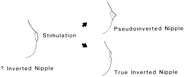
Examination of the nipples is of particular importance during early pregnancy to identify those patients with a truly inverted nipple as early as possible, because this condition can make breast-feeding difficult or impossible. Subnipple adhesions caused by a low-grade mastitis or trauma may bind the nipple to the underlying breast stroma so that eversion does not occur when the nipple is stimulated (Fig. 1). The pseudoinverted nipple becomes erect and protuberant when stimulated, whereas the true inverted nipple retracts. The true inverted nipple is uncommon but should be identified and treated during pregnancy. As often as possible, the patient should stretch the areolae with opposing fingers placed at the 9 and 3 o'clock positions and at the 12 and 6 o'clock positions to break down the subnipple adhesions (Hoffman's exercises). In addition, a nipple shield should be worn under the brassiere during pregnancy to force the nipple slowly forward and outward. When this treatment is begun early in pregnancy, it is almost always corrective and allows nursing without difficulty. Although a variety of surgical techniques have been described for the correction of inverted nipples, no sustainable good solution yet exists that avoids the risk for aesthetic and functional problems.1
|
The final size of the mammary glands at term depends on many factors (e.g. preformed size before pregnancy, distribution of mammary fat, number of initial lobules established, parity, age). It is important to reassure the patient that the size of the gland has little relation to its functional capacity. Such reassurance during pregnancy may go a long way toward alleviating many of the lactation-inhibiting effects of anxiety and feelings of inadequacy, especially in the primigravida who has never attempted to nurse.
Hormonal Effects
Total estrogen excretion increases from 20–20,000 mg per 24 hours between early and late pregnancy. This reflects the rising plasma estrogen levels, which greatly stimulate the ductal arborization begun at puberty and the differentiation of epithelial cells into ductal, acinar, and myoepithelial elements. As the acinar-ductal system expands, it replaces much of the fatty tissue of the breast and is organized into mature, functional, lobular-alveolar-ductal units surrounded by hypertrophied myoepithelial elements (Fig. 2). In addition to its effect on the mammary cells themselves, estrogen stimulates the synthesis and release of prolactin from the pituitary lactotrophs. Rising prolactin levels appear to be necessary for estrogen to exert its biologic effects on the mammary gland. In addition, prolactin induces the enzymes necessary for the acinar secretory activity seen after delivery. Prolactin levels increase from 20–200 ng/ml during pregnancy.
|
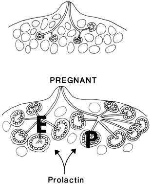
Progesterone secretion increases from 3–300 mg/day during pregnancy. In the presence of estrogen and prolactin, progesterone stimulates acinar proliferation and inhibits lactose synthesis. The high plasma concentrations of estrogen and progesterone present before delivery inhibit the active secretory effects of prolactin on mammary alveolar epithelium.
In addition to the regulatory role of sex steroid hormones, an increasing list of local growth factors has been shown to modulate survival and apoptosis of the mammary gland. A stimulating role in the proliferation and/or differentiation of mammary epithelial cells is suggested for most growth factors, including epidermal growth factor, transforming growth factor-alpha, and insulin like growth factors.2 The specific effects of insulin and placental lactogen on mammogenesis have yet to be fully elucidated. It has been clearly shown in tissue culture that insulin is necessary for estrogen, progesterone, and prolactin to stimulate the growth of mammary epithelial cells.3 Alloxan-treated diabetic rats have also been shown to have diminished milk secretion. Human placental lactogen (HPL), also called human chorionic somatomammotropin, is a placental protein hormone that has both lactogenic and somatotropic effects that may facilitate mammogenesis directly or act by competitively inhibiting prolactin receptors in the mammary tissue during pregnancy to delay milk production until after delivery.4
The initial stimulation of mammary epithelium occurs during the first few weeks of pregnancy. By the second trimester, colostrum, the first milk, appears in the alveoli of the acinar glands in small quantities, reflecting the beginning of protein synthesis under the influence of prolactin. By the third trimester, the alveoli contain significant amounts of colostrum, the epithelial cells are laden with fat droplets, and the adipose tissue of the breast has been markedly reduced and replaced by functioning glandular units.
LACTOGENESIS
The mammary epithelium remains a presecretory tissue until the abrupt diminution in plasma estrogen and progesterone concentration that occurs at the time of delivery. Without the inhibitory influence of progesterone on mammary epithelium, prolactin and the other hormones active in the initiation of milk production can exert their effects on acinar cells. By 4–5 days postpartum, estrogen and progesterone concentrations in the plasma are less than normal follicular phase levels and the transition in the acinar epithelium from a presecretory to a secretory state is complete. The ovaries apparently are not necessary for the initiation or maintenance of lactation, because oophorectomy has no effect on this process.
The initiation of milk production (lactogenesis) requires 2–5 days in the human being. This is the length of time necessary for complete secretory maturation of acinar epithelium. The inhibition of lactogenesis before delivery appears to be a consequence of high circulating levels of progesterone, which competitively inhibits the binding of cortisol to an intracellular receptor. This prevents cortisol from acting synergistically with prolactin to initiate milk production.5 The rapid fall in progesterone after deliver allows cortisol binding to occur and lactogenesis to proceed. Administration of large doses of progesterone in the immediate postpartum period inhibits milk production. However, once the secretory transformation of the acinar epithelium is completed, sex steroids are ineffective in halting lactogenesis.
Prolactin and cortisol are essential for lactogenesis, and growth hormone, insulin, and thyroxin play facultative roles. Prolactin, a peptide hormone with a molecular weight of 23,500, is produced by the lactotrophs of the pituitary gland. Although it was initially thought to be almost identical to growth hormone and HPL, further studies have demonstrated many structural and functional characteristics unique to prolactin. Prolactin specifically binds to a receptor on the surface of the alveolar epithelium, stimulating synthesis of messenger RNA (mRNA) molecules that are necessary for the production of milk proteins and other required enzymes.6, 7 For example, production of the mRNA for casein synthesis, an important milk protein, is initiated by the synergistic actions of prolactin and cortisol. Prolactin also causes increased activity of galactosyltransferase, lactose synthetase, and γ-lactalbumin.
The high prolactin levels reached under the influence of estrogen during pregnancy are not maintained after delivery. There is a rapid decrease in prolactin concentration after delivery, and normal nonpregnant levels are attained by approximately 7 weeks postpartum in both lactating and nonlactating mothers. Prolactin 'surges' occur within 15 minutes of nipple stimulation during nursing, however. These surges peak at levels of 100–200 ng/ml during the first week, 25–250 ng/ml during the second to fourth weeks, and less than 20–40 ng/ml thereafter.
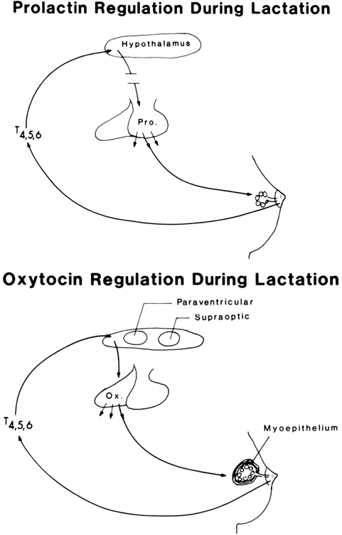
Suckling stimulates sensory receptors in the nipple that activate nerve impulses; these impulses are transmitted through thoracic nerves 4, 5, and 6 to the spinothalamic tracts in the spinal cord, terminating in neurons in the mesencephalon. Impulses from the mesencephalon are transmitted to the hypothalamus, resulting in a decrease in prolactin-inhibiting factor (probably dopamine) that releases the lactotrophs from the inhibitory influence of catecholamines. This permits the synthesis and release of prolactin.
In the absence of prolactin, lactation does not occur. Hypophysectomy, postpartum pituitary necrosis, destructive diseases of the hypothalamic pituitary system, and ingestion of dopamine agonists (e.g. bromocriptine, L-dopa) result in failure of lactation. Increased concentrations of prolactin appear to be of particular importance in the process of lactogenesis, whereas only normal nonpregnant levels seem to be necessary for the maintenance of lactation once begun.
The only other specific hormone required for lactogenesis is oxytocin. Oxytocin is an octapeptide produced in the supraoptic and paraventricular nuclei of the hypothalamus and stored in the posterior lobe of the pituitary gland. It is released after suckling stimulates sensory fibers in the nipple. Impulses that activate its release are transmitted along the same pathways as those that carry impulses for prolactin release up to the level of the mesencephalon (Fig. 3). At that point, the pathways divide and the impulses that control oxytocin release travel to the supraoptic and paraventricular nuclei, where they stimulate both synthesis and release of oxytocin. Oxytocin is released from neurovesicles (Herring bodies) within the neuronal terminals of the posterior pituitary gland. These neurovesicles are located close to the dense vasculature that drains this area. Via beta-receptors, oxytocin causes the myoepithelial cells to contract, which results in release of milk into the lactiferous ducts and sinuses so that it can be removed by suckling. The release of oxytocin becomes a conditioned response in the lactating woman, requiring only visual stimulation or conscious thought. No such conditioned release of prolactin has been demonstrated.
|
Only the hormones mentioned have been shown to be essential to lactogenesis. Normal levels of thyroid hormone, insulin, growth hormone, and parathyroid hormone appear to be facilitatory but are not required in other than normal, nonpregnant concentrations.8
INHIBITION OF LACTATION
The success of a purposeful suppression of lactation depends on inhibiting the process of lactogenesis. Because lactogenesis does not begin in the human being until the rapid decrease in estrogen and progesterone that occurs at delivery and because it requires 2–3 days to be completed, it is possible to inhibit lactogenesis through the use of exogenous steroids to maintain relatively high circulating levels during this critical period after delivery. The administration of sex steroids after lactogenesis is completed has little effect on lactation. Estrogen or progesterone administered alone have little or no effect on lactogenesis or lactation; when given in combination, their effectiveness in decreasing milk production is dose dependent. One possible mode of action already suggested is that progesterone, in the presence of estrogen, may competitively inhibit the cortisol-receptor complex necessary for the formation of rough endoplasmic reticulum and protein synthesis. Once this complex has been formed, progesterone is without effect on lactation.9
In the 1980s, bromocriptine was suggested as a medical therapy for the suppression of lactation. As a dopamine receptor agonist, bromocriptine is highly effective at lowering prolactin levels postpartum and inhibiting lactation.10 The typical dose is 2.5 mg two to three times daily for up to 2 weeks. More recently, cabergoline, a long-acting prolactin-lowering medication, was found to give results comparable to those of bromocriptine. Cabergoline is given as a single 1-mg dose within 24 hours after delivery.11 These medications are not currently recommended for suppression of lactation.
Side effects with both medications include dizziness, hypotension, headache, nausea, and drowsiness. More women report adverse events with bromocriptine (26%) than with cabergoline (16%). These medications can be associated with serious adverse reactions, and nonpharmacologic methods are the method of choice for lactation suppression.
Certain general supportive measures to inhibit lactation are believed by many to be as effective as medical therapy. These supportive measures greatly facilitate successful suppression of lactation. Use of breast binders or a tight brassiere worn 24 hours/day; intermittent application of an ice pack; use of analgesics when necessary; avoidance of tea, coffee, and phenothiazine tranquilizers; and, most importantly, the avoidance of nipple stimulation, ordinarily result in cessation of lactation by 1 week postpartum. Nipple stimulation is an extremely potent factor in lactation. Even after receiving medication such as estrogen or progesterone, it is possible for most patients to nurse satisfactorily, because persistent suckling eventually overrides the inhibitory influence of the medication.
Currently, when breast-feeding is not desired, conservative supportive measures are usually instituted. Steroid medications, bromocriptine, or cabergoline are rarely prescribed today to inhibit lactation.
GALACTOPOIESIS
Galactopoiesis is the maintenance of milk production once it has been established by completion of lactogenesis. The single most important factor in successful galactopoiesis is regular and frequent milk removal from the mammary gland. Milk removal stimulates further milk secretion by at least three mechanisms. First, regular suckling promotes the regular synthesis and release of both prolactin and oxytocin, which are necessary for continued milk secretion. Second, the breast has the capacity to store milk for a maximum of 48 hours before there is a substantial decrease in production. This reduced milk production is caused by the diminished stimulation of the glandular epithelium by prolactin and the vascular stasis caused by increased intramammary pressure resulting from distention of the mammary ducts and alveoli with stored milk. Blood flow to the mammary glands is significantly reduced by this increased intramammary pressure, which diminishes the nutrient and hormonal supply necessary for milk production. Third, as in other milk-producing animals, the amount of milk produced daily is fairly closely related to the demand (i.e. the amount of milk removed the previous day), as long as the nutritional and hormonal requirements are met. Normal levels of prolactin (5–20 ng/ml), with surges of prolactin and oxytocin at the time of suckling, are also necessary for the maintenance of normal milk production.
The catecholamines released at times of stress and anxiety directly antagonize the action of oxytocin on the myoepithelial cells, and norepinephrine causes vasoconstriction, which has an effect on milk production similar to that of the failure of milk removal. Therefore, the psychological state of the lactating woman is of critical importance in the maintenance of adequate milk production.
The connection between maternal behavior and lactation has been clarified further in studies of the suckling reflex in rodents. Using selective placement of lesions in the serotonergic tracts in the rat brain, investigators have found that the suckling stimulus initiates both reflex maternal nursing behavior and bursts of prolactin release. These two reflexes occur through different afferent pathways. Selective destruction of either pathway results in a dramatic decrease in the growth of the rat pups, documenting the necessity of appropriate maternal behavior for successful lactation.12
Finally, the importance of an adequate diet for the lactating woman cannot be overemphasized. The dietary requirements for lactation are even greater than those for the third trimester of pregnancy.13 Significant increases in protein, carbohydrate, lipid, minerals, and caloric intake must be maintained throughout lactation if adequate volumes of milk are to be produced.14 Inadequate diet affects the volume of milk produced rather than the constituents of milk. The milk has the same concentrations of basic ingredients when dietary intake is inadequate, but these ingredients are drawn from maternal stores. If there is inadequate calcium in the diet, for example, maternal bone stores are tapped until normal levels are reached in the secreted milk. However, the volume of milk produced is significantly diminished if maternal nutrition is poor.
AMENORRHEA AND INFERTILITY
Studies have documented slower return of ovulation and regular menses in lactating women after pregnancy. The duration of lactational amenorrhea varies considerably and is based in part on the frequency and duration of suckling. There is no pattern that guarantees anovulation and infertility for all women. A consensus statement on the use of breast-feeding for family planning concluded that up to 98% of women have protection from pregnancy for 6 months after delivery if they are fully breast-feeding and are amenorrheic.15 While breastfeeding results in amenorrhea and delays the return of fertility, the length of the delay cannot be reliably predicted or detected.16 As such, most lactating women will resume menses within 6–9 months. Because lactating women can ovulate as early as 6–8 weeks postpartum, contraception should be considered for all breast-feeding women.
HUMAN MILK
Colostrum and Transitional Milk
After removal of the inhibitory influence of estrogen and progesterone, prolactin stimulates the alveolar epithelial cells to begin active secretion of the first milk, colostrum. Colostrum is moved into the lactiferous ducts and sinuses by the contraction of the myoepithelial cells, under the influence of oxytocin, and is removed by suckling. The aqueous phase of milk is predominantly a lactose solution. When progesterone is withdrawn, postpartum lactose is secreted into the acinar lumen and osmotically incorporates water, resulting in a solution that is isosmotic with plasma. Therefore, lactose is the primary controlling influence on milk volume. The electrolyte content of aqueous milk is like that of intracellular fluid (i.e. high in potassium and relatively low in sodium and chloride). An adequate supply of glucose to the alveolar cells is also essential for continued milk production.
Colostrum is produced during the first 3–5 days postpartum by both apocrine (apical degeneration) and merocrine (transmembrane transport) secretion. It contains proteins, minerals and vitamins, lipids, carbohydrates, and both chemical and cellular immunologic factors of great importance to the survival of the newborn, even though transplacental passage of immunoglobulins confers passive immunity on the fetus while in utero and for the first few weeks after birth. In many species, such as ruminants, there is no transplacental transfer of immunoglobulins and the newborn is entirely dependent on the immunoglobulins present in colostrum. Although the human infant is less vulnerable initially, postpartum colostrum provides extremely important immunoglobulins and other antimicrobial substances that act locally within the intestinal tract against potential pathogens.
Grossly, colostrum is a yellow, viscous fluid amounting to approximately 25 ml/day. It is high in vitamin A, the pigment of which gives it its yellow color. The protein and mineral content of colostrum is significantly greater than that of mature milk, and one half of the total protein is identical to γ-globulin, primarily of the immunoglobulin A type. This immunoglobulin is a unique double molecule linked with disulfide bridges that greatly diminish its hydrolysis and metabolism in the intestinal tract. Colostrum contains immunologically active lymphocytes and monocytes, interferon, a factor that facilitates the removal of intestinal meconium, and a factor for stimulation of the beneficial Lactobacillus bifidus microorganisms in the intestinal tract.
Production of transitional milk begins after the first week of lactation and continues through the third week postpartum. The immunoglobulin and total protein content of this milk is less than that of colostrum, whereas the content of lactose, lipid, water-soluble vitamins, and calories is greater. The volume of milk produced increases from 100–500 ml by the end of the second week postpartum.
Mature Milk
In many respects, mature human milk differs from cow's milk and cow's milk-based milk replacers. Human milk contains 1.2 g/100 ml protein, which is less than half that in cow's milk. The major difference is in the content of casein, which is substantially less in human milk. The relatively low casein content facilitates the formation of curd in the infant's intestinal tract. This increases the digestibility of milk and keeps the gastric acidity high, resulting in an increased antimicrobial action. Because the content of essential amino acids in human milk is essentially the same as that found in human plasma, the transfer from intestinal tract to plasma is quite efficient. In addition, human milk is high in the amino acid, taurine, which may be of great importance in neural development, especially in premature infants. Finally, human milk contains several specific growth factors that are missing from bovine milk, as discussed later.17
The carbohydrate content of human milk is approximately 7 g/100 ml, nearly twice that of cow's milk. The primary carbohydrate of milk is lactose, which seems to serve several functions. It promotes the growth of L. bifidus flora, which maintains a high intestinal acidity, inhibits growth of pathogenic bacteria, and maximizes calcium absorption. Hydrolysis of lactose results in the formation of galactose, which is necessary for the production of cerebrosides utilized in myelin synthesis.
Lipids are present in human milk in a concentration of 3.7 g/100 ml. They act as the primary energy source for the growing infant. The emulsified lipid in human milk is much finer than that of cow's milk, and the lipase activity is much greater as well. Both these factors make human milk easier for the infant to digest.
Human milk has approximately 200 mg/100 ml mineral content, which is one third that of cow's milk. The primary difference is in the concentration of sodium.
Vitamin A may be insufficient in human milk if the mother is poorly nourished. Similarly, vitamin D probably should be supplemented by 3–4 months postpartum. Because cow's milk contains half as much vitamin A, vitamin D, and iron as human milk, there is an earlier tendency toward growth deficiencies and anemia when an infant is maintained on cow's milk. Vitamin C in cow's milk is lower than in human milk initially, and the heating process further reduces its concentration.
Human colostrum and mature milk contain several growth factors. The most important of these factors, constituting approximately 75% of the growth-promoting activity of milk, is human epidermal growth factor (EGF). The concentration of EGF is highest in colostrum and falls during the transition to mature milk but remains detectable throughout lactation.17 Insulin-like growth factor I (IGF-I), another potent growth factor, is also present in human milk. In contrast to EGF, the concentration of IGF-I in breast milk increases during the first 6 weeks postpartum.18 Evidence indicates that breast milk from women who deliver preterm infants may contain much higher concentrations of EGF and other growth factors.19 This represents a unique adaptation to preterm delivery and implies that the premature infant may benefit from maternal breast milk, as opposed to pooled human milk or formula.
Antimicrobial Factors
The antimicrobial constituents of human milk have only recently begun to be appreciated. Human milk contains numerous anti-inflammatory cytokines, antioxidants, protease inhibitors and prostaglandins to help protect the nursing infant.20 As mentioned earlier, the immunoglobulin content has a local action against pathogenic bacteria and viruses. The production of immunoglobulin in the mother is a dynamic process that continuously produces immunoglobulins both against pathogens to which the mother is independently exposed and against those transferred from the infant to the mother before clinical infection of the infant has occurred. This gives rise to a local immune system in the infant's intestinal tract that remains up-to-date in relation to the potential pathogens in the environment.
The bifidus factor in human milk is 40–100 times that in cow's milk. It consists of a group of saccharides that stimulate the growth and dominance of L. bifidus flora in the infant's intestinal tract. By the end of the first week, 55% of the bacteria that can be cultured from the breast-fed infant's intestinal tract are L. bifidus, whereas coliform organisms predominate in the bottle-fed infant. By acidifying the gut, the bifidus flora produce resistance to Staphylococcus aureus, Shigella, protozoans, and pathogenic coliform organisms.
Lactoferrin and transferrin, by their iron-binding and chelating properties, respectively, are bacteriostatic against coliform organisms. Finally, lysozyme (muramidase), present in significant concentrations in human breast milk, is bacteriolytic to Enterobacteria and gram-positive organisms.
BREAST-FEEDING: PRINCIPLES AND PRACTICE
Among children born in 2004, 21 states in the United States achieved the national Healthy People 2010 objective of 75% of mothers initiating breastfeeding.21 The great resurgence in breast-feeding has required the physician who cares for patients of reproductive age to acquire an in-depth knowledge of lactation and breast-feeding. Enlightened and sound advice to the lactating mother greatly increases the satisfaction with nursing for the mother and the overall benefit to the infant.
Nursing should begin at or shortly after delivery for the healthy, mature infant. In earlier times, the infant was put to the mother's breast immediately after delivery to induce uterine contractions for the expulsion of the placenta and for hemostasis. In addition, placing the infant at the mother's breast after delivery also may increase the strength of maternal-infant bonding.
The initial nursing period should be short (2–3 minutes on each breast at 2–4 hour intervals), with a slight increase in duration each day. Nursing may be carried out on a regular basis (every 4 hours) or on demand. During the first week to 10 days of nursing, the infant will not take all of the milk in the breast, and the remainder should be removed by suction to maximize milk production and minimize symptoms of pain and engorgement. By 2 weeks postpartum, supply and demand regulates the volume. Night feedings are usually necessary during the first month postpartum but can be gradually eliminated during the second month for most infants. Nursing time should be increased to a maximum of 10 minutes per breast, because 5–6 minutes is all that is usually required to empty a breast. Excessive nursing can produce nipple fissures and mastitis.
Causes of Difficulty
The greatest cause of difficulty with nursing or milk supply is anxiety. This anxiety may be secondary to fears of inadequacy related to breast size, to concern about the ability to produce sufficient milk (usually for the first pregnancy or if such a problem arose in nursing a previous infant), or to unconscious or conscious negative feelings about nursing. The stress and anxiety caused by these feelings are associated with increased catecholamine production, which inhibits the stimulation of the myoepithelial cells by oxytocin and causes vasoconstriction that diminishes the supply of both oxytocin and prolactin to the acinar cells of the breast. This condition is similar to psychological impotence; the physiologic mechanism is intact, but there is an autonomic block that originates in the psyche and inhibits the normal physiologic function. The treatment for this difficulty is counseling, education, and support. Such groups as the La Leche League, Nursing Mothers, and Childbirth Education Association are extremely useful support groups for the young nursing mother or the mother having difficulty nursing. The support provided by such groups are usually all that is necessary to promote successful breast-feeding. When medical therapy is needed, chlorpromazine, 25 mg (three times a day) for 3–10 days, has been successful both in relaxing the anxious mother and in increasing prolactin production and, thus, milk production. Similarly, the theophylline content of coffee and tea in moderation can stimulate prolactin production and increase milk secretion. A frustrated, anxious mother and a hungry infant are a bad combination. Good prenatal education and support from a sensitive nurse-midwife or support group are highly recommended for the mother who is nursing for the first time.
In addition to anxiety, there are several other, less common causes of inadequate milk supply. Combination birth control pills have been implicated in diminishing milk supply and may be detrimental to the infant, although the standard moderate- or low-dose combination pills in use today have not been shown to diminish milk supply in quality or quantity or to cause any untoward effects in the infant. The high-dose estrogen pills formerly used did diminish milk secretion to some extent.
The heavy smoker (one or more packs per day) has circulating nicotine levels sufficient to inhibit prolactin synthesis and release. Significant concentrations of nicotine are also found in the breast milk and are undesirable in the developing infant. Therefore, cigarette smoking is contraindicated for the nursing mother.
Occasionally, a mother has an inadequate milk supply as a result of inadequate dietary intake of calories and nutrients. Dietary restrictions may result in insufficient caloric intake to meet the 3000 calorie-per-day requirement for lactating women (Table 1). If there is concern about decreased milk supply, it is important to consider the woman's dietary habits. Recent studies indicate that a modest weight loss (less than 2 kg monthly) and exercise do not affect milk volume or composition.22
Table 1. Nutritional requirements during early pregnancy and lactation
Daily Requirements | Early Pregnancy | Lactation |
Calories (kcal) | 2100 | 3000 |
Protein (g) | 55 | 85 |
Fat (g) | 60 | 80 |
Carbohydrate (g) | 330 | 480 |
Calcium (g) | 0.8 | 1.3 |
Iron (mg) | 15 | 20 |
Vitamin A (IU) | 5000 | 8000 |
Vitamin D (IU) | 200 | 400 |
There are rare cases of oligogalactia and agalactia caused by failure of the pituitary gland to produce prolactin. This failure may occur as a result of hypothalamic-pituitary disease or postpartum pituitary necrosis. If this condition is suspected, a series of provocative tests are required to determine pituitary sufficiency. A corticotropin-releasing hormone (CRH) stimulation test determines the responsiveness of the pituitary-adrenal axis. Similarly, a thyrotropin-releasing hormone (TRH) stimulation test determines whether the pituitary gland can produce and release thyroid-stimulating hormone and prolactin. TRH is given in a dose of 100 μg, and blood is drawn for prolactin after 0, 0.5, 1, 2, and 3 hours. If the prolactin level in the blood at least doubles, the pituitary lactotrophs are intact.
One of the most common conditions arising during lactation is nipple soreness. Nipples can become cracked or bruised and can bleed. Prenatal preparation can reduce soreness. Grasping the areola, gently pulling outward, and rolling the nipple between the thumb and forefinger prepares the breast for suckling and reduces soreness. Once breast-feeding has begun, soreness can be decreased by using proper infant positioning, limiting nonnutritive suckling time, avoiding breast engorgement, and applying a commercially available breast-feeding ointment.
Blocked ducts, manifesting as a tender lump in the breast, can occur with incomplete emptying of the breast. Heat and massage, along with frequent nursing, may help promote duct drainage.
Engorgement generally occurs 3–4 days after delivery and can affect the ability of the infant to latch on and feed. Management includes regular feeding from both breasts and the use of a pump or manual expression before feeding. If discomfort is significant, analgesics may be required.
Mastitis affects up to 3% of breast-feeding women.23 When a defined area of firmness and redness develops without systemic symptoms (e.g. fever, malaise), the condition usually can be treated satisfactorily with the local application of heat and continued nursing from both breasts. If systemic symptoms develop, antibiotic therapy against resistant staphylococcal strains should be used for 7–10 days. There is no need to stop nursing during treatment, and nursing should continue from both breasts. Occasionally, mastitis may organize into a discrete abscess that is not responsive to antibiotic or local therapy and requires surgical drainage. Even in this situation, the determined mother can safely continue to breast-feed.
Benefits
The potential benefits of breast-feeding are many. The infant's acquisition of resistance to intestinal and respiratory bacterial and viral infections has been described.24 Infants at risk for the development of allergies may also benefit from breast-feeding. The delay in exposure to foreign proteins may be beneficial. Additionally, breast milk contains secretory immunoglobulin A that may limit the absorption of potentially allergenic compounds from the infant's gut. Breast-fed infants only rarely acquire the debilitating condition of acrodermatitis enteropathica, a rare autosomal recessive disorder of zinc metabolism causing a vesicular eruption of the lower extremities and body orifices with recurrent diarrhea. Although cow’s milk contains higher concentrations of zinc, the bioavailability of zinc in human milk is greater and confers temporary protection against the disorder. Infants who are bottle fed will manifest the condition within the first few days of life, or within days to weeks after weaning.25 The unique amino acid of breast milk, taurine, and its possible benefit to the premature infant in regard to neural development have already been described. Maternal thyroxine is transported in breast milk to the infant and prevents the devastating development of cretinism, which occurs when hypothyroidism is undiagnosed during the infant's continued neural development in the critical early months after birth.
The high cholesterol concentrations of human milk have been the subject of intensive investigation over many years. Infants who are breast fed tend to have higher total lipid levels than infants who are bottle fed. However multiple cross sectional and cohort studies show that adults who were exclusively breast-fed have significantly lower circulating cholesterol concentrations than do control patients who were bottle-fed.26 It is suspected that the exposure of the infant to high cholesterol levels induces enzymes necessary in later life for the handling of dietary cholesterol loads.27 Whether this modest effect has any long term clinical impact remains uncertain.
Obesity in childhood and adulthood may have its roots in early infant feeding. Overfeeding of the infant and consequent later obesity appear to be much less common in the breast-fed than in the bottle-fed child. However, the studies leading to this conclusion did not control for important variables such as solid food intake.
The concept of maternal-infant bonding has been defined relatively recently. The behavior patterns involved are apparently established quite soon after birth if they are established at all, and the effects are long lasting. Several studies have indicated that maternal-infant bonding may influence psychosocial behavior, learning ability, and linguistic facility in later life. If nothing else, it produces a much happier mother-child relationship from the beginning and, possibly, a healthier, happier parent-child relationship in later years.
Prematurity
Whether or not to breastfeed the premature infant has been debated for many years. Certainly, the weak and sick premature infant who is not physically capable of adequate suckling should be fed by the least exerting and most beneficial method possible. However, most neonatal care units now recognize the potential immunologic, developmental, economic and psychological benefits of breastfeeding even in the very premature. Clinical studies in nurseries worldwide have suggested a decreased rate of various infections in premature infants fed human milk compared with those fed infant formula.28 Because of their unique physiologic needs, fortification of expressed human milk may be indicated for very low birth weight infants. Hospitals and physicians should recommend human milk for premature and other high-risk infants either by direct breastfeeding and/or by using the mother's own expressed milk, and this should be encouraged as early as feasible.29
Induced Lactation
The possibility of surrogate lactation for an adopted infant has generated growing interest. Although supplementation may be required, the primary goal of induced lactation is nurturance, not nutrition. Stimulation of the breast and nipples for 2 months before the arrival of the infant is recommended. Medications that increase serum prolactin levels, such as chlorpromazine or metaclopromide, can be useful when breast-feeding is initiated. Intranasal oxytocin, to trigger a let-down response, may also be beneficial for the first several weeks. Similar techniques may be useful for relactation when prematurity or infant illness delays breast-feeding.30
The composition of breast milk appears to be normal in women with induced lactation. The volume of breast milk may be limited, and supplementation with donated breast milk or formula may be required. Considerable support from the family, the practitioner, and a breast-feeding consultant are vital for the success of induced lactation.
Weaning and Involution
The process of involution that begins with diminishing frequency of nursing or weaning requires approximately 3 months. A slow rather than abrupt cessation of lactation is recommended for the sake of both the infant and the mother. There is less resistance on the part of the infant and less symptomatic engorgement and discomfort for the mother if the weaning is gradual. Decreasing frequency of nursing increases the amount of breast milk retained in the breast, resulting in vascular stasis and alveolar atrophy. Less prolactin is also produced with diminished suckling, which further decreases milk synthesis. The major change that occurs during involution is a decrease in the size of the alveolar-lobular-ductal units rather than a quantitative loss of these units, such as that which occurs during menopausal involution. Menopausal involution also includes a loss of elastic supportive tissues and a decrease in the fat pad of the breast, neither of which occurs to any significant degree in postlactational involution. By 3 months after cessation of lactation, the breasts have regressed maximally. Because they retain some of the increased fatty tissue and connective tissue elements developed during pregnancy, they remain slightly larger than their pre-pregnancy size.
DRUGS AND BREAST MILK
Relatively little is known about the appearance in breast milk of the myriad of drugs on the market today.31 The little information available indicates that drugs are deposited in breast milk in the same way that they are deposited in other organs and tissues. Drugs ingested by the mother diffuse or are transported from the maternal plasma to the alveolar cells of the breast. The rate and amount of transport depends on the concentration of the unbound drug in the maternal circulation, its molecular size, and its lipid solubility or state of ionization. Drugs in the plasma are either bound to carrier proteins or free. It is only the free compound that is active in the diffusional sense. Small (molecular weight less than 200), water-soluble drugs pass freely from the plasma of the mother to the alveolar cells of the breast and into the breast milk in the alveolar lumen. Because the major barrier to drug diffusion is the exposed lipid of the cell membrane, the less ionized and/or the more lipid-soluble a molecule is, the more easily it passes to the alveolar cells and then into the milk. As a general rule, it can be assumed that any drug ingested by the mother appears to some degree in breast milk; usually, the total amount of drug in breast milk is less than 1% of that ingested by the mother. For most drugs, chronic ingestion by the mother, which exposes the infant to a constant level of the medication, is likely to create the greatest harm.
The predominant symptom for many drugs is either vomiting, diarrhea, or skin rash. Whenever these symptoms occur in the nursing infant, a history of drug ingestion by the mother should be sought. Breast-feeding is not recommended for women receiving radioactive isotopes or chemotherapeutic agents. Alcohol intake can cause sedation, or even impaired motor function, in the infant. Because alcohol is lipid soluble, it is found in the same concentration in milk as in the maternal serum. Both sedatives and stimulants readily appear in breast milk and give rise to either hyperactive or hypoactive infants. The excretion of antithyroid compounds in breast milk is significant and may produce hypothyroidism and goiter in the chronically exposed infant. One of the more detrimental drugs that appears in breast milk is reserpine, which causes difficulty in breathing and poor suckling due to nasal stuffiness. Heparin, because of its high molecular weight, and coumadin, because of its high degree of protein binding, are unlikely to cause a bleeding dyscrasia in the infant; infant prothrombin times and partial thromboplastin times are reportedly normal.
Lithium is excreted in breast milk, producing a potential for lithium toxicity in the infant. Ergotamine has been reported to cause vomiting, diarrhea, and convulsions in breast-fed newborns. Serum levels of antiseizure medications such as Tegretol and phenytoin should be monitored closely in the mother to avoid excessive infant exposure. Phenobarbital is very slowly excreted by the newborn and can cause significant sedation when used by the mother.
Combination oral contraceptives were contraindicated in the past for the lactating mother because the chronic exposure to rather high doses of estrogen and progesterone compounds significantly suppressed milk production. Since the introduction of low-dose pills and progesterone-predominant contraceptives, there is less concern about the effect on milk production. Some studies do suggest a limited effect on milk volume, even with low-dose oral contraceptives. Once lactation is established, the effect appears to be minimal, allowing lactating women to use oral contraceptives. Progesterone-only contraceptives have also been chosen to avoid any suppressive effect of combination pills on lactation. The slight infant breast stimulation caused by ingested estrogen and seen with use of higher-dose pills in the past is rarely seen with low-dose pills. When it does occur, it is reversible after cessation of the pill. No long-term effects in the infant exposed to combination estrogen and progestins are known. The infants nursed by mothers in the 1950s during the original trials of birth control pills in Puerto Rico have not been found as young adults to have a greater incidence of any abnormality than the unexposed control population.
In general, however, all medications that are not absolutely necessary should be avoided by the lactating mother. When medication is necessary, the concentration ingested and the duration of treatment should be kept to a minimum. When there is doubt about the possible effect of a drug, nursing should be temporarily discontinued, and the breast should be pumped.
POTENTIAL HAZARDS OF BREAST MILK
Most women can breast-feed their infants. With proper education, support, and reassurance, only a small minority are unable to breast-feed satisfactorily. On the other hand, there are certain deficiencies of breast milk, certain groups of patients who should not breast-feed, and certain potentially detrimental long-term effects of breast-feeding that are not yet clearly defined.
From the nutritional point of view, vitamin A, vitamin B12, and folic acid all tend normally to be present at borderline levels in breast milk and are significantly reduced in patients with poor dietary intake. Vitamin A, for example, is necessary for epithelial growth and the formation of visual pigments and may be reduced by as much as one half of the required amount in the breast milk of economically deprived mothers. These vitamins, along with iron, should probably be added as supplements to the diet of the breast-feeding infant.
Certain infants have a genetic deficiency in the enzyme necessary to metabolize galactose, which results in galactosemia with its characteristic clinical symptoms of mental deficiency, liver and spleen enlargement with ascites, and cataracts. This condition is reversible in large part when exposure of the infant to galactose or to its precursor, lactose, is ended. A family history of galactosemia should be sought, and infants with such a history should be tested early in life for the condition.
Women who take potentially toxic medications on a chronic basis should not plan to breast-feed. Such medications include antithyroid compounds, antimetabolites, lithium, and reserpine. Alcoholics, drug addicts, and food faddists are all likely to have an inadequate dietary intake for successful and healthful breast-feeding, and the transfer of alcohol and addictive drugs to the infant in breast milk is an additional contraindication in these groups.
Breast-feeding in the setting of hyperbilirubinemia has created some confusion. The original association of breast-feeding with the appearance of severe and prolonged hyperbilirubinemia at 10–15 days of age is uncommon and should mandate cessation of breast-feeding until the bilirubin concentration returns to normal levels. A much more common physiologic hyperbilirubinemia appears on the second or third day postpartum, during the critical time of milk let down and completion of lactogenesis, but this is not adversely affected by breast-feeding and should not be a contraindication to it.
A relatively new area of concern is the effect of a polluted environment on breast milk and the breast-fed infant. The possibility of contamination of human milk by such detrimental compounds as pesticides, polychlorinated biphenyls, and polybrominated biphenyls remains largely unknown, and the incidence and long-term significance have not been determined. The well-educated and conscientious mother concerned about providing her infant with the most natural and uncontaminated environment for growth is faced with a dilemma: she does not know whether to bottle-feed in the hope of avoiding possible exposure to these compounds, or to risk nursing her infant despite possible environmental contamination in order to provide that myriad of beneficial substances found in human milk. At this point, there is no indication that the possible presence of these compounds in breast milk has any detrimental effect; until such data are forthcoming, breast milk remains the nutrient of choice for the growth of the human infant.
Breast-feeding in women who are infected with the human immunodeficiency virus (HIV) has raised a new concern about infant safety. Viral elements can be isolated in human milk, and numerous reports have documented the transmission of HIV through breast-feeding. Although the risk of transmission during pregnancy is difficult to separate from the risk from breast-feeding, a meta-analysis demonstrated that breast-fed infants had higher rates of infection than did bottle-fed infants.32 A primary HIV infection in a postpartum woman is likely to present the greatest risk to the infant, owing to the high viral load during the initial infection. In the developing world, where infant nutrition is a critical problem, concern about HIV infection creates a serious health dilemma. Protection of the infant with antiviral therapy is being investigated, but the ability of these medications to prevent transmission is not yet known.
The World Health Organization (WHO) recommends that women and health care providers be aware of the potential risk of HIV infection during pregnancy and lactation. Protection against infection is critical for the breast-feeding woman who is at risk. All women are encouraged to have HIV testing. WHO does not recommend breast-feeding for HIV-positive women. Caution should be exercised in neonatal intensive care units when handling breast milk because of the possibility of HIV contamination. Finally, it is recommended that human milk banks use stringent testing for milk donors.33
REFERENCES
Ritz M, Silfen R, Morgan D, Southwick G: Simple Technique for Inverted Nipple Correction. Aesth Plast Surg 29: 24, 2005 |
|
Lamote I, Meyer E, Massart-Leen A, Burvenich C: Sex steroids and growth factors in the regulation of mammary gland proliferation, differentiation, and involution. Steroids 69: 145, 2004 |
|
Turkington RW: Molecular biological aspects of prolactin. In Wolstenholme GEW, Knight J (eds): Lactogenic Hormones, p 111. London: Churchill Livingstone, 1972 |
|
Vorherr H: The Breast: Morphology, Physiology and Lactation. New York: Academic Press, 1974 |
|
Rosen JM, Jones WK, Rogers JR et al: Regulatory sequences involved in the hormonal control of casein gene expression. Ann N Y Acad Sci 464: 87, 1986 |
|
Shiu RP: The prolactin target cell and receptor. Prog Reprod Biol 6: 97, 1980 |
|
Topper YJ: Multiple hormone interactions in the development of mammary gland in vitro. Recent Prog Horm Res 26: 287, 1970 |
|
Cowie AT: Hormonal factors in mammary development and lactation. In Stoll BA (ed): Mammary Cancer and Neuroendocrine Therapy, p 3. London: Butterworths, 1974 |
|
Wynn RM, Harris JA, Chatterton RT: Interaction of progesterone and adrenocorticoids in ultrastructural development of the mammary gland of the rat. Am J Obstet Gynecol 126: 920, 1976 |
|
Cooke I, Jenkins A, Foley M et al: The treatment of puerperal lactation with bromocriptine. Prostgrad Med J 1 (Suppl 52): 75, 1976 |
|
Rolland R, DeGoeij W, Nappi C et al: Single dose cabergoline versus bromocriptine in inhibition of puerperal lactation. Br Med J 302: 1367, 1991 |
|
Barofsky A-L, Taylor J, Massari VJ: Dorsal raphehypothalamic projections provide the stimulatory serotonergic input to suckling-induced prolactin release. Endocrinology 113: 1894, 1983 |
|
Butte N, King J: Energy requirements during pregnancy and lactation. Public Health Nutrition 8: 1010, 2005 |
|
Vorherr H: Lactation, puerperal mastitis, and inappropriate lactation (galactorrhea). In Rovinsky JJ (ed): Davis' Gynecology and Obstetrics, p 11. Vol 1. New York: Harper & Row, 1972 |
|
Kennedy KI, Rivera R, McNeilley AS: Consensus statement on the use of breast-feeding as a family planning method. Contraception 39: 477, 1989 |
|
Van der Wijden C, Kleijnen J, Van den Berk T: Lactational amenorrhea for family planning. Cochrane Database of Systematic Reviews. Issue 4, Art No:CD001329, 2003 |
|
Shing YW, Klagsbrun M: Human and bovine milk contain different sets of growth factors. Endocrinology 115: 273, 1984 |
|
Corpos AN, Brown DK, Rees LH et al: The insulin-like growth factor I content in human milk increases between early and full lactation. J Clin Endocrinol Metab 67: 25, 1988 |
|
Read LC, Francis GL, Wallace JC et al: Growth factor concentrations and growth-promoting activity in human milk following premature birth. J Dev Physiol 7: 135, 1985 |
|
Newburg D, Walker A: Protection of the neonate by the innate immune system of developing gut and of human milk. Pediatr Res 61: 2, 2007 |
|
U.S. Department of Health and Human Services (DHHS) National Center for Health Statistics. The 2006 National Immunization Survey. Hyattsville, MD: Center for Disease Control and Prevention, 2007 |
|
Dewey KG: Effects of maternal caloric restriction and exercise during lactation. J Nutr 128: 386S, 1998 |
|
Melnikow J, Bedinghaus JM: Management of common breast-feeding problems. J Fam Pract 39: 56, 1994 |
|
Jelliffe DB, Jelliffe EFP: Nutrition and human milk. Postgrad Med 60: 153, 1976 |
|
Maverakis E, Fung M, Lynch P, et al: Acrodermatitis enteropathica and an overview of zinc metabolism. J Am Acad Dermatol 56: 116, 2007 |
|
Owen C, Whincup P, Odoki K, et al: Infant feeding and blood cholesterol: A study in adolescents and a systematic review. Pediatrics 110: 597, 2002 |
|
Friedman G, Goldberg ST: Concurrent and subsequent serum cholesterol of breast- and formula-fed infants. Am J Clin Nutr 28: 42, 1975 |
|
Schanler R: The use of human milk for premature infants. Ped Clin N Am 48:3-6, 2001 |
|
American Academy of Pediatrics Policy Statement: Breastfeeding and the use of human milk. Pediatrics 115: 497, 2005 |
|
Brown RE: Relactation: An overview. Pediatrics 60: 116, 1977 |
|
Dillon AE, Wagner CL, Wiest D et al: Drug therapy in the nursing mother. Obstet Gynecol Clin 24: 675, 1997 |
|
Dunn DT, Newell MC, Ades AE et al: Risk of human immunodeficiency virus type 1 transmission through breastfeeding. Lancet 340: 585, 1992 |
|
Committee on Pediatric AIDS: Human milk, breastfeeding, and transmission of human immunodeficiency virus type I in the United Sates. Pediatrics 112: 1196, 2003 |